F
ailed tracheal intubation is a concerning event that may lead to increased patient morbidity and mortality. Our current prediction techniques are imperfect, leading to its occurrence despite careful preprocedural airway risk assessment. a true medical emergency exists when difficult intubation occurs in combination with difficult bag-mask ventilation that impairs oxygenation. This emergency canoccur when failed laryngoscopy attempts are persistent.1,2
Safe management requires timely decision-making and may require the use of alternative techniques to ensure successful tracheal intubation. Current intubation rescue techniques include video laryngoscopy, flexible bronchoscopic intubation, the use of a lighted stylet, or insertion of a
What We Already Know about This Topic
• Airway management guidelines suggest various options after failed direct laryngoscopy
• We lack knowledge of the effectiveness of each alternative technique for achieving successful tracheal intubation What This Article Tells Us That Is New
• In this large, multicentered, retrospective observational study, 1,619 rescue attempts after a failed tracheal intubation were performed in 1,427 out of 346,861 cases
• Video laryngoscopy was the most frequently chosen rescue technique (69%) and resulted in the highest success rate (92%) among the alternative techniques
Copyright © 2016, the American Society of Anesthesiologists, Inc. Wolters Kluwer Health, Inc. All Rights Reserved. Anesthesiology 2016; 125:656-66
ABSTRACT
Background: Multiple attempts at tracheal intubation are associated with mortality, and successful rescue requires a
structured plan. However, there remains a paucity of data to guide the choice of intubation rescue technique after failed initial direct laryngoscopy. The authors studied a large perioperative database to determine success rates for commonly used intuba-tion rescue techniques.
Methods: Using a retrospective, observational, comparative design, the authors analyzed records from seven academic centers
within the Multicenter Perioperative Outcomes Group between 2004 and 2013. The primary outcome was the comparative success rate for five commonly used techniques to achieve successful tracheal intubation after failed direct laryngoscopy: (1) video laryngoscopy, (2) flexible fiberoptic intubation, (3) supraglottic airway as part of an exchange technique, (4) optical stylet, and (5) lighted stylet.
Results: a total of 346,861 cases were identified that involved attempted tracheal intubation. a total of 1,009 anesthesia
providers managed 1,427 cases of failed direct laryngoscopy followed by subsequent intubation attempts (n = 1,619) that employed one of the five studied intubation rescue techniques. The use of video laryngoscopy resulted in a significantly higher success rate (92%; 95% Ci, 90 to 93) than other techniques: supraglottic airway conduit (78%; 95% Ci, 68 to 86), flexible bronchoscopic intubation (78%; 95% Ci, 71 to 83), lighted stylet (77%; 95% Ci, 69 to 83), and optical stylet (67%; 95%
Ci, 35 to 88). Providers most frequently choose video laryngoscopy (predominantly GlideScope® [Verathon, USa]) to rescue
failed direct laryngoscopy (1,122/1,619; 69%), and its use has increased during the study period.
Conclusions: Video laryngoscopy is associated with a high rescue intubation success rate and is more commonly used than
other rescue techniques. (Anesthesiology 2016; 125:656-66)
This article is featured in “This Month in Anesthesiology,” page 1A. Corresponding article on page 615. This work was an oral abstract presentation at the American Society of Anesthesiologists Annual Meeting in New Orleans, Louisiana, on October 14, 2014.
Direct Laryngoscopy in Adults
A Retrospective Comparative Analysis from the Multicenter
Perioperative Outcomes Group
Michael F. Aziz, M.D., Ansgar M. Brambrink, M.D., Ph.D., David W. Healy, M.D., M.R.C.P., F.R.C.A., Amy Wen Willett, M.D., Amy Shanks, Ph.D., Tyler Tremper, B.S., Leslie Jameson, M.D., Jacqueline Ragheb, M.B.B.Ch., F.F.A.R.C.S.I., F.J.F.I.C.M.I., Daniel A. Biggs, M.D., William C. Paganelli, M.D., Ph.D., Janavi Rao, M.D., Jerry L. Epps, M.D., Douglas A. Colquhoun, M.B.Ch.B., M.Sc., M.P.H., Patrick Bakke, M.D., Sachin Kheterpal, M.D., M.B.A.
PeRiOPeRaTiVe MediCiNe
This article has been selected for the Anesthesiology CME Program. Learning objectives
and disclosure and ordering information can be found in the CME section at the front of this issue.
supraglottic airway (SGa) as a conduit to tracheal intuba-tion. Widely used algorithms do not provide clear guidance
on which techniques to use after direct laryngoscopy fails.3 if
ventilation is adequately restored, the guidelines do suggest the use of alternate devices to maintain ventilation or secure the airway with a tracheal tube.
Several techniques have been advocated to facilitate suc-cessful tracheal intubation after failed direct laryngoscopy. One retrospective effectiveness study across two centers
dem-onstrated that rescue using the GlideScope® video
laryngo-scope (Verathon, USa) was successful in 94% (224 of 239)
of cases after failed direct laryngoscopy.4 according to a large
observational study, the Pentax aWS® (Pentax, Japan) was
found to achieve successful intubation in 99% (268 of 270) of cases where direct laryngoscopy failed to achieve an adequate
laryngeal view for intubation.5 in a large observational study
of a new algorithm involving a small group of providers and select group of patients, failed direct laryngoscopy was
res-cued utilizing the airtraq® (Prodol, Spain) system in 27 of
28 cases.6 Others reported that when mask ventilation and
intubation are both difficult, SGas restore ventilation in 94%
of cases (16 of 17).7 Other devices have been advocated as
use-ful rescue intubation tools in smaller case series.8–11 However,
collectively, the aforementioned studies do not provide the evidence necessary for a direct comparison between individual rescue techniques as they either focus on only one technique, the practice in one single center, or represent small case series.
Therefore, we designed a study to help determine the comparative effectiveness of intubation rescue techniques suggested by the american Society of anesthesiologists
dif-ficult airway algorithm3 by analyzing a large database of
perioperative medical records from seven large tertiary care centers across the United States. We tested the hypothesis that video laryngoscopy is associated with a higher success rate compared to other recommended techniques (flexible fiberoptic intubation, lighted stylet, optical stylet, and SGa as a conduit to tracheal intubation).
Methods
The experimental design was a multicentered, retrospective observational study utilizing the Multicenter Perioperative
Outcomes Group (MPOG) database. MPOG is a consor-tium of institutions formed in 2008 with a shared data set facilitating the investigation of perioperative outcomes. The structure of this database has been described previously in
great detail.12,13 in brief, each institution uses an
intraop-erative electronic medical record and routinely extracts data into a common research database structure and common clinical lexicon to enable comparison of medications, pro-cedures, techniques, and outcomes across centers. Rigorous data visualization techniques and case validation are used to maximize data quality and consistency in the centralized database.
institutional Review Board approval was obtained from each MPOG (ann arbor, Michigan) center to contribute and analyze deidentified data in a central data repository. a requirement for written informed consent was waived for these purposes. The clinical investigation protocol, includ-ing planned primary and secondary outcomes and statisti-cal analysis plan, was presented to the MPOG Perioperative Clinical Research Committee in 2012, reviewed and cri-tiqued by the members, and then approved for data extrac-tion. a detailed proposed query plan, data collection sheet, and data definitions are included in the MPOG protocol that was registered with the MPOG research committee.
The analysis was based on the systematic evaluation of electronic medical records collected from seven large ter-tiary care academic institutions in the MPOG consortium between January 2004 and January 2013: University of Michigan, Oregon Health & Science University, Oregon, Michigan; University of Colorado, aurora, Colorado; Uni-versity of Virginia, Charlottesville, Virginia; UniUni-versity of Oklahoma, Oklahoma City, Oklahoma; University of Ver-mont, Burlington, Vermont; and University of Utah, Salt lake City, Utah. These centers were selected among many MPOG contributing centers based upon the availability of structured documentation elements for key variables neces-sary for analysis: intubation device(s) used, number of laryn-goscopy attempts, laryngeal view achieved, and bag-mask ventilation assessment. details of the intubation procedures are documented in a structured format at each institution and mapped to a shared MPOG data structure.
included in the final analysis were electronic medical records from all adult patients (greater than 18 years of age) who had tracheal intubation attempted initially with direct laryngoscopy and then rescue attempted with the technique of interest (video laryngoscopy, flexible fiberoptic intubation, lighted stylet, optical stylet, and SGa as a conduit to tracheal intubation). The following events were excluded from fur-ther analysis: the use of direct laryngoscopy alone, primary technique that was not a direct laryngoscopy, or pediatric patients (less than 18 years of age). Cases in which direct laryngoscopy was performed only to document laryngeal view for future purposes were considered not to be direct laryngoscopy as the primary technique as intubation was not intended by direct laryngoscopy.
Submitted for publication June 20, 2015. Accepted for publication June 14, 2016. From the Department of Anesthesiology, Oregon Health & Science University, Portland, Oregon (M.F.A., A.M.B.); Department of Anesthesiology, University of Michigan Medical School, Ann Arbor, Michigan (D.W.H., A.S., T.T., J. Ragheb, S.K.); Department of Anesthe-siology, Yale School of Medicine, New Haven, Connecticut (A.W.W.); Department of Anesthesiology, University of Colorado, Aurora, Colorado (L.J.); Department of Anesthesiology, University of Oklahoma Health Sciences Center, Oklahoma City, Oklahoma (D.A.B.); Department of Anesthesiology, University of Vermont College of Medicine, Burlington, Vermont (W.C.P.); Department of Anesthesi-ology, Washington University, St. Louis, Missouri (J. Rao); Department of Anesthesiology, University of Tennessee Graduate School of Medi-cine, Knoxville, Tennessee (J.L.E.); Department of Anesthesiology, University of Virginia, Charlottesville, Virginia (D.A.C.); and Depart-ment of Anesthesiology, University of Utah, Salt Lake City, Utah (P.B.).
The primary outcome was the successful tracheal intu-bation rate of (1) video laryngoscopy, (2) flexible fiberoptic laryngoscopy, (3) SGa as a conduit to intubation, (4) opti-cal stylet, or (5) lighted stylet performed after failed initial direct laryngoscopy. although the documentation templates of the institutions did not include a standard definition of an “intubation attempt,” for purposes of this analysis, we defined a “failed attempt” as the documented use of a device with or without attempted tube passage that did not result in successful tracheal intubation. The use of a bougie or an introducer with direct laryngoscopy was not considered a failure unless direct laryngoscopy was abandoned. an “intu-bation rescue technique” was recorded as successful when it resulted in successful tracheal intubation, regardless of the number of attempts with that technique. in contrast, the “intubation rescue technique” was recorded as a failure if the record showed that the provider switched to a different rescue technique or reverted to direct laryngoscopy. Rescue techniques were then categorized into one of the identified groups (table 1). Cases that did not clearly discriminate an “intubation attempt” or “intubation rescue technique” as defined here were excluded from further analysis (fig. 1).
a secondary outcome was the tracheal intubation success rate of the five intubation rescue techniques of interest in the subgroup of patients with “difficult or impossible mask ven-tilation.” Patients were included into this subgroup analysis
if a mask ventilation scaled score of 3 or 414 was documented
or if the mask ventilation narrative indicated “two-hand mask ventilation,” or documented that the patient could not be ventilated by mask at all. an additional secondary outcome was any documentation of airway-related trauma assessed as (1) no injury, (2) dental trauma, (3) pharyngeal injury, (4) tracheal injury, and (5) death.
The primary automated query (fig. 1) identified all cases that involved multiple attempts at laryngoscopy and the use of alternative intubation techniques, or cases with four or more laryngoscopy attempts. in all identified electronic records, the intubation narrative was queried as to whether one or more of the five devices of interest were mentioned by searching for the following terms: “video,” “could not intubate,” “could not ventilate,” “lightwand,” “fiberoptic,” “video,” “CMaC,” “C-MaC,” “stylet,” “storz,” “glidescope,” “glide,” “mcgrath,” “shikani,” “bullard,” “bonfils,” “aintree,” “fiberoptic,” “intubating lMa,” “airq,” “air-q,” “gscope,” “fast track,” “fast trach,” “cricothyrotomy,” “trach,” “lma,”
and “sga.” each institution has a structured airway man-agement template, but not all templates had discreet fields for which device was used first. Therefore, each anesthesia record, in its entirety, was manually reviewed by investigators (M.F.a., d.W.H., a.W.W., l.J., J. Ragheb, d.a.B., W.C.P., J. Rao, J.l.e., P.B., d.a.C., S.K.) to establish the primary and secondary outcomes. First, the intraoperative record was reviewed in detail to determine whether the patient did indeed undergo initial direct laryngoscopy followed by an alternative intubation technique. The case was excluded from further analysis if the documentation was unclear regarding the sequence of events. Cases in question were reviewed by a second reviewer in order to determine inclusion versus exclu-sion of the case and finally by a third reviewer if any debate remained. all other successful airway management strategies were also recorded, which included “surgical airway,” “patient awoken and case cancelled,” “patient awoken and flexible fiberoptic endoscopy,” “supraglottic airway used for the case,” and “return to direct laryngoscopy after failed rescue.” Before reviewer analysis of individual anesthesia records, the data collection definitions were communicated and a tutorial on data definitions was performed for all reviewers. The primary query captured many intubations that were not included, but appropriately screened because multiple attempts were noted along with free text notations of rescue devices of interest. an example of this exclusion was a successful intubation utilizing direct laryngoscopy after multiple attempts, but a broncho-scope was used for further diagnostic purposes, not as a rescue for intubation.
in an attempt to further characterize the affected patient population, severable variables were also recorded. elements of the airway exam from those institutions that had provided these elements as a part of the electronic medical record to MPOG were included. For the purposes of this analysis, cases were determined to be “at higher risk of difficult direct laryngoscopy” when the presence of the following objective criteria were identified: “Mallampati classification score of 3 or 4,” “limited cervical motion,” “limited mouth opening” (i.e., less than 3 cm), “limited jaw protrusion” (i.e., unable to protrude the lower teeth in front of the upper teeth), “short thyromental distance” (i.e., less than 6 cm), or “radiation changes to the neck.” Furthermore, counts of the number of previous direct laryngoscopy attempts before rescue were recorded. also, hypoxemia associated with airway
manage-ment was recorded and defined as SpO2 less than 90% for
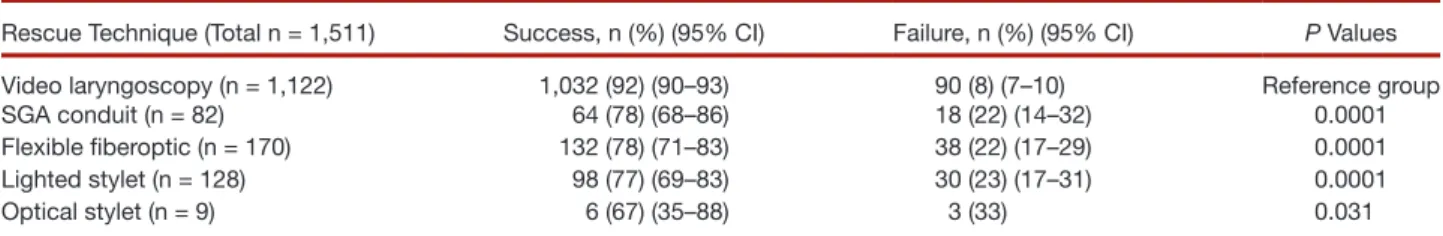
Table 1. Airway Rescue Techniques and Comparative Success Rates of the Common Rescue Strategies
Rescue Technique (Total n = 1,511) Success, n (%) (95% CI) Failure, n (%) (95% CI) P Values Video laryngoscopy (n = 1,122) 1,032 (92) (90–93) 90 (8) (7–10) Reference group
SGA conduit (n = 82) 64 (78) (68–86) 18 (22) (14–32) 0.0001
Flexible fiberoptic (n = 170) 132 (78) (71–83) 38 (22) (17–29) 0.0001
Lighted stylet (n = 128) 98 (77) (69–83) 30 (23) (17–31) 0.0001
Optical stylet (n = 9) 6 (67) (35–88) 3 (33) 0.031
at least 1 min. Finally, a summative count of “higher risk” attempts was recorded and defined by attempts involving
either SpO2 less than 90% for 1 min or longer, concomitant
difficult/impossible bag-mask ventilation or after at least two previous failed attempts at direct laryngoscopy.
Statistical Analysis
Statistical analysis was performed using SPSS version 21 (USa). To determine if there was a statistically significant difference among the proportions of successful tracheal intu-bations and the five identified groups, a chi-square test was used. To compare the successful tracheal intubation rates with the five devices in the setting of failed direct laryngos-copy and difficult or impossible mask ventilation (secondary outcome), the data were recategorized based upon the stated outcome and chi-square test was used. P < 0.05 was consid-ered statistically significant. Proportions are represented with exact 95% Cis.
a mixed-effects logistic regression model was performed to determine if there was any variation among the individual institutions. The binary outcome was success or failure using video laryngoscopy. due to low sample-size issues, video laryngoscopy was the only model we were able to perform. The fixed effect was having two or more of the preoperative
airway predictors as a binary concept. The random effect was the institution. The variance estimate across the institutions was used to calculate the median odds ratio (MOR). The MOR calculates the variation for a random effect (insti-tution) similar to fixed-effects odds ratios. an MOR of 1
means that there is no variation across the institutions.15
Stata Se version 13 (Chicago, illinois) was used for this por-tion of the analysis. a convenience sample size of the seven MPOG institutions’ clinical volume was chosen due to the absence of existing data regarding direct laryngoscopy rescue rates and usage patterns of rescue devices at these seven cen-ters. On post hoc analysis (using nQuery), when the sample size in each of the five groups is 64, a 0.050-level chi-square test will have 80% power to detect a difference in propor-tions characterized by a variance of proporpropor-tions of 0.006344 and an average proportion of 0.784.
Results
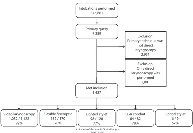
a total of 346,861 cases across seven institutions in the United States were identified that involved attempted or successful tracheal intubation using direct laryngoscopy and had adequate reporting of the necessary airway documen-tation fields (fig. 1). Out of these, 7,259 cases (2%) were identified by automated primary query to involve multiple
Exclusion: Primary technique was
not direct laryngoscopy 2,951 Exclusion: Only direct laryngoscopy was performed 2,881 Intubations performed 346,861 Primary query 7,259 Met inclusion 1,427 Video laryngoscopy 1,032 / 1,122 92% 132 / 17078% Lighted stylet 98 / 128 77% SGA conduit 64 / 82 78% Optical stylet 6 / 9 67%
# of successful attempts / # of attempts % successful
Fig. 1. Flow diagram of case inclusion and airway outcome. Among a large cohort of patients requiring tracheal intubation in the Multicentered Perioperative Outcome Group data set, the “primary query” was identified by electronic search that included air-way management details of interest. After manual review of these records, the final sample of cases was grouped into common airway management rescue techniques after failed direct laryngoscopy. Each case resulted in one or more attempted rescue techniques. Success rates for these techniques are included. SGA = supraglottic airway.
attempts at laryngoscopy and notation of rescue techniques of interest. Manual review of each of these records identified 1,427 cases (20%) that met inclusion criteria of an initial attempt(s) of unsuccessful direct laryngoscopy followed by rescue intervention(s) using some other means. These air-ways were managed by 1,009 distinct anesthesia providers (353 attending anesthesiologists, 449 resident anesthesiolo-gists, and 207 certified registered nurse anesthetists). among the 1,427 cases, there were 1,619 attempts at intubation rescue. The majority of these rescues (n = 1,511 of 1,619; 93%) involved one of the five rescue strategies related to the primary hypothesis (video laryngoscopy, flexible fiberoptic intubation, lighted stylet, optical stylet, and SGa as a con-duit to tracheal intubation). The patients included in the analysis were 60% of male gender and had a mean age of 57 ± 14 yr.
Table 1 summarizes the primary outcome data. Provid-ers most frequently choose video laryngoscopy to rescue the airway (n = 1,122 out of 1,619 attempts; 69%). in far fewer cases, flexible fiberoptic (n = 170; 11%), lighted sty-let (n = 128; 8%), SGa conduit (n = 82; 5%), or optical stylet (n = 9; 0.6%) were chosen as the rescue technique. The SGa conduits were used for tracheal intubation performed either blindly (n = 43) or with the aid of a bronchoscope (n = 39). Other management attempts included a return to direct laryngoscopy again (n = 61; 4%); surgical airway (n = 11; 0.7%); an SGa to maintain ventilation throughout the entire case (n = 26; 2%); waking up the patient followed by awake fiberoptic intubation (n = 8; 0.7%); or case cancel-ation (n = 2; 0.1%). Using video laryngoscopy resulted in a high success rate (92%, 95% Ci, 90 to 93) that was signifi-cantly higher than that for the other four primarily studied rescue techniques: SGa conduit (78%; 95% Ci, 68 to 86; P < 0.001), flexible fiberoptic intubation (78%; 95% Ci, 71 to 83; P < 0.001), lighted stylet (77%; 95% Ci, 69 to 83; P < 0.001), or optical stylet (67%; 95% Ci, 47 to 99; P < 0.001). We demonstrated that there was very small variance (0.2%) of successful video laryngoscopy across the institu-tions when controlling for preoperative airway risk factors. However, the MOR was 1.00, which indicates no significant variation across institutions.
Table 2 lists the different video laryngoscopy systems used to rescue failed direct laryngoscopy (n = 1,122) and their
respective success rates. Most rescues using a video laryngos-copy system (n = 1,003) involved the GlideScope (89%); in
6%, the Storz dCi® or C-MaC® video laryngoscopes (Karl
Storz, Germany); in 4%, the Bullard scope (Circon aCMi,
USa), and in less than 1% the Pentax, McGrath® (aircraft
Medical, United Kingdom), and airtraq systems. The suc-cess rates of the three most frequently used video laryngos-copy techniques were similar (90 to 92%). The frequency of the use of other devices was very low in this sample, which precluded a meaningful comparative analysis.
Figure 2 illustrates the proportional increase in the use of video laryngoscopy for the rescue of failed direct laryn-goscopy during the period that is reflected in these data. in contrast, the use of flexible fiberoptic intubation or optical stylets in this event has proportionally waned.
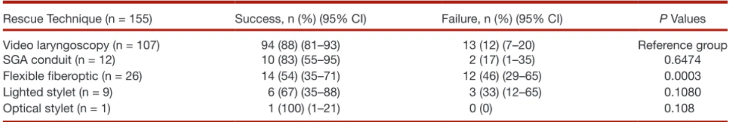
Table 3 summarizes the results from a subgroup analysis of those cases of failed direct laryngoscopy that also involved difficult or impossible mask ventilation (n = 145/1,427; 10%). Similar to the results for the whole sample, video laryngoscopy was chosen most frequently for the attempt to rescue failed direct laryngoscopy (69%). Video laryngoscopy resulted in a higher success rate (88%; 81 to 93) than flexible fiberoptic intubation (54%; 35 to 71; P = 0.0003).
When video laryngoscopy failed as rescue means (n = 90; 8%), the airway was most often successfully secured when using flexible fiberoptic intubation (n = 30) or by return to direct laryngoscopy (n = 29), often with the use of a bougie (n = 15).
Table 4 summarizes the number of failed direct laryn-goscopy attempts before conversion to any of the five rescue techniques of interest. The majority of rescue intubations occurred after one failed direct laryngoscopy attempt in 1,023 of 1,511 cases (68%). For 78% (1,116 of 1,427) of these studied failed direct laryngoscopy cases, information was available for the type of direct laryngoscopy blade used. in 606 of 1,116 (54%) cases, only a Macintosh blade was used, whereas in 180 of 1,116 (16%) cases, only a Miller blade was used. in 330 of 1,116 (30%) cases, both blades were used during the initial attempt. after only Macintosh laryngoscopy, alternatives were approached after one attempt in 463 of 606 cases (76%).
Table 5 describes the details of the preoperative airway examination recorded and episodes of hypoxemia associated
Table 2. Video Laryngoscopy Devices Used and Comparative Success Rates
Device Used (n = 1,238) Rescue Success, n (%) 95% CI Comparison of Device vs. GlideScope, P Values
GlideScope (n = 1,122) 1,032 (92) 90–93 Reference group
C-MAC/Storz DCI (n = 66) 61 (92) 83–97 0.907*
Bullard (n = 40) 36 (90) 77–96 0.645*
Pentax AWS (n = 7) 7 (100) N/A 1.000†
McGrath (n = 1) 1 (100) N/A 1.000†
Airtraq (n = 2) 1 (50) 9–91 0.155†
with airway management. depending on the variable and contributing institution, 79% (n = 1,122) of the 1,427 cases of failed direct laryngoscopy had preoperative airway exami-nations available for review. The final tally for data comple-tion demonstrated that 54 to 63% of the 1,427 cases had valid preoperative airway examination details, depending on the variable. Based on the available data, 28% (n = 313 of 1,122) of the cases where direct laryngoscopy failed had two or more predetermined predictors of difficult direct laryn-goscopy recorded. an episode of hypoxemia as defined by
SpO2 of less than 90% for at least 1 min around the time
of intubation was observed in 372 of 1,511 (25%) rescue attempts. a total of 782 of 1,511 (52%) cases were consid-ered “higher risk” as defined by an episode of hypoxemia, or after difficult/impossible mask ventilation, or after two failed previous attempts at direct laryngoscopy.
acute complications were rare: pharyngeal injury was reported in 12 cases, and all occurrences were associated with the use of video laryngoscopy as rescue (n = 12/1,122; 1.1%). Pharyngeal injuries noted frank blood coming from the mouth (n = 8 GlideScope, n = 1 airtraq, n = 1 C-MaC)
or a laceration to the tonsillar pillar (n = 1 GlideScope) or a laceration to the epiglottis (n = 1 GlideScope). dental injury was reported in four cases (0.3%). Tracheal trauma was not documented for any patient.
Discussion
This multicentered, retrospective observational study has revealed important new information. First, the use of video laryngoscopy in the setting of failed direct laryngoscopy is associated with a statistically significant higher success rate than the other commonly performed alternative techniques, such as flexible fiberoptic intubation, intubation through SGa devices, or optical stylets. Second, the use of video laryngoscopy, predominantly with the GlideScope during the period studied, has become the preferred method to achieve successful tracheal intubation after this event. Third, video laryngoscopy retains a high success rate after failed tracheal intubation by initial direct laryngoscopy combined with difficult/impossible mask ventilation. Fourth, the data con-firm that failure of initial direct laryngoscopy remains difficult to predict, as only 28% of the affected patients had more than
Fig. 2. Rescue techniques attempted over time. This diagram illustrates the proportional use of each studied rescue technique of interest over time. During this period, the use of video laryngoscopy has substantially grown, while the use of all other rescue techniques has proportionally decreased. SGA = supraglottic airway.
Table 3. Airway Rescue Techniques and Comparative Success Rates in Patients with Difficult or Impossible Mask Ventilation Rescue Technique (n = 155) Success, n (%) (95% CI) Failure, n (%) (95% CI) P Values Video laryngoscopy (n = 107) 94 (88) (81–93) 13 (12) (7–20) Reference group
SGA conduit (n = 12) 10 (83) (55–95) 2 (17) (1–35) 0.6474
Flexible fiberoptic (n = 26) 14 (54) (35–71) 12 (46) (29–65) 0.0003
Lighted stylet (n = 9) 6 (67) (35–88) 3 (33) (12–65) 0.1080
Optical stylet (n = 1) 1 (100) (1–21) 0 (0) 0.108
Table 4.
Pr
evious Number of Attempts at Dir
ect Laryngoscopy Befor
e Rescue T echniques of Inter est No. of Pr evious Dir ect
Laryngoscopy Attempts/ Blades Used
Video Laryngoscopy (n = 1122) Flexible Fiber optic (n = 170) Lighted Stylet (n = 128) Supraglottic Airway (n = 82) Optical Stylet (n = 9) No. of Attempts Total (%) Success (%) Failur e (%) Total (%) Success (%) Failur e (%) Total (%) Success (%) Failur e (%) Total (%) Success (%) Failur e (%) Total (%) Success (%) Failur e (%) 1 760 (68) 699 (92) 61 (8) 112 (66) 85 (76) 27 (24) 92 (72) 67 (73) 25 (27) 53 (65) 40 (75) 13 (25) 6 (67) 5 (83) 1 (17) 2 253 (23) 237 (94) 16 (6) 29 (17) 25 (86) 4 (14) 30 (23) 27 (90) 3 (10) 15 (18) 12 (80) 3 (20) 1 (11) 1 (100) 0 (0) 3 88 (8) 77 (88) 11 (13) 24 (14) 19 (79) 5 (21) 7 (5) 4 (57) 3 (43) 12 (15) 10 (83) 2 (17) 1 (11) 0 (0) 1 (100) 4 20 (%2) 18 (90) 2 (10) 3 (2) 1 (33) 2 (67) 2 (2) 2 (100) 0 (0) 0 (0) 0 (0) 0 (0) 5 1 (0) 1 (1) 0 (0) 1 (1) 1 (100) 0 (0) 1 (11) 0 (0) 1 (100) 6 1 (1) 1 (100) 0 (0) Blade type (fr om 1,181 of 1,485 cases) Macintosh only 509 477 (94) 32 (6) 55 45 (82) 10 (18) 38 26 (68) 12 (32) 26 21 (81) 4 (19) 1 0 (0) 1 (100) Miller only 149 134 (90) 15 (10) 26 15 (58) 11 (42) 15 6 (40) 9(60) 10 7 (70) 3 (30) 0 0 (0) 0 (0) Both 246 216 (88) 30 (12) 49 43 (88) 6 (12) 26 22 (85) 4 (15) 27 19 (70) 8 (30) 4 2 (50) 2 (50) Table 5.
Airway Management Conditions During Rescue Attempts
Intubation Condition Video Laryngoscopy (n = 1,122) Flexible Fiber optic (n = 170) Lighted Stylet (n = 128) Supraglottic Airway (n = 82) Optical Stylet (n = 9) Total (%) Success (%) Failur e (%) Total (%) Success (%) Failur e (%) Total (%) Success (%) Failur e (%) Total (%) Success (%) Failur e (%) Total (%) Success (%) Failur e (%) Hypoxemia (Sp O2 < 90% for at least 1 min) 226 (20) 191 (85) 35 (15) 64 (38) 43 (67) 21 (33) 41 (32) 27 (66) 14 (34) 38 (46) 29 (76) 9 (24) 3 (33) 2 (67) 1 (33) Anticipated dif ficulty (>2 pr edictors of dif ficulty) 219 (20) 205 (94) 14 (6) 49 (29) 38 (78) 11 (22) 29 (23) 22 (76) 7 (24) 13 (16) 11 (85) 2 (15) 3 (33) 2 (67) 1 (33) Higher risk 540 (48) 490 (91) 50 (9) 109 (64) 79 (72) 30 (28) 73 (57) 56 (77) 17 (23) 53 (65) 42 (79) 11 (21) 7 (77) 4 (57) 3 (43)
Higher risk = summary of events that involved either hypoxemia for at least 1
min, dif
ficult or impossible bag-mask ventilation, or occurr
ed after at least two pr
evious dir
two classical predictors for difficult airway. Finally, despite its multiple risks, complications were rare after failed initial direct laryngoscopy with a 1% risk for pharyngeal injury when video laryngoscopy was attempted during rescue.
Our findings are the result of systematic analysis of a large number of perioperative tracheal intubations from sev-eral large anesthesia practices. The resulting data are highly relevant because they not only allow the inclusion of a uniquely large number of failed initial direct laryngoscopies (n = 1,427) and the analysis of their subsequent manage-ment, but they also reflect the current practice of periopera-tive airway management practice in tertiary medical centers across the United States. To our knowledge, this is the larg-est and most diverse study of its kind, and we consider the results highly relevant for clinical anesthesiologists and the field of perioperative airway management at large.
in essence, our new findings quantify the success of video laryngoscopy in routine clinical practice: video laryn-goscopy is used in the vast majority of airway rescue events where initial direct laryngoscopy has failed and its use is associated with a high likelihood of success. This success rate was maintained during times of increased urgency such as during threatened or actual failed ventilation, after mul-tiple failed direct laryngoscopy attempts, and in the setting of hypoxemia. additionally, we found that the use of video laryngoscopy for the rescue of failed initial direct laryn-goscopy has increased over the past decade from less than 30% in 2004 to over 80% of rescues more recently (fig. 2). it appears that clinical practice may be gravitating toward a reduction in the number of laryngoscopy attempts, as these rescues mostly occurred after only one failed direct laryngoscopy attempt. although persistence with direct laryngoscopy may have resulted in ultimate success, our data suggest that in recent years, providers are avoiding this practice. However, the data do not allow us to deter-mine why practitioners now more frequently prefer video laryngoscopy over other rescue strategies. We speculate that this preference reflects today’s widespread availability of video laryngoscopy, an anticipated high success rate, and growing comfort and familiarity with using this technique. Nevertheless, we consider it a practice improvement that the growing use of video laryngoscopy is associated with a reduced incidence of multiple attempts at direct laryn-goscopy. This work builds on previous studies examining rescue techniques after failed direct laryngoscopy that had limited relevance due to single-center data, small number of providers, limited sample size, or lack of comparisons. Our new findings confirm those of the existing studies, sug-gesting that video laryngoscopy rescues initial failed direct
laryngoscopy with success rates between 80 and 95%.4,5,11,16
Similarly, SGas have been previously proposed as effec-tive rescue means when used as a conduit for intubation with reported success rates of 87 to 94% in 23 or 15 cases,
respectively.17,18 a previous prominent single-center study
evaluating that 12,225 patients proposed a novel difficult
airway algorithm incorporating video laryngoscopy was limited to only 29 failed direct laryngoscopy events
per-formed by 15 anesthesiologists.6 For the first time, we
have a multicenter perspective on the performance of the new-generation video laryngoscope and alternate intuba-tion techniques. Furthermore, after a center-effects analysis, we observed little variance across institutions regarding the success rate of video laryngoscopy in rescuing failed direct laryngoscopy. We believe that this data set demonstrates that modern day video laryngoscopy is used with a high suc-cess of tracheal intubation when initial direct laryngoscopy fails. However, despite the very large number of cases ana-lyzed in this study, interpretation of comparative success rates is limited since the choice of the rescue device was not randomized but rather at the discretion of each provider. it is possible that specific patient features, personal preferences, or immediate availability have biased the practitioner’s decision to use one rescue technique over another.
Our observation of a high use of video laryngoscopy (with a maintained high rate of successful intubation rescue) in the presence of difficult or impossible mask ventilation describes a practice in variance to existing guidance. The current failed ventilation pathway of the american Society of anesthesiologists airway algorithm suggests consideration
of an SGa.3 This suggestion is based upon expert opinion
and supplemented by a single small study predating modern video laryngoscopy in which the use of the SGa restored ventilation in 16 of 17 cases of difficult mask ventilation
and difficult intubation.7 Our study similarly reports 10
cases of SGa airway rescue with two reported failures to suc-cessfully intubate in this setting. in eight of these cases, a tracheal tube was effectively placed, and in one case, the SGa was used for definitive airway management. However, it is surprising that in contrast to established guidelines, pro-viders more frequently (n = 107 of 155; 69%) chose to use video laryngoscopy in the setting of difficult or impossible ventilation rather than other efforts to restore ventilation, and this practice retained a high success rate of successful tracheal intubation of 88% (n = 94 of 107). indeed, the low incidence of using SGas to restore ventilation when difficult intubation and difficult ventilation are encountered echoes the findings of a recent MPOG study specifically examining
this event.19
although our data are informative regarding the effective-ness of intubation rescue devices, they must be cautiously applied to individual patient care. Provider experience, device availability, and patient-specific airway and cardio-pulmonary features must drive the choice of rescue device. These data are impactful because they significantly advance our knowledge of success for the techniques analyzed in a practice setting that is diverse and allow provider choice among different rescue strategies. Nevertheless, our results do not preclude other practice settings from achieving high rescue success rates with alternate strategies that are well established in those environments.
We recorded 12 cases of pharyngeal injury in this data set and all occurred when video laryngoscopy was attempted after the failure of initial direct laryngoscopy. Pharyngeal injury during primary airway management with video
laryngoscopy has been described previously.20–26 While
pre-vious reports indicate a low incidence, our analysis indicates an increased risk of pharyngeal injury of 1% (12 of 1,122 cases) when video laryngoscopy is used after failed direct laryngoscopy. Moreover, based on the study design, it can-not be ruled out that true incidence is higher since the data did not include postoperative observations or longer term outcome data. While the true incidence remains unclear, video laryngoscopy may require specific precautions to reduce the risk of pharyngeal injury. in particular, providers should focus attention on the oral and pharyngeal cavities during blade insertion and/or tube placement, not just on the video screen.
Our analysis has several limitations. First, we could not determine provider experience with the given devices since the record remains unclear as to which of the pro-viders failed with direct laryngoscopy and which one of them was involved in any of the subsequent intubation attempts. Procedural experience of the laryngoscopist cannot be used to further interpret the data. Second, the data provided regarding hypoxemia cannot be precisely coincided with the intubation event, but rather was gath-ered in association with the time of intubation, as docu-mented by the anesthesia provider. Consequently, it is unclear whether rescue techniques were chosen because of hypoxemia or if the hypoxemia was a result of persis-tence with a given rescue technique. Third, data about the results of preoperative airway assessment were not avail-able for every study patient. Therefore, it is not possible to conclude whether or not all failed direct laryngosco-pies represented patients with unanticipated difficult air-ways. Fourth, the data did not allow interpretation of the timing, type, or dosage of muscle relaxation around the time of airway interventions. Fifth, post hoc sample size estimates confirm that the study may not have been suf-ficiently powered to detect outcome differences between all groups. Finally, the retrospective observational nature of this study limits the interpretation of the frequency of airway rescue and the rescue success rates observed in this study population.
in summary, we found that video laryngoscopy was asso-ciated with a higher rate of successful tracheal intubation compared to the other commonly performed techniques in the perioperative practice of large tertiary care aca-demic medical centers. Furthermore, video laryngoscopy (especially use of the GlideScope) was the most frequent technique chosen to rescue failed direct laryngoscopy. in contrast to current recommendations, we found video laryngoscopy to be used frequently for airway rescue when difficult mask ventilation occurred after failed direct laryn-goscopy. Nevertheless, its use was associated with a high
success rate for rescue. We found the use of video laryn-goscopy in rescue larynlaryn-goscopy to be associated with a 1% risk for pharyngeal injury. These findings may help guide equipment provision and clinical use when managing cases of failed direct laryngoscopy. The data may also serve as evidence when reviewing existing airway algorithms or developing new guidelines. Further research is necessary to identify specific factors of patients and provider experience that might determine airway rescue success when using specific devices.
Acknowledgments
The authors would like to thank the members of Periopera-tive Clinical Research Committee from the Multicenter Peri-operative Outcomes Group (Ann Arbor, Michigan; members are listed in the Appendix).
Research Support
Supported by a Foundation for Anesthesia Education and Research Medical Student Anesthesia Research Fellowship (Schaumburg, Illinois; to Dr. Willett).
Competing Interests
Dr. Aziz and Dr. Brambrink have received research fund-ing from Karl Storz Endoscopy (El Segundo, California). Dr. Aziz has also received honoraria for speaking from this vendor. Dr. Healy is a paid scientific advisor to Brio Device LLC (airway device development) (Ann Arbor, Michigan). The other authors declare no competing in-terests.
Correspondence
Address correspondence to Dr. Aziz: Mail Code KPV 5A, 3181 Sam Jackson Park Rd., Portland, Oregon 97239. azizm@ohsu. edu. This article may be accessed for personal use at no charge through the Journal Web site, www.anesthesiology.org.
References
1. Peterson GN, Domino KB, Caplan RA, Posner KL, Lee LA, Cheney FW: Management of the difficult airway: A closed claims analysis. ANesthesiOLOGy 2005; 103:33–9
2. Mort tC: emergency tracheal intubation: Complications asso-ciated with repeated laryngoscopic attempts. Anesth Analg 2004; 99:607–13, table of contents
3. Apfelbaum JL, hagberg CA, Caplan RA, Blitt CD, Connis Rt, Nickinovich DG, hagberg CA, Caplan RA, Benumof JL, Berry FA, Blitt CD, Bode Rh, Cheney FW, Connis Rt, Guidry OF, Nickinovich DG, Ovassapian A; American society of Anesthesiologists task Force on Management of the Difficult Airway: Practice guidelines for management of the difficult airway: An updated report by the American society of Anesthesiologists task Force on Management of the Difficult Airway. ANesthesiOLOGy 2013; 118:251–70 4. Aziz MF, Healy D, Kheterpal S, Fu RF, Dillman D, Brambrink
AM: Routine clinical practice effectiveness of the Glidescope in difficult airway management: An analysis of 2,004 Glidescope intubations, complications, and failures from two institutions. ANesthesiOLOGy 2011; 114:34–41
5. Asai t, Liu eh, Matsumoto s, hirabayashi y, seo N, suzuki A, toi t, yasumoto K, Okuda y: Use of the Pentax-AWs in
293 patients with difficult airways. ANesthesiOLOGy 2009; 110:898–904
6. Amathieu R, Combes X, Abdi W, housseini Le, Rezzoug A, Dinca A, slavov V, Bloc s, Dhonneur G: An algorithm for difficult airway management, modified for modern optical devices (Airtraq laryngoscope; LMA Ctrach™): A 2-year pro-spective validation in patients for elective abdominal, gyne-cologic, and thyroid surgery. ANesthesiOLOGy 2011; 114:25–33 7. Parmet JL, Colonna-Romano P, horrow JC, Miller F, Gonzales
J, Rosenberg h: the laryngeal mask airway reliably provides rescue ventilation in cases of unanticipated difficult tracheal intubation along with difficult mask ventilation. Anesth Analg 1998; 87:661–5
8. Bein B, yan M, tonner Ph, scholz J, steinfath M, Dörges V: tracheal intubation using the Bonfils intubation fibre-scope after failed direct laryngoscopy. Anaesthesia 2004; 59:1207–9
9. Maharaj Ch, Costello JF, McDonnell JG, harte Bh, Laffey JG: the Airtraq as a rescue airway device following failed direct laryngoscopy: A case series. Anaesthesia 2007; 62:598–601 10. shippey B, Ray D, McKeown D: Use of the McGrath
videolar-yngoscope in the management of difficult and failed tracheal intubation. Br J Anaesth 2008; 100:116–9
11. Noppens RR, Möbus s, heid F, schmidtmann i, Werner C, Piepho t: evaluation of the McGrath series 5 videolaryngoscope after failed direct laryngoscopy. Anaesthesia 2010; 65:716–20 12. Freundlich Re, Kheterpal s: Perioperative
effective-ness research using large databases. Best Pract Res Clin Anaesthesiol 2011; 25:489–98
13. Kheterpal s: Clinical research using an information system: the Multicenter Perioperative Outcomes Group. Anesthesiol Clin 2011; 29:377–88
14. Han R, Tremper KK, Kheterpal S, O’Reilly M: Grading scale for mask ventilation. ANesthesiOLOGy 2004; 101:267
15. Larsen K, Merlo J: Appropriate assessment of neighborhood effects on individual health: integrating random and fixed effects in multilevel logistic regression. Am J epidemiol 2005; 161:81–8
16. Malin e, Montblanc Jd, ynineb y, Marret e, Bonnet F: Performance of the Airtraq laryngoscope after failed conven-tional tracheal intubation: A case series. Acta Anaesthesiol scand 2009; 53:858–63
17. Ferson DZ, Rosenblatt Wh, Johansen MJ, Osborn i, Ovassapian A: Use of the intubating LMA-Fastrach in 254 patients with difficult-to-manage airways. ANesthesiOLOGy 2001; 95:1175–81
18. Combes X, Le Roux B, suen P, Dumerat M, Motamed C, sauvat s, Duvaldestin P, Dhonneur G: Unanticipated dif-ficult airway in anesthetized patients: Prospective valida-tion of a management algorithm. ANesthesiOLOGy 2004; 100:1146–50
19. Kheterpal s, healy D, Aziz MF, shanks AM, Freundlich Re, Linton F, Martin LD, Linton J, epps JL, Fernandez-Bustamante A, Jameson LC, tremper t, tremper KK; Multicenter Perioperative Outcomes Group (MPOG) Perioperative Clinical Research Committee: incidence, pre-dictors, and outcome of difficult mask ventilation combined with difficult laryngoscopy: A report from the Multicenter Perioperative Outcomes Group. ANesthesiOLOGy 2013; 119:1360–9
20. Aziz MF, Abrons RO, Cattano D, Bayman eO, swanson De, hagberg CA, todd MM, Brambrink AM: First-attempt intubation success of video laryngoscopy in patients with anticipated difficult direct laryngoscopy: A multicenter ran-domized controlled trial comparing the C-MAC D-Blade ver-sus the Glidescope in a mixed provider and diverse patient population. Anesth Analg 2016; 122:740–50
21. Cooper RM: Complications associated with the use of the GlideScope videolaryngoscope. Can J Anaesth 2007; 54:54–7 22. Cross P, Cytryn J, Cheng KK: Perforation of the soft palate
using the Glidescope videolaryngoscope. Can J Anaesth 2007; 54:588–9
23. hirabayashi y: Pharyngeal injury related to Glidescope videolaryngoscope. Otolaryngol head Neck surg 2007; 137:175–6
24. Hsu WT, Hsu SC, Lee YL, Huang JS, Chen CL: Penetrating injury of the soft palate during Glidescope intubation. Anesth Analg 2007; 104:1609–10; discussion 1611
25. Nestler C, Reske AP, Reske AW, Pethke h, Koch t: Pharyngeal wall injury during videolaryngoscopy-assisted intubation. ANesthesiOLOGy 2013; 118:709
26. Vincent RD Jr, Wimberly MP, Brockwell RC, Magnuson Js: soft palate perforation during orotracheal intubation facilitated by the Glidescope videolaryngoscope. J Clin Anesth 2007; 19:619–21
Rescue of Failed Direct Laryngoscopy
Appendix
Study group members of Multicenter Perioperative Outcomes Group are as follows: ana Fernandez-Bustamante, M.d., Ph.d., associate Professor, department of anesthesiology, University of Colorado, aurora, Colorado; leslie C. Jameson, M.d., associate Professor, department of anesthesiology, University of Colorado, aurora, Colorado; daniel a. Biggs, M.d., associate Professor, department of anesthesiology, University of Oklahoma Health Sciences Center, Oklahoma City, Oklahoma; Jonathan Wanderer, M.d., Ph.d., assistant Professor, department of anesthesiology, Vanderbilt University Medical Center, Nashville, Tennessee; Jerry l. epps, M.d., associate Professor, department of anesthesiology, University of Tennessee Graduate School of Medicine, Knoxville, Tennessee; Robert M. Craft, M.d., Professor and Chair, depart-ment of anesthesiology, University of Tennessee Graduate School of Medicine, Knoxville, Tennessee; Mitchell F. Berman, M.d., Profes-sor, department of anesthesiology, Columbia University Medical Center, New York, New York; Kevin l. Wethington, M.d., Profes-sor, department of anesthesiology, University of Utah, Salt lake
City, Utah; Nathan l. Pace, M.d., M.Stat., Professor, department of anesthesiology, University of Utah, Salt lake City, Utah; Wil-liam C. Paganelli, M.d., Ph.d., associate Professor, department of anesthesiology, University of Vermont College of Medicine, Burlington, Vermont; Wilton Van Klei, M.d., Ph.d., Professor, department of anesthesiology, University Medical Center, Utrecht, Netherlands; Peter Fleischut, M.d., associate Professor, depart-ment of anesthesiology, Weill Cornell Medical College, New York, New York; Timothy Morey, M.d., Professor, department of anesthesiology, University of Florida Medical School, Gainesville, Florida; Marcel durieux, M.d., Ph.d., Professor, department of anesthesiology, University of Virginia School of Medicine, Char-lottesville, Virginia; Bhiken Naik, M.B.B.Ch., assistant Profes-sor, department of anesthesiology, University of Virginia School of Medicine, Charlottesville, Virginia; Bala Nair, Ph.d., assistant Professor, department of anesthesiology, University of Washington Medical School, Seattle, Washington; Robert Schonberger, M.d., assistant Professor, department of anesthesiology, Yale School of Medicine, New Haven, Connecticut.
Copyright © 2016, the American Society of Anesthesiologists, Inc. Wolters Kluwer Health, Inc. All Rights Reserved. Anesthesiology 2016; 125:656-66
insertbreak after "series-text/italic"
Inspired by Shakespeare’s The Tempest, an unknown artist painted ca.1780 the Bard’s characters Duke Prospero (center), his daughter Miranda (left) and the spirit Ariel (right). Also from The Tempest, the brother of the King of Naples, Sebastian, observed that Prospero’s brother “dost snore distinctly; There’s meaning in … snores.” The author of Shakspeare [sic] and the Bible, nitrous oxide pioneer G.Q. Colton (1814–1898) used the snoring of partial airway obstruction to judge whether his Manhattan patients were anesthetized deeply enough by “Colton gas” for dental extraction. And quoting the title character from Shakespeare’s Henry IV, Colton had no intent to administer “vapours … to strangle” or smother hapless patients. (Copyright © the American Society of Anesthesiologists’ Wood Library-Museum of Anesthesiology.)
George S. Bause, M.D., M.P.H., Honorary Curator, ASA’s Wood Library-Museum of Anesthesiology, Schaumburg, Illinois, and Clinical Associate Professor, Case Western Reserve University, Cleveland, Ohio. [email protected].