HAL Id: tel-02879562
https://tel.archives-ouvertes.fr/tel-02879562
Submitted on 24 Jun 2020
HAL is a multi-disciplinary open access archive for the deposit and dissemination of sci-entific research documents, whether they are pub-lished or not. The documents may come from teaching and research institutions in France or abroad, or from public or private research centers.
L’archive ouverte pluridisciplinaire HAL, est destinée au dépôt et à la diffusion de documents scientifiques de niveau recherche, publiés ou non, émanant des établissements d’enseignement et de recherche français ou étrangers, des laboratoires publics ou privés.
Sandra Bos
To cite this version:
Sandra Bos. Undestanding the viral molecular factors involved in Zika virus pathogenicity in humans. Health. Université de la Réunion, 2019. English. �NNT : 2019LARE0005�. �tel-02879562�
Université de la Réunion
Ecole doctorale ″Sciences, Technologies et Santé" E.D. 542
THESE DE DOCTORAT
Présentée en vue de l’obtention du grade de :
DOCTEUR EN BIOLOGIE, MEDECINE, SANTE
Spécialité : Virologie
UNDERSTANDING THE VIRAL MOLECULAR
FACTORS INVOLVED IN ZIKA VIRUS
PATHOGENICITY IN HUMANS
SANDRA BOS
Soutenue le 18 Avril 2019
Composition du Jury :
Pr. Diane GRIFFIN Johns Hopkins Bloomberg School of Public Health Rapporteur
Dr. Jean DUBUISSON Institut Pasteur de Lille Rapporteur
Dr. Catherine CETRE-SOSSAH CIRAD, UMR ASTRE Examinateur
Dr. Gilles GADEA INSERM, UMR PIMIT Directeur de thèse
Pr. Marie-Lise GOUGEON Institut Pasteur Co-directeur de thèse
À vous,
qui m’avez tout appris
Et à vous que j’estimais beaucoup
et qui, même à bout de souffle,
avez su me faire rire
« Science is a way of thinking
much more than it is a body of knowledge »
REMERCIEMENTS
Avant toute chose, je voudrai remercier chaque entité ayant consciemment ou inconsciemment participé à ce travail. Tout au long de ma thèse j’ai pu bénéficier du soutien indéfectible de mes proches et rencontrer des gens venus des quatre coins du monde, qui m’ont inspirée, enseignée, épaulée, et encouragée dans mes recherches. Amis, Paul, Zoé, Dorian, B2, Famille.s, Arnaud, collaborateurs et collègues, professeurs et néophytes à qui j’ai pu faire découvrir ma passion pour la Virologie, je n’aurai pas assez de ces quelques lignes pour vous écrire un petit mot à tous mais je tiens sincèrement à vous dire merci !
‒‒
Je tiens également à remercier Patrick Mavingui, directeur de l’unité PIMIT qui m’a accueillie pour mon doctorat, ainsi que Diane Griffin, Jean Dubuisson et Catherine Cetre-Sossah d’avoir accepté d’être rapporteur ou examinateur de ma thèse.
Dear Diane, I particularly want to express my profound respect and gratitude to you. Since we met, you have always been there for me and available during my visits to Baltimore. Many thanks for coming to my defense. Having you on my jury is a great privilege for me.
‒‒
Je voudrai particulièrement remercier mes directeurs et encadrants de thèse, pour leur confiance et leur bienveillance à mon égard et saluer leurs qualités pédagogiques, humaines, et scientifiques. Nul ne peut rêver d’un meilleur soutien et encadrement. Tout au long de ces années, vous m’avez offert une multitude d’opportunités, la possibilité d’assister à des congrès, de nous y représenter seule, mais également une grande liberté vis-à-vis de mes recherches.
Gilles, je suis fière d’avoir été ta première thésarde. Grâce à toi j’ai appris la vie en équipe, la rigueur de la paillasse, et le « on » non-participatif ! Plus
sérieusement, j’ai adoré travailler avec toi. Au-delà d’être mon directeur de thèse tu as également été un « lab partner » idéal ! Je ne sais même plus combien de fous rire on a eu, mais repenser à la pipette (qui coule) du P2+ suffit à me refaire rire. J’espère sincèrement que notre collaboration continuera après mon doctorat.
Philippe, vous représentez tellement pour moi. Apprendre à vos côtés fut un honneur et je ne vous remercierai jamais assez pour tout le précieux temps que vous m’avez accordé. Je ne connaissais rien aux Flavivirus avant de vous rencontrer, ni même à la Virologie. Les yeux pétillants de la passion qui vous anime, vous m’avez enseigné tant de choses. Outre vos qualités scientifiques indéniables, votre humanité est admirable. J’espère du plus profond de moi pouvoir devenir une virologue aussi respectable, et perpétuer tout ce que vous m’avez appris avec autant de passion et de patience.
Marie Lise, vous êtes également une grande dame. Merci d’avoir accepté la co-direction de ma thèse, de m’avoir accueillie à l’Institut Pasteur, soutenue, et fait découvrir votre domaine de recherche durant ces quelques mois. Cette période peut sembler courte, surtout lorsqu’elle est rapportée à l’échelle d’une vie où elle représenterait un peu moins de 1%, mais elle est tout de même équivalente à la fréquence des pDCs dans le sang ; lorsque l’on voit tout ce qu’elles sont capables de faire on se rend compte que moins de 1% c’est important ! Et, qui sait, je continuerai sans doute (plutôt peut-être ?) à étudier les pDCs car malgré la difficulté inhérente à leur rareté et leur culture, c’est un challenge que j’ai beaucoup aimé relever. Enfin, travailler à l’Institut fut un honneur, et c’est avec mille plaisirs que j’y venais chaque matin, boostée par une pensée à ceux et celles qui étaient là avant moi.
‒‒
Enfin, Papa, Maman, Gaëlle, Papis et Mamies, je ne serai rien sans l’amour et les valeurs que vous m’avez tous donné. Qu’importe la couleur de notre ciel, vous m’avez supporté (dans les deux sens du terme) et tant appris. Aujourd’hui ni moi, ni ce travail, n’aurions été ce que nous sommes sans vous.
― PREFACE ―
Avant de commencer cette préface de thèse tout à fait inhabituelle, évidemment, je voudrais vous poser cette question : le hasard existe-t-il ? Les rencontres que l’on fait, les choses qui nous arrivent, les livres que l’on lit et les virus que l’on découvre, ne sont-ils pas des rendez-vous, des synchronicités ?
La rencontre. Tout a commencé lors d’un anodin trajet en covoiturage avec mon encadrant de stage de Master 1 : “ David, j’ai découvert un virus incroyable ! C’est un Flavivirus, il s’appelle Zika. Il a l’air différent des autres, il peut faire ça, ou ça, et encore ça ! C’est incroyable tu ne trouves pas ? Je veux absolument en apprendre plus !”. Par chance le Pr Philippe Desprès, dont je ne connaissais alors que vaguement le nom apposé sur des publications scientifiques, venait d’arriver au laboratoire pour diriger l’équipe I2T ; l’équipe sœur de DySIIS, à laquelle j’étais rattachée à ce moment-là. Grâce à David, j’ai pu rencontrer Philippe et discuter un moment avec lui. Bien décidé à en savoir plus sur Zika je lui ai demandé s’il ne connaissait pas quelqu’un qui travaillait sur ce virus chez qui je pourrai postuler pour mon stage de master. Et, comme vous devez vous en douter, ce quelqu’un c’était lui. Car oui, Philippe avait emporté Zika dans ses valises. L’Aventure commença.
The proof of concept. C’est la boule au ventre mais pleine de motivation que je suis arrivée au laboratoire pour mon premier jour de stage. Je ne connaissais rien à la Virologie ni à ses techniques, et je me souviens que “plaque forming assay”, ma technique favorite, sonnait à ce moment-là comme un mot issu d’une langue totalement incompréhensible. A part ça tout se passait bien jusqu’à ce que Philippe me dise que ce n’est pas lui qui allait m'encadrer mais son collègue Gilles Gadea. Maintenant je peux dire que je n’étais pas vraiment super emballée par cette nouvelle ; et il me semble que toi non plus Gilles tu ne l’étais pas... Finalement, le choix du chef était le bon (comme toujours n’est-ce pas ?) et je crois que l’on a réussi à former une super
équipe. Après tout, comment cela aurait-il pu être autrement quand il s’agit de deux êtres matinaux, organisés et pleins de tocs ?
Durant mes débuts au laboratoire, une parole de Philippe résonnait dans ma tête : “La Virologie c’est comme la cuisine. Soit tu as le truc, soit tu ne l’a pas. C’est elle qui te choisit, pas l’inverse.” Quel stress ! Moi qui au plus profond de moi avait le sentiment d’être faite pour ça. Et si je m’étais trompée ? Si je ne l’avais pas, ce truc ? De toute façon il n’y avait qu’un seul moyen de le savoir :
essayer. Et puis, le 15 mars 2016, le petit Zika-GFP est né. Même Philippe, qui
l’a pourtant conçu, ni croyait pas. Grâce à ce clone, je me suis rendu compte combien j’aimais voir les virus pousser et combien cela m'émerveille. Comme
l’a dit Richard Dawkins : “There's real poetry in the real world.” Et cette poésie
elle était là, sous mes yeux.
La thèse. J’ai pleuré de joie pendant des heures (littéralement) lorsque j’ai compris que j’avais été reçue au concours de la bourse MENRT. Après toutes les épreuves surmontées, avoir l’opportunité de faire une thèse représentait beaucoup pour moi. C’était comme me délester de tous les poids qui me pèsent et ouvrir la porte sur un nouvel Univers : un Univers où je me sens légère et à ma place, et où mes différences sont une force. J’allais enfin avoir la possibilité de faire mes preuves, de devenir chercheur. Ce sentiment reste encore très vif aujourd’hui et difficile à décrire mais j’étais profondément heureuse et reconnaissante d’avoir cette chance. Et puis ma chance ne s’est pas arrêtée là, faisant de ma thèse une aventure humaine et scientifique exceptionnelle dont j’ai beaucoup appris. Tout au long de ces années, j’ai pu rencontrer et travailler avec des gens adorables qui ont cru en moi et ont tout fait pour m’aider et m’encourager dans mes recherches et mon futur. Mais, au-delà de tout, j’ai eu la chance de bénéficier d’un encadrement en or que je souhaite à chaque thésard.
C’est dans un contexte exceptionnel, en plein cœur de l'épidémie de Zika, que j’ai réalisé ma thèse. Car nous, à I2T, nous avons connu Zika avant le grand Boum, pendant, et maintenant que les choses se calment. Au début très peu de données étaient disponibles dans la littérature mais depuis notre caillou
du bout du monde, nous commencions déjà à apprendre à connaître la souche de Polynésie Française. Lorsqu’il explosa, et devint la « star des médias » début 2015, nous étions là, à la fois acteurs et spectateurs, au milieu de ce tourbillon. Nous vivions et faisions la recherche, les découvertes en “live”. En quelques mois à peine, de nouvelles données étaient publiées. Des données à lire, à trier, qui renforçaient nos hypothèses, en soulevaient de nouvelles, ou donnaient une nouvelle perspective d’analyse de nos résultats. C’était incroyable et tellement passionnant. Comprendre ce virus au jour le jour fut un énorme challenge que de nombreuses équipes de recherche partout dans le monde ont relevé. Aujourd’hui une productivité scientifique titanesque résulte de cet effort ; productivité à laquelle nous avons humblement participé.
ABBREVIATIONS
ADE : Antibody-dependent enhancement
Ae. : Aedes
ATP : Adénosine triphosphate
BR15 : Zika virus strain BeH819015
C : Capsid protein
CCR-7 : C-C chemokine receptor type 7
CD : Cluster of differenciation
CDC : Center for disease control and prevention
CDP : Common dendritic cell progenitor
CHIKV : Chikungunya virus
CLPs : Common lymphoid progenitors
CLR : C-type lectin receptor
CMPs : Common myeloid progenitors
CS : Conserved sequence
CZS : Congenital Zika syndrome
Da : Dalton
DC : Dendritic cells
DENV : Dengue virus
DNA : Deoxyribonucleic acid
E : Enveloppe protein
ED : Ectodomain
EID : Emerging infectious disease
ELISA : Enzyme linked immunosorbent assay
ER : Endoplasmic reticulum
FLT3L : FMS-like tyrosine kinase 3 ligand
GBS : Guillain-Barré syndrome
GFP : Green fluorescent protein
HAVcr-1 : Hepatitis A virus cellular receptor
HIV : Human immunodeficiency virus
ICTV : International committee on taxonomy of viruses
IFN : Interferon
IFNAR : Interferon-α/β receptor
Ig : Immunoglobulin
IL : Interleukines
ILT : Immunoglobulin-like transcript
IRAK : Interleukin-1 receptor-associated kinase
IRF : Interferon regulatory factor
ISA : Infectious sub-genomic amplicons
ISFV : Insect specific flaviviruses
ISG : Interferon stimulated gene
KFDV : Kyasanur Forest disease virus
LRR : Leucine-rich repeat
M : Membrane protein
MBFV : Mosquito-borne flaviviruses
mDC : myeloid DC
MERS : Middle East respiratory syndrome
MHC : Major histocompatibility complex
MMR : Macrophage mannose receptor
mRNA : messenger RNA
Mtase : Methyltransferase activity
M-TBFV : Mammalian tick-borne flaviviruses
NK : Natural killer lymphocyte
NKV : No-known vector
NLS : Nuclear location signal
NPC : Neuronal progenitor cells
NS : Non-Structural protein
ODN : Oligodeoxynucleotides
OHFV : Omsk Hemorrhagic Fever virus
ORF : Open reading frame
PAMP : Pathogen associated molecular pattern
PBMC : Peripheral blood mononuclear cell
PCR : Polymerase chain reaction
pDC : Plasmacytoid dendritic cell
pH : Hydrogen potential
POWV : Powassa virus
PRNT : Plaque reduction neutralization test
PRR : Pattern recognition receptor
pTalpha : pre-T cell receptor alpha
RCP : Representative concentration pathway
RCS : Repeated conserved sequence
RdRp : RNA-dependent RNA polymerase activity
RLRs : RIG-I-like receptors
RNA : Ribonucleic acid
ROS : Reactive oxygen species
RT-PCR : Reverse-transcriptase polymerase chain reaction
SARS : Severe acute respiratory syndrome
sfRNA : Subgenomic flaviviral RNA
SL : Stem-loop
SPOV : Spondweni virus
STAT : Signal transducer and activator of transcription
S-TBFV : Seabird tick-borne flaviviruses
TAK : transforming-growth-factor-β-activated kinase
TBEV : Tick-borne Encephalitis virus
TBFV : Tick-borne flaviviruses
TGN : Trans-Golgi Network
TIR : Toll-IL-1 receptor
TLR : Toll-like receptor
TM : Transmembrane
TNF : Tumor necrosis factor
TRAF : TNF receptor associated factor
TRAIL : TNF-Related Apoptosis-Inducing ligand
Treg : regulatory T lymphocyte
UTR : Untranslated transcribed region
WHO : World health organization
WNV : West Nile virus
Xrn-1 : 5'-3' exorivonuclease 1
YFV : Yellow Fever virus
LIST OF TABLES
Table 1: Selected arboviruses of medical importance ______________________________________ 21 Table 2: Reported suspect Zika and GBS cases per location ________________________________ 60 Table 3: Determinants of viral pathogenesis and diseases _________________________________ 64 Table 4: Factors responsible for cell injuries _______________________________________________ 66 Table 5: Zika virus cellular targets and receptors ___________________________________________ 68 Table 6: Human cell lines permissive to Zika virus infection ________________________________ 69 Table 7: Cellular antiviral responses against Zika virus infection ____________________________ 74 Table 8: Phenotypic characterization of human myeloid and plasmacytoid dendritic cells __ 169 Table 9: Zika virus counteraction of antiviral response ____________________________________ 220 Table 10: Overview of Zika virus-induced cellular pathogenesis ___________________________ 221
LIST OF FIGURES
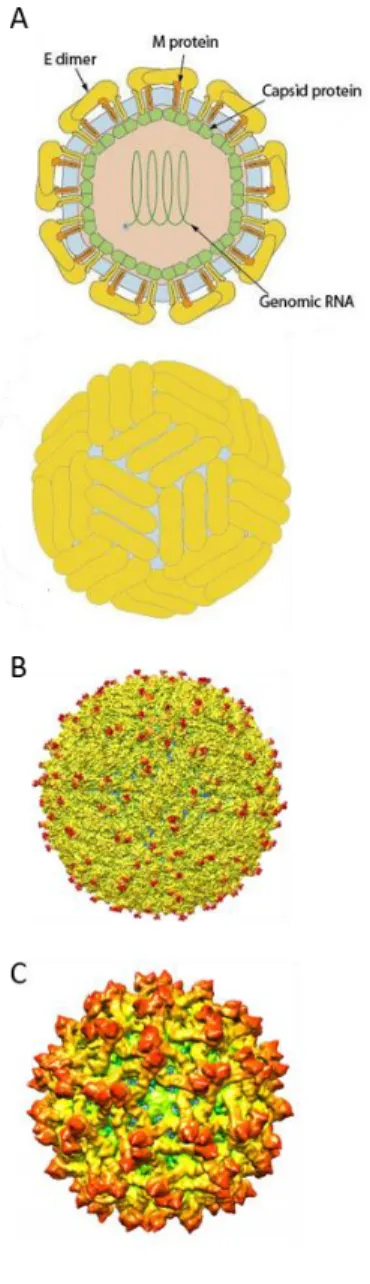
Figure 1: Classification of viruses from capsid shape to viral RNA ___________________________ 2 Figure 2: The convergence model __________________________________________________________ 4 Figure 3: Climate change and health ________________________________________________________ 8 Figure 4: Projected climate changes _______________________________________________________ 10 Figure 5: Health maps _____________________________________________________________________ 17 Figure 6: Distribution of extreme poverty in the world ______________________________________ 18 Figure 7: Post-earthquake Zika virus surge ________________________________________________ 19 Figure 8: Global distribution of emerging and re-emerging arboviruses _____________________ 20 Figure 9: Predicted global distribution of Ae. aegypti and Ae. albopictus. ____________________ 26 Figure 10: Phylogenetic reconstruction of Flaviviridae ______________________________________ 29 Figure 11: Structure of Zika virus __________________________________________________________ 32 Figure 12 : Flavivirus genome structure and protein expression ____________________________ 34 Figure 13 : Flavivirus life cycle _____________________________________________________________ 36 Figure 14: How Zika virus spread to the Americas __________________________________________ 46 Figure 15: How Zika virus enters the human population ____________________________________ 54 Figure 16: Zika symptoms _________________________________________________________________ 56 Figure 17: Zika virus vaccine platforms in clinical studies __________________________________ 57 Figure 18: Time until the clearance of Zika virus RNA ______________________________________ 58 Figure 19: Congenital Zika syndrome cases in Brazil _______________________________________ 60 Figure 20: Zika virus tropism ______________________________________________________________ 67 Figure 21: Schematic diagram of extrinsic antibody-dependent enhancement of infection __ 76 Figure 22: Notable amino acid changes since ZIKV discovery ______________________________ 79 Figure 22: Overall structure of ZIKV C protein _____________________________________________ 106 Figure 24: Comparison of ZIKV C with other known flavivirus C structures _________________ 106 Figure 25: Flavivirus particle at different maturation stages _______________________________ 108
Figure 28: Surface-exposed amino acids conservation in flavivirus E protein ______________ 112 Figure 29: The fusogenic conformational change of the E protein during cell entry _________ 113 Figure 30: Diverse functions of plasmacytoid dendritic cells _______________________________ 173 Figure 31: Activation pathway in plasmacytoid dendritic cells responding to nucleic acids _ 177 Figure 32: Signaling of CpG ODN classes in different endosomal compartments ___________ 179 Figure 33: Contact between infected cells and plasmacytoid dendritic cells ________________ 181
LIST OF CONTENT
INTRODUCTION
INTRODUCTION TO VIRUSES AND EMERGENCE ______________________________ 1 Viruses ______________________________________________________________________ 1 I1.1. Definition of Viruses ___________________________________________________ 1 I1.2. Classification system _________________________________________________ 1 Emergence of infectious diseases and viruses _______________________________ 3 I2.1. Microbial adaptation __________________________________________________ 3 I2.2. Human susceptibility to infection ______________________________________ 5 I2.3. Climate and Ecological changes _______________________________________ 8 I2.4. Human behaviors and economic development ______________________ 12 I2.5. Breakdown or absence of Public Health measures ___________________ 17 ARBOVIRUSES TO FLAVIVIRUSES __________________________________________ 20 The threat of Arboviruses _________________________________________________ 20 II1.1. The cladistic paradox of Arboviruses ________________________________ 22 II1.2. Epidemiology of Human Arbovirosis _________________________________ 22 II1.3. The emergence triangle of mosquito-borne arboviruses _____________ 25 FLAVIVIRUSES ___________________________________________________________ 28 II2.1. Flavivirus burden on Human Health _________________________________ 28 II2.2. Classification and Phylogeny of Flaviviruses _________________________ 30 MOLECULAR BIOLOGY OF FLAVIVIRUSES ________________________________ 32 II3.1. Virion and Genome Structure ________________________________________ 32 II3.2. Viral Cycle ___________________________________________________________ 35 II3.3. Features and Role of the Viral Proteins ______________________________ 38 II3.4. Evolutionary advantage of Flaviviruses ______________________________ 42 THE FIERY TALE OF ZIKA VIRUS ___________________________________________ 45 Classification ____________________________________________________________ 45 Emergence and Global spread____________________________________________ 45 III2.1. Zika in the Pacific and Asia __________________________________________ 47
Zika virus Ecology ________________________________________________________ 52 III3.1. Host and Reservoir __________________________________________________ 52 III3.2. Vectors and vector-borne transmission _____________________________ 53 III3.3. Non vector-borne transmission _____________________________________ 54 Clinical features of Zika Fever ____________________________________________ 56 III4.1. Symptomatology ____________________________________________________ 56 III4.2. Treatment ___________________________________________________________ 56 III4.3. Diagnosis and Detection ____________________________________________ 58 III4.4. Complications _______________________________________________________ 59 Social Impact ____________________________________________________________ 62 UNRAVEL THE PUZZLE OF ZIKA VIRUS PATHOGENICITY ___________________ 63 Reminder on Viral Pathogenesis _________________________________________ 63 IV1.1. Terminology and Principle of viral pathogenesis _____________________ 63 IV1.2. Pathogenic Mechanisms and determinants _________________________ 65 First insight on Zika virus Pathogenesis __________________________________ 67 IV2.1. Target cells and tissues _____________________________________________ 67 IV2.2. Attachment factors and Entry Receptors ____________________________ 70 IV2.3. Host responses to Zika virus infection _______________________________ 73 Viral molecular factors involved in Zika virus pathogenesis _______________ 77 IV3.1. Molecular epidemiology _____________________________________________ 78 IV3.2. Objective of the Doctoral Research __________________________________ 81
PART I : DEVELOPMENT OF MOLECULAR VIROLOGY TOOLS
FOR THE STUDY OF ZIKV BIOLOGY AND PATHOGENICITY
RATIONALE ______________________________________________________________ 96 ARTICLE n°1 ____________________________________________________________ 98
PART II : CONTRIBUTION OF ZIKV STRUCTURAL PROTEINS
FEATURES OF ZIKA VIRUS STRUCTURAL PROTEINS _____________________ 105 Capsid ________________________________________________________________ 105 Pr and Membrane _____________________________________________________ 107 I2.1. The role of prM in the maturation process _________________________ 107 I2.2. Particle heterogeneity _____________________________________________ 108 I2.3. Feature of ZIKV prM _______________________________________________ 109 Envelope ______________________________________________________________ 110 I3.1. Feature of ZIKV E protein __________________________________________ 112 I3.2. Potential impact on ZIKV entry and cellular tropism ________________ 112 I3.3. The role of ZIKV E in the fusion process ____________________________ 114 Investigate the impact of ZIKV structural proteins _____________________ 114 ARTICLE n°2 ___________________________________________________________ 117 ARTICLE n°3 __________________________________________________________ 126 ARTICLE n°4 __________________________________________________________ 147
PART III : ZIKV IMPACT ON PLASMACYTOID DENDRITIC
CELLS ANTIVIRAL RESPONSE
INTRODUCTION TO PLASMACYTOID DENDRITIC CELLS _________________ 167 Overview of Dendritic Cells ____________________________________________ 167 I1.1. Main features of Dendritic cells ____________________________________ 167 I1.2. Dendritic cell diversity _____________________________________________ 168
Plasmacytoid Dendritic Cells: Sentinels and Orchestrators of the
Antiviral Immune Response ______________________________________________ 170 I2.1. Chronology of the plasmacytoid dendritic cells discovery __________ 170 I2.2. Ontogeny of pDCs _________________________________________________ 171 I2.3. Distribution of plasmacytoid dendritic cells ________________________ 172 I2.4. Physiological role of plasmacytoid dendritic cells __________________ 172 Virus detection and IFN production signaling pathway _________________ 175
Flavivirus infection and Plasmacytoid dendritic cell response __________ 180 I4.1. Indirect sensing of flavivirus infection ______________________________ 180 I4.2. Plasmacytoid dendritic cell activation upon flavivirus infection _____ 182 ARTICLE n°5 ___________________________________________________________ 189
DISCUSSION
Asian versus African ZIKV lineages: Can less be more? ____________________ 216 Phenotypic differences between ZIKV African and Asian lineage ________ 216 The unforeseen effect of ZIKV structural proteins _______________________ 218 Molecular determinant of differential binding rate _______________________ 218 Determinants of ZIKV virulence ___________________________________________ 220 The E-glycosylation: a key factor for a broad diffusion __________________ 221 Is there only one mutation in prM involved in microcephaly? ____________ 223 188V: ZIKV boarding pass by NS1 ______________________________________ 225 Concluding remarks: Did Zika mutate to cause severe diseases in humans and outbreaks? ___________________________________________________________________ 226 Personal remarks _________________________________________________________ 228
ANNEX
Annex n°1 ________________________________________________________________ 233 Annex n°2 ________________________________________________________________ 235 Annex n°3 ________________________________________________________________ 237― INTRODUCTION TO VIRUSES AND EMERGENCE ―
― 1―
INTRODUCTION
TO
VIRUSES
AND
EMERGENCE
Viruses
Viruses are everywhere, in every imaginable corner of the planet and have been living on Earth for hundreds of millions of years. Long before humans discovered and accepted their existence, at the end of the 19th century, viruses and infectious diseases shaped the history of Humanity; and will undoubtedly continue to do so. Nonetheless, humans, at all times, expressed reservations about them. Does a pathogen smaller than a bacterium exist? Are the viruses alive? What is the Origin of viruses, where do they come from? If some questions have been answered, the contemporary History of Virology, still enlivened by persistent and unanswered debates, illustrates the enigmatic nature of viruses. I1.1. Definition of Viruses
More than in any other field, terminologies in virology are not an easy exercise as it aims to set boundaries on phenomena that remain misconceived; which sometimes lies on the cusp of philosophy of biology and Science. Viruses are not exempted from that, and have always been difficult to define since their discovery.
Viruses are obligate parasites, infecting all living organisms, and including themselves. They do not possess the ability to capture and store energy. From this arises the fundamental characteristic of their absolute dependence on a living host for reproduction. This way, a “virus” can be defined as an infectious agent composed of an RNA or DNA genome that replicates only within the cells of living hosts. It is an organism producing virions - namely a particle - that protects viral genome during the extracellular phase and allows viruses to infect new cells. I1.2. Classification system
›
The Classical SystemIn 1962, Lwoff, Robert Horne, and Paul Tournier, proposed a comprehensive scheme for the classification of all viruses (bacterial, plant, and animal) under the classical Linnaean hierarchical system consisting of phylum, class, order, family, genus, and species. The major principle of this classification is that viruses
― 2―
should be grouped according to their shared properties rather than the one of the cells or organisms they infect. A second principle was a focus on the nucleic acid genome as the primary criterion for classification. Four characteristics were used for the classification of viruses: (i) nature of the nucleic acid genome in the virion, (ii) symmetry of the protein shell (capsid), (iii) presence or absence of a lipid membrane (envelope) and (iv) dimensions of the virion and capsid.
However, the International Committee on Taxonomy of Viruses (ICTV) did not adopt this system in toto. Designation of families, genera, and species was applied in both the scientific and medical literature but was only used for the classification of animal viruses (plant virologists use group names derived from the prototype virus of each group).
›
The Baltimore Classification SystemFrancis Crick conceptualized the central dogma in which cellular genes are encoded in a double stranded nucleic DNA that will be converted into working proteins carrying out all the functions necessary for life. To be done, information in DNA is first transcribed into a messenger RNA (mRNA) molecule, then mRNAs are transported to the cytoplasm where they are translated by ribosomes and
C.
Figure 1: Classification of viruses from capsid shape to viral RNA
(A) Diversity of capsid shape. H helicoidal HE helicoidal enveloped icosahedral IE icosahedral enveloped C complex. Legend: blue =capsid, red = genome, grey = envelop, orange = glycoproteins (science photo library)
(B) Virus structure in colored transmission electron micrographs. 1 tobacco mosaic virus, helicoidal structure, unenveloped, 2 Yellow fever virus polyhedral structure, enveloped (Dennis Kunkel microscopy). 3 Bacteriophage T4, complex structure (science photo library)
(C) The Baltimore classification of viruses. This classification was created by David Baltimore, based on the nature of the viral genome and is on the method of viral mRNA synthesis. ss = single stranded; ds = double stranded (Figure source: Wikipedia commons)
― INTRODUCTION TO VIRUSES AND EMERGENCE ―
― 3―
associated machinery into proteins. But, because viral protein synthesis is completely dependent on the cell’s translational machinery, all viruses must direct the synthesis of mRNA to produce proteins. According to the obligatory relationship between the viral genome and its mRNA, David Baltimore proposed an alternative classification that groups viruses into families, depending on their type of genome, strand polarity, and their method of replication. This classification provides virologists with immediate insight into the steps that must take place to initiate replication and expression of viral genome.
Emergence of infectious diseases and viruses
In the book "A Planet of Viruses", Carl Zimmer wrote: "Viruses are the smallest living thing known to science, yet they hold the entire planet in their sway"1.
However, many of them will remain unknown and will never emerge, raising the question of which parameters facilitate their introduction and spread in human populations.
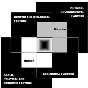
Emerging infectious diseases (EIDs) are defined as diseases caused by an infectious origin and whose incidence in humans has increased over the last two decades or is likely to increase in the near future. In the past 30 years, more than 30 new infectious diseases have been reported highlighting the growing threat of EIDs to populations. The emergence and spread of infectious diseases are driven by the convergence of a complex set of factors that promote the initiation of an epidemic process. To understand which variables contribute to these
phenomenon, Smolinski et al developed a model, “The Convergence Model”,
which illustrates how factors related to (i) genetic and biology, (ii) physical environment, (iii) ecology, and (iv) social, political and economic status, can
impact on the human-pathogen interaction2. The following paragraphs will
address some of the factors that contribute to the emergence of infectious diseases, particularly viral diseases.
I2.1. Microbial adaptation
In the "Germ Theory" and Koch's postulates, the scientists of the time assumed that the diseases were caused by a "fixed" microbe species, monomorphic and invariant. Currently, the tremendous mutation capacity of microbes under
― 4―
selection pressures is well established and can be an important determinant in the emergence or resurgence of many infectious diseases.
Microbes developed many sophisticated survival strategies that allow them to co-evolve with their host and environment, as well as to protect themselves from degradation by the immune system.
Among these strategies, several
microbes have developed an ability to exchange or incorporate new genetic material into their own genome. This horizontal gene transfer, or lateral transfer, is well described in bacteria and is recognized as a driving process involved in the acquisition of antibiotic
resistance genes3. However, recent
studies suggest that this process is not limited to this domain of life. Indeed, we now suspect a viral genus, a priori non-pathogenic to humans, the Mimiviruses, to be capable of such a process, as the acquisition of the topoisomerase gene would suggest4.
Another way for microbes to adapt very quickly to their environment lies in the speed of the mutation process. The champions in this field are viruses, especially RNA viruses which exhibit mutation rates that are higher in order of magnitude
than any other replicative entity5. For instance, it is estimated that the mutation
rate of RNA viruses is up to a million times higher than their hosts, the record
being held (to the best of our knowledge) by the Bacteriophage Qβ with ~10-3
mutations per nucleotide per replication cycle6. If RNA viruses are probably the
most intriguing biological entities for studying mutation rates, it's because they Figure 2: The convergence model
At the center of the model is a box representing the convergence of factors leading to the emergence of an infectious disease. The interior of the box is a gradient flowing from white to black; the white outer edges represent what is known about the factors in emergence, and the black center represents the unknown (similar to the theoretical construct of the “black box” with its unknown constituents and means of operation). Interlocking with the center box are the two focal players in a microbial threat to health—the human and the microbe. The microbe–host interaction is influenced by the interlocking domains of the determinants of the emergence of infection: genetic and biological factors; physical environmental factors; ecological factors; and social, political, and economic factors.
(Figure and caption from the book "Microbial Threats to Health: Emergence, Detection, and Response")
― INTRODUCTION TO VIRUSES AND EMERGENCE ―
― 5―
encode their own replication machinery. As a result, they are able to optimize their mutation rate for their fitness (compared to DNA viruses that use hosts polymerases). This incredible mutation capacity, inherently high, combined with their replication rate yields a progeny that differs from the parents by one or two
mutations7, and thus generates a mutant cloud of descendants. Interestingly, the
increase and decrease in the mutation rate of a virus leads to a reduction in the
virulence of the viral population8. These results suggest a close relationship
between the mutation rate of a virus, the diversity created in a viral population, and pathogenesis in an infected host. The perpetual genetic variation of microbes
gives them a wide range of strategies to bypass the immune system9. Some of
these include the antigenic variation, the hiding from the immune system10
(either by masking key surface antigens or by coating their surface with compounds mimicking host tissue to prevent recognition as "nonself"), the mechanisms to downregulate immune system and finally the ability to cause latent infection.
Overall, the high evolutionary potential of microbes makes them organisms able to adapt and develop resistance to even the most potent therapies. Nowadays, microbial adaptation seriously challenges our therapeutic response capacity and stands as the main obstacle to the development of protective vaccines and the discovery of new effective drugs.
I2.2. Human susceptibility to infection
If pathogens evolve, so do their hosts. The human body, like a fortress, is full of barriers to prevent invasion by pathogens, which have been selected and conserved by hundred thousand years of co-evolution. These barriers are physical, such as the skin and mucous membranes, or cellular and molecular through the immune system and its effectors. Susceptibility to infection can occur when these defenses are by-passed, altered or compromised by the following factors.
›
Transmission routeInfectious agents are transmitted from one host to another by specific means mainly determined by the site of excretion and the physiological stability of a
― 6―
pathogen. Transmission routes play an important role in the fate of a pathogen and its ability to induce disease. While some pathogens have to deal with a limited number of transmission possibilities, others may use several routes to infect their hosts.
Transmission routes are divided into two main types. (i) Horizontal transmission refers to the spread of a pathogen to other organisms of the same or different species by non-hereditary means. This category includes the vectorial transmission by which an infectious agent is transmitted by the bite of an infected hematophagous vector, usually an insect. In this case, the pathogen is injected directly into the subcutaneous tissues and blood, thus bypassing the first defense of the host: the skin. (ii) Vertical transmission refers to the transfer of a pathogen between parent and offspring. It includes the transmission that occurs during pregnancy, when the infectious agent crosses the placental barrier, or during birth. When an agent is transmitted as a part of the host genome, as in the case of a retrovirus infection, we talk about germline transmission.
›
Immunity and AgingWhen a foreign organism is detected by the immune system, the innate immune response is induced to eliminate the invader. When this non-specific response is inadequate to control the infection, the adaptive immune response is triggered. The latter one, specific to a given pathogen, is essential to establish the immune memory. Induction and preservation of this response ensures individual immunization against subsequent infections and is the core principle of vaccination. If we consider the example of a viral infection, two major alternatives are possible: either the virus is cleared and the individual is immunized against infection with the same variant, or the virus settles in and persists. Immunization is triggered either following a natural infection or as a result of preventive vaccination. Being aware of this, the prevalence of infected people is a critical parameter in the emergence of viral disease and their epidemic potential, as only the naive immune people will be susceptible to infection.
However, susceptibility to infection varies throughout an individual's life and is strongly influenced by age, with the very young and the elderly at increased risk
― INTRODUCTION TO VIRUSES AND EMERGENCE ―
― 7―
of infection. While young people are at high risk of infection because of a fragile immune status, older people are more vulnerable to infection because of a weakened immune system due to chronic diseases and medical treatments. In addition, the senescence process significantly reduces cell-mediated immunity, immunization level, and impairs host defenses. This is highlighted by recent Dengue fever epidemics, in which a significant increase in the incidence of cases
among the elderly has been reported11.
›
Immunocompromised populationsThanks to medical advances, the life expectancy of patients has considerably been improved in recent years. But this invaluable benefit is not always priceless and some therapies used to treat or limit the progression of a disease reduce the overall immune capacity of the patients. The most striking example is cancer, whose burden is steadily increasing. Based on data from the World Health Organization, it is estimated that the global incidence of cancer will reach 29.5
million in 2040 compared to 18 million today12. These patients, usually
undergoing heavy treatment such as chemotherapy, are in addition to an ever-growing population of people living with HIV (human immunodeficiency
viruses)13. This upsurge in immunocompromised people is concomitant with the
emergence of opportunistic pathogens, some of which, previously uncommon or
unrecognized as pathogenic for humans (e.g. Aspergillus spp.14), are now
extending the list.
›
Genetic polymorphismEach of us has a different capacity for immunological reactivity and not all of us are equal against a pathogen. Indeed, individuals and populations are more or less susceptible to infection depending on their genetic determinants. J.B.S. Haldane was the first to suggest that people living in areas where a disease is historically persistent and endemic, such as malaria-laden areas, evolved
genetically in order to enhance their ability to survive15. Research conducted in
recent decades supports this hypothesis and allows the discovery of new important genetic determinants. For instance, it’s now known that the “S” allelic variant of hemoglobin, which, if homozygous, causes sickle cell disease, confers
― 8―
Furthermore, the persistence of this allele, despite its potential disadvantage, attests to the potent selection pressure exerted by pathogens.
I2.3. Climate and Ecological changes
In general, environmental changes, both climatic and physical, have a large influence on the transmission dynamics and spread of microbes. Indeed, these perturbations directly impact the biology of pathogens, and the behavior of their hosts. Environmental factors are usually deeply involved in the emergence of vector-borne diseases, which are among the most sensitive.
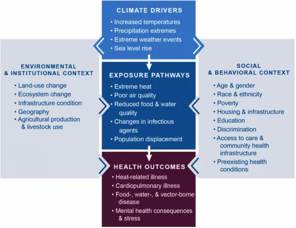
Figure 3: Climate change and health
Conceptual diagram illustrating the exposure pathways by which climate change affects human health. Exposure pathways exist within the context of other factors that positively or negatively influence health outcomes (gray side boxes). Key factors that influence vulnerability for individuals are shown in the right box, and include social determinants of health and behavioral choices. Key factors that influence vulnerability at larger scales, such as natural and built environments, governance and management, and institutions, are shown in the left box. All of these influencing factors can affect an individual’s or a community’s vulnerability through changes in exposure, sensitivity, and adaptive capacity and may also be affected by climate change (Figure and caption from “U.S. Global Change Research Program" section Health 2016)
― INTRODUCTION TO VIRUSES AND EMERGENCE ―
― 9―
›
Weather and ClimateThe seasonal occurrence of certain infectious diseases, in particular respiratory and gastrointestinal diseases, rightly underlines the role of climate. Some pathogens display a different sensitivity to humidity or temperature, for example, which may be involved in the seasonal pattern of infection. It has been demonstrated that the influenza A virus, causing the flu, is more effective at low
temperatures and low humidity16. These properties can explain why, during the
winter months, the influenza A virus remains infectious and causes massive flu outbreaks. But, as mentioned above, the range of climate effects is not limited to the pathogen directly, and plays an important role in the biology and distribution of some hosts. Vectors and reservoirs need conditions favorable to their survival that first of all define their own ecological niche and range, but also, as a consequence, those of their infecting pathogens. Thus, geography and climate are a powerful determinant of vector-borne diseases, acting as strong barriers to their distribution; most of the time insurmountable for many species.
›
Change in vector ecologyA vector is a living organism that can transmit pathogens, including viruses, bacteria or parasites, from one vertebrate to another when infected itself. Many vectors are blood-sucking insects, which ingest the pathogen(s) by feeding blood from an infected host and inject it to a new host during subsequent blood meals. Among them are mosquitoes, ticks, fleas, sandflies, flies, triatomine bugs and
some freshwater aquatic snails17.
Each vector can be defined through the part of the environment it inhabits, namely in which it fits and is adapted, as well as through the way it interacts with that environment and the other organisms living there. Interactions between vectors and hosts are highly dependent on environmental and climatic factors, and are the root of vector disease transmission and persistence. Temperature (which determines how long it takes for parasites to develop and the survival of mosquito larvae), precipitation (which creates egg-laying sites), wind speed (which affects feeding frequency), abundance and diversity of vegetation, and alternative hosts (which impacts blood meal rates on humans) are all drivers of pathogen transmission.
― 10―
Figure 4: Projected climate changes
Average of the model projections available for the 2081–2100 period under the evolution scenario of the earth radiative balance RCP2.6 (lower) and RCP8.5 (higher) for (a) change in annual mean surface temperature and (b) change in annual mean precipitation, in percentages, and (c) change in average sea level. Changes are shown relative to the 1986–2005 period. The number of models used to calculate the multi-model mean is indicated in the upper right corner of each panel. Stippling (dots) on (a) and (b) indicates regions where the projected change is large compared to natural internal variability (i.e., greater than two standard deviations of internal variability in 20-year means) and where 90% of the models agree on the sign of change. Hatching (diagonal lines) on (a) and (b) shows regions where the projected change is less than one standard deviation of natural internal variability in 20-year means. (Figure and caption from “Summary for Policymakers of IPCC Report 2013")
― INTRODUCTION TO VIRUSES AND EMERGENCE ―
― 11―
Although the evolution of a pathogen is usually a key factor in the emergence or resurgence of an infectious vector-borne disease, it’s in many cases an environmental co-factor that catalyzes the phenomenon, providing the vector with ideal conditions to ensure its spread. With this in mind, it is natural to worry about the effects of global warming, which could have devastating consequences on the incidence of infectious diseases. Currently, vector-borne diseases account for about 17% of all infectious diseases, with a burden of more than 700,000
deaths per year17, and are estimated to represent about 30% of EIDs over the past
two decades18.
›
Reservoir abundance and distributionIn epidemiology, a reservoir refers to the principal habitat of a pathogen that allows its reproduction and maintenance within an ecosystem. In this way, it can be environmental (e.g. soil for Clostridium tetani and the fungus Histoplasma, or
water for Legionella pneumophila) or animal (including humans and vectors
species) depending on the given pathogen. In this section, particular emphasis will be placed on animal reservoirs, which represent one of the main reservoir of viruses.
Diseases of non-human animal origin, termed zoonosis, account for the majority of the EIDs events. It is estimated that more than 70% of them are caused by pathogens from wildlife, and this number is expected to rise. As a matter of fact, reservoir species are widely subject to climate change, which in some cases affects their abundance and leads to changes in their behaviors. The emergence of a large number of viruses over the past few decades is a clear indication of this, and highlights the growing threat posed by these changes. As an example, many viruses are maintained in migratory species, such as birds or bats, which live closer to humans and have the potential to spread pathogens over long distances. Changes in migration routes or territory of such reservoir species
threaten to occur in response to the global warning19. As a result, the distribution
of the carried-pathogens will be modified and interactions with other species will take place, increasing the risk of transmission and emergence of new pathogens
or variants19,20. Another example illustrating how ecological changes of a
― 12―
Sin Nombre21. In 1993, in the United States, the southwestern region of the
country was hit by an outbreak of acute respiratory distress disease. A lot of Native Americans were affected. The case fatality rate was approximatively 60%; people were dying. To everyone's surprise, a virus belonging to the Hantaviruses, until now not considered as pathogenic for humans, is identified as the etiological agent of the new disease: the virus Sin Nombre. A few years later, the emergence of Sin Nombre virus was correlated with an El Nino Southern Oscillation event in
previous years22. This climatic phenomenon, which caused ample rainfalls and
warm winters, considerably increased the forage availability resulting in a
dramatic high density of deer mouse, the reservoir of Sin Nombre23.
In a same way as for Hantaviruses, a strong link exists between the emergence
and resurgence of Arenaviruses and the density of their rodent reservoir24. The
epidemiological history of these two viral groups is enough to highlight the importance of reservoir populations (including vectors) and the disastrous consequences that ecological changes can have on human health. Political will to create programs and public health strategies for the study and control of such populations would be a significant asset in the fight against these diseases. However, although the mechanisms of transmission of a pathogen to humans are generally well described, the identification of its reservoir(s) is complex and remains unclear in many cases. This lack of knowledge seriously hinders our ability to anticipate outbreaks. More epidemiological surveillance studies in wildlife could help to bridge this gap. Furthermore, it would provide new insights that could improve the scientific community's responsiveness in the event of a new zoonosis emergence, as was the case for Zika.
I2.4. Human behaviors and economic development
In his book “Spillover: Animal Infections and the Next Human Pandemic”, David Quammen argues that the increase in zoonosis observed in the past decades can be directly linked to human behavior and the ways in which we are irrevocably
altering the world’s ecosystems25. At a time of globalization, most activities
related to the economic and demographic development of human populations, from the consumption of natural resources to deforestation, have an impact on the environment and enhance the risk of pathogens emergence. Furthermore,
― INTRODUCTION TO VIRUSES AND EMERGENCE ―
― 13―
international exchanges through travel and commerce facilitate a broad spread of pathogens and vectors throughout the world.
›
Land useNow that there are seven billion of us on the planet, humans are changing land use to find more materials, to build or to produce more food. Indeed, humans have extended their territory into wild areas like never before, multiplying interactions with wildlife and, ultimately, finding new infections. In this context, it seems relevant to mention that a growing number of EIDs arise from increased interactions between humans and animal reservoirs due to land-use change. Deforestation and habitat fragmentation in favor of the expansion of living areas or new crops are behind the emergence or recrudescence of several infectious diseases. Ironically, reforestation efforts can also be the origin of these diseases,
as shown by the emergence of Lyme disease in the United States26,27.
Environmental changes related to water use infrastructure can also be involved in these processes. Dam building and irrigation systems that change water level and flow and create stagnant water pools are, among other things, often associated with the resurgence of mosquito-borne diseases and the spread of
schistosomiasis to new areas28–30.
›
Animal Husbandry and Food IndustryThe ever-growing human population is associated with an expanding need for food and clothes. To meet this demand livestock farming intensifies and farms are established in new areas. These new husbandry practices promote the risks of amplification and emergence of new pathogens enhancing the host-pathogen interaction dynamics. In Australia, the establishment of horse and pig farms in fruit bats living areas led to bats urbanization, due to the degradation of their natural habitat, and the emergence of Hendra and Menangle viruses
respectively31–33. At the same time, livestock populations have grown
exponentially in response to the demand for meat protein. Animal density per feedlot has increased dramatically, as well as the threat of zoonotic disease, the risk of which inevitably increases in proportion to the animal population. China is probably the country that experienced the most spectacular increase in livestock populations in the last few decades. Poultry and pigs are among the
― 14―
most prevalent, both of which are hosts of influenza viruses. Intensive poultry production in confined feedlots is a boon for viral amplification. Moreover, poultry markets are a long-standing tradition in China and attract a large number of Chinese people. On this occasion multiple farmers are gathered to sell their poultry, alive, thereby ensuring the virus maintenance and dramatically increasing the probability of its transmission to humans. This optimal combination certainly explains why most of the influenza pandemics of the 20th century originated in China.
›
Population mobilityThrough the development of new and efficient means of transportation, natural borders, as were the oceans, gradually fade away. The potential for rapid spread of pathogens – and their reservoirs including vectors – around the world is growing as people keep ongoing international travels and expanding global trade markets. Nowadays, an infected host can travel the world, simply by taking a plane, and encounter naive populations that could be extremely susceptible to the carried pathogen. With technological advances, the transportation means are faster and faster and allow to travel long distances within the timeframe of a viremia. In particular, human-to-human infections can easily be spread from one geographical area to another. Pathogens that infect humans asymptomatically or are transmissible before the symptomatic period pose a real threat in the absence of a recognized infection and protective measures. Fortunately, vector-borne diseases are in principle less prone to this dissemination pathway. In most cases, not all the factors necessary for the transmission cycle of the pathogen are present (vectors, insufficient number of subsequent infections). Nevertheless, this property tends to disappear gradually due to the increasing distribution of some vectors, typically mosquitoes.
›
UrbanizationCurrently, more than half of the world's population lives in urban areas34. By
2030, it is estimated that the global urban population will exceed 4.5 billion35 and
almost all of this urbanization will take place in the cities of developing countries. The relocation of rural populations to urban areas is a major demographic trend of the 21st century, yet migration to cities not properly equipped with adequate
― INTRODUCTION TO VIRUSES AND EMERGENCE ―
― 15―
infrastructure has serious health consequences. Population density is a critical parameter for the maintenance of certain virus populations, especially when the host has a viral immune memory. Depending on the virus route of transmission, vectorial or respiratory, to name a few, the potential for interaction is a limiting factor. Person-to-person transmission of some acute viral infections only occurs if the host population is large and interactive. For example, Measles virus can be maintained only in human populations over 200,000 people, most likely because there is no animal reservoir and infected individuals develop complete and long-lasting immunity. Furthermore, urbanization prompts people into frequent travel between large cities and their villages to visit their relatives. In this way, increasing interactions blur the boundaries of both areas and enhance the risk of pathogen transmission. Lastly, many rural migrants live in overcrowded conditions as a consequence of housing costs and family size. Poor sanitary conditions and lack of access to safe water frequently observed in these areas greatly enhance the probability of disease and facilitate their spread.
›
Human High Risk BehaviorsAs described in the previous sections, human behavior, individual or collective, plays a major role in the emergence of infectious diseases. Behavior modification is an essential strategy in the prevention of infectious diseases; and for some, it’s the only option. However, despite considerable efforts deployed to educate populations, high-risk behaviors are still a burden on public health. New cases of sexually transmitted diseases, such as HIV, contracted as a result of unprotected sex or drug injection appear at first sight easily preventable; yet their incidence is persistent. On the other hand, the current mistrust of vaccination and the rise of "anti-vaxxers" have severe consequences for public health. Heavy lobbying by anti-vaccine activists has led to a drop in the vaccination rate in several countries, mainly among children. Ironically, the immunization campaigns were so well conducted that this generation of parents probably never had to worry about the gravity of these diseases. Nevertheless, although they probably do not fully realize the risks involved in refusing vaccination, the latter actually do exist. Such behavior has already led to the re-emergence of vaccine-preventable diseases in
― INTRODUCTION TO VIRUSES AND EMERGENCE ―
― 17―
several countries. The death due to measles infection of more than 30 people in Europe in 2018 is a tragic illustration of this. If the refusal to vaccinate intensifies, populations could face a serious threat, since a breakdown in immunization coverage would facilitate the re-emergence of virtually eradicated diseases such as polio, involving dramatic consequences.
I2.5. Breakdown or absence of Public Health measures
Developing countries bear a disproportionate burden of infectious diseases and emerging events compared to the rest of the world. Poverty creates conditions favorable to the spread of infectious diseases and prevents affected populations from appropriate access to prevention and care. Failure of health systems, instability or lack of political will, as well as natural disasters, are often pointed out as contributing factors to epidemic emergence.
›
PovertyAccording to the latest World Bank report, nearly 1.1 billion people fewer than in 1990 live in extreme poverty36. In 2015 the overall poverty rate dropped to a
record level of 10%. But the decrease was not equal in all regions. While Europe, Central Asia, East Asia and the Pacific, successfully reduced the poverty rate below 3%, the number of poor people in Sub-Saharan Africa has increased. Of the 736 million people still living on less than $1.90 a day, more than half of them lives in Sub-Saharan Africa. Nevertheless, while the fight against extreme poverty is globally on track, one should not forget that nearly half of the world's population - 3.4 billion people - still struggle to meet their basic needs. Actually, it is estimated that more than a quarter of the world's population lives on $3.20 a
day and nearly half of the world's population on less than $5.50 a day37.
From a health point of view, I would suggest that poverty should be estimated in terms of more than just a monetary value. The lack of sufficient food, access to safe drinking water, sanitation and proper medical infrastructure are
Figure 5: Health maps
Evolution of the ProMED health maps from 2016 to 2018. The international society for infectious diseases publishes annual word clouds based on ProMED’s infectious disease surveillance. In the maps the size of each word indicate the number of report but word location does not always correspond to the exact location. (Maps and caption from “ProMed")
― 18―
prerequisites that, if not available, seriously burden human lives. Sanitary poverty is the real plague, opening wide to the emergence of new pathogens and to their persistence in populations.
›
War and Natural disasterWars and natural disasters shake the sanitary conditions of the countries affected, which are in too many cases already precarious. These two situations are not only disastrous, but also create conditions highly conducive to the emergence or resurgence of infectious diseases. Victims of natural disasters and war refugees gather in overcrowded emergency camps to survive. There, sanitary conditions and personal hygiene are very limited, water sources unprotected and often in close proximity to fecal repositories. This chaotic environment has repeatedly been at the core (and is so in Yemen and Zimbabwe currently), of cholera and tuberculosis outbreaks, among others.
EID events are based on multiple factors, the convergence of which leads to epidemics with more severe consequences than if they were dependent on a single factor. The emergence of Ebola virus in Guinea, Liberia and Sierra Leone in 2014 is a recent example. The lack of rapid disease detection, availability of basic care, efficient quarantine, and mobilization of international rescue forces with which people had to cope, demonstrated how the convergence of political instability and poor socio-economic conditions made outbreak management difficult38.
Figure 6: Distribution of extreme poverty in the world
Of the world’s 736 million extreme poor (those living on less than $1.90 a day) in 2015, half of the total lived in just five countries. The five countries with the highest number of extreme poor are (in descending order): India, Nigeria, Democratic Republic of Congo, Ethiopia, and Bangladesh. (Figure and caption from “World Bank.org")
― INTRODUCTION TO VIRUSES AND EMERGENCE ―
― 19―
›
Political willThe financial resources that a country can allocate to health and research are also a key component in the fight against infectious diseases and the prevention of emergence. When insufficient budget can lead to inappropriate use of medical equipment and contribute to the spread of a pathogen (e.g. Lassa fever in Nigeria in 1989 due to the reuse of needles in hospitals39), it can also lead to the
interruption of surveillance and vector control programs. Additionally, governments tend to fund research activities only once the pathogens or associated diseases are an obvious threat and, more especially, when the latter threatens their own populations. This tardive rise in awareness clearly impairs scientific community responsiveness and could be minimized if research programs focusing on "outsider" pathogens were valued.
Lastly, when national economies are highly dependent on tourism, the political will may be lacking to report a miscontrolled outbreak to the WHO. Some examples include Mauritius, which in 2006 alleged in a press release that the danger of Chikungunya spread was “overplayed”, Saudi Arabia, which took a
long time to report the emergence of MERS (Middle East Respiratory Syndrome)40,
and China, which overlooked the emergence of SARS (Severe Acute Respiratory
Syndrome) in 200341.
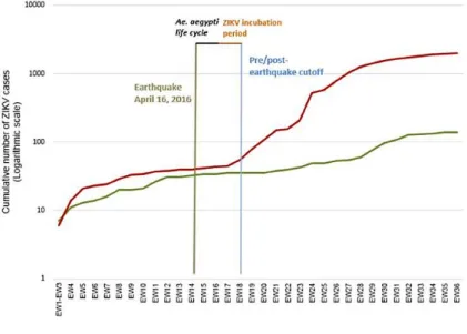
Figure 7: Post-earthquake Zika virus surge
In April 2016, Ecuador experienced a massive 7.8 M earthquake — the strongest seism in almost four decades. Shown are the cumulative number of autochthonous ZIKV cases after a major earthquake (M7.8) that affected Ecuador in 2016. (Cumulative number cases at week 36 of 2016 for mildly affected cantons (green) and severely affected cantons (red). (Figure and caption from Ortiz et al., 2017)
― 20―
ARBOVIRUSES
TO
FLAVIVIRUSES
The threat of Arboviruses
Arboviruses are a very heterogeneous group, well known in public health, as many of them cause high morbidity and mortality in both human and animal populations. The term "arbovirus" refers to a virus transmitted to vertebrate hosts by blood-sucking arthropod vectors. It comes from laboratory jargon used in the early 1940s by Californian researchers in reference to "arthropod-borne-virus"42.
The study of arboviruses was developed at the initiative of the Rockefeller Foundation, with the collaboration of a reference laboratory at Yale University and various laboratories located in tropical countries. The network of Pasteur Institutes from overseas played a central role in this field of research. In 1930, only 6 arboviruses were known. In 1980, 500 had been identified. Currently 537 arboviruses are listed in the international arbovirus catalogue, but this number is likely to be revised upwards in the near future based on the results of studies investigating virosphere.
Figure 8: Global distribution of emerging and re-emerging arboviruses
The color symbols indicate the global distribution of the major pathogenic arboviruses for humans as published in 2012, except for Zika virus. Zika virus distribution (brown) has been updated and represented by a circle in view of its recent emergence. (Figure adapted from Anez et al., 2012)