HAL Id: tel-01149058
https://tel.archives-ouvertes.fr/tel-01149058
Submitted on 6 May 2015
HAL is a multi-disciplinary open access archive for the deposit and dissemination of sci-entific research documents, whether they are pub-lished or not. The documents may come from teaching and research institutions in France or abroad, or from public or private research centers.
L’archive ouverte pluridisciplinaire HAL, est destinée au dépôt et à la diffusion de documents scientifiques de niveau recherche, publiés ou non, émanant des établissements d’enseignement et de recherche français ou étrangers, des laboratoires publics ou privés.
the cerebellar cortex
Antoine Valera
To cite this version:
Antoine Valera. Spatial and temporal integration of granular inputs in the cerebellar cortex. Neuro-biology. Université de Strasbourg, 2013. English. �NNT : 2013STRAJ111�. �tel-01149058�
Thèse de doctorat de l’Université de Strasbourg
Ecole Doctorale des Sciences de la Vie et de la Sante
Spécialité : Neurosciences
Présentée par Antoine VALERA
En vue d’obtenir le grade de docteur de l’université de Strasbourg
Intégration spatiale et temporelle des entrées granulaires dans le cortex cérébelleux
Soutenue publiquement le 28 novembre 2013
Membres du jury
Pr. Henrik JÖRNTELL Rapporteur externe
Dr. Dominique DEBANNE Rapporteur externe
Pr. Rémy SCHLICHTER Examinateur
Dr. Philippe ISOPE Directeur de thèse
Spatial and Temporal Integration of Granular
Inputs in the Cerebellar Cortex
"Un bureau bien rangé est le signe d’un esprit dérangé"
ACKNOWLEDGMENTS
First and foremost, I would like to thank my committee members Professor Henrik Jörntell, Doctor Dominique Debanne and Professor Rémy Schlichter for their precious time.
Merci à toi aussi Philippe pour tout le temps que tu m’as consacré depuis mon master en 2009 (et oui, 5 ans déjà !). Merci de m’avoir transmis ton enthousiasme pour les neurosciences et les analyses compliquées, mais surtout, merci pour la confiance que tu m’as accordée au cours de cette thèse à chaque fois que je voulais essayer une manip ou une analyse. Je crois que c’est cette liberté qui m’a le plus motivé pendant ces années.
Ensuite, je me dois de remercier infiniment la dream team. Merci à Laetitia pour toutes ces années passées à me supporter (depuis la L1, on attaque la 10ème année là !). Je te souhaite plein de bonnes choses pour la suite. Si tu te retrouves à Bale, ne prend pas l’accent suisse! Merci à Joseph, inébranlable, infatigable (sauf le matin), toujours prêt à filer un coup de main ! Et merci pour cette leçon de vie : un bureau peut effectivement rester rangé pendant toute une thèse. Respect ! Merci à ma Padawan, Anaïs, pour la joie, l’enthousiasme et le dynamisme que tu apportes dans notre bureau! Et puis bonne chance avec SynaptiQs... Merci aussi à la petite dernière, Flavia qui m’a sauvé des génotypages (et quand je dis sauvé, je le pense).
Merci aussi à tous les membres de notre chouette équipe. Tout d’abord, Jean-Luc, pour toutes ces passionnantes discussions politiques, culturelles et historiques qu’on a eu au cours de ces années. Et sache que Gustav Mahler m’a dignement accompagné durant cette rédaction! Merci aussi à Fred pour ta bonne humeur permanente, et ton stock secret de nourriture caché dans le placard du bas, porte de droite… (d’ailleurs il est vide, il faudrait que tu refasses les courses). Merci aussi à Bernard pour tes discussions précieuses, et tes connaissances encyclopédiques. Tu auras en tout cas réussi à me faire apprécier l’analyse de variance, et la chasse aux champignons. Merci aussi à tous les autres membres de l’équipe Jeff (prend soin de toi !), Didier, Jean, Jean-Louis. Grazie per la tua motivazione Super Francesca, e ricordati: non tocchi l'occhio!
Merci à tous les autres copains de l’institut avec qui j’ai passé tellement de soirées rock-happy-barbar…Merci à Coco, Pauline, Seb, Edith, David, Vy, Annie, Laureen, Marion, Aurélie, Audrey, JBS, et à tous les copains de la promo de master qui rôdent encore dans le coin ou qui viennent de partir : Alex, Romain, Michael, Paul, Laurent, Vivien, Bruce… Merci à tous ceux que je n’ai pas cité mais avec qui j’ai passé pleins de bons moments !
Merci à mes amis de longue date, que je devrais aller voir plus souvent (et qui me le rappellent régulièrement !) Julien, Noëlie, Tiphaine (nous sommes collègues désormais !), Lowik, Caro, Nono, Hélène, Sarah, Imane. Vous êtes les bienvenus à Londres ! Comme ça on est sûr de se croiser… Pleins de merci à ma toute ma grande famille, et tout particulièrement à mes parents, mon petit frère Vincent, et à mes grands parents! Merci aussi à Armel, Carole et la Clara pour leur soutien. Enfin, merci à ma Charline pour ta patience infinie, ta douceur, la force de tes convictions, et ta compréhension lorsque je rentre aussi tard qu’en ce moment. Zoubi la Nanine!
TABLE OF CONTENTS Acknowledgments ... 3 Table of contents ... 4 Table of figures... 6 Abbreviations ... 7 Summary ... 9 Preface ... 11 Introduction ... 13
A short history of the cerebellar physiology ... 13
First anatomical descriptions ... 13
Early concepts on Cerebellar functions ... 13
1 General anatomy and histology of the cerebellum ... 17
1.1 Cerebellar functions ... 17
1.1.1 Roles of the cerebellum ... 17
1.1.2 Semiology ... 17
1.2 General structure of the cerebellum ... 18
1.2.1 Multiple organisation levels in the cerebellar cortex ... 18
1.2.2 cerebellar inputs: inferior olive and precerebellar nuclei ... 22
1.2.3 cerebellar outputs: cerebellar nuclei and vestibular nuclei ... 22
1.2.4 Inter-species variations and homology ... 24
1.3 Cellular structure of the cerebellar cortex ... 25
1.3.1 Cell types and information pathway ... 25
1.3.2 The Purkinje cell ... 28
1.3.3 The granule cell ... 29
1.3.4 The Golgi cell ... 33
1.3.5 The glomerulus ... 36
1.3.6 molecular layer interneurons ... 36
1.4 Functional connectivity in the cerebellar cortex ... 38
1.4.1 Marr-Albus-Ito theory of the cerebellar cortex ... 38
1.4.2 Silent synapses at the parallel Fibre to Purkinje cell synapse ... 40
1.4.3 Golgi cell functions ... 43
1.4.4 Molecular Layer Interneurons Functions ... 44
2 Spatial organisation of the cerebellum ... 45
2.1 Architectonic variations ... 45
2.2 Organisation of the climbing fibre pathway ... 46
2.2.1 climbing fibre zones and microzones ... 46
2.2.2 Olivo-cortico-nuclear loops ... 48
2.3 Histochemical compartmentation ... 48
2.3.1 Zebrin bands: a marker of Purkinje cells compartmentation ... 49
2.3.2 Histochemical compartmentation of the other cortical cell types ... 52
2.4 mossy fibre afferences and fractured somatotopy ... 54
2.4.1 Topography of mossy fibre inputs ... 54
2.4.2 Matching between mossy fibre inputs and climbing fibre inputs ... 59
2.4.3 Functional relevance of fractured somatotopy ... 61
2.5 From the beam the hypothesis to the patch hypothesis ... 61
2.5.1 The beam hypothesis ... 61
3 High frequency transmission in the cerebellum: short term plasticities and temporal coding ... 69
3.1 Temporal organisation of high frequency cerebellar inputs ... 69
3.1.1 Firing patterns in the mossy fibre à granule cell à Purkinje cell pathway ... 69
3.2 Modulation of Purkinje cell discharge ... 73
3.2.1 Discharge patterns ... 74
3.2.2 Purkinje cells synchrony ... 77
3.3 Short term plasticities at the granule cells to Purkinje cells synapse during high frequency bursts ... 78
3.3.1 Presynaptic Ca2+ and vesicular release ... 78
3.3.2 Short term facilitation and short term depression ... 80
3.3.3 Vesicle refilling ... 82
3.4 A few precisons on the acute slice model ... 82
4 Article I ... 83
4.1 Supplementary introduction to Variance-Mean analysis ... 83
4.1.1 The quantal parameters N, P and Q ... 83
4.1.2 Analytic method ... 86
4.1.3 Multple Probability Fluctuation Analysis... 86
4.2 Presentation of the first article ... 90
4.3 Adaptation of granule cell to Purkinje cell synapses to high-frequency transmission ... 91
4.4 Supplementary results ... 93
4.4.1 Supplementary methods ... 93
4.4.2 Supplementary Results ... 95
5 Article II ... 97
5.1 Presentation of the second article ... 97
5.2 Functional precisions about our region of interest ... 98
5.2.1 Mediolateral and anteroposterior position of the recordings ... 98
5.2.2 Inputs and outputs in our region of interest ... 98
5.3 Cerebellar microzones are coordinated by granule cell inputs ... 102
6 Discussion ... 103
6.1 Heterogeneous synaptic organisation of granule cell inputs ... 103
6.1.1 Purkinje cells are excited by specific hotspots of granule cells ... 103
6.1.2 Hypothesis on the origin of connected hotspots ... 105
6.1.3 Neighbouring cells share input patterns ... 105
6.1.4 Shared patterns between animals ... 106
6.1.5 The Molecular layer interneurons distinct pattern ... 107
6.2 Golgi cells are local interneurons ... 108
6.2.1 Function of the apical dendrites ... 108
6.2.2 Functional relevance of Golgi cells subpopulations ... 109
6.3 High frequency bursts allow reliable information transfer ... 109
6.3.1 Reluctant vesicles allow Reliable transmission at the parallel fiber to Purkinje cell synapse ... 110
6.3.2 Fast vesicle replenishment allows sustained transmission of the signal ... 111
6.3.3 On beam bursts influence the cerebellar output ... 111
6.3.4 Bursts and plasticity inductions; Relevance of the Purkinje cell proteic profile ... 112
6.4 Conclusion and future directions... 113
7 References ... 115
8 Appendix ... 135
8.1 Clusters of cerebellar Purkinje cells control their afferent climbing fiber discharge. ... 135
TABLE OF FIGURES
Figure 1. Anatomical planes in the cerebellum ... 19
Figure 2. General anatomy of the cerebellar cortex ... 21
Figure 3. Cerebellar inputs and outputs ... 23
Figure 4. Structure of the cerebellar cortex and cellular orientation depends on the slicing plane ... 26
Figure 5. Inputs and outputs of the cerebellar cortex ... 27
Figure 6. Excitatory and inhibitory inputs onto Purkinje cells ... 30
Figure 7. Golgi cells morphology and connectivity ... 32
Figure 8. Golgi cells subtypes ... 34
Figure 9. Molecular layer interneurons ... 37
Figure 10. Adapted perceptron in the cerebellar cortex ... 39
Figure 11. Plasticities at the parallel fibre to Purkinje cell synapse ... 42
Figure 12. Organisation of the climbing fibres inputs ... 47
Figure 13. Myeloarchitecture and Zebrin Bands ... 50
Figure 14. Other parasagittal markers in the cerebellum ... 53
Figure 15. Fractured somatotopy in the cerebellar cortex ... 56
Figure 16.Organisation of the mossy fibres inputs ... 58
Figure 17. Mossy fibre and climbing fibre convergence ... 60
Figure 18. Beam hypothesis and lateral inhibition ... 62
Figure 19. Functional discrepancies between local and distal granule cells ... 65
Figure 20. Large scale imaging of patch-like and beam-like activity in the cerebellar cortex ... 67
Figure 21. High frequency transmission in the mossy fibre pathway ... 70
Figure 22. Simple spikes and complex spikes in Purkinje cells ... 73
Figure 23. Modulation of the spiking activity by different Purkinje cell inputs ... 75
Figure 24. Spiking activity in Purkinje cell and bistability ... 76
Figure 25. Presynaptic Ca2+ controls neurotransmitter release ... 79
Figure 26. Calcium transients during high frequency bursts ... 81
Figure 27. Release is dependent on extracellular Ca2+ concentration ... 84
Figure 28. Binomial and multiple probability fluctuation analysis ... 88
Figure 29. Supplementary results. Two distinct low frequency depression protocols silence either basal release pool or both basal and reluctant release pool ... 94
Figure 30. Inputs and outputs in the anterior cerebellar vermis ... 101
ABBREVIATIONS
- A -
AMPA(R): 2-amino-3-(3-hydroxy-5-methyl-isoxazol-4-yl) propanoic acid (Receptor)
- D -
DAG: Diacylglycerol
DAO: Dorsal Accessory Olive
- E -
EAAT: Excitatory Amino Acid Transporter EPSCs: Excitatory Postsynaptic Currents
- G -
GABA: gamma-Aminobutyric acid GAD67: glutamic acid decarboxylase GlyT2: Glycine transporter 2
- H -
Hsp25: Heat shock protein 25
- I -
IP3: Inositol triphosphate
- L -
LFD: Low frequency depression LTD: Long term depression LTP: Long term potentiation
- M –
MAO: Medial Accessory Olive
mGluR: metabotropic glutamate receptor MPFA: Multiple-probability fluctuation analysis
- N -
Nfh: Neurofilament heavy chain
NMDA(R): N-Methyl-D-aspartate (Receptor) nNOS: neuronal Nitric Oxyde Synthase
- P -
pcp2: Purkinje cell protein 2 PK: Protein Kinase
PLC: Phospholipase C PP: Protein Phosphatase
- R -
RRP: Readily releasable pool
- S -
S1: Primary somatosensory cortex
- V -
VGLUT: Vesicular Glutamate Transporter
- Z -
SUMMARY
The cerebellum is a structure involved in the control of posture, gait, motor coordination and motor learning. It integrates both sensory and motor information through two pathways: the climbing fibre-Purkinje cell pathway and the mossy fibre-granule cell-fibre-Purkinje cell pathway. Mossy fibre inputs carry sensorimotor context such as somesthesic information from specific receptive fields. Projections arrive in scattered locations in the cerebellar cortex which result in a fractured somatotopy. Sensory information carried by mossy fibres is transmitted to granule cells, which can fire high frequency bursts up to 1000 Hz. Many aspects of the spatiotemporal integration in the mossy fibre pathway structure are still unknown. During my PhD, I addressed two questions:
1: What are the short term plasticities that occur at the granule cell axon (parallel fibre) to Purkinje cell synapse during high frequency bursts and sustained trains? Is the information reliably transmitted in the whole physiological range of frequencies?
2: How are granule cell to Purkinje cell connections spatially organised? Are granule cells homogeneously connected along the mediolateral axis, or are there some hotspots of higher connection probability? Can we describe the cerebellar functional module, that is, the smallest processing unit, at the microcircuit level? This question was further extended to the other granule cell targets: molecular layer interneurons and Golgi cells.
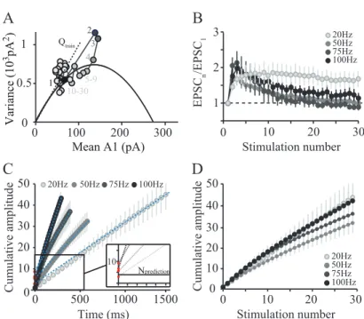
Using whole cell patch clamp recordings in rat cerebellum acute slices, I found that high frequency information processed in the mossy fibre-granule cell pathway is conserved at the parallel fibre to Purkinje cell synapse. Small bursts of action potential could evoke strong Excitatory Postsynaptic Currents (EPSCs) at the Purkinje cell soma. The reliable transmission at the parallel fibre to Purkinje cell synapse can follow high frequency rates, with high initial release probability, paired-pulse facilitation up to 700 Hz, and sustained facilitation during tens of pulses. We found, by using variance mean analysis, that this fast release is possible during bursts through the recruitment of reluctant vesicles that boost vesicular release. Moreover, fast release can be sustained through fast vesicles reloading.
In a second study, by using precise RuBi-Glutamate uncaging onto granule cells, and by recording either Purkinje cells, molecular layer interneurons or Golgi cells, I found that in the anterior vermis of the mouse cerebellum, granule cell to Purkinje cell connection follows a precise spatial organisation. Specific sets of Purkinje cell, that can be identified using histochemical markers, receive inputs from small granule cell hotspots. Local granule cells generally elicit a strong input, but distal granule cells located in specific regions can also be strongly connected. The connection pattern between two neighbouring cells is highly correlated in a single animal. Inter-individual variability is important, probably because of individual motor learning performed in each animal, but similarities can be found, suggesting a shared general organisational map in the cerebellar cortex. Similar experiments performed on molecular layer interneurons showed an distinct pattern, suggesting that a given patch of granule cell either directly activate or indirectly inhibit distal Purkinje cells. Recordings of Golgi cells expressing glycine transporter 2 (GlyT2+), a subpopulation of Golgi cells that are not homogeneously distributed, revealed that Golgi cells essentially receive inputs from local granule cells, whereas EPSCs from parallel fibres were weak and rare. This result suggests that basolateral dendrites and apical dendrites perform distinct computations in the network.
PREFACE
During my thesis, I worked on two main projects. The first one focused on short term plasticities that occur at the parallel fibre to Purkinje cell synapse in the cerebellar cortex, during high frequency bursts of action potentials. This work resulted in a first article “Adaptation of granule cell to Purkinje
cell synapses to high-frequency transmission” (Valera et al., 2012). Frederic Doussau, in our team,
continued this work and found further evidences supporting our data. I will describe some of these new results as supplementary results.
During the third and fourth year of my PhD, I started a second project that focused on the spatial organisation of the cerebellar cortex. The goal was to describe the cerebellar module, that is, the smaller functional unit of the cerebellum, at the microcircuit level. Could I delimitate its functional borders? And if it exists, which cells are actually included in this microcircuit, and which ones are not? This project required a long period of coding and technical adjustments. The result of this work is exposed in a second article, which has been recently written. It is thus presented in its non-definitive form.
In parallel, in the continuity of a previous PhD student, Heloïse Cruveiller, along with heroic histological quantifications of Jean-Luc Dupont, a third article is in preparation, which will be more focused on the spatial distribution of the different Golgi cells subtypes in the cerebellar cortex. In this project, which is complementary to my second paper, my contribution was essentially in data analysis. A small part of this work is presented in the second article of this manuscript.
For clarity, my two projects will be introduced in a non-chronological order. Thus, in the first chapter, I will describe the general organisation of the cerebellum, its main functions, the major cell types and subtypes, and some general consideration about the cortical network connectivity. The remarkable spatial organisation of the structure will be detailed in a second chapter, in which different somatotopical, histochemical and functional aspects of the cerebellar physiology will be detailed. Finally, in a third chapter, some temporal aspects of the neurotransmission will be developed: high frequency bursts and their consequences on firing patterns and short term plasticities.
INTRODUCTION
A SHORT HISTORY OF THE CEREBELLAR PHYSIO LOGY
The first known mention of the cerebellum dates back to the Greco-Roman literature from the 4th century B.C., when a structure distinct from the cerebrum was described. This structure, located at the back of the brain, was named paracephalon by Aristotle and Praxagoras, and enpenkranis by Erasistratus. Galen, in the 2nd century A.D. described the cerebellum with more details, and especially its medial part, later named vermis. Cerebellum was proposed to be the source of the cranial nerves and spinal cord. In accordance with the current opinion of that time, Galen suggested that the cerebellum could be a pump involved in the regulation of the flow of the animal spirit, that is, the liquid giving the movement to the muscles. Although Galen’s observations were a big step forward at that time, their interpretation through the Greek theory of humorism restricted medical progresses for almost 15 centuries.
FIRST ANATOMICAL DESCRIPTIONS
Very little progress was made in neuroanatomy until Vesalius in the 16th century and his human anatomy textbook: De humani corporis fabria, published in 1543, when the work of Galen was reanalysed with new methodological approaches. Vesalius represented the human cerebellar structure, but without commenting on cerebellar function. His seminal work influenced all the following anatomists of the Renaissance. In 1575, Varolio brought more details in the external description of the cerebellum, and notably its link with the pons. One century later, Malpighi (1665) described the cerebellar cortex and the white matter fibres, noticing that they “seem to have origin
from the trunk of the spinal marrow contained within the cranium”. Raymond de Vieussens
mentioned a few years after an “ash grey glandular area” that turned to be the cerebellar nuclei, a structure that Félix Vicq d’Azyr described more in details in 1805, although their modern names were only fixed later in the 19th century.
The first precise report on cerebellar cortex appears at the end of the 18th century in Malacarne work (1776). He described and named the lobes and lobules for the first time. He chose names based on lobule shape. Some of these names are still in use, like the lingula (from linguetta - “small tongue” in Italian), the uvula or the tonsil. This descriptive work was later refined by Reil (1807-1808) and Burdach (1819-1826). At this time, the first anatomical bases of the cerebellar anatomy were established.
EARLY CONCEPTS ON CEREBELLAR FUNCTIONS
All attempts to describe cerebellar function before the beginning of the 19th century were highly speculative. Rolando (1809), Flourens (1826), and later Luciani (1891) were the first to assess the link
partial and total ablation of the cerebellum, observed severe impairment of motor functions, but not of sensory or intellectual ones. He concluded from this work that the cerebellum was involved in the initiation of movements. One year after the translation of Rolando’s work in French, Flourens (1924) developed the idea that movement initiation was actually not affected, but that regularity and coordination were lost. Thus, Flourens was the first to separate motor initiation from motor coordination, and to associate them with distinct brain regions. Luciani further detailed the semiology of cerebellar lesions, discriminating transient from permanent effects of a cerebellar lesion. According to Luciani, a cerebellar lesion is followed by an asthenia or muscular weakness, an atonia or lack of normal muscle tone, and an astasia or unstable muscular contraction. For him, these three symptoms explain all the behaviours associated with a cerebellar lesion, including “tremor,
titubation and rhythmically oscillating movements”. Later on, more precision in the symptoms were
added by Babinsky (1902) like dysdiadochokinesis - the inability to perform rapid sequences of movements - or asynergia - the inability to coordinate groups of muscles in complex movements. These observations were confirmed and clarified in the following years by Holmes (1917, 1922) who studied soldiers wounded during the First World War, and patients with cerebellar tumours. He observed many occurrences of tremors and dyskinesia, and developed the concept of ataxia. At this time, the major implication of the cerebellum in the control of complex and voluntary movements was established.
The concept of functional organisation only emerged at the end of the 19th century in the cerebral cortex, and was extended to the cerebellum a few years later. After the early description of the cerebellar histology (Ramon y Cajal, 1911) and the first recordings of electrical activity in the cerebellum (Adrian, 1935), the idea of a structured cerebellar cortex was progressively refined, with major breakthrough at each technological progress. In the 60’s, electronic microscopy allowed the description of the fine structure of the cerebellar tissue (Palay and Chan-Palay, 1974), while electrophysiological recordings permitted to describe the network and the cellular connections (Eccles et al., 1967). During the 70’s and the 80’s, the first precise maps of cerebellar somatotopy were performed, and immunohistochemistry revealed new cellular populations and cerebellar compartments (Voogd, 1967; Shambes et al., 1978b; Hawkes et al., 1985). More recently, patch clamp recordings and Ca2+ imaging techniques helped to understand synaptic integration, while genetically modified mice revealed the function of specific proteins in the cerebellar physiology. Though, one century after the first description of the cerebellar circuitry, and despite the great number of laboratories that worked - and are still working - on the cerebellar physiology, the precise description of the cerebellar spatial organisation is yet in debate, like many other aspects of the cerebellar functions. To date, we probably lack an overview of all the cellular subtypes, the complete wiring diagram of the different cerebellar regions, the precise electrophysiological profile of the cerebellar neurons with their respective integration properties and metaplasticities profiles, as well as a comprehensive computational model of the cerebellar cortex, and even a full list of cerebellar functions such as its involvement in cognition.
1 GENERAL ANATOMY AND HISTOLOGY OF THE CER EBELLUM
1.1 CEREBELLAR FUNCTIONS
Most of the cerebellar functions are known from clinical observations, some of them for more than a century. Since then, pharmacological, lesion-induced, or more recently mutant animal models were developed to study cerebellar functions. In this chapter, I will present the main cerebellar function and the general circuitry of the cerebellar cortex. Then, in a second time, I will detail morphological and functional features of some of the cerebellar cell types, since we will discuss about cerebellar connectivity in the following chapters.
1.1.1 ROLES OF THE CEREBELLUM
The cerebellum is a structure related, first and foremost, to sensorimotor functions. It is considered to be involved in the control of gaze, gait and posture, fine motor coordination, motor learning, prediction of movements and correction of motor errors. To perform these tasks, the cerebellum integrates both sensory and motor signals coming from both the cerebral cortex and from multiple sensory receptors throughout the body, sometimes after multiple and complex integrative steps. Sensory information coming from the whole body give contextual clues like skin pressure, muscle or skin stretch, articulations position or head inclination. Visual, auditory and somesthetic information are carried to the cerebellum by distinct pathways. In parallel to real-time sensory information, the cerebellar cortex receives inputs coming from several areas of the neocortex such as the prefrontal or the parietal cortex, carrying a copy of the motor command sent to the spinal cord termed corollary
discharges (Sperry, 1950; Bell, 1981; Wolpert et al., 1998).
In order to efficiently control and adjust the movement while it is executed, the cerebellum is thought to predict the future sensory state of the body (Wolpert et al., 1998; Bastian, 2006). In this view, the prediction is then compared with the real sensory state, and any discrepancy would indicate a motor error or a novel sensory stimulus. In this process, the cerebellum is also able to adjust the movement, by sending a corrected motor order both to the motor cortex and to other motor structures like the red nucleus. In order to sort only relevant sensory information, the cerebellum is able to suppress the sensory feedback induced by the execution of self-generated movements (Blakemore et al., 1998).
1.1.2 SEMIOLOGY
Illustration of the cerebellar functions can come from the observation of cerebellar lesions. For example, a lesion in the flocculonodular lobe can result in a nystagmus, but also in impairments of the body balance. These symptoms illustrate the implication of that region of the cerebellar cortex in occulomotor reflexes and in the visual and vestibular control of movements. Lesions located in the
vermis essentially impair the gait (“drunken sailor” gait) whereas damages in the cerebellar hemispheres can result in ipsilateral impairment of voluntary multi-joint movements and intention tremor.
More surprising evidences suggest an implication of the cerebellum in various cognitive functions (Ivry et al., 1988; Ito, 2008; D’Angelo and Casali, 2012) like time perception, autonomous responses in fear conditioning, schizophrenia, dyslexia, autism and even tinnitus (Bauer et al., 2013). With the major difficulty that each lesion in the cerebellum can also induce motor disturbances, which can affect the measurement of the performances during cognitive tasks, these experiments revealed broader cerebellar functions than initially hypothesised. Several authors suggested that the cerebellum might use the same framework to perform cognitive tasks as those used for sensorimotor processes (Glickstein et al., 2011; D’Angelo and Casali, 2012).
1.2 GENERAL STRUCTURE OF THE CEREBELLUM
Inherited from the phylogenetic evolution of the vertebrates, the structure of the cerebellum essentially differs from one species to another in term of size and shape. It evolved from a leaf-like structure in amphibians and reptiles to the complexly foliated structure we can observe in upper mammals (Voogd and Glickstein, 1998). Nonetheless, the cerebellum is an evolutionary conserved structure, keeping striking similarities in development, histochemical compartmentation, structure and functions between vertebrates, suggesting its conserved involvement in sensorimotor tasks. In mammals, the size of the cerebellum is linearly correlated with the body surface, another argument for its role in sensorimotor processing (Sultan and Braitenberg, 1993; Heck and Sultan, 2002).
In all mammals, the cerebellum is composed of two main structures: three pairs of cerebellar nuclei and the cerebellar cortex, a three-layer cortex of about 500 µm thick. As an indication of its important foliation, in humans, the unfolded cerebellum is 15 cm large and 2 m long.
1.2.1 MULTIPLE ORGANISATION LEVELS IN THE CEREBELLAR CORTEX
The spatial organisation of the cerebellar cortex follows two main axes (Figure 1): the anterio-posterior one separates the cortex in lobes and lobules (Figure 2A1-2A3) whereas the mediolateral one separates it between a vermis and two hemispheres (Figure 2B). The mediolateral axis is sometimes named long axis of the folium. This term actually embraces both the coronal and the horizontal/transverse plane (Figure 1B2, 1C2). Because of the cerebellar folding, acute slices in either coronal or horizontal/transverse plane have similar tissular organisation (Figure 1B3, 1C3, see also Figure 4). However, each slicing plane will allow the study of a different set of lobules. This point is of primary importance in my second article, in which the experiment required the recording of analogous cells (i.e. at the same set of parasagittal and mediolateral coordinates). More generally, the use of the horizontal plane is more convenient because it allows an easier recognition of the lobules, compared to the coronal plane which often results in twisted regions with more damaged cells.
1.2.1.1 ANATOMICAL SEGMENTATION
Several grooves cross the cortex transversally, segmenting the cerebellum along the anterio-posterior axis. The two deepest ones divide the cerebellum in three lobes (Figure 2A1, 2A2, 2A3): the anterior lobe (anterior zone), the posterior lobe (central zone and posterior zone) and the flocculonodular lobe (nodular zone). A finer subdivision based on human anatomy (Larsell, 1952), splits the cerebellum in ten lobules, but the exact number of folia can actually vary from one species to another. For instance, in the mouse, lobules IV and V are fused whereas lobule VI is separated in VIa and VIb-c.
The mediolateral organisation (Figure 2B) separates the cerebellum in a vermal part, surrounded by two cerebellar hemispheres which can each be further split into an intermediate hemisphere or
paravermis, and a lateral hemisphere.
1.2.1.2 FUNCTIONAL SEGMENTATION
A third approach to divide the cerebellum is based on a general segmentation of cerebellar functions. Inherited from the phylogeny, the cerebellum has three major regions, each one receiving major inputs from different regions of the nervous system, and each cortical region projecting to separate cerebellar nuclei (Figure 2C). Although often used, this segmentation can be misleading, because most regions actually receive inputs from several origins.
1.2.1.2.1 VESTIBULOCEREBELLUM
The oldest phylogenetic part, the vestibulocerebellum or archeocerebellum, is already present in the chondrichthyes (cartilaginous fishes). Besides vestibular information, the primitive cerebellum also receives cutaneous and proprioceptive inputs, especially from the lateral line. In higher vertebrates, this region corresponds to the flocculonodular lobe (Lobule X), which controls balance and eye movements. Because the posterior part of the Lobule IX performs similar functions, it is also considered as part of the vestibulocerebellum. The vestibulocerebellum projects directly out of the cerebellum, in the lateral vestibular nuclei (see Figure 3C).
1.2.1.2.2 SPINOCEREBELLUM
On the phylogenetic timescale, the second part of the cerebellum to appear is the vermis. Major inputs are coming from the spinal cord, giving the name of spinocerebellum to that region. The vermis receives somatic sensory inputs as well as visual, auditory and vestibular information, in order to control posture, locomotion and gaze. Efferences from the vermis of lobules I to IXanterior mainly target structures related to these functions, in the brain stem through the fastigial nucleus, directly to vestibular nuclei as in the flocculonodular lobe, and to the cerebral cortex (Sugihara, 2011). The muscles targeted by spinocerebellar outputs are principally located in the trunk and in the proximal parts of the limbs.
The paravermis or lateral hemisphere can be considered as part of the spinocerebellum. This region integrates information coming from the limbs and projects first to the interposed nucleus, which in turn projects to the red nucleus or to the cortex through the thalamus.
1.2.1.2.3 CEREBRO-CEREBELLUM
The third part of the cerebellar cortex, the neocerebellum or cerebrocerebellum corresponds to the cerebellar hemispheres. Somesthetic, sensory and motor afferences coming from the cerebral cortex enter the cerebellum after a relay in the pontine nuclei. The cerebrocerebellum is particularly developed in primates in parallel to the massive development of the neocortex. The cerebrocerebellum sends back information to the cerebral cortex via the dentate nuclei.
1.2.2 CEREBELLAR INPUTS: INFERIOR OLIVE AND PRECEREBELLAR NUCLEI
Two main inputs project to the cerebellum, (Figure 3). Climbing fibres all originate from the inferior olive, an extensive nucleus located in the medulla, while mossy fibres are of various origins, grouped under the name of precerebellar nuclei.
The inferior olive receives sensory and motor information from very broad origins (cortex, spinal cord, precerebellar nuclei as well as a feedback from the cerebellar nuclei). It is an integrative structure that sends its projection to the cerebellum (Figure 3A). Its function is not clearly defined, but it was proposed to be involved in movement error detection and movement timing control (De Zeeuw et al., 1998; Llinás, 2009). Briefly the inferior olive is subdivided in three major subnuclei: Principal Olive, Dorsal Accessory Olive (DAO), Medial Accessory Olive (MAO) and four smaller subnuclei. Each part can be further subdivided based on its afferences and efferences.
Precerebellar nuclei (Figure 3B) carry sensorimotor information through mossy fibres either from the spinal cord, brainstem or from the cortex after a variable number of relays. Some precerebellar nuclei like the external cuneate nucleus or the gracile nucleus are direct relays of sensory information coming from the body whereas other nuclei like the pontine nuclei relay more integrated information coming from the cortex. The organisation of the inputs will be described more in details in chapter 2.
1.2.3 CEREBELLAR OUTPUTS: CEREBELLAR NUCLEI AND VESTIBULAR NUCLEI
The cerebellar nuclei (often: deep cerebellar nuclei) are the sole output of the cerebellum (Figure 3C). They receive collaterals from the cerebellar inputs, and are also targeted by all the axons leaving the cerebellar cortex from Purkinje cell axons. They are composed of three pairs of nuclei, located in the depth of the cerebellum, and surrounded by white matter. Cerebellar nuclei are named medial, interposed (anterior and posterior part) and lateral nuclei (respectively fastigial,
emboliform & globose and dentate in humans). To these three pairs of nuclei, we should add the vestibular nuclei, located at the junction between the cerebellum and the brain stem. Vestibular nuclei are similar to other cerebellar nuclei in the way that they receive both Purkinje cells and mossy fibres collaterals.
All these nuclei are topographically organised. They all receive projections from a specific part of the cerebellar cortex, and appear to be involved in separate processes. It must be noticed that except for some projections of the vestibulocerebellum, the deep cerebellar nuclei never project directly onto motoneurons, but rather to the motor cortex or to spinal local motor networks through the thalamus or the red nucleus (Figure 3C). This observation underlines the highly integrated position of the cerebellum in motor systems, which is involved in motor coordination and modulation but not in direct movement execution.
1.2.4 INTER-SPECIES VARIATIONS AND HOMOLOGY
Because of the apparent conservation of cerebellar structure across species, observation made in one animal model is often generalised to other models. My first project was performed on rats, whereas we used mice in the second project. Can we take into account observations performed in different species in a common cerebellar model?
Historical studies started with observations on humans, and the first animal models were essentially monkeys, cats and rabbits. In the 80’s, the broader use of in vitro experiments introduced the rat model in the laboratories. The rat is a convenient animal model because of its fast reproduction rate, small size and homogeneous genetic background. Moreover, the animal is adapted to behavioural studies, although it cannot perform as complex tasks as monkeys do. Many of the initial observations in relation to the neuronal physiology in the cerebellum were performed in rats. Later, the development of transgenic mice pushed the lab to use the murine model. Fortunately, when experiments are realised in different species, similarities are striking. Sometimes inter-species or background-dependent discrepancies can be observed. For instance, it is possible to find very different firing frequencies for cerebellar nuclei neurons or for Purkinje cells between mice and rats (Rowland and Jaeger, 2005; Person and Raman, 2012a). However, when the study is performed in homogeneous conditions (same anaesthetics, same animal facility,…), values are remarkably similar (Shin et al., 2007). Most of the differences might be due to experimental variations. Some differences are however certain, like subtle variations in the protein expression patterns. For example between the rat and the mouse (Sugihara and Shinoda, 2004; Sugihara and Quy, 2007).
In this manuscript, I will give alternatively values coming from the cat, the rat and the mouse. When differences are documented, I will precise species in which the experiment was performed, but when no alternative data are available, we will have to postulate that the physiology between those species is close enough to allow generalisation. Based on the inter-species data already available, the risk is probably acceptable for the study of the cerebellar physiology because of the highly conserved features of that structure.
1.3 CELLULAR STRUCTURE OF THE CEREBELLAR CORTEX
In this section, I will briefly describe the cerebellar cortical structure and the cerebellar inputs. In a second time, I will detail the four cell types I recorded during my PhD, both in term of morphology and connectivity.
1.3.1 CELL TYPES AND INFORMATION PATHWAY
1.3.1.1 CELLULAR ELEMENTS OF THE CORTEX
Unlike the cerebral cortex, the architectonic and histological structure of the cerebellar cortex is constant throughout all lobules. This noteworthy feature led many authors to outline cerebellar cortex anisotropic, almost crystalline organisation. The number of morphologically distinguishable neuronal types is very low; only seven major types of neurons are described, all defined by a very specific morphology and localisation, and positioned precisely in one of the three cortical layers: Purkinje cells, granule cells, Golgi cells, basket cells, stellate cells, Lugaro cells and unipolar brush cells. A few other cell types were proposed, but for now they are still speculative, or too poorly characterised. In the seven identified neuronal types, several subtypes were described even if subtypes distinction remains essentially biochemical rather than functional or morphological. Except for unipolar brush cells, all the cell types are present all over the cerebellar cortex, without obvious variation in number, density or shape, strengthening this apparent homogeneity.
The three layer cortex is composed of, from the more internal to the more external layer: the granular layer, the Purkinje cells layer and the molecular layer (Figure 4A1, 4A2). Under the granular layer is the white matter, containing all the fibres entering and leaving the cerebellar cortex.
· The granular layer contains granule cells - which are by far the most numerous cells in the cortex (Figure 4B1) -, Golgi cells, Lugaro cells, and only in the posterior lobules, unipolar brush cells. Other cell types such as candelabrum or globular cells are sometimes mentioned in the literature (Lainé and Axelrad, 2002; Hirono et al., 2012). They are both supposed to be inhibitory interneurons, but they will not be developed here, as they are poorly characterised and impossible to identify in our experiments. In the rat, granular layer has an average thickness of 149 µm (Harvey and Napper, 1991), but unlike the molecular layer, this thickness is not perfectly constant and can present local variation in the sulcus or at the apex of each lobule (Braitenberg, 1967), probably because of the foliation itself.
· The Purkinje cells form a monolayer, sending their axon through the granular layer and their planar dendrites in a narrow parasagittal plane in the molecular layer (see Figure 4B1-4B3 and 6A2).
· The molecular layer, the most external layer of the cerebellar cortex is around 225 µm thick (Harvey and Napper, 1991). It is composed essentially of the parallel fibres which are the
cell types are present in this layer: basket cells and stellate cells, which will both be pooled under the name of molecular layer interneurons in this manuscript. The parallel fibres run along a mediolateral plane, crossing perpendicularly the flat dendritic tree of the Purkinje cells, the apical dendrites of the Golgi cells and the dendrites of the molecular layer interneurons. The molecular layer is bounded on its external part by the pia matter.
1.3.1.2 MOSSY FIBRES AND CLIMBING FIBRES
The information entering the cerebellar cortex follows two main pathways that are both converging onto Purkinje cells, the sole output of the cerebellar cortex (Figure 5A, 5B). Climbing fibres are coming from the inferior olive and projecting directly onto Purkinje cells, and indirectly through spillover to molecular layer interneurons (Szapiro and Barbour, 2007). Climbing fibre evokes a particular type of spike in the Purkinje cell, termed complex spike. Mossy fibres originating from various precerebellar nuclei project on granule cells and Golgi cells through a structure called glomerulus. Granule cells send their axon, the parallel fibre, in the molecular layer. A parallel fibre activates Purkinje cells, but also interneurons like Golgi cells, basket cells and stellate cells (Figure 5A,6B). Those four cell types will be described more in details in the following part of this chapter.
1.3.2 THE PURKINJE CELL
The Purkinje cell, discovered by the Czech physiologist Johannes Purkinje in 1837, is a central cell in the cerebellar circuitry. Indeed, this cell is the sole output of the cerebellar cortex and is therefore the final step in signal integration in the cerebellar cortex. Any relevant information computed in the cerebellar cortical network should have a detectable effect on its discharge (Figure 5B). Understanding how a given set of mossy fibres and climbing fibres inputs will modulate the Purkinje cell firing is a key point to understand how the cerebellar cortex compute sensorimotor information.
1.3.2.1 MORPHOLOGY
The Purkinje cell receives two excitatory inputs: one single climbing fibre coming from the inferior olive, and about 175 000 parallel fibres inputs coming from the granule cells (Napper and Harvey, 1988), themselves activated by mossy fibres (Figure 5A, 6A1). Inhibition on the Purkinje cell is performed by the molecular layer interneurons (Figure 6B) and neighbouring Purkinje cell collaterals (Figure 6A1).
Purkinje cells have a soma of about 20 µm in the rat, 15 µm in the mouse, forming a monolayer between the granular and the molecular layer (Figure 6A1, 6A2). The typical Purkinje cell has a tree-shaped planar dendritic tree of about 200 X 200 µm in the sagittal plane in rodents - 217 µm on average in the adult rat (Harvey and Napper, 1991) - , and a thickness of only 15 µm to 20 µm in the mediolateral plane (Figure 6A2). In higher mammals (cats, dogs or humans for instance), these cells can be twice as big as in rodents (Braitenberg and Atwood, 1958). Purkinje cells usually have one primary, more rarely two dendritic trunks that split in secondary and thin tertiary branchlets. These tertiary branchlets are covered with dendritic spines receiving only parallel fibre inputs and organised in a helical pathway with a short pitch (Palay and Chan-Palay, 1974; O’Brien and Unwin, 2006). These spines are rarely contacted by more than one parallel fibre (Napper and Harvey, 1988). Inhibitory molecular layer interneurons synapse preferentially on the dendritic shaft, and climbing fibres contacts directly smooth dendrites of the Purkinje cells on thorny spines, forming hundreds of release sites that appear to wrap the dendritic trunk (Palay and Chan-Palay, 1974) (Figure 5A).
1.3.2.2 TARGETS
The Purkinje cell is a GABAergic inhibitory projection neuron. It sends a myelinated axon that ultimately reaches several neurons in the cerebellar nuclei. Conversely, in the cerebellar nuclei, one neuron receives inhibition from several Purkinje cells. Thus, there are both a high degree of convergence and divergence of Purkinje cells onto cerebellar nuclei neurons (Person and Raman, 2012b).
The Purkinje cell axon can branch and form collaterals that contact other Purkinje cells (Palay and Palay, 1974; Orduz and Llano, 2007; Bornschein et al., 2013) and Lugaro cells (Palay and Chan-Palay, 1974; Hirono et al., 2012), but also basket cells (Palay and Chan-Chan-Palay, 1974; O’Donoghue et al., 1989), and possibly Golgi cells (Palay and Chan-Palay, 1974; Hirono et al., 2012) (Figure 6A).
Purkinje cell collaterals distribution is poorly understood. Briefly, two axonal plexus were described, one located just above or at the level of the neighbouring Purkinje cells, and a second deeper in the granular layer (Palay and Chan-Palay, 1974). The Purkinje cell collateral plexus was often described as more parasagittally oriented, but not exclusively. Purkinje cell collaterals can reach cells up to 200 µm in this axis (Braitenberg and Atwood, 1958). However, Richard Hawkes observed that Purkinje cells send collaterals up to five cells laterally (̱80 to 100 µm) in the mediolateral axis, in the anterior lobe (Hawkes and Leclerc, 1989). A recent single cell reconstruction study (Sugihara et al., 2009) confirmed those early observations: even if most of the collaterals remains in a parasagittal axis, some of them occasionally travel along a mediolateral axis for short distances.
1.3.3 THE GRANULE CELL
If Purkinje cells are the sole output of the cerebellar cortex, granule cells are the major input stage cells, at least in number and density. Moreover, with the exception of unipolar brush cells in posterior lobules, granule cells are the only excitatory neurons in the cerebellar cortex. After migrating radially from the external granular layer between the first and the third postnatal weeks, granule cells establish their position in the inner part of the cerebellar cortex, forming the internal granular layer (Wang and Zoghbi, 2001). As we are working in the adult or juvenile (i.e. after P17) mouse or rat, any mention of the granular layer in this manuscript will thus refer to the inner granular layer.
1.3.3.1 MORPHOLOGY
The granule cells of the cerebellum are the most numerous neurons in the brain and might represent 60% of the neurons in the mouse brain and up to 70% in the rat brain. Granule cells are also among the smallest neurons, with an average somatic size of 4.82 µm (Harvey and Napper, 1991), and they have consequently a very high but constant density (1.92.106 cells mm3 in (Harvey and Napper, 1988), 2.85 106 in (Palkovits et al., 1971)). They usually display 4 to 5 short dendrites terminating in the glomerulus (Cathala et al., 2003), which receives excitatory transmission form the mossy fibres (Figure 5A). On average, the granule cell dendritic extension is about 30 µm in the cat, and is probably almost the same in the rat and the mouse.
Granule cells send a very long axon in the molecular layer. This axon can be divided in two elements (Figure 6C): the ascending axon, which is the initial part of the axon rising vertically from the granule cell soma to the molecular layer, and the parallel fibre which run along the mediolateral plane in the molecular layer.
1.3.3.2 THE ASCENDING AXON
On an anatomical basis, the ascending part of the granule cell axon - ascending axon - makes several synapses before the parallel fibre bifurcation (Napper and Harvey, 1988; Gundappa-Sulur et al., 1999). The density of these synapses was estimated to be higher in the ascending axon (one synapse every 4.0 µm) compared to the density in parallel fibres (one synapse every 7.4 µm). As the ascending axon follow the dendritic plane of a unique Purkinje cell, it was estimated that one granule cell can do on average up to 31 synapses on a Purkinje cell (Gundappa-Sulur et al., 1999). Napper and Harvey proposed that 3% of the total granule cell to Purkinje cell synapses are coming from the ascending axon, whereas the value of 7 to 24% of the total granule cells inputs was proposed by a more recent study (Gundappa-Sulur et al., 1999). Although the exact number of synapses is still a matter of debate, even the lower estimate represent a significant number of synapses (3.5% would represent more than 6000 inputs per Purkinje cell). The high number of synapses does however not predict their functional properties. Finally, the Purkinje cell is not the only cell type to be contacted by the ascending part of the axon. Based on electronic microscopy, the ascending axon might also contact Golgi cells (Hámori, 1981) and molecular layer interneurons (Sultan and Bower, 1998).
1.3.3.3 THE PARALLEL FIBRES
Once in the molecular layer, the ascending axon bifurcates in a T-shaped manner. The two branches of the T run in the mediolateral plane, taking the name of parallel fibres. They are thin unmyelinated axons, making en passant synapses with Purkinje cells, stellate cells, basket cells and Golgi cells they cross. The exact longitudinal extend of the parallel fibres depends on species. Available data gives an estimated length between 4.2 and 4.7 mm in the rat (Pichitpornchai et al., 1994). The vermis measures 3 mm to 4 mm in the anterior lobules in the rat, and a quick estimate would suggest that although parallel fibres probably do not cross the whole cerebellum from one hemisphere to the other, most of the parallel fibres located on the midline are likely to be long enough to cross the vermis and even to reach the paravermis.
A study performed in 2005 (Zong et al., 2005) focused on the post-developmental final position of the granule cells coming from a single original clone during the development. It appeared that axons of granule cells coming from a same clone are bundled in the molecular layer in a restricted sublayer. On the other hand, their soma are scattered in all the depth of the granular layer. Neighbouring cells in the granular layer do not share neighbouring parallel fibres, but neighbouring parallel fibres come from the same clone. Determining whether neighbouring parallel fibres in the molecular layer correspond to a functionally related group of granule cells is actually a major technical concern, because many experiments use direct electrical stimulation of the parallel fibres to study granule cell input. In all my experiments, I always stimulated granule cells in the granular layer.
1.3.3.4 PARALLEL FIBRE TO PURKINJE CELL SYNAPSE
In the molecular layer, parallel fibres cross orthogonally the dendritic tree of the Purkinje cells and make contact with more than half the Purkinje cells they pass through (Napper and Harvey, 1988) (Figure 6A, 6C). Electronic microscopy studies suggest that a parallel fibre contact one only, sometimes two dendritic spines in a Purkinje cell (double synapse in 2 to 11% of the cases, depending on the distance from the ascending axon bifurcation site (Pichitpornchai et al., 1994)). Dendritic spines contacted by two parallel fibres or more are on the other hand infrequent. A varicosity present in most cases one single active zone, with 8 docked vesicles, and 480 vesicles in the whole varicosity. The synapse is strongly ensheathed by glia (Xu-Friedman et al., 2001).
Thus, on a purely anatomical basis, a Purkinje cell can receive more contacts from a local granule cell through its ascending axon than from a distal granule cell through its parallel fibres, suggesting a dichotomy between inputs from the local network and distal inputs.
1.3.4 THE GOLGI CELL
The granular layer is considered as the input stage of the cerebellar cortex. Any attempt to model its functions will require a good understanding of all its cell types. If the major excitatory cell type is the granule cell, the major inhibitory interneuron in term of number and extension is the Golgi cell (Figure 7A, 7C, 7D).
1.3.4.1 MORPHOLOGY
In 1873, Camillo Golgi developed the silver nitrate method and opened the way to the morphological study of neurons. His first study focused on the cerebellum, in which he described two types of interneurons in the granular layer. The type I interneurons were described as “long and narrow cells irregularly fusiform” and were probably Lugaro cells. The type II interneurons were irregularly round or polygonal cells […] both these types have a large number of prolongations.” They were named Golgi cells by Ratzius in 1892. Santiago Ramon y Cajal gave more detailed description of their morphology in his Histology of the Nervous System of Man and Vertebrate (Ramon y Cajal, 1911), with particularly precise descriptions of the axonal plexus and dendrites. Far more details were brought by Palay and Chan Palay (Palay and Chan-Palay, 1974). Based on optical and electronic microscope observations, they tried to define the Golgi cell connectivity. They observed a single postsynaptic target, the granule cell, but multiple presynaptic inputs: mossy fibres, granule cells, climbing fibres, Purkinje cells collaterals, and basket and stellate cells.
The most impressive part of the Golgi cell is its extensive axonal plexus that branch hundreds, maybe thousands of times, inhibiting granule cells in a large volume of cerebellar cortex (Figure 7A, 7C, 7D). The mean extent of this plexus was estimated to be 650 µm sagittally and 180 µm mediolaterally in the mouse (Barmack and Yakhnitsa, 2008). The dendritic tree is actually separated between apical dendrites, which have, like Purkinje cells, an extensive parasagittal orientation but a poor mediolateral extension of only 82 µm (Sillitoe et al., 2008) and the basolateral dendrites, which are
restricted to the granular layer (Figure 7A). Basolateral dendrites spread also in the medio lateral axis, but their extent was never quantified. Since they are contained in the axonal plexus, we can only say that they are at most as long as the total neuritic extent (180 µm).
Golgi cells are directly activated by mossy fibres (Kanichay and Silver, 2008), evoking feedforward inhibition onto granule cells. Golgi cells can also be activated by granule cells, both on basolateral and apical dendrites (Midtgaard, 1992; Dieudonne, 1998; Cesana et al., 2013), and consequently perform feedback inhibition onto granule cells (Figure 7A). The existence of Golgi cell inhibition by molecular layer interneurons to Golgi cells was recently questioned by Wade Regehr (Hull and Regehr, 2012). Massive electrical stimulations or optogenetically-induced excitation of the molecular layer interneurons did not elicit inhibitory postsynaptic current that could be recorded in Golgi cells (Hull and Regehr, 2012). Moreover, since Palay and Chan-Palay study, new evidences suggest the existence of both Golgi cell to Golgi cell GABA transmission and Lugaro cell to Golgi cell corelease of GABA and Glycine (Dieudonné and Dumoulin, 2000; Dumoulin et al., 2001).
Finally, a major discovery is the strong electrical coupling between Golgi cells in the first part of the apical dendrites through connexin-36 gap junctions (Dugué et al., 2009; Vervaeke et al., 2012).
1.3.4.2 GOLGI CELL SUBTYPES
Golgi cells present important variations in size, shape or biochemical markers. One major difficulty is to distinguish Golgi cells from the other granular layer interneurons. No universal and specific marker has been identified yet. The most used markers also label Lugaro cells (polyclonal antibody rat-303), or some unipolar brush cells (somatostatin), or both some unipolar brush cells and Lugaro cells (calretinin) (Geurts et al., 2001).
Nevertheless, among the cells that are most probably Golgi cells, we can observe several subtypes based on both their molecular markers and their morphology (Figure 8).
For instance, most of the Golgi cells express the metabotropic glutamate receptor 2 (mGluR2), but 10% express mGluR5 instead (Neki et al., 1996). A further study suggested that mGluR2+ cells are mGluR3+ too, and that mGluR5+ cells are mGluR1+ too (Jaarsma et al., 1998).
Neurogranin, a calmodulin-binding protein that participates in the Protein Kinase C (PKC) signalling pathway, is another marker expressed in only a subset of Golgi cells of the mouse (Singec et al., 2003). Besides the fact that only one part of the Golgi cells express the protein, the authors observed spatial variations in the average density between the vermis and the hemispheres. In our morphological study, we performed a more detailed analysis of the distribution of this Golgi cell subtype.
Golgi cells can perform glycine uptake (Wilkin et al., 1981) or GABA uptake (Ottersen et al., 1987). The first team to study this aspect of cerebellar physiology indicated that 72% of the Golgi cells are immunopositive for both GABA and GlyT2, whereas 14% are only GABAergic, and 14% only glycinergic (Ottersen et al., 1988). In this first study, Lugaro cells - which are 90% both GABAergic and glycinergic (Dumoulin et al., 2001; Crook et al., 2006) - were probably counted with Golgi cells, and integrated in the total number. GABAergic cells express glutamic acid decarboxylase (GAD67).
In a first attempt to classify Golgi cell subtypes, a classification in 5 groups was recently proposed (Simat et al., 2007) based on the presence of markers and on cell morphology (Figure 8).
- Group 1 cells (65% of the cells) are [GAD67+, GlyT2+, mGluR2+ and Neurogranin+] - Group 2 cells (10% of the cells) are [GAD67+, GlyT
2+, mGluR2+]. Group 1 and group 2 cells perform both GABA and Glycine release.
- Group 3 are small sized cells and correspond to 3 to 5% of the total. Cells are either [GAD67+, GlyT2+, mGluR2+] like group 2 Golgi cells or only [GlyT2+, mGluR2+].
- Group 4 cells release only GABA, and constitute 15% of the total population. They are only [GAD67+, Neurogranin+].
- Group 5 cells are the last 5%, and are pure glycinergic cells [GlyT2+], expressing none of the three other makers.
Pure glycinergic cells do not express mGluR2, and according to Neki (Neki et al., 1996), they are mGluR5+. It should be noticed that Neki found 10% of mGluR2- cells whereas Simat found 20%. These divergences between the results raise the question of a possible bias in such quantifications. There is no precise indication of the region of the cerebellum that were used (along the mediolateral axis, the whole cerebellar vermis was considered, but lobules were different in the two studies). Any spatial
heterogeneity in the subtype distribution would dramatically modify the values. We quantified these spatial heterogeneities for the GlyT2+ and Neurogranin+ subpopulation (see second article).
Finally, we must keep in mind that this complex clustering of the Golgi cells is only based on the few identified markers that were shown to be heterogeneously expressed. As all proteins expressed in Golgi cells, it is possible that some other markers are expressed in specific subpopulations, either by respecting the subpopulation described above, or by clustering Golgi cells into even more subgroups.
1.3.5 THE GLOMERULUS
Now that mossy fibres, granule cells and Golgi cells are described, we should mention the particular structure in which all these three cell types interact: the glomerulus. When the myelinated mossy fibre enters the cerebellar cortex, it sends collaterals that will produce an axonal structure called rosette (Figure 7B). The rosette can either be an en passant synaptic structure, or the terminal part of the mossy fibre. The glomerulus was studied in details in the rat (Jakab and Hámori, 1988). It is a 10 µm spherical structure in which the mossy fibre terminal (the rosette), the granule cell dendrites and the Golgi cell axon tightly interact. The whole structure is wrapped with a glial sheet that isolates the glomerulus from the outside, and confers specific electrophysiological properties, favouring notably excitatory and inhibitory neurotransmitter spillover between synapses located within the glomerulus (Rossi and Hamann, 1998; DiGregorio et al., 2002) (Figure 7A, inset).
Granule cells send all their 4-5 dendrites in different glomeruli. Each dendrite further split into several protrusions (dendritic digits) in the glomerulus. Each granule cell dendritic digit receives one or more excitatory mossy fibre synapse (67%), inhibitory Golgi cell synapse (25%), or both (8%) (Jakab and Hámori, 1988). However, as there each dendrite has several digits, 60% of the dendrites is inhibited by at least one Golgi cell. On average one glomerulus can contact 53 granule cells, which indicates a high divergence in the transmission of the information carried by one single mossy fibre.
1.3.6 MOLECULAR LAYER INTERNEURONS
Until now, we only described Purkinje cells excitatory inputs, through parallel fibres, ascending axon, or climbing fibres. Molecular layer interneurons are dedicated to the inhibitory control of Purkinje cells, since they are their only source of inhibition, besides collaterals of other Purkinje cells. Moreover, according to recent study, Purkinje cells might be the only target besides other molecular layer interneurons (Hull and Regehr, 2012).
1.3.6.1 MORPHOLOGY
Molecular layer interneurons were initially described by Ramon y Cajal (Ramon y Cajal, 1911). They are inhibitory interneurons displaying a parasagittally oriented axodendritic tree. The axon can project up to 450 µm from the cell body. Thus, when activated by a beam of parallel fibres, molecular layer interneurons perform lateral inhibition onto off beam targets (Eccles et al., 1967; Cohen and Yarom, 2000). The cellular morphology actually varies depending on the interneuron position in the molecular layer; cells located at the very top or at the very bottom show a more asymmetrical morphology. Molecular layer interneurons are classically distributed in two populations: stellate cells are located in the outer two-third of the molecular layer (Figure 9A, 9C2), whereas basket cells are located in the two inner thirds (Figure 9A, 9C1). Both basket and stellate cells perform feedforward inhibition following granule cells activation onto Purkinje cells and other molecular layer interneurons (Figure 6B, 9A). The basket cells present a supplementary axonal specialisation compared to stellate cells: lower part of the axon branches in the vicinity of a Purkinje cell and forms a basket around its soma (Figure 9B). The axon then wraps the axon initial segment, forming another structure called the brush (Figure 9B, inset). An interesting theory suggested that the brush does not contain any chemical contact, but rather acts directly through field potential to prevent action potential initiation without synaptic delay (Korn and Axelrad, 1980). This ephaptic contact would be very interesting in term of temporal coding as it would prevent Purkinje cell firing without synaptic delay. Basket cell contacts several Purkinje cells along the parasagittal axis, and sometimes two to three in the mediolateral one (Figure 9B). The separation of basket cells and stellate cells in two distinct populations is still in debate, with numerous authors suggesting a continuum of morphologies from purely stellate cells to purely basket cells (Ramon y Cajal, 1911; Palay and Chan-Palay, 1974; Sultan and Bower, 1998; Mittmann and Häusser, 2007; Schilling et al., 2008).
Molecular layer interneurons receive several hundreds of excitatory parallel fibres inputs (Palay and Chan-Palay, 1974) and between one and 20 inputs from other inhibitory interneurons (Lemkey-Johnston and Larramendi, 1968; Llano and Gerschenfeld, 1993). There are on average 10 molecular layer interneurons for one Purkinje cell, and in vitro recordings showed that one Purkinje cells receives on average 9 interneurons inputs (Häusser et al., 2004). Both their parasagittal morphology and their restricted connectivity suggest that molecular layer interneurons are contacting very few Purkinje cells, and that they are consequently restricted to a small local network.
1.4 FUNCTIONAL CONNECTIVITY IN THE CEREBELLAR CORTEX
1.4.1 MARR-ALBUS-ITO THEORY OF THE CEREBELLAR CORTEX
A few years after the first description of the cerebellar neuronal circuitry by John Eccles group (Eccles et al., 1967), David Marr developed a theoretical model of the cerebellar cortex (Marr, 1969) that was extended a few years later by James Albus (Albus, 1971) and functionally demonstrated by Masao Ito (Ito and Kano, 1982). In this theory, the cerebellum is described as a perceptron (Figure 10). Basically, a perceptron is a structure in which all the inputs are connected to the output in a feedforward manner, and in which the message is coded by a linear summation of the inputs
(Rosenblatt, 1958). If a threshold is reached, then the output cell fires and transmits the message to the next neuron in the network.
In Marr-Albus-Ito model, extended by Dean and Porrill (Dean et al., 2010), the network can compute supervised learning, which means that the selection of specific pattern is under the control of a
teaching signal: the climbing fibre activity. Learning induces the selection of particular input patterns
(adaptative filter), by changing the weight of the individual connections.
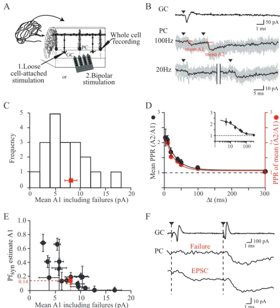
![Figure 1 D, in which PPRs were averaged (n 5 18; PPR A2/A1 at 50 Hz 5 1.7 6 0.14, PPR A2/A1 at 100 Hz 5 2.1 6 0.15; t 5 29.4 6 8.49 ms; [Ca 21 ] e 5 2.5 m M ), shows that facilitation is modest and decays more quickly than reported previously using compoun](https://thumb-eu.123doks.com/thumbv2/123doknet/14738836.754685/97.892.113.531.137.484/figure-averaged-facilitation-modest-quickly-reported-previously-compoun.webp)
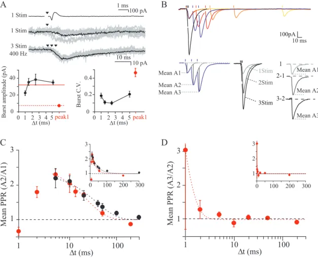
![Figure 4. Estimation of initialPr site at GC–PC synapses. A1 , Time course of A1 as [Ca 21 ] e was varied: 2.5, 4, and 1.5 m M](https://thumb-eu.123doks.com/thumbv2/123doknet/14738836.754685/100.892.364.785.135.877/figure-estimation-initialpr-site-synapses-time-course-varied.webp)
![Figure 5. Additional release sites during high-frequency triplet stimulation. A , Example of variance–mean analysis of A1 (filled black circle) and A2 (open black circle) for one experiment at 75 Hz and 2.5 m M [Ca 21 ] e](https://thumb-eu.123doks.com/thumbv2/123doknet/14738836.754685/101.892.71.436.153.255/figure-additional-frequency-stimulation-example-variance-analysis-experiment.webp)