HAL Id: tel-01887784
https://tel.archives-ouvertes.fr/tel-01887784
Submitted on 4 Oct 2018HAL is a multi-disciplinary open access archive for the deposit and dissemination of sci-entific research documents, whether they are pub-lished or not. The documents may come from teaching and research institutions in France or abroad, or from public or private research centers.
L’archive ouverte pluridisciplinaire HAL, est destinée au dépôt et à la diffusion de documents scientifiques de niveau recherche, publiés ou non, émanant des établissements d’enseignement et de recherche français ou étrangers, des laboratoires publics ou privés.
Role of AMPK in aging and age-related loss of
behavioral plasticity in C. elegans
Caroline Escoubas-Güney
To cite this version:
Caroline Escoubas-Güney. Role of AMPK in aging and age-related loss of behavioral plasticity in C. elegans. Health. Université Côte d’Azur, 2018. English. �NNT : 2018AZUR4029�. �tel-01887784�
Role of AMPK in aging and age-related loss of behavioral
plasticity in C. elegans
Caroline Escoubas-Güney
Laboratoire William Mair / Laboratoire Eric Gilson
Présentée en vue de l’obtention
du grade de docteur en Sciences de la Vie et de la Santé de l’Université Côte d’Azur Dirigée par : Dr. William Mair
Co-encadrée par : Dr. Eric Gilson Soutenue le : 4 Mai 2018
Devant le jury, composé de :
Eric Gilson, PU-PH, Universite Cote d’Azu William Mair, Assistant Professor, Harvard University
Colin Ewald, Professor, ETH
Jennifer Tullet, HDR, University of Kent
THÈSE DE DOCTORAT
2 Université de Nice Sophia Antipolis – UFR Sciences
Ecole Doctorale Sciences de la Vie et de la Santé
Thèse
Pour obtenir le diplôme de Docteur en Sciences de l’Université de Nice Sophia Antipolis
Role of AMPK in aging and age-related loss of behavioral
plasticity in C. elegans
(Le rôle de l’AMPK dans le vieillissement et la perte de plasticité neuronale liée au
vieillissement chez C. elegans)
Caroline Escoubas-Güney
2018
Dirigée par Dr. William Mair
Harvard School of Public Health, Department of Genetics and Complex Diseases, Co-dirigée par Dr. Eric Gilson
Université de Nice Sophia Antipolis, IRCAN
Comité / Committee:
Dr. Eric Gilson PU-PH Université de Nice Sophia Antipolis (Nice) Examinateur Dr. William Mair Assistant Professor Harvard School of Public Health (Boston) Examinateur
Dr. Colin Ewald Professor ETH (Zurich) Rapporteur
3 “Your profession is not what brings home your weekly paycheck, your profession is what you're put here on earth to do, with such passion and such intensity that it becomes spiritual in calling”
4 Acknowledgements
My PhD was a journey. A journey filled with ups and downs, with dreams, hopes, and excitement for science as well as disappointments, doubts, and solitude, but never regrets. I truly believe that interrupting my medical studies to pursue a PhD was the best decision I ever made.
For that, I have to thank Dr. William Mair, who welcomed me in his lab and has, for the past 4 years, taught me everything about science, from bench work and critical thinking to presenting in meetings. You have been an incredible mentor and I feel extremely lucky and grateful to have trained in your lab. I think the most valuable thing you pass on to your lab members is your endless passion and excitement for science. I hope I can keep the same interest and curiosity throughout my future career.
I would like to thank Dr. Eric Gilson who made it possible for me to do a PhD. You have been supportive of the MD-PhD path when very few people did, you accepted me in your lab and provided me with funding opportunities. I am very grateful for your support.
MD-PhD opportunities are still too rare in France and very little to no support exist for students willing to pursue this career. I therefore would like to acknowledge the MD-PhD program Ecole de l’INSERM Lilian Bettencourt which has been a pioneer in this field and has been giving the opportunity to medical students to train in basic research for the last 15 years. Thank you for your endless support, both financially and academically. This small (but growing!) MD-PhD community you created is very valuable and I would like to thank all my 2012 classmates! And a big hug to the Team, Hugo, Antoine and Simon, the whole US adventure started with you.
I would also like to acknowledge my committee, Dr. Ewald and Dr. Tullet. It is an honor to have you judge the quality and relevance of my work.
I would like to thank past and current members of the Mair Lab, Kris, Caroline, Heather, Nessa, Yue, Gio, Annie, Sneha, Pallas, Nicole, Emina, Juan, Staci, it was so much fun and pleasure to work and collaborate with such driven scientists! A special thanks to Kris, Caroline, Heather, Yue and Nessa, you guys made me
5 want to stay for my PhD. Yue, you have been an amazing friend to have in the lab. You were the first to teach me all about worms and I am so happy that I had someone to share my struggles with during those (many) late nights at the bench! Annie, thank you for your endless support and happiness! A big thank you to my former students, Staci, Nicole, Juan and Emina for contributing to my project in many ways and a special thanks to Emina, it was a great pleasure to see you grow as a scientist.
I would also like to thank the members of the Hotamisligil Lab, for the use of the confocal microscope and more generally technical help and advice and always very interesting scientific discussions. A big thank you to my favorite Turkish/Brazilian couple from the 6th floor, Ana and Gunes, you guys are amazing
scientists but more than that, you have become family.
The one person who has gone through the whole PhD with me and shared all my personal and scientific joys and deceptions is Maria. It was friendship at first sight and I feel extremely lucky to have met you. I had the most fun with you and Staci (#bridetribe) and I can’t wait to see what next adventures life will bring us!
I would like to thank to thank my family, Bab, Mam, Alex, and especially my parents. I know you did not embrace the idea of me doing a PhD at first but you have always shown strong support, no matter what, and followed me in each of my decisions. As Bab would say “A journey of a thousand li begins with a single step” (Lao Tzu) and you have been there at every one of them. And finally, I would like to thank an incredibly smart, kind and passionate man, my husband Ekin. You have always been there for me, thank you for loving me, believing in me and always pushing me to be my best.
6 Declaration
I declare that the work presented in this thesis is my own except where duly noted.
7 Abstract - English
The dramatic increase in life expectancy during the 20th century was accompanied by a resultant epidemic
of age-related pathologies including neurodegenerative diseases. Unfortunately, current therapeutics primarily focusing on protein misfolding aspects of diseases such as Alzheimer’s Disease (AD) have been unsuccessful in the clinical trials. Recent epidemiological studies have suggested a strong association between metabolic dysfunction and neurodegeneration. Therefore, an alternative approach is to target metabolic pathways disrupted in AD models for therapeutics. AMP activated protein kinase (AMPK) is activated in a low energy state via sensing the AMP:ATP ratio. Once active, AMPK promotes longevity in model organism and protects against a wide range of age related diseases including neurodegenerative diseases. In addition, AMPK regulates mitochondrial homeostasis and mitochondrial networks in mammals. However, whether mitochondrial regulation causally links AMPK to protection against neurodegenerative disease is unknown. Here we use a learning and memory protocol in C. elegans as readout of neuronal function. We show that nematodes expressing the toxic amyloid peptide Aβ1-42 in the
neurons display impaired learning ability, which can be rescued by constitutive activation of AMPK (CA-AMPK). We further show that CA-AMPK enhances learning ability in wild type nematodes by promoting mitochondrial fusion. Indeed, fusion deficient worms show impaired learning, which can be rescued by restoring mitochondrial fusion specifically in the neurons. Additional results suggest that AMPK might promote its beneficial effects on neuronal function via inhibition of CREB-regulated transcriptional co-activator 1 (CRTC-1). Our results show that targeting neuronal metabolism may be a viable therapeutic option to restore neuronal function in the context of neurodegenerative diseases.
8 Résumé - Français
La progression de l’espérance de vie observée au cours du XXième siècle a été accompagnée par une
augmentation massive de l’incidence des maladies liées à l’âge et en particulier des maladies neurodégénératives. Malheureusement, les thérapeutiques actuelles ciblant principalement les anomalies d’agrégation protéique caractérisant ces maladies, tel que la maladie d’Alzheimer, ont échoué au niveau des essais cliniques. De récentes études épidémiologiques ont suggéré un lien entre la dysfonction métabolique et les maladies neurodégénératives. Par conséquent, une approche alternative pour développer des nouveaux médicaments serait se cibler les voies de signalisation métaboliques perturbées dans les modèles de maladie d’Alzheimer. L’AMPK (AMP activated protein kinase) est une enzyme activée par les bas niveaux d’énergie cellulaire via la détection du taux AMP:ATP. Une fois activée, l’AMPK allonge la durée de vie d’organismes modèles et protège contre le développement de pathologies liées à l’âge telle que les maladies neurodégénératives. De plus, l’AMPK régule l’homéostasie mitochondriale et les réseaux mitochondriaux chez les mammifères. Cependant, il reste à savoir si l’AMPK protège contre le développement de pathologies neurodégénératives via la régulation de la structure mitochondriale. Lors de ces travaux, nous avons utilisé un protocole d’apprentissage et de mémoire chez C. elegans pour mesurer la fonction neuronale. Nous avons montré que les nématodes exprimant le peptide amyloïde Aβ1-42 dans les
neurones avait une capacité d’apprentissage détériorée. Ce déficit a pu être restauré par l’activation constitutionnelle de l’AMPK. Nous montrons également que l’activation de l’AMPK améliore les capacités d’apprentissage des nématodes sauvages en induisant la fusion des mitochondries. En effet, les vers mutés pour le gène responsable de la fusion mitochondriale ont une capacité d’apprentissage diminuée, laquelle peut être restaurée par le rétablissement de la fusion mitochondriale, spécifiquement dans les neurones. Des résultats supplémentaires suggèrent que l’AMPK induirait ses effets bénéfiques sur la fonction neuronale en inhibant le facteur de transcription CRTC-1 (CREB-regulated transcriptional co-activator 1). Nos résultats tendent à montrer que cibler le métabolisme cellulaire neuronal représenterait une option thérapeutique viable afin de maintenir les fonctions neuronales dans le cadre de pathologies neurodégénératives.
Mots clés: vieillissement, C. elegans, maladie d’Alzheimer, mémoire associative, AMPK, dynamique mitochondriale
9 Table of Contents Acknowledgements ... 4 Declaration... 6 Abstract - English ... 7 Résumé - Français ... 8 Table of Contents ... 9
Index Figures & Tables ... 13
List of abbreviations ... 15
1. Introduction ... 16
1.1. The challenges of an aging population ... 16
1.1.1. Aging as a risk factor for chronic diseases ... 16
1.1.2. Alzheimer’s Disease and the need to find new targets ... 19
1.1.3. Benefits of dietary restriction in the aging brain ... 25
1.2. Targeting AMPK in the context of neurodegeneration ... 32
1.2.1. AMPK structure ... 32
1.2.2. AMPK downstream targets ... 33
1.2.3. AMPK neuronal functions ... 38
10
1.3.1. C. elegans as an aging model ... 49
1.3.2. C. elegans learning and memory / nervous system ... 50
2. Chapter II Materials and Methods ... 56
2.1. C. elegans husbandry ... 56 2.1.1. C. elegans culture ... 56 2.1.2. Freezing/thawing strains ... 57 2.1.3. Media preparation ... 58 2.1.4. RNAi ... 59 2.2. Strains ... 60 2.2.1. C. elegans trains ... 60
2.2.2. Extrachromosomal arrays generation ... 60
2.2.3. Genotyping ... 62
2.3. Behavioral assays ... 64
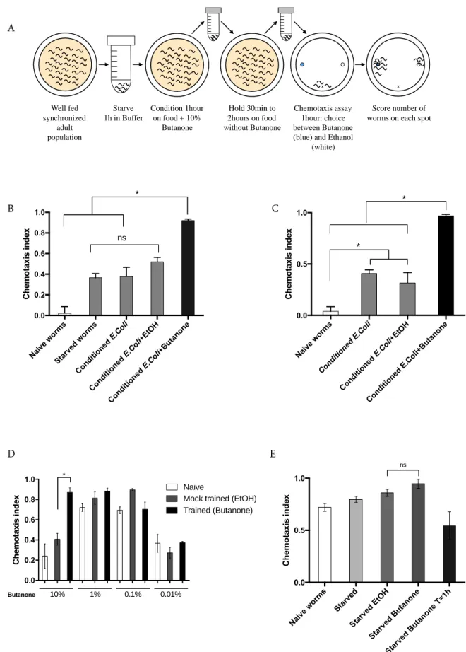
2.3.1. Chemotaxis assay ... 64
2.3.2. Optimization of the short-term memory assay: pairing of butanone and food ... 66
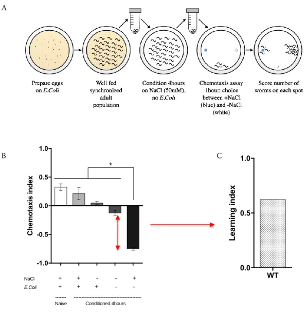
2.3.3. Optimization of the NaCl negative association assay ... 72
2.4. Lifespans ... 75
2.4.1. Synchronization of a C. elegans population ... 75
2.4.2. Lifespan ... 76
11 2.5.1. Sample collection ... 78 2.5.2. Protein extraction ... 78 2.5.3. Protein visualization ... 79 2.6. RT-qPCR ... 80 2.6.1. Sample collection ... 80 2.6.2. RNA extraction ... 81
2.6.3. Reverse Transcriptase - Quantitative PCR ... 82
2.7. Image acquisition ... 83
2.7.1. Neuronal mitochondrial reporter ... 83
2.7.2. Glutamatergic neurons reporter ... 85
2.8. Statistical analysis ... 85
3. Chapter III AMPK is required to maintain behavioral plasticity with age ... 86
3.1. Loss of AMPK function mimics age associated and Aβ1-42 induced learning deficit ... 86
3.2. AMPK activation improves learning and maintains neuronal function with age ... 96
4. Chapter IV AMPK requires mitochondrial fusion to improve behavioral plasticity 103 4.1. AMPK requires mitochondrial fusion to promote learning ... 108
4.2. AMPK modulates neuronal mitochondrial morphology and requires fusion in the neurons for learning ... 117
5. Chapter V Targeting CRTC-1 (CREB regulated transcription coactivator 1), a downstream mediator of AMPK ... 127
12
5.1. Introduction ... 128
5.2. Results ... 138
5.3. Discussion ... 150
6. Chapter VI Modulation of pre-mRNA splicing as a downstream mediator of DR .... 154
6.1. Introduction ... 154
6.2. Results ... 157
6.3. Discussion ... 165
7. Chapter VIII Conclusion: significance and future directions ... 168
7.1. Conclusion ... 168
7.2. Using C. elegans as a model for cognitive decline ... 170
7.3. Targeting AMPK ... 172
7.4. Other downstream mediator of AMPK contributing to beneficial effect ... 174
8. Supplemental Publications ... 176
13 Index Figures & Tables
FIGURE 1.1: US POPULATION >65YO REPRESENTS BOTH A PUBLIC HEALTH AND ECONOMIC BURDEN1 ... 17
FIGURE 1.2: PROJECTED LIFE EXPECTANCY AND PERCENTAGE OF AGING POPULATION IN THE WORLD1 ... 18
FIGURE 1.3: AMYLOIDOGENIC (2) VS. NON-AMYLOIDOGENIC (1) PATHWAYS. ADAPTED FROM BERRIDGE ET AL.12 ... 22
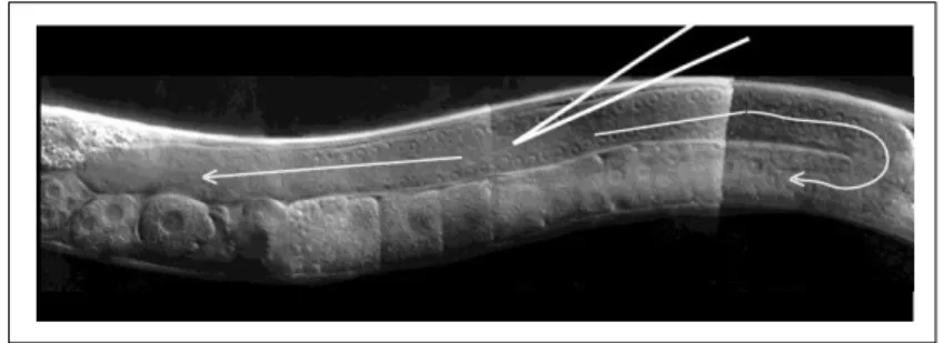
FIGURE 1.4: C. ELEGANS NERVOUS SYSTEM HIGHLIGHTED BY PAN NEURONAL GFP EXPRESSION215 ... 51
FIGURE 2.1: C. ELEGANS DISTAL GONAD, THE OPTIMAL LOCATION FOR DNA MICROINJECTION246 ... 61
FIGURE 2.2: SCHEMATIC OF CHEMOTAXIS PLATES PREPARATION249,250 ... 65
FIGURE 2.3:OPTIMIZATION OF A SHORT-TERM MEMORY ASSAY PAIRING BUTANONE AND E.COLI ... 70
FIGURE 2.4: OPTIMIZATION OF THE SHORT-TERM NACL NEGATIVE ASSOCIATION ASSAY ... 74
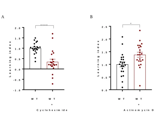
FIGURE 3.1: BEHAVIORAL PLASTICITY IN THE NACL BASED ASSAY REQUIRES TRANSLATION BUT NOT TRANSCRIPTION ... 88
FIGURE 3.2: AGE-DEPENDENT LOSS OF BEHAVIORAL PLASTICITY IN WT C. ELEGANS ... 89
FIGURE 3.3:LOSS OF AAK-2 ENHANCES AGE-RELATED DECLINE IN BEHAVIORAL PLASTICITY ... 91
FIGURE 3.4: ALZHEIMER’S DISEASE MODEL IN C. ELEGANS EXPRESSING PAN NEURONAL AΒ1-42 ... 93
FIGURE 3.5: CONSTITUTIVE ACTIVATION OF AMPK IMPROVES BEHAVIORAL PLASTICITY IN YOUNG WORMS ... 96
FIGURE 3.6: CONSTITUTIVE ACTIVATION OF AMPK RESCUES AGE-RELATED AND AΒ1-42 INDUCED NEURONAL DEFECTS ... 98
FIGURE 3.7:PHARMACOLOGICAL ACTIVATION OF AMPK VIA PHENFORMIN RECAPITULATES THE GENETIC ACTIVATION MODEL RESULTS ... 100
FIGURE4.1: ILLUSTRATION OF MITOCHONDRIAL FUSION/FISSION PROCESS IN C. ELEGANS ... 105
FIGURE 4.2: A CONNECTED MITOCHONDRIAL NETWORK IS REQUIRED FOR BEHAVIORAL PLASTICITY ... 109
14
FIGURE 4.4: AMPK REQUIRES MITOCHONDRIAL FUSION TO ENHANCE NEURONAL PLASTICITY ... 112
FIGURE 4.5: MODULATION OF MITOCHONDRIAL MORPHOLOGY DOES NOT MITIGATE AΒ1-42 INDUCED DEFICITS ... 113
FIGURE 4.6: GENERATING A PAN NEURONAL MITOCHONDRIAL REPORTER IN C. ELEGANS ... 117
FIGURE 4.7: MITOCHONDRIAL FRAGMENTATION IN THE NEURONS CAUSES THE LOSS OF BEHAVIORAL PLASTICITY ... 119
FIGURE 4.8: AMPK REQUIRES NEURONAL MITOCHONDRIAL FUSION TO ENHANCE NEURONAL PLASTICITY ... 121
FIGURE 4.9: CA-AMPK DOES NOT RESCUE MUTANT HTT INDUCED BEHAVIORAL DEFECTS ... 124
FIGURE 5.1:AMPK INHIBITS CRTC-1 IN NEURONS TO EXTEND LIFESPAN.ADAPTED FROM BURKEWITZ ET AL.296 ... 127
FIGURE 5.2: CRTC-1 PROTECTS AGAINST AΒ1-42 INDUCED CELL DEATH IN GLUTAMATERGIC NEURONS ... 140
FIGURE 5.3: CRTC-1 ACTIVATION IN NEURONS IMPAIRS BEHAVIORAL PLASTICITY ... 145
FIGURE 5.4: NEURONAL CRTC-1 S>A BLOCKS AMPK MEDIATED BEHAVIORAL PLASTICITY ... 149
FIGURE 6.1:SPECIFIC SPLICING FACTORS ARE REQUIRED FOR DR MEDIATED LONGEVITY ... 158
15 List of abbreviations
ALS: amyotrophic lateral sclerosis AMPK: AMP activated protein kinase AD: Alzheimer’s disease
C. elegans: Caenorhabditis elegans
CREB: cAMP response element binding protein
CRISPR: Clustered Regularly Interspaced Short Palindromic Repeats CRTC: CREB regulated transcription coactivator
DR: dietary restriction
DRP-1: dynamin related protein 1, C. elegans homolog of DRP1 FZO-1: fuzzy onion 1, C. elegans homolog of MFN1/2
GFP: green florescent protein HD: Huntington’s disease
ND: neurodegenerative disorders PD: Parkinson’s disease
S6K: ribosomal protein S6 kinase SFA-1: splicing factor 1
16 1. Introduction
1.1. The challenges of an aging population
1.1.1. Aging as a risk factor for chronic diseases
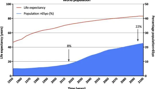
Dramatic increases in life expectancy was one of the most distinctive demographic events
of the 20th century, with 27 years added to average longevity due to advances in medicine and
public health measures. In 1955, worldwide average life expectancy was 46 years old, but by 2015
this number had risen by nearly 20 years to 65 (United Nations Report, 2015). This increase in
world life expectancy is projected to continue, as developing countries improve their health care
systems. By 2100, at least half of the human population worldwide can expect to live to 83 years
of age (United Nations Reports, 2015). Increased life expectancy will inevitably mean a change in
demographic, with a significant percentage of the population being over the age of 65 an estimated
1.5 billion people by 2050 (United Nations Report, 2015). However, a downside to the aging
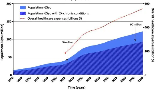
17 system which is rapidly becoming untenable. Indeed, overall healthcare expenses are expected to
exceed 500 billion dollars in the US by 2100 (Fig1.1).
Figure 1.1: US population >65yo represents both a public health and economic burden1
Age is the biggest risk factor for the vast majority of chronic diseases including some of
the biggest killers, such as metabolic diseases, cardiovascular disease and neurodegenerative
disorders. Worse still, many of these conditions often occur simultaneously in the same individual
– in the USA over half of people older than 65 years have two or more chronic conditions (Fig1.2).
Current therapeutic strategies rely on understanding the underlying mechanisms of individual
18 condition will only provide a limited increase in the “disease-free” lifespan of an elderly individual
as this person will most likely continue to suffer from multiple other age-related conditions.
Figure 1.2: Projected life expectancy and percentage of aging population in the world1
An emerging alternative approach is that of ‘geroscience’2. Instead of focusing on proximal
mechanisms of individual age-related diseases in isolation, geroscience focuses on aging as a
common risk factor, and on the cellular components that might link patient age to overall disease
risk. Although aging research is a rather young field, several genetic, environmental, and
pharmacological interventions that slow aging and decrease age-related pathology of model
organisms have been characterized. Among those, Dietary Restriction or “reduction of food intake
19 during dietary restriction (DR) increases longevity in species ranging from yeast to primates3 and
has protective effects against multiple age-onset conditions. Conserved mechanisms that integrate
upstream energy-sensing pathways with targeted downstream transcriptional targets might
therefore be ideal targets to specifically recapitulate the physiological benefits of DR for
therapeutic purposes.
1.1.2. Alzheimer’s Disease and the need to find new targets
Neurodegeneration, and in particular dementia, is one of the biggest contributors to age
related diseases and its increasing prevalence represents a major burden as these patients display
increased susceptibilities to other comorbidities and usually become dependent on care-givers
many years before death4. The most prevalent neurodegenerative disorder is Alzheimer’s Disease
(AD), a disease which currently affects 5.5 million Americans. Given the growing proportion of
elderly people in the general population (Fig1.2), an estimated 16 million Americans will suffer
from AD by 2050. AD is a leading cause of disability and poor health, representing the 6th most
prevalent cause of death in the USA and effectively costing 259 billion dollars in health care in
20
decay in cognitive abilities and in particular learning and memory. Patients also exhibit spatial and
temporal disorientation, changes in mood and behavior and at the later stages of the disease,
difficulties speaking or walking, all of which leading to a loss of autonomy (www.alz.org). Brains
of individuals suffering from AD display extracellular accumulation of amyloid fibrils and plaques
made of the toxic amyloid peptide Aβ, hyperphosphorylation of intracellular tau and neuronal cell
death5. The amyloid cascade hypothesis stipulates that AD pathogenesis is caused by the abnormal
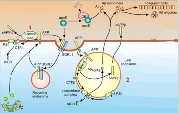
processing of APP (Amyloid Precursor Protein) thus leading to the formation and accumulation of
the toxic amyloid peptide Aβ6-8. APP is synthetized and transferred to the plasma membrane
through the ER-Golgi secretory pathway, where it can then be processed through the
amyloidogenic or non-amyloidogenic pathways (Fig1.3). Through the non-amyloidogenic
pathway, APP is cleaved at the plasma membrane by α-secretases resulting in an extracellular
fragment, the soluble APPα, and an intracellular fragment CTFα (C-Terminal Fragment). The CTFα can be hydrolyzed inside the cytoplasm by γ-secretases to release the transcription factor
peptide AICD (APP intracellular domain), the physiological role of which is still not fully
21
SORL1 leads to its internalization in recycling endosomes9. This physiological processing of APP
does not result in formation of toxic amyloid peptides and is thus called the nonamyloidogenic
pathway. When processed through the amyloidogenic pathway, cytoplasmic membrane bound
APP is internalized into late endosome where it is cleaved by the β-secretase (BACE), releasing
the N-terminal sAPPβ region into the endosome and leaving the C-terminal fragment CTFβ in the
membrane. CTFβ is then hydrolyzed at the membrane by the γ-secretase complex containing the
presenilin enzymes, yielding the toxic peptides Aβ1-40 or Aβ1-42 depending on the cleavage site.
These amyloid peptides are released in the extracellular space where they aggregate to form
oligomers or fibrils and plaques, disrupting synaptic functions by binding to cytoplasmic
membrane bound receptors and ion channels and inducing a constant state of inflammation in the
brain5,10. The brain can physiologically degrade amyloid peptides either through autophagy of the
Aβ-containing endosomes11 or through microglia-mediated phagocytosis of extracellular Aβ and
IDE (insulin-degrading enzyme)-mediated degradation. Apolipoprotein E plays a prominent role
22
endocytosis process, explaining why polymorphisms of the APOE gene, such as ApoE4 isoform,
can increase susceptibility to develop AD13,14.
Figure 1.3: Amyloidogenic (2) vs. non-amyloidogenic (1) pathways. Adapted from Berridge et al.12
Despite strong evidence that Aβ accumulation in the brain, as stated by the amyloid cascade
hypothesis, is responsible, or at least tightly linked, to the onset of AD, an increasing number of
alternative hypotheses have emerged to explain the pathogenesis of AD, as it is still unclear how
Aβ oligomers alter neuronal function and cause neurodegeneration. One strong argument in favor
of the amyloid cascade hypothesis is that the autosomal dominant familial form of AD,
1
23
characterized by early onset cognitive decline (<60 years old), is caused by mutations in key
components of the amyloidogenic pathway such as APP, ApoE4, presenilin 1-2 and SORL1. These
mutations directly link Aβ metabolism to the rapid onset of AD symptoms15-18. Interestingly,
Down’s Syndrome (or Trisomy 21) patients display accumulation of AD-like amyloid plaques, a
phenotype which could be linked to the extra copy of APP gene they carry on chromosome 2119.
In the last two decades, AD-related research has represented almost 18% of neuroscience research
efforts20 but unfortunately, since 2003, neither a new medication or an effective prevention method
has been approved by the FDA for AD21 (www.alz.org). Research has focused on the amyloid
cascade hypothesis as the main driver of AD pathogenesis and although targeting components of
the amyloidogenic pathway has yielded promising results in animal models, it has failed in human
clinical trials22.
Several observations challenge the Aβ centered view of AD: the brains of some cognitively
healthy human subjects have been described to contain amyloid plaques similar to those normally
encountered in AD patients, and Aβ actually accumulates long before clinical symptoms
hyper-24
phosphorylated tau, although present in other forms of dementia25, actually better correlate with
degradation of cognitive functions24. It has therefore become apparent that alternative hypotheses
have to be explored to determine novel therapeutic targets to test in clinical trials. Since 2009,
several GWAS studies have substantiated this idea by identifying over 20 different SNPs across
the genome mediating AD risk, involved in various cellular functions such as inflammation, lipid
metabolism or endocytosis26. Among those alternative hypotheses regarding AD pathogenesis are
i) the calcium hypothesis, linking amyloid metabolism to neuronal dysfunction12,27-30, ii) the
bioenergetics hypothesis, stating that the switch from a glucose-dependent to a ketone-based
metabolism or fatty acid oxidation is accompanied in the brain by oxidative stress, decline in
mitochondrial function, calcium overload, and general cellular malfunction31, iii) the inflammation
hypothesis, proposing chronic brain inflammation as the initiator of AD32, and iv) the microglial
hypothesis, suggesting that Aβ induced alteration in glial cells contribute to impaired glutamate
transport, calcium dysregulation and release of pro-inflammatory cytokines33.
Since current therapeutics primarily focusing on the protein misfolding aspect of AD have
25 which could participate in the pathogenesis of the disease. Epidemiological studies have shown
that obese or diabetic patients are at higher risk of developing AD, suggesting a strong association
between metabolic dysfunction and neurodegeneration34. Furthermore, animal models have
provided insight into neuronal metabolic dysregulation observed in AD and have demonstrated the
critical causal role of mitochondrial dysfunction leading to neuronal dysfunction and cellular
death35. An alternative AD therapeutic approach might therefore be to target metabolic pathways
disrupted in AD, rather than targeting the protein folding pathways or the ER stress response
induced by accumulation of toxic aggregates. To explore this hypothesis, I decided to focus on
dietary restriction, an intervention which not only extends lifespan but reduces the risk of
developing age-related diseases including neurodegenerative disorders by modulating cellular
metabolism.
1.1.3. Benefits of dietary restriction in the aging brain
McCay and colleagues were the first to demonstrate that caloric restriction (CR) could lead
to lifespan extension in rats compared to ad libitum fed rats36. This important finding was then
26 model organism tested as well as promotion of healthy aging3,37. Throughout this dissertation, we
will use the term Dietary Restriction (DR) for any intervention with reduces food intake, including
caloric restriction. DR promotes healthy aging by delaying the onset or improving the outcome of
major age-related chronic diseases such as cancer, cardiovascular disease metabolic disorder or
neurodegenerative diseases38.
Besides its beneficial effects on lifespan and the general physiology of the organism,
modulation of diet can impact brain structure, plasticity and function and affect markers of brain
physiology such as synaptic function, trophic factors or hippocampal neurogenesis39,40. The
hippocampus, a brain region known to be highly sensitive to environmental changes and
considered to be central in regulating learning and memory processes can be modulated by dietary
restriction suggesting potential benefits on cognitive functions41-43. DR seems to benefit neuronal
plasticity by improving synapse adaptation to oxidative or metabolic stress44, potentially through
differential expression of genes implicated in synaptic plasticity45. In addition, DR stabilizes the
levels of glutamate receptors and synaptic proteins required for excitatory transmission, a key
27 working memory, suggesting that the cellular changes induced by DR can translate into cognitive
benefits48,49. Similar beneficial effects have been reported upon intermittent fasting (alternate
periods of ad libitum intake with complete or partial restriction of calories) suggesting that other
interventions that modulate diet can impact cognitive outputs50.
Given that the aging brain is characterized by metabolic changes, including increased
vulnerability to toxic or metabolic insults eventually leading to impaired synaptic function such as
long-term potentiation51,52, a number of researchers have explored the possibility that DR mediated
cellular changes in the normal brain could delay or reverse age related neuronal decline. The
benefits of DR on the aging brain include the prevention of age related decline in learning and the
preservation of spatial and working memory in mice upon late onset DR53,54. Similar
improvements can be observed with either midlife onset DR55 or late stage short exposures to
DR56,57. The mechanisms of these effects could be explained by the prevention of age-related loss
28 for potential translation to humans although a life-long DR regimen is not an easily sustainable
therapeutic or preventative measure in humans.
These data linking dietary restriction to healthy brain aging have prompted health care
authorities to test the translational potential to humans. Two major population studies have been
initiated: the National Institute of Aging CALERIE trial (Comprehensive Assessment of
Long-Term Effects of Reducing Intake of Energy) and the ENCORE study (Exercise and Nutrition
Interventions for Cardiovascular Health)60,61. Although the 6 months DR regimen experimented in
the CALERIE trial did not lead to improvement of cognitive functions, the ENCORE study was
able to demonstrate enhanced executive-function learning when DR was combined with exercise62.
Moreover, Witte et al. reported that a daily 30% DR regimen in a population of elderly subjects
(>60yo) improved by ~20% the verbal memory scores compared to an ad libitum group63. This is
the first study to show an added benefit of DR on cognitive function in an elderly human population
and therefore opens new questions for potential beneficial effects of DR on age-related brain
29 Dietary interventions have been reported to enhance cognitive or neuronal functions not only
in the normal aging brain but also in the context of mood changes or anxiety and neurodegenerative
diseases such as Alzheimer’s disease50. Aβ mediated synapse dysfunction is a potential key
contributor to the disease and it has therefore been hypothesized that DR could alleviate AD
pathogenesis by preserving synaptic function64,65. A 30% DR diet was able to alleviate memory
deficits and pathophysiology of the disease in a mouse AD model, likely through the upregulation
of genes associated with neurogenesis, synaptic plasticity and downregulation of inflammatory
markers66. These results have the potential to be translated to humans as epidemiological studies
demonstrate an association between high caloric diets and risk of developing AD or suffering from
mild cognitive impairment67-69. Likewise, intermittent fasting, another dietary intervention shown
to extend lifespan, started prior to the onset of the disease phenotype, can rescue the age-related
cognitive decline in 17 months old 3xTg-AD mice70 as measured by the Morris water maze assay71.
Unfortunately, DR induces a number of detrimental side effects on growth, reproductive
30 to harness the beneficial effects of DR without suffering its negative side effects, we need to
elucidate pathways underlying the effects of DR on aging and more specifically in the brain.
Uncovering new cellular mechanisms will provide novel targets which can potentially be targeted
pharmacologically for treatment of human diseases such as AD. DR can be performed in various
ways in laboratory settings and even though different protocols of reduction of food intake can
extend lifespan, they might not all rely on the same genetic pathways3. However, most DR
mediators are metabolic regulators involved in nutrient signaling and nutrient sensing pathways
such as insulin, sirtuins or TOR/AMPK (Target of Rapamycin, AMP activated protein kinase)3.
Given the well-documented role of AMPK in the context of aging and age-related
neurodegenerative diseases, we focused our work on the role of the central energy sensor and
metabolic regulator, AMPK, as a downstream mediator of DR. Indeed, the aging process has been
associated with an alteration of AMPK signaling which normally plays a key role in maintaining
cellular homeostasis in response to various stresses. Evidence shows an age-dependent reduction
31 as changes in expression levels of the different subunits required to form the AMPK complex
(α,β,γ)74, whereas other studies point towards an alteration of AMPK capacity to respond to stress
in old animals73. The aging process is associated with a decline in AMPK activation in old versus
young rat muscles in response to activation insults75 or electrical stimuli74. Similarly, AMPK’s
response to stroke in the brain or hypoxia in the liver is lost in old mice. These observations in
mice correlate with data in the model organism the nematode Caenorhabditis elegans76, in which
the AMP:ATP ratio can be used as a lifespan predictor77. Increasing the expression of AMPK
catalytic subunit (aak-2 gene) is sufficient to increase lifespan77 and similar lifespan extensions in
C. elegans have been reported via transgenic expression of a constitutive active form of AMPK
generated by a mutation in the AMP-binding site mutant of the regulatory subunit γ78 or expression
of the truncated catalytic subunit AAK-2 (aa 1-321)79. This effect is also conserved in Drosophila,
32
1.2. Targeting AMPK in the context of neurodegeneration
1.2.1. AMPK structure
The AMP-activated protein kinase (AMPK) is a highly conserved key sensor of cellular
energy status, present in essentially all eukaryotic cells81-84. Cellular energy deficiency, as
characterized by ATP depletion, activates AMPK, which in turn inhibits anabolic reactions and
stimulates catabolic reactions in order to increase energy production and decrease cellular ATP
usage. AMPK is a heterotrimeric complex which consists of an α catalytic subunit and β and γ
regulatory subunits85. In mammals, these subunits have several isoforms, which are differently
expressed in different tissues. Phosphorylation of the α catalytic subunit at the Threonine-172
residue (Thr-172) is required for the activation of AMPK86 and induces its kinase activity greater
than 100-fold. Three upstream kinases target this phosphorylation site to activate AMPK: Liver
Kinase B1 (LKB1) which is predominant, especially in the context of energy stress87-90,
Ca2+/Calmodulin-dependent kinase kinase β (CAMKKβ)91,92 and transforming growth
factor-β-activated kinase 1 (Tak1)93,94. On the other hand, the phosphorylated Thr-172 residue can be
33 2A (PP2A) and Protein Phosphatase 2C (PP2C)95-97. The activity of AMPK is not only dependent
on upstream stimulatory and inhibitory signals but is also dependent on cellular energy status, as
nucleotide binding acutely tunes AMPK activation. The γ regulatory subunit contains four
potential nucleotide-binding sites, which can either bind AMP, ADP or ATP. Under low energy
conditions when the AMP/ADP:ATP ratio increases, AMP and ADP compete with ATP to bind
AMPK85. Three consequences arise from this nucleotide binding: allosteric activation of AMPK
2-10 fold, promotion of Thr-172 phosphorylation by upstream kinases98 and inhibition of Thr-172
de-phosphorylation by protein phosphatases most likely through conformational changes85,99. The
binding of ATP antagonizes these effects, therefore making AMPK a very sensitive sensor of
AMP:ATP and ADP:ATP levels.
1.2.2. AMPK downstream targets
When activated, AMPK modulates downstream signaling pathways in order to replenish
cellular ATP supplies on one side and reduce ATP-consuming biosynthetic processes on the other
side. Studies of AMPK downstream targets led to the identification of a conserved sequence motif
-34 3 positions relative to the phospho-acceptor site and hydrophobic residues at -5 and +4 positions
seem to be important for the motif recognition100-103. In order to generate more ATP, AMPK
enhances glucose uptake in muscles by inducing the translocation of the glucose transporter
GLUT4 to the plasma membrane82. The intracellular storage vesicles containing GLUT4 require
the RAB family of proteins to be GTP-bound104. In the contracting muscle, AMPK phosphorylates
the RAB-GAP protein TBC1D1, which normally maintains the RAB proteins in the inactive
GDP-bound state. Phosphorylation by AMPK induces its association with 14-3-3 protein and subsequent
dissociation from the vesicles, triggering the conversion of the RAB protein to its active
GTP-bound form and the fusion of the GLUT4 carrying vesicles to the plasma membrane104-107. In
addition to GLUT4, AMPK also promotes glucose entry via activation of another plasma
membrane-located glucose transporter, GLUT1, expressed in most cells other than muscle, liver
and adipose tissue108. Finally, AMPK activates glycolysis via phosphorylation of PFKFB
(PFKFB2 isoform, 6-phosphofructo-2-kinase/fructose-2,6-biphosphatase) in cardiac myocytes or
monocytes and macrophages. Active phosphorylated PFKFB in turn, catalyzes the generation of
(6-35 phosphofructo-1-kinase)109,110. In addition to upregulating glucose catabolism, AMPK inhibits its
anabolism by inhibiting the glycogen synthesis enzyme, glycogen synthase111 as well as other
gluconeogenic enzymes such as phospho-enolpyruvate carboxykinase and
glucose-6-phosphatase112,113. AMPK downregulates the expression of these gluconeogenic enzymes by
phosphorylating and excluding from the nucleus the transcription factor CRTC2 (CREB-regulated
transcription co-activator 2)1 and the class IIa HDAC (histone deacetylase), which normally
deacetylates and activates the FOXO transcription factors113. Regarding lipid metabolism, AMPK,
on one hand, upregulates fatty acid uptake and β-oxidation and on the other hand downregulates
triglycerides and fatty acids synthesis pathways and lipogenesis. AMPK promotes fatty acid uptake
first in the cell, as shown in cardiomyocytes via translocation of CD36-containing vesicles114 and
second, into the mitochondria, by phosphorylation and subsequent inactivation of ACCβ
(acetyl-CoA carboxylase), leading to a reduction in the inhibitor of mitochondrial fatty acid entry,
malonyl-CoA115. AMPK phosphorylates and inhibits the lipogenic transcription factor SREBP1c
(sterol regulatory element binding protein)116. In addition to this master regulator of lipogenesis,
acyl-36 transferase (GPAT) or 3-hydroxy-3-methylglutaryl CoA reductase (HMGR) in order to inhibit
fatty acid synthesis, triglyceride and phospholipid synthesis or cholesterol synthesis
respectively117-119. AMPK modulates protein metabolism by reducing protein synthesis on one side
and increasing protein degradation through autophagy on the other side. AMPK represses protein
synthesis both directly, by phosphorylation of eEF2 (eukaryotic elongation factor 2) upstream
inhibitory kinase120 and indirectly, by inhibiting the mTOR pathway through phosphorylation of
TSC2 (tuberous sclerosis 2) or RAPTOR (regulatory-associated protein of mTOR)121,122.
Subsequent inhibition of mTOR downstream targets S6K and 4E-BP impacts translation initiation,
therefore affecting protein synthesis123. AMPK also acts at the transcriptional level by inhibiting
the transcription factor TIF-IA (transcription initiation factor IA), responsible for the expression
of the rRNA producing enzyme, RNA Polymerase I124. Recycling of organelles and cytosolic
content through autophagy is a general catabolic response to starvation, also activated by AMPK,
both in mammals and in yeast125. AMPK is able to form a stable complex with ULK1/2 proteins
(UNC-51-like kinase 1)126 and phosphorylate them127,128. This triggers autophagy through the
37 AMPK has been shown to phosphorylate Beclin-1 to promote the formation of “pro-autophagy”
VPS34 complex at the expense of “non-autophagy” VPS34 complexes involved in intracellular
vesicle trafficking130.
Loss of mitochondrial homeostasis has been correlated with the aging process and it is
therefore not surprising to find interactions between AMPK and mitochondrial biology. AMPK
drives the switch from carbohydrates to lipids as the main energy source in response to fasting and
exercise in skeletal muscle cells131. AMPK transcriptionally induces mitochondrial biogenesis by
modulating the activity of the “master regulator” of mitochondrial biogenesis, PGC-1α
(Peroxisome proliferator-activated receptor Gamma Coactivator-1 α), a transcription factor which
enhances the expression of nuclear-encoded mitochondrial genes132. AMPK directly
phosphorylates PGC-1α, which leads to its own transcription through a positive feedback loop133.
A second mechanism has been described, where AMPK promotes SIRT1 (sirtuin1) activity by
increasing the cellular concentrations of NAD+, leading to increased deacetylation and activation
38 mitochondria, a process known as mitophagy. Indeed, inhibition of ULK1 phosphorylation by
AMPK leads to the accumulation of mitochondria with abnormal morphology and reduced
membrane potential, suggesting that AMPK has a role in maintaining efficient, ATP-generating,
mitochondria by removal and recycling of the damaged mitochondria127. Finally, it was more
recently demonstrated in our laboratory that AMPK regulates mitochondrial morphology (fusion
and fission process) to maintain mitochondrial network homeostasis during aging and preserve
functional coordination with peroxisome to promote fatty acid oxidation135. As a consequence of
this regulation, loss of muscle AMPK activity in a murine model (AMPKβ1/AMPKβ2 double
knockout) leads to reduced mitochondrial content and diminished muscle performance136, whereas
activation of AMPK via AICAR (5-AminoImidazole-4-CArboxamide Ribonucleotide) increases
mitochondrial genes expression in muscles and confers beneficial effects in term of exercise
endurance to “sedentary mice”137,138.
1.2.3. AMPK neuronal functions
An increasing number of publications suggest a very prominent role for AMPK both for
39 neurodegenerative diseases76. A cross sectional population study in Korean subjects aged >60
years old shows a significant association between the AMPK γ2 gene polymorphism (PRKAG2
-26C/T) and cognitive impairment in old age, independent of the presence of diabetes. Consistent
with the hypothesis that metabolic dysregulation is tightly link to cognitive dysfunction, this SNP
has previously been associated with diabetes139. While the role of AMPK as energy sensor and
metabolic regulator in tissues like liver, muscle and adipose tissue has been described, it is also
present in the mammalian brain 140-142 and its functions in the nervous system and more specifically
in memory formation have only recently gained attention143-145. It seems intuitive to hypothesize
an important role for AMPK in neurons due to the fact that the brain combines a high metabolic
rate with a poor nutrient storage capacity, making it vulnerable to large energy variations. AMPK
is a modulator of Long Term Potentiation (LTP)146, and has been shown to increase
NMDA-induced K-ATP currents by a Ca2+-dependent process, which as a consequence diminishes the
excitatory effect of glutamate-mediated transmission in rat neurons147. Further characterizing the
synaptic role of AMPK, a group showed a decrease in the levels of LKB1 – an AMPK upstream
40 formation, deletion of either kinase led to aberrant axonal retraction, extension of postsynaptic
dendrites and formation of ectopic synapses in retinal neurons whereas genetic and
pharmacological (metformin, caloric restriction) activation of AMPK protects against age-related
synaptic alterations in those neurons148. Consistent with its role as a key metabolic modulator,
AMPK has protective effects against metabolic and excitotoxic insults. The AMPK agonist
AICAR protects rat hippocampal neurons from various stresses such as glucose deprivation,
chemical hypoxia, glutamate exposure and Aβ toxicity141. GABA (γ-AminoButyric Acid), the
major inhibitory neurotransmitter in the brain, might mediate AMPK’s protective effect. AMPK
activates GABAB receptors which then modulate post-synaptic K+ signaling as well as
pre-synaptic Ca2+ channels, to suppress neuronal excitation, thus exerting a neuroprotective effect
during metabolic insults149,150.
Despite these publications, the beneficial or detrimental role of AMPK in neuronal function
still needs to be further addressed, as there is evidence of a deleterious role of AMPK activation
in the context of stroke damage in a murine model of cerebral artery occlusion151. Highlighting
41 young and old mice and correlates with the induction of neuronal development and plasticity
genes152, or impair the formation of long term fear memory142. Indeed, a decrease in AMPK
activity following contextual fear conditioning is required for fear memory formation142. These
discrepancies might be explained by the different mechanisms involved in the formation of spatial
versus fear memory. AMPK proved to have an even more prominent role in the context of
neurodegenerative diseases and more specifically Alzheimer’s Disease. AMPK is required downstream of two interventions known to ameliorate the pathology of Alzheimer’s Disease:
leptin153 and resveratrol154. Inhibition of Aβ production and tau phosphorylation by leptin and
extracellular Aβ accumulation by resveratrol in neuronal cultures both required AMPK and could
be replicated using pharmacological activators of AMPK153,154. Likewise, AMPK activation
inhibits palmitate-induced apoptosis and tau hyperphosphorylation in SH-SY5Y cells155.
Contradicting with the potential beneficial role of AMPK in Alzheimer’s Disease, AMPK is a tau
kinase, which could potentially contribute to disease pathogenesis by altering microtubule binding
of tau156. AMPK abnormally accumulates in tangle and pre-tangle bearing neurons in major
42 (Ca2+/calmodulin-dependent protein kinase kinase β) leads to increased phosphorylation of tau at
Ser262/Ser356/Ser396156,157. These observations were confirmed in vivo in an AMPKα2 deficient
mouse model, which displayed reduced levels of endogenous tau phosphorylation158. Supporting
a contribution to the pathology of Alzheimer’s Disease, AMPK activation, through the
transcriptional upregulation of BACE1, significantly increases the generation of intracellular and
extracellular Aβ peptides159. Despite convincing evidence that AMPK is a tau kinase, these
findings do not address the functional impact of such phosphorylation in vivo on cognitive
functions. It is therefore difficult to assess whether this increased tau phosphorylation by AMPK
contributes to the memory loss or neurodegeneration observed in Alzheimer’s Disease. Ma et al.
sought to study the effect of AMPK inhibition on long term potentiation (LTP) and long-term
depression (LTD) in APP/PS1 transgenic mice, a process considered as the cellular basis for
learning and memory formation. They showed a negative contribution of AMPK as treatment of
this murine Alzheimer’s model with the AMPK inhibitor compound C or genetic deletion of the
43 behavioral data clearly showing beneficial effects of activation of AMPK on memory
formation161,162.
These discrepancies between the behavioral and the cellular effects of AMPK in neurons
point to the gap in the understanding of the mechanisms of action of AMPK and the need to further
characterize the downstream targets of AMPK mediating its behavioral benefits. Further
highlighting the complex role of AMPK in cognitive functions, polyQ-expanded mutant huntingtin
(HTT) protein causing Huntington’s Disease induces abnormal activation of AMPK in both human
and mice striatum neurons. The aberrant AMPK activation contributes to the progression of the
disease by increasing oxidative stress, brain atrophy, neuronal loss and mutant HTT
aggregation163,164. Similarly, in Parkinson’s Disease cellular models or Drosophila models, AMPK
activation was both characterized as detrimental165 or beneficial166,167 in disease pathogenesis as
measured by neuronal cell death. Modulation of AMPK in amyotrophic lateral sclerosis (ALS),
another neurodegenerative disease caused by selective loss of motor neurons, also generated
conflicting results, making it unclear whether AMPK activity is abnormally activated or inhibited
44 the question of AMPK’s impact on cognitive function is not restricted to neurodegeneration
models as pharmacological activation of AMPK by metformin can rescue memory deficits induced
by brain ischemia172 or traumatic brain injury173.
Given the evidence that Type 2 Diabetes (T2D), or more broadly metabolic dysfunction, is
a risk factor for the development of neurological diseases such as stroke, vascular dementia or
Alzheimer’s Disease, anti-diabetic drugs such as metformin are being explored as potential
therapies for neurodegenerative conditions174. Metformin is a medication belonging to the
biguanide class175-177, used to treat diabetes. Metformin lowers hyperglycemia by inhibiting
hepatic glucose production, increasing insulin sensitivity and enhancing peripheral glucose uptake
in muscle178. Even though the mechanism of action of metformin is not fully characterized179, it
has very clear benefits on the risk of metabolic syndrome and T2D180,181 and is often used to
enhance AMPK activity by increasing its Thr-172 phosphorylation. Metformin per os crosses the
blood brain barrier and activates AMPK in mouse brain182 and can therefore be used in animal
45 the impact of metformin in T2D mice models (High Fat Diet or db/db mutants) and show improved
behavioral response in protocols such as the Morris Water Maze, which measures spatial memory
formation, both after short term and long term metformin supplementation183-185. In db/db mutants
(Leptin Receptor deficient mice), metformin treatment reduces the accumulation of neuronal Aβ
1-42 and phosphorylated tau (p-tau) in the brain186, potentially by decreasing Aβ1-42 influx and
rescuing Aβ1-42 efflux through the blood brain barrier185. Similarly, metformin can reduce cellular
stress markers induced by Aβ1-42 in human neuronal stem cells187. In a Type 1 Diabetes (T1D)
model (induced by streptozotocin injections), metformin treatment only slightly improves the mice
response in the T-maze test, however it normalizes a number of neuronal stress markers such as
FOX-1 and NeuN (neuronal survival markers) or IL-1beta (inflammation marker)188,189. To further
confirm the beneficial role of metformin in the context of neurodegenerative diseases, evidence
shows that metformin may preserve against dopaminergic neurons degeneration in a MPTP
(1-methyl-4-phenyl-1,2,3,6-tetrahydropyridine) induced Parkinson’s Disease mouse model190 and
protect striatal cells from mutant huntingtin protein-induced toxicity and mitochondrial
46 of the cholinergic neuronal pathway and memory circuits in the brain, impairing memory, thus
simulating Alzheimer’s Disease cholinergic deficits191. Mostafa et al. used this model to test the
effect of metformin on cognitive function in a rat model of memory deficit independently of insulin
dysfunction. Metformin treatment was associated with improved response in spatial memory tests
as well as a reduction of inflammation in the hippocampus and cortex192. Supporting the idea that
metformin might have beneficial effects on cognitive functions more broadly, metformin treatment
can rescue memory deficits induced by injections of pentylenetetrazole (epilepsy mice model)193,
chronic L-methionine administration (model of oxidative stress in the brain)194 or transient
forebrain global ischemia195. In this latter model, the authors demonstrated that metformin required
AMPK to improve the behavioral response. Despite these positive observations, metformin
administration does not always improve cognitive functions, whether in a db/db186 or WT
background183,193,196. Metformin is able to penetrate the brain and activate AMPK, however the
downstream mechanisms required for its effects on memory formation remain unknown. Wang et
al. were the first to show that metformin can improve spatial memory formation via enhanced
47 activation of the atypical PKC-CBP pathway - a pathway already known for its role in neural
precursors differentiation198 - both in rodent and human cell cultures, and show that this
neurogenesis is required for metformin-induced beneficial effects on neural functions.
Interestingly, in the liver, the aPKC-CBP pathway is downstream of the metformin-activated
kinase, AMPK197,199.
Finally, these results have translated to humans as several studies now point to the potential
beneficial effect of metformin on cognitive performance. In a clinical study including patients
suffering from depression and T2D, metformin improved cognitive performance and the
depressive symptoms200. More recently, results of a pilot clinical trial for metformin in treating
mild cognitive impairment (NCT00620191) suggest a beneficial effect of metformin treatment in
a cohort of overweight or obese patients, aged over 55 years old, suffering from amnestic mild
cognitive impairment (AMCI). These results have to be interpreted with caution due to the small
sample size, however they justify the conduct of a larger clinical trial to fully assess the beneficial
role of metformin in cognitive disorders201. Likewise, a longitudinal cohort study including 1814
48 suggests that metformin can provide some level of protection against cognitive impairment and
more specifically in regards of verbal learning, working memory and executive function202. Not
all results are positive though and the effects of long term usage of metformin should be described
as a case-control study suggests a potentially increased risk of developing Alzheimer’s Disease
after long term usage of metformin in a diabetic population203. Another clinical study in patients
aged over 50 years old showed an alteration of cognitive performance in patients with T2D
compared to healthy patients. Metformin seemed to further impair the cognitive functions as
assessed by the MMSE (Mini Mental State Examination) score. However, this study fails to
properly control for the severity of T2D and metformin intake for each patient204.
Thesis overview
In order to study the metabolic role of AMPK in the context of neurodegenerative disease, we
sought a model organism which could recapitulate age related phenotypes and especially age
related neuronal function decline, where human neurodegenerative diseases could be modelled and
49
1.3. C. elegans as a model organism
1.3.1. C. elegans as an aging model
C. elegans is a ~1mm soil, free-living nematode found all over the globe that primarily
feeds on bacteria such as E. Coli. They have a very short reproductive cycle of 3 days and are able
to lay several hundred eggs per individual, which is an advantage in lab settings where large
synchronized populations can be generated very quickly205. C. elegans has a lifespan of about 3
weeks and recapitulates a number of age related phenotypes during that time which explains why
it became a major model system in the aging research field. These nematodes are usually found as
hermaphrodites but males can also be generated and exploited for genetic crosses. Although C.
elegans has a rather simple morphology and is made up of only 959 somatic cells, it displays
specialized tissues such as cuticle, muscle tissue, neuronal tissue or intestinal tissue serving as a
metabolic organ (wormatlas.org). The worm’s compact genome has been mapped and sequenced
which led to the generation by various research labs of thousands of publicly available genetic
mutants as well as a genome wide RNAi library (Ahringer Library). More recently, the
50 effectively adapted to C. elegans use206. Besides these tools, C. elegans genome bears between
40% and 70% homology with the human genome, making it a relevant tool for basic research.
Finally, C. elegans is a transparent organism and allows for in vivo imaging of any fluorescently
labelled protein in the animal, a major advantage comparing to mouse studies. Finally, given that
this model organism can recapitulate key features of aging or neurodegenerative disease and is
readily amenable to small molecule screening studies, it can be effectively used for high
throughput drug screens.
1.3.2. C. elegans learning and memory / nervous system
Over the past few decades, C. elegans has been widely used as a neuroscience model as
well a powerful model system to uncover novel mechanisms underlying neurodegenerative
processes. Malleable model systems such as C. elegans are much needed in a field where few
efficient curative therapies exist due to the insufficient characterization of cellular pathways
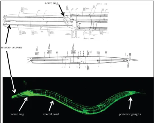
contributing to pathogenesis. The adult hermaphrodite C. elegans nervous system is comprised of
302 neurons belonging to two distinct groups: a large somatic nervous system (282 neurons) and
51 of most neurons are located in the nerve ring or the various ganglia in the head, body, tail of the
worm. Despite the widespread prejudice (even among scientists!) that nematodes are not capable
of complex behaviors, C. elegans actually display a variety of elaborate behaviors, ranging from
basic locomotion, foraging or feeding211 to discrimination between different chemicals, odorants,
temperatures or other external stimuli which can lead to a modification of their overall behavior
212-214.
52 Each of the sensory system in C. elegans (mechanosensation, chemosensation, and
thermosensation) can be involved in forms of learning and memory and lead to plasticity in the
behavior as reported in adaptation, habituation or associative learning assays216. Besides the
advantages of using C. elegans as an aging model, the nematode has a nervous system easily
accessible to genetic manipulation or in vivo imaging and bears striking similarities with vertebrate
neurons. Indeed, C. elegans uses the same major neurotransmitters, acetylcholine, glutamate,
γ-aminobutyric acid (GABA), serotonin, and dopamine and its vesicular transporters and ion
channels function in a similar fashion as in vertebrates217,218. Furthermore, the location of chemical
synapses is 75% reproducible between individuals thus decreasing variability when experimenting
on large number of worms (www.wormatlas.org). Since Kandel and colleagues’ work on the
marine mollusk Aplysia219, the biological basis of learning has been proven to be highly conserved
between invertebrates and mammals220,221. Learning and memory behavior in C. elegans has been
categorized in 3 groups: habituation – or non-associative learning- whereby the worm’s initial
response to a stimulus progressively declines as it is exposed to the stimulus repeatedly, associative
53
imprinting, a process taking place during larval development222. C. elegans has been used as a
model to study a number of human neurodegenerative diseases such as: Alzheimer’s Disease (AD),
Huntington’s disease (HD), Amyotrophic lateral sclerosis (ALS), Parkinson’s disease (PD),
tauopathy or other forms of protein aggregation223. These models constitutively overexpress either
the wild type, mutated or truncated forms of the human proteins involved in the neurodegenerative
disease they model. Depending on the strains, their expression is driven either by a body wall
muscle promoter or a neuronal promoter. In case of body wall muscle promoter (eg: unc-54p),
protein aggregation as well as dysfunctional motility or progressive paralysis can be assayed
224-227. More interestingly, in case of neuronal expression, protein can be expressed in all neurons (eg:
rab-3p, unc51p, snb-1p, aex-3p)228-234 or in a specific subset of neurons. By expressing the protein
of interest in specific neurons such as mechanosensory neurons, motor neurons (acr-2p, mec-3p,
mec-7p, unc-30p) or sensory neurons (osm-10p), targeted functional assays can be performed, in
this case, scoring worms for their response to mechanic stimuli235-238. Likewise, taking advantage
of the fact that the C. elegans cell lineage is fully characterized, the protein of interest can be driven
54 (dopaminergic neurons) or eat-4p (glutamatergic neurons), which allows for precise scoring of
neuronal loss and characterization of neurodegenerative processes such as abnormal neuron
morphology and axonal defects237,239. Genetic screens can then be performed on different worm
models to identify conserved genes modifying disease progression with the ultimate goal of
55
The failure in human clinical trials of therapeutic approaches targeting primarily the aggregation and protein misfolding aspect of neurodegenerative diseases such as Alzheimer’s Disease has prompted the scientific community to search for novel targets. Evidence from the literature points towards dysregulation of cellular metabolism as a potential causal factor in these brain diseases. Dietary Restriction, a potent intervention for lifespan extension, can also delay the onset of a number of age related disorders, potentially through its effect on multiple metabolic pathways. Unfortunately, dietary restriction does not seem like a viable therapeutic option given its detrimental side effects and compliance difficulty. I therefore focused on one downstream mediator of DR, AMPK, which not only extends lifespan in C.
elegans and Drosophila but also seems to have a prominent role in the mammalian brain. In
my thesis work, I investigated the role of AMPK in the nervous system in C. elegans in the context of aging and age-related disorders such as Alzheimer’s Disease. Given the contradictory evidence from the literature, I first asked whether activation of AMPK, though beneficial for longevity, was beneficial or detrimental in a C. elegans model of age-induced neuronal dysfunction. I then sought to understand which of AMPK’s multiple downstream targets potentially mediated its effects in neurons.