HAL Id: hal-02493967
https://hal.sorbonne-universite.fr/hal-02493967
Submitted on 28 Feb 2020
HAL is a multi-disciplinary open access
archive for the deposit and dissemination of
sci-entific research documents, whether they are
pub-lished or not. The documents may come from
teaching and research institutions in France or
abroad, or from public or private research centers.
L’archive ouverte pluridisciplinaire HAL, est
destinée au dépôt et à la diffusion de documents
scientifiques de niveau recherche, publiés ou non,
émanant des établissements d’enseignement et de
recherche français ou étrangers, des laboratoires
publics ou privés.
ameliorates Duchenne muscular dystrophy in mouse
models
Lu Yu, Xiaoli Zhang, Yexin Yang, Dan Li, Kaiyuan Tang, Zifan Zhao,
Wanwan He, Ce Wang, Nirakar Sahoo, Kimber Converso-Baran, et al.
To cite this version:
Lu Yu, Xiaoli Zhang, Yexin Yang, Dan Li, Kaiyuan Tang, et al.. Small-molecule activation of
lysoso-mal TRP channels ameliorates Duchenne muscular dystrophy in mouse models. Science Advances ,
American Association for the Advancement of Science (AAAS), 2020, 6 (6), pp.eaaz2736.
�10.1126/sci-adv.aaz2736�. �hal-02493967�
D I S E A S E S A N D D I S O R D E R S
Small-molecule activation of lysosomal TRP channels
ameliorates Duchenne muscular dystrophy
in mouse models
Lu Yu1, Xiaoli Zhang1, Yexin Yang1, Dan Li1,2, Kaiyuan Tang1, Zifan Zhao1, Wanwan He1,2,
Ce Wang1, Nirakar Sahoo1,3, Kimber Converso-Baran4, Carol S. Davis4, Susan V. Brooks4,
Anne Bigot5, Raul Calvo6, Natalia J. Martinez6, Noel Southall6, Xin Hu6, Juan Marugan6,
Marc Ferrer6, Haoxing Xu1*
Duchenne muscular dystrophy (DMD) is a devastating disease caused by mutations in dystrophin that com-promise sarcolemma integrity. Currently, there is no treatment for DMD. Mutations in transient receptor potential mucolipin 1 (ML1), a lysosomal Ca2+ channel required for lysosomal exocytosis, produce a DMD-like phenotype. Here, we show that transgenic overexpression or pharmacological activation of ML1 in vivo facilitates sarcolemma repair and alleviates the dystrophic phenotypes in both skeletal and cardiac muscles of mdx mice (a mouse model of DMD). Hallmark dystrophic features of DMD, including myofiber necrosis, central nucleation, fibrosis, elevated serum creatine kinase levels, reduced muscle force, impaired motor ability, and dilated cardiomyopathies, were all ameliorated by increasing ML1 activity. ML1-dependent activation of transcription factor EB (TFEB) corrects lysosomal insufficiency to diminish muscle damage. Hence, targeting lysosomal Ca2+ channels may represent a promising approach to treat DMD and related muscle diseases.
INTRODUCTION
Duchenne muscular dystrophy (DMD), an X-linked inherited muscle disease (1), is caused by loss-of-function mutations affecting dys-trophin, a large cytoplasmic protein that connects the cytoskeleton with extracellular matrix proteins via the muscle membrane (sarco-lemma) (2, 3). The fragile sarcolemma that makes human DMD muscles prone to contraction-induced muscle damage is mimicked in the striated muscles of the mdx mouse, a murine model of DMD (4–6). Although gene therapy approaches, including CRISPR-Cas9 technology (7), have provided hope for potential treatment to some specific variants of DMD, the existence of thousands of DMD-causing dystrophin mutations remains a major challenge (1). To develop a therapeutic approach that would be more broadly applicable to all forms of DMD, it is necessary to understand the common pathological mechanisms underlying the condition (4).
Muscle cells are especially sensitive to membrane damage, and recent studies have identified impairment of membrane repair ca-pability as an important cause of muscular dystrophies (8–10). For instance, we recently found that mice lacking transient receptor potential mucolipin 1 (TRPML1/MCOLN1; ML1), a lysosomal Ca2+ release channel required for lysosomal exocytosis (11, 12), display early-onset, progressive dystrophies similar to DMD (13). When ML1 is pharmacologically inhibited or genetically inactivated, membrane resealing is impaired in skeletal muscles (13). Hence, lysosomes may
provide a major source of membranes for repairing damaged sarco-lemma, and ML1 is essential for Ca2+-dependent delivery of lyso-somal membranes (i.e., lysolyso-somal exocytosis) to damaged sites (13). Studies of other muscle diseases have also connected lysosomal dys-function with muscle pathogenesis (14).
In the current proof-of-concept study, we examined whether up- regulation of ML1, by genetic or pharmacological methods, is sufficient to increase lysosomal functions and sarcolemma repair. If so, ML1 may be a putative target for amelioration of muscle damage in vivo. RESULTS
Transgenic overexpression of ML1 ameliorates muscular dystrophies in mdx mice
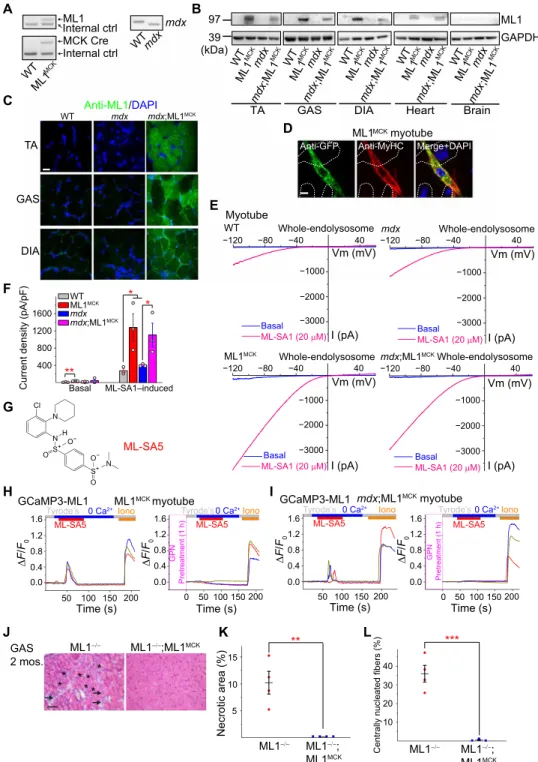
To achieve muscle-specific overexpression of ML1 in vivo, we crossed a mouse line carrying a GCaMP3-ML1 transgene (ML1 ROSA-lSl) (15) with a Cre line driven by the muscle-specific creatine kinase (CK) promoter (MCK-Cre) (16). The resultant ML1 ROSA-lSl;MCK-Cre (abbreviated as ML1MCK) progeny were then crossed with mdx mice with a loss-of-function dystrophin mutation (17) to generate mdx;ML1MCK mice (Fig. 1A and fig. S1A). Western blotting and im-munofluorescence analyses with anti-ML1 antibody revealed ML1 overexpression in both skeletal and cardiac muscle tissues of the mdx;ML1MCK mice; isolated primary myotubes were ML1 immuno-positive, whereas nonmuscle tissues showed barely detectable im-munoreactivity (Fig. 1, B to D, and fig. S1, B and C).
Whole-lysosomal ML1 currents, activated by TRPML-specific synthetic agonists (ML-SAs) (12, 15, 18), were 4 to 10 times larger in the ML1MCK myotubes than in wild-type (WT) controls (Fig. 1, E and F). ML-SAs induced robust glycyl-l-phenylalanine 2-naphthylamide–
sensitive lysosomal Ca2+ release (15) in ML1MCK myotubes, suggesting that genetically overexpressed ML1 channels were functionally localized on the late endosomal and lysosomal membranes of muscle cells (Fig. 1, G to I, and fig. S1, D and E). Introducing ML1MCK into the
1Department of Molecular, Cellular, and Developmental Biology, University of Michigan,
4114 Biological Sciences Building, 1105 North University, Ann Arbor, MI 48109, USA.
2Collaborative Innovation Center of Yangtze River Delta Region Green
Pharmaceu-ticals, Zhejiang University of Technology, Hangzhou 310014, China. 3Department of
Biology, The University of Texas Rio Grande Valley, 1201 W University Dr., Edinburg, TX 78539, USA. 4Department of Molecular and Integrative Physiology, University of
Michigan, Ann Arbor, MI 48109, USA. 5Sorbonne Université, INSERM, AIM, Center for
Research in Myology, UMRS974, GH Pitié-Salpétrière, 75651 Paris Cedex 13, France.
6NIH/NCATS/NCGC, 9800 Medical Center Drive, Rockville, MD 20850, USA.
*Corresponding author. Email: haoxingx@umich.edu
Copyright © 2020 The Authors, some rights reserved; exclusive licensee American Association for the Advancement of Science. No claim to original U.S. Government Works. Distributed under a Creative Commons Attribution NonCommercial License 4.0 (CC BY-NC). on February 28, 2020 http://advances.sciencemag.org/ Downloaded from
B C D WT WTmdx ML1 Internal ctrl MCK MCK Cre Internal ctrl mdx ML1 H
Anti-GFP ML1Anti-MyHC Merge+DAPI
MCKmyotube E mdx;ML1MCK DIA GAS WT Anti-ML1/mdxDAPI TA I A WT Whole-endolysosome −120 −80 −40 −120 −80 −40 −120 −80 −40 −120 −80 −40 40 −1000 −2000 −3000 Basal ML-SA1 (20 M) Vm (mV) I (pA) 40 ML1MCK Whole-endolysosome Basal ML-SA1 (20 M) Vm (mV) I (pA) ML1MCK myotube Tyrode’s ML-SA5 0 Ca2+Iono F/ F0 0.0 0.4 0.8 1.2 1.6 50 100 150 200 Time (s) F/ F0 0.0 0.4 0.8 1.2 1.6 Tyrode’sML-SA50 Ca 2+Iono 50 100 150 200 Time (s) 0 GPN Pretreatment (1 h)
GCaMP3-ML1 GCaMP3-ML1 mdx;ML1MCK myotube
Tyrode’s ML-SA5 0 Ca2+ Iono F/ F0 0.0 0.4 0.8 1.2 1.6 50 100 150 200 Time (s) F/ F0 0.0 0.4 0.8 1.2 1.6 Tyrode’sML-SA50 Ca 2+Iono 50 100 150 200 Time (s) 0 GPN Pretreatment (1 h) F mdx;ML1MCKWhole-endolysosome 40 Basal ML-SA1 (20 M) Vm (mV) I (pA) mdx Whole-endolysosome 40 −1000 −2000 −3000 −1000 −2000 −3000 −1000 −2000 −3000 Basal ML-SA1 (20 M) Vm (mV) I (pA) WT ML1MCK mdx mdx;ML1MCK
Current density (pA/pF)
400 800 1200 1600 Basal ML-SA1–induced ** * *
TA GAS DIA Heart Brain WT ML1 MCKmdx mdx ;ML1 MCK 39 97 ML1 MCK ML1 MCK ML1 MCK mdx mdx mdx WT WT WT mdx ;ML1 MCK mdx ;ML1 MCK mdx ;ML1 MCK GAPDH ML1 (kDa) ML1 MCKmdx WT mdx ;ML1 MCK Myotube ML1 ML1 ;ML1MCK GAS 2 mos. * ** **** J K L Necrotic area (%) 5 10 15 ML1 ML1 ; ML1MCK **
Centrally nucleated fibers (%)
10 20 30 ML1 ML1 ; ML1MCK 40 *** N N S+ O O− S+ N O O− Cl H ML-SA5 G
Fig. 1. Muscle-specific transgenic overexpression of ML1. (A) PCR genotyping of the mdx mutation, GCaMP3-ML1 transgene, and MCK-Cre. (B) Western blotting with anti-ML1 antibody in brain and various skeletal muscle tissues, including GAS, TA, and DIA from WT, ML1 ROSA-lSl;MCK Cre (ML1MCK), mdx, and mdx;ML1MCK mice (see
fig. S1B for the source files). GAPDH served as the loading control. Note that MCK-Cre is selectively expressed in differentiated myotubes (16), and its expression in the muscle tissue might be affected by the degree of dystrophies. (C) Immunofluorescence analysis of TA, GAS, and DIA cryosections from various transgenic mice. Scale bar, 10 m. (D) Immunofluorescence analysis of primary myotubes isolated from ML1MCK mice. Scale bar, 10 m. (E) Whole-endolysosome ML1 currents (I
ML1) were activated by
ML-SA1 (20 M), a synthetic agonist of ML1, in primary myotubes harvested from WT, ML1MCK, mdx, and mdx;ML1MCK mice. Currents were stimulated with a ramp voltage
protocol from −120 to +60 mV. Holding potential = 0 mV. (F) IML1 current densities of myotubes from (E). Each open circle represents one cell/patch. (G) Structure of
ML-SA5. (H and I) ML-SA5–induced lysosomal Ca2+ release, measured with GCaMP3 imaging, in primary myotubes isolated from ML1MCK mice. GPN (glycyl-l-phenylalanine
2-naphthylamide), a dipeptide causing osmotic lysis of lysosomes, was used as a negative (depleting lysosomal Ca2+ stores) control. (J) Representative images showing
H&E staining of GAS isolated from 2-month-old ML1−/− and ML1−/−;ML1MCK mice. Arrows label necrotic areas, and asterisks show centrally nucleated myofibers. Scale bar, 100 m.
(K and L) Quantification of necrosis (K) and central nucleation (L) in GAS sections from 2-month-old ML1−/− and ML1−/−;ML1MCK mice. All data are means ± SEM; *P < 0.05,
**P < 0.01, ***P < 0.001.
on February 28, 2020
http://advances.sciencemag.org/
ML1 knockout (KO) mice resulted in a complete rescue of the dys-trophic phenotype, as manifested by the decreases in myofiber necrosis and central nucleation (Fig. 1, J to L) (13). Hence, the ML1MCK mice were considered suitable for investigating the in vivo effects of ML1 overexpression on striated muscles.
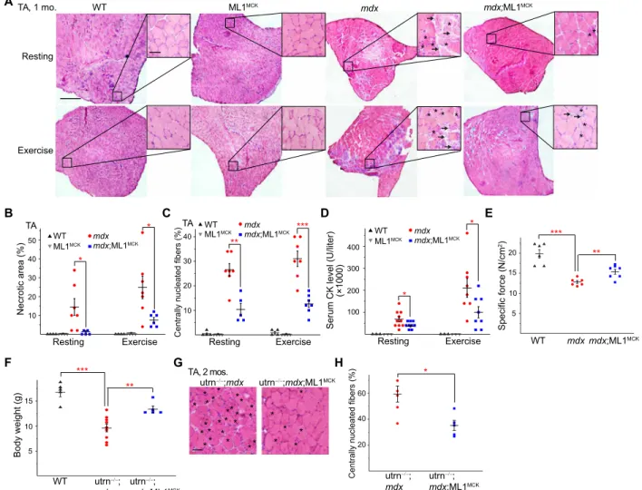
Mdx mice, either homozygous females or hemizygous males, exhibit early-onset muscular dystrophies, as evidenced by myofiber necrosis (myonecrosis) and degeneration/regeneration cycles, which were readily observed by postnatal day 14 (P14) (19). In 1-month-old mdx;ML1MCK mice, hematoxylin and eosin (H&E) staining revealed that myonecrosis, quantified by the percentage of the necrotic area (i.e., the presence of necrotic myofibers, immune cells, and fibro-blasts) in whole cross sections, was markedly reduced in tibialis anterior (TA) muscles compared with age-matched mdx mice, especially after downhill treadmill exercise (Fig. 2, A and B). The
number of centrally nucleated fibers, caused by repeated myocyte degeneration and regeneration, was also significantly reduced in the mdx;ML1MCK muscles (Fig. 2, A and C). Similar anti-dystrophic effects were seen in other skeletal muscles, including the gastrocnemius (GAS) and diaphragm (DIA) (fig. S2, A to G). Serum CK levels, a diagnostic biomarker of DMD (13), were also reduced by ML1 overexpression (Fig. 2D). Consistent with the histological and biochemical results, physiological assays showed improved specific muscle force in mdx mice following ML1MCK overexpression (Fig. 2E).
In most skeletal muscles of mdx mice, the dystrophic phenotype did not appear to be progressive, perhaps due to compensatory expres-sion of utrophin, a functional homolog of dystrophin (20). Relative to mdx mice, utrophin−/−;mdx mice have a much more severe and pro-gressive muscular dystrophy that resembled human DMD (20). Both dystrophy and body weight loss characteristic of the utrophin−/−;mdx A B C H F D E G WT mdx Resting Exercise ** * * * TA, 1 mo. * * * ** * * * * * ML1MCK mdx;ML1MCK TA Necrotic area (%)10 20 30 40 50 WTML1MCK mdx;ML1MCK mdx Resting Exercise * * Specific force (N/cm 2) 5 20 10 WT mdxmdx;ML1MCK *** ** 15 Body weight (g) *** ** WT utrn ; mdx;ML1MCK utrn ; mdx 5 10 15 TA Resting Exercise
Centrally nucleated fibers (%)
10 20 30 40 ** *** WT mdx;ML1MCK ML1MCK mdx
Serum CK level (U/liter)
100 200 300 400 * * Resting Exercise (×1000) WT mdx;ML1MCK ML1MCK mdx utrn ;mdx utrn ;mdx;ML1MCK TA, 2 mos. * **** * ** * * ** * * ** * * * * * ** * * * * * * *
Centrally nucleated fibers (%)
20 40 60 utrn ; mdx utrn ;mdx;ML1MCK *
Fig. 2. Transgenic overexpression of ML1 reduces muscle pathologies in young mdx mice. (A) H&E staining of TA sections from WT, ML1MCK, mdx, and mdx;ML1MCK
mice at the age of 1 month, before (rest) and after treadmill exercise. Both male and female mice were randomly assigned into different groups. Arrows label necrotic areas and asterisks show centrally nucleated myofibers. Scale bar, 500 m or 50 m (zoom-in images). (B) Percentage of necrotic area in TA muscles from various trans-genic mice. Each datum (n indicates the number of the muscle) represents the averaged result from at least five representative images randomly selected from at least three sections. Statistical analyses were performed by experimenters who were blind to animal genotypes. (C) Percentage of centrally nucleated fibers in TA muscles from different transgenic mice. (D) Serum CK levels in 1-month-old WT, ML1MCK, mdx, and mdx;ML1MCK mice before and after treadmill exercise. (E) Specific force test of GAS
from multiple 1-month-old mice. (F) Body weight measurements of WT, utrophin−/−;mdx (utrn−/−;mdx), and utrn−/−;mdx;ML1MCK male mice at the age of 1 month. (G) Effect
of ML1 overexpression on histology of TA muscles isolated from 2-month-old utrn−/−;mdx mice. Scale bar, 50 m. (H) Quantification on central nucleation of muscle histology
from (G). All data are means ± SEM; *P < 0.05, **P < 0.01, and ***P < 0.001.
on February 28, 2020
http://advances.sciencemag.org/
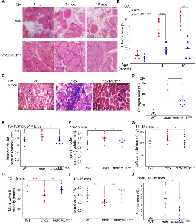
phenotypes (20) were improved by ML1MCK overexpression (Fig. 2, F to H). Dystrophy of the DIA in mdx mice was also progressive, as seen in human DMD, and respiratory failure is a major cause of death in DMD (6). Consistent with previous studies (6), we observed massive necrosis and subsequent fibrous and adipose tissue
replace-ment (i.e., fibrosis) in mdx DIA muscles (Fig. 3, A and B). At all ages examined (1, 4, and 10 months), fibrosis was reduced significantly by ML1MCK overexpression (Fig. 3, A and B). The content of collagen, a major component of fibrous scar tissue (13), was also decreased by ML1MCK overexpression (Fig. 3, C and D). A Interventricular septum (diastole, mm) WT 0.4 0.8 1.0 0.6 0.2 mdx mdx;ML1MCK * P = 0.07 13–15 mos. Interventricular septum (systole, mm) 0.4 0.8 1.4 0.6 0.2 1.2 1.0 WT mdx mdx;ML1MCK * * 13–15 mos. B D C
Left ventricle mass (mg)
150 100 50 * * WT mdx mdx;ML1MCK 13–15 mos.
Mitral valve E velocity (mm/s) 600 400 200 WT mdx mdx;ML1MCK ** *** 13–15 mos.
Mitral valve E/A
1.0 2.0 1.5 0.5 *** ** WT mdx mdx;ML1MCK 13–15 mos. F G E WT mdx mdx;ML1MCK DIA 4 mos. DIA * 20 40 60 Collagen area (%) WT mdx mdx;ML1MCK mdx mdx;ML1MCK
DIA 1 mo. 4 mos. 10 mos. DIA
Age (month) 1 4 10 mdx mdx;ML1MCK 20 40 60 Fibrotic area (%) *** ** H I J Heart, 13–15 mos. 1 5 9 Fibrotic area (%) 23 4 6 7 8 * * WT mdx mdx;ML1MCK
Fig. 3. Transgenic ML1 overexpression ameliorates myopathies in aged mdx mice. (A) H&E staining of DIA isolated from mdx and mdx;ML1MCK mice at the age of
1, 4, and 10 months. Both male and female mice were used in this experiment. Scale bar, 50 m. (B) Age-dependent progressive fibrosis in DIA muscles isolated from mdx and mdx;ML1MCK mice. (C) Trichrome collagen staining of DIA from 4-month-old mice. Scale bar, 100 m. (D) Quantification of results (n indicates the number of the animal)
averaged from multiple randomly selected images as shown in (C). (E and F) Thickness of IVS was measured by echocardiography (see fig. S2H) at the end diastole (E) and end systole (F) from 13- to 15-month-old WT, mdx, and mdx;ML1MCK male mice. The echocardiographer was blind to the mouse genotype. (G) Calculated left ventricle
mass in 13- to 15-month-old WT, mdx, and mdx;ML1MCK male mice. (H) Peak velocity of E wave measured by LV pulse-wave Doppler (see fig. S2I) in 13- to 15-month-old
WT, mdx, and mdx;ML1MCK male mice. (I) Ratios between peak velocity of E and A waves (see fig. S2I) in WT, mdx, and mdx;ML1MCK male mice. (J) Quantification for the
percentage of fibrotic area in WT, mdx, and mdx;ML1MCK mice at the age of 13 to 15 months (see fig. S2J). All data are means ± SEM; *P < 0.05, **P < 0.01, and ***P < 0.001.
on February 28, 2020
http://advances.sciencemag.org/
Cardiac failure is another major cause of death in DMD, but cardio-myopathies are observed only in aged (e.g., >10-month-old) mdx mice (21, 22). We performed echocardiograms in 13- to 15-month-old WT, mdx, and mdx;ML1MCK male mice (fig. S2H). Compared with WT mice, mdx mice had thickened interventricular septum (IVS) and increased left ventricle mass (Fig. 3, E to G), both of which are characteristic of dilated cardiomyopathies (21). Both echocardio-graphic abnormalities were ameliorated by ML1 overexpression (Fig. 3, E to G). We also performed pulse-wave Doppler echocardio-graphy to analyze the contractile function of the left ventricle (fig. S2I). Consistent with previous studies (23), mdx hearts had reduced E wave velocity and E/A ratio (Fig. 3, H and I), suggestive of ventric-ular dysfunction. Both parameters were corrected by ML1MCK over-expression (Fig. 3, H and I). Histological analyses showed that cardiac fibrosis in aged (15-month-old) mdx mice was also reduced by ML1MCK expression (Fig. 3J and fig. S2J). Together, these results suggest that transgenic overexpression of ML1 is sufficient to attenuate dystrophies of both skeletal and cardiac muscles in DMD-like mouse models.
Pharmacological activation of ML1 in vivo ameliorates muscular dystrophy in mdx mice
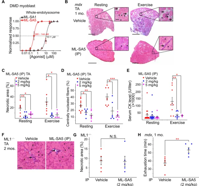
Next, we tested whether small-molecule ML1 agonists have a muscle protective effect in mdx mice. ML-SA5, a potent ML-SA compound with an in vitro EC50 (median effective concentration) value in the nanomolar range for endogenous ML1 (Figs. 1G and 4A), exhibited pharmacokinetic properties suitable for in vivo studies (fig. S3A). Mdx mice received daily intraperitoneal injection of ML-SA5, start-ing at P14 and continustart-ing for at least 2 weeks. Like the vehicle [10% dimethyl sulfoxide (DMSO) + 40% polyethylene glycol (PEG) + 50% phosphate-buffered saline (PBS)]–treated group, no patholog-ical signs were evident following ML-SA5 injection, manifested by normal body weight, complete blood count, liver biochemistry and histology, and kidney histology (fig. S3, B to J), suggesting the minimal toxicity effects. However, at the intraperitoneal dose of 2 to 5 mg/kg, daily ML-SA5 injection for 2 weeks decreased TA muscle necrosis by more than 70%, both at rest and after treadmill exercise (Fig. 4, B and C). Centrally nucleated fibers were also decreased in the ML-SA5–injected mdx mice (Fig. 4, B and D). Similar rescue effects were also seen in the GAS and DIA muscles (fig. S4, A to F). Consistently, serum CK level was also reduced in mdx mice that received ML1 agonist injection (Fig. 4E). In contrast, intraperitoneal injection of ML-SI6, a potent ML1 inhibitor (13, 24), worsened the dystrophic phenotype in mdx mice (fig. S4, G to K). Furthermore, in ML1 KO mice that also exhibit a DMD-like phenotype such as necrosis and central nuclei (13), no obvious ML-SA5 rescue effects were seen (Fig. 4, F and G). Hence, the actions of ML-SA5 are likely to be mediated through ML1 (i.e., on target). Last, at the behavioral level, motor performance in a downhill treadmill test improved markedly following ML-SA injection in mdx mice (Fig. 4H). Therefore, similar to the genetic overexpression studies, pharmacological activation of ML1 in vivo using small molecules was also muscle protective.
ML1 facilitates sarcolemma repair, thereby reducing skeletal and cardiac muscle damage in mdx mice
One of the major causes of muscular dystrophy is defective sarco-lemma repair, and lysosomal exocytosis is a primary route for reseal-ing damaged membranes (25). Given the essential role of ML1 in lysosomal exocytosis and membrane resealing (13), it is likely that ML1’s muscle protective effects are mediated by sarcolemma repair.
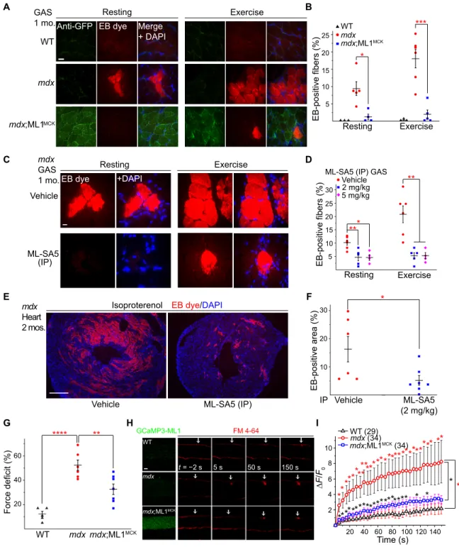
Skeletal muscle damage was experimentally induced in vivo with a short-term treadmill exercise protocol (26) and confirmed with the entry of the membrane-impermeable Evans blue (EB) dye (13). The percentage of EB-positive fibers, which never exceeded 2% in WT muscles, reached 9% at rest and 18% after treadmill exercise in mdx muscles (Fig. 5, A and B). Notably, mdx;ML1MCK mice had less than 2% of EB-positive muscle fibers, even after treadmill exercise (Fig. 5, A and B, and fig. S5, A and B). EB uptake was also much reduced in ML-SA–treated mdx muscle (Fig. 5, C and D, and fig. S5, C and D) but increased in ML-SI–treated mdx mice (fig. S4, J and K). Like-wise, when cardiomyocyte damage was induced in young (2-month-old) mdx mice with isoproterenol, EB uptake in the heart tissues was much reduced by ML-SA5 injection (Fig. 5, E and F). Collectively, these results suggest that compromised membrane integrity in both skeletal and cardiac muscles can be improved by ML1 up-regulation.
We also performed an ex vivo muscle damage test, in which in situ force in GAS muscles was measured before and after mechanical stretch by blind experimenters (27). The muscle force deficit seen in mdx mice was markedly reduced with ML1 overexpression (Fig. 5G), showing that ML1 expression has the potential to protect muscles from contraction-induced muscle damage in vivo. To study mem-brane repair in vitro, we used FM 4-64 dye to detect memmem-brane dis-ruptions (8) in single myofibers isolated from the flexor digitorum brevis (FDB) muscle (8, 9). Upon laser irradiation, mdx fibers continued to take up FM dye at the injury sites for several minutes, whereas FM dye uptake was much reduced in ML1-overexpressed mdx muscle (Fig. 5, H and I). These results suggest that ML1 activation facilitates membrane repair to reduce muscle damage in mdx mice.
Up-regulation of ML1 in muscle activates transcriptional factor EB and corrects lysosomal insufficiency
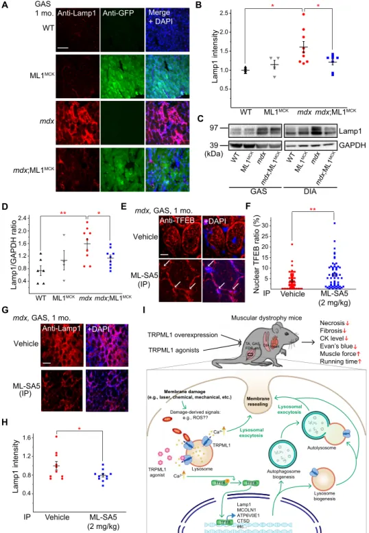
Expression of lysosome-associated membrane protein 1 (Lamp1) is an indicator of lysosome function in vivo such that Lamp1 up-regulation accompanies lysosomal dysfunction or insufficiency (28). Aberrant Lamp1 expression has been reported in several mdx studies (29, 30). We found that Lamp1 levels were increased in mdx compared with WT mice in both GAS and DIA isolated from 1-month-old animals (Fig. 6, A to D). In our immunofluorescence analysis, Lamp1 up-regulation was apparent in both muscle fibers and surrounding cells (e.g., infiltrated macrophages; Fig. 6A and fig. S5E), suggesting that inflammation, which is known to be associated with necrosis (31), may also contribute to Lamp1 up-regulation. Collectively, these results suggest that there is lysosome insufficiency in the muscle tissues of mdx mice and that lysosome biogenesis might have been weakly activated to compensate for the deficiency.
ML1 activation has been reported to increase lysosome biogenesis and Lamp1 levels through nuclear translocation of transcriptional factor EB (TFEB), a master regulator of lysosomal genes (18, 32). Lamp1 expression was significantly lower in mdx;ML1MCK mice compared to mdx mice (Fig. 6, A to D). Likewise, the aberrant up-regulation of TFEB was also corrected by ML1 overexpression or activation (see fig. S5, F to H). These seemingly puzzling results may be explained by the fact that ML1 expression boosts lysosome function and decreases necrosis in muscle, obviating the need for compensatory changes via the expression of lysosomal genes. In mdx;ML1MCK mice or ML-SA5–injected mdx mice, TFEB nuclear translocation was increased (Fig. 6, E and F, and fig. S5F), but Lamp1 and TFEB up-regulation and other compensatory lysosomal changes, e.g. increased LC3-II levels, were suppressed (Fig. 6, G and H, and
on February 28, 2020
http://advances.sciencemag.org/
fig. S5, F to I). Hence, ML1 activation may further boost lysosome function to override lysosome insufficiency, yielding muscle protec-tive effects. Similar scenarios have been reported in several lysosomal storage disorders (LSDs), in which TFEB activation has been found to increase lysosome biogenesis in WT cells while suppressing Lamp1 up-regulation in LSD animal models (14, 28).
ML1 protects human DMD muscle cells from damage through TFEB and lysosomal exocytosis
To study the role of ML1 activation in lysosome biogenesis directly, we used DMD cells, an immortalized myoblast line developed from a DMD patient’s muscle cells (fig. S6, A and B) (33). Only mild
Lamp1 up-regulation was observed in DMD myoblasts (fig. S6C), suggesting that the lysosome insufficiency occurring in mdx mice might be caused by extensive in vivo muscle damage. Consistent with our lysosomal patch-clamp studies (Fig. 4A), nanomolar con-centrations of ML1 agonists were sufficient to induce notable nuclear translocation of TFEB (fig. S6, D and E) and the related protein TFE3 (fig. S6, F and G). Consistently, expression of TFEB/TFE3 target genes, including those required for lysosome biogenesis and func-tion, were elevated by ML-SA5 treatment (fig. S6H). Notably, TFEB mRNA and protein expressions were significantly reduced in ML-SA5–treated WT and DMD cells (fig. S6, H to K), suggestive of the negative feedback regulation. Hence, ML1 activation may lead A C B D E F ML-SA5 (IP) TA Necrotic area (%)20 40 60 Resting Exercise *** ** 2 mg/kgVehicle 5 mg/kg ML-SA5 (IP) TA
Centrally nucleated fibers (%)
10 20 30 40 50 Resting Exercise * * ** *** 2 mg/kg Vehicle 5 mg/kg
Exhaustion time (min)
30 40 50 60 ** Vehicle ML-SA5 (2 mg/kg) IP mdx, 1 mo. Resting Exercise mdx Vehicle ML-SA5 (IP) TA 1 mo. * * * * * ** * * * * Necrotic area (%) 10 20 15 5 Vehicle ML-SA5 (2 mg/kg) N.S. IP ML1 Normalized response 0.25 0.50 0.75 1.00 [Agonist] ( M) 0.01 0.1 1 Whole-endolysosome DMD myoblast ML-SA5 285 144 nM 10 100 ML-SA1 10 1 M
Serum CK level (U/liter)
50 100 150 200 250 Resting Exercise *** (×1000) 2 mg/kg Vehicle 5 mg/kg ML-SA5 (IP) G ML1 TA 2 mos.
Vehicle ML-SA5 (IP)
** * * * * * * * ** * H
Fig. 4. Small-molecule ML1 agonists ameliorate muscular dystrophies in mdx mice. (A) ML-SA1 and ML-SA5 dose-dependently activated whole-endolysosomal ML1 currents in DMD myoblasts. (B) H&E staining of TA from 1-month-old mdx mice that received daily intraperitoneal injection of ML-SA5 (2 mg/kg) for 14 days starting at P14. Arrows and asterisks indicate necrotic areas and centrally nucleated fibers, respectively. Scale bar, 500 or 50 m (zoom in). (C) Percentages of necrotic area in ML-SA5–injected mice. Each datum (n indicates the number of the muscle; n ≥ 4) represents the averaged result from at least three representative images randomly selected from at least three sections. Statistical analyses were performed by experimenters who were blind to treatment conditions. (D) Percentage of centrally nucleated fibers in ML-SA5–injected mice. (E) Serum CK levels in ML-SA5–treated mdx mice at the age of 1 month before and after treadmill exercise. (F and G) Effect of ML-SA5 intraperitoneal injection on TA from 2-month-old ML1 KO mice. Injection starts from P14. N.S., not significant. (H) Treadmill exhaustion time of mdx mice treated with ML-SA5 versus vehicle control. N, number of tested animals. The experimenter was blind to the treatment conditions. In the experiments shown in this figure, both male and female mice were randomly assigned into different treatment groups. All data are means ± SEM; *P < 0.05, **P < 0.01, and ***P < 0.001.
on February 28, 2020
http://advances.sciencemag.org/
B A F E D C G H EB-positive fibers (%) 5 10 15 20 25 Resting Exercise * *** WT mdx;ML1MCK mdx F/ F0 2 4 6 8 10 Time (s) 20 40 60 80 100 120 140 WT (29) mdx (34) mdx;ML1MCK (34) *** *********** ************ *** ** ** * * * ** * * * * * * * * * * * * * * * * * I WT mdx mdx;ML1MCK t = −2 s 5 s 50 s 150 s GCaMP3-ML1 FM 4-64 20 40 60 Force deficit (%) **** ** WT mdx mdx;ML1MCK Resting Exercise Anti-GFP EB dye WT mdx mdx;ML1MCK Merge + DAPI GAS 1 mo. Resting Exercise mdx GAS 1 mo. Vehicle EB dye +DAPI ML-SA5
(IP) EB-positive fibers (%) 5
10 15 20 25 30 ** Resting Exercise 2 mg/kg Vehicle 5 mg/kg ***
ML-SA5 (IP) GAS
EB dye/DAPI ML-SA5 (IP) Vehicle mdx Heart 2 mos. Isoproterenol EB-positive area (%) 10 20 30 ML-SA5 (2 mg/kg) Vehicle IP *
Fig. 5. ML1 activation facilitates sarcolemma repair to reduce muscle damage in mdx mice. (A) Representative images of EB dye uptake in GAS isolated from WT, mdx, and mdx;ML1MCK mice at the age of 1 month, before (rest) and after treadmill exercise. Scale bar, 10 m. GCaMP3-ML1 expression was detected using an anti-GFP
antibody. (B) Quantification of EB-positive fibers from (A). Each datum (n, number of the muscle) represents the averaged result from at least five representative images selected from at least three sections. (C) EB dye uptake in GAS isolated from ML-SA5–treated mdx mice. Scale bar, 10 m. (D) Quantification of EB dye uptake in GAS from ML-SA5–treated mdx mice. (E and F) EB dye uptake in cardiac muscles isolated from 2-month-old ML-SA5 (2 mg/kg)–injected mdx mice that were stimulated with -isoproterenol to cause cardiac damage. Injection of ML-SA5 started from P14. Scale bar, 500 m. (G) Muscle force deficit of mechanically stretched GAS from 1-month-old mice. (H) Representative images of laser damage–induced FM dye accumulation in isolated FDB fibers. Arrows highlight damage sites. Scale bar, 20 m. (I) Time-dependent laser damage–induced FM dye accumulation in FDB fibers isolated from WT, mdx, and mdx;ML1MCK mice at the age of 1 month. n indicates the number of the FDB fibers
for each genotype. In all the experiments shown in this figure, both male and female mice were randomly assigned into different experimental groups. All data are means ± SEM; *P < 0.05, **P < 0.01, and ***P < 0.001.
on February 28, 2020
http://advances.sciencemag.org/
A B E F G H C D WT ML1MCK mdxmdx;ML1MCK Lamp1 intensity 0.5 1.5 2.0 2.5 * * 1.0 WT ML1MCK mdxmdx;ML1MCK Lamp1/GAPDH ratio0.4 0.8 1.2 1.6 2.0 2.4 ** * Vehicle ML-SA5 (IP) Anti-Lamp1 +DAPI mdx, GAS, 1 mo. Vehicle ML-SA5 (2 mg/kg) Lamp1 intensity * 0.4 0.8 1.2 1.6 IP 97 39 GAS ML1 MCK mdx WT mdx ;ML1 MCK (kDa) GAPDH Lamp1 DIA ML1 MCK mdx WT mdx ;ML1 MCK I Vehicle ML-SA5 (2 mg/kg) 5 10 15 20 25 30 Nuclear TFEB ratio (%) ** IP Anti-TFEB +DAPI Vehicle ML-SA5 (IP) mdx, GAS, 1 mo. ML1MCK mdx mdx;ML1MCK WT
Anti-Lamp1 Anti-GFP Merge
+ DAPI
GAS 1 mo.
TFEB TFEB
Membrane damage (e.g., laser, chemical, mechanical, etc.)
Damage-derived signals: e.g., ROS?? TRPML1 agonist Ca2+ Lysosome Lamp1 MCOLN1 ATP6V0E1 CTSD etc... Autophagosome biogenesis Lysosome biogenesis Autolysosome Lysosomal exocytosis Membrane resealing TRPML1 Lysosomal exocytosis Ca2+ TRPML1 overexpression TRPML1 agonists Necrosis Fibrosis CK level Evan’s blue Muscle force Running time Muscular dystrophy mice
TA, GAS, FDB, etc.DIA Heart
e.g. ML1 nist Ca2+ Ca2+ TFEB P ROS ROS ROS
Fig. 6. ML1 agonist activates TFEB to correct lysosomal insufficiency in mdx muscles. (A) Lamp1 immunofluorescence staining of GAS from WT, ML1MCK, mdx, and
mdx;ML1MCK mice at the age of 1 month. GCaMP3-ML1 expression was detected using an anti-GFP antibody. Scale bar, 100 m. Both male and female mice were used in
the experiments shown in this figure. (B) Quantitative analyses of images in (A). For each datum representing each muscle, at least five representative images from at least three sections were analyzed. (C) Western blotting analysis of Lamp1 protein expression in GAS and DIA from 1-month-old mice. (D) Quantitation of Western blotting results in (C). (E) TFEB immunolabeling in GAS from ML-SA5–treated mice at the age of 1 month. (F) Quantitative analysis of nuclear versus total TFEB ratio from (G). n indicates muscle fibers in randomly selected images from at least four muscles in each group. (G) Lamp1 immunostaining in GAS from ML-SA5 (2 mg/kg)–treated mdx mice at the age of 1 month. Scale bar, 100 m. (H) Quantification of Lamp1 immunolabeling in GAS from ML-SA5–treated mdx mice. For each datum representing each muscle, at least five representative images from at least three sections were analyzed. (I) Genetic or pharmacological up-regulation of ML1 ameliorates myopathies in vivo through TFEB-dependent lysosome biogenesis, Ca2+-dependent lysosomal exocytosis, and sarcolemma repair. Muscle damage may generate a yet-to-be-defined signal
(e.g., reactive oxygen species) (18) to activate ML1 channels on lysosome membranes. Subsequent lysosomal Ca2+ release triggers Ca2+-dependent lysosomal exocytosis
and nuclear translocation of TFEB, which then activates transcription of a unique set of genes related to lysosome biogenesis. Subsequently, lysosome function is boosted and sarcolemma repair may be facilitated to reduce muscle damage in DMD cells in vitro and in vivo. All data are means ± SEM; *P < 0.05 and **P < 0.01.
on February 28, 2020
http://advances.sciencemag.org/
to TFEB nuclear translocation, thereby increasing lysosome bio-genesis, which, in turn, reduces lysosome stress and restores lyso-some homeostasis.
Whereas increased TFEB activation and lysosome biogenesis may amplify lysosomal trafficking events including lysosomal exocytosis (34), ML1 activation alone can trigger lysosomal exocytosis directly (35). Membrane damage [i.e., chemical injury caused by streptolysin O (SLO) toxin] triggers lysosomal exocytosis and membrane repair (25). In myofibers, which contain a large cytoplasm, ML1-mediated lyso-somal Ca2+ release, but not Ca2+ influx mediated by overexpressed ML1 channels (36) (see fig. S7, A to E), may preferentially activate lysosome-localized Ca2+ sensors to facilitate lysosomal exocytosis and biogenesis. Propidium iodide (PI) staining is a common read-out for membrane damage and cell viability (37). In DMD myoblasts and differentiated myotubes, both membrane damage and ML-SA treatment induced lysosomal exocytosis (fig. S9, A and B), suggesting that ML-SAs may trigger membrane repair processes by mimicking a yet-to-be-identified damage signal. Consistent with this hypothesis, ML1 agonists and inhibitors decreased and increased, respectively, PI-positive cells following SLO treatment (fig. S8, A to D). The cyto-protective effects of ML-SAs were abolished by knockdown of TFEB expression (fig. S8, E to H) or blockage of lysosomal exocytosis with dominant-negative synaptotagmin VII (Syt-VII) (fig. S9, C and D). Collectively, these results suggest that boosting ML1 activity may promote myocyte survival by reducing membrane damage via a direct and acute mechanism (i.e., lysosomal exocytosis), as well as through an indirect and more sustained mechanism (i.e., lysosome biogenesis; Fig. 6I).
DISCUSSION
ML1 deficiency causes a DMD-like muscle phenotype in both humans and rodents (13). In the current study, we found that both genetic and pharmacological activation of ML1 had muscle protec-tive effects in mdx mice and human DMD muscle cells. ML1 activa-tion led to TFEB nuclear translocaactiva-tion and increased lysosomal biogenesis while also increasing lysosomal trafficking (e.g., exocytosis), thereby facilitating sarcolemma repair to reduce muscle damage (Fig. 6I). Hence, our proof-of-concept study has provided strong evidence that small-molecule ML1 agonists can be developed to treat DMD. Upon withdrawal of ML-SA compounds, the muscle protec-tive effects of the drugs disappeared (see fig. S10), suggesting that sustained or periodic activation of lysosome enhancement is required for DMD treatment. Although DMD is not an LSD per se (28), the observed compensatory changes are suggestive of lysosome insuffi-ciency. In both cases, augmenting ML1/TFEB-dependent lysosome biogenesis can alleviate lysosome insufficiency. Since ML1 is a ubiq-uitously expressed lysosomal channel, there could be “on-target” adverse effects of ML1 activation on nonmuscle issues in mdx mice. However, ML1/TFEB activation is most effective in cells/tissues with lysosome dysfunction/insufficiency, and ML1/TFEB-mediated lyso-some biogenesis is suppressed by multiple mechanisms in healthy cells (28, 38). Hence, the on-target, “overstimulating” effects of ML1 may be minimal on healthy cells/tissues/animals.
The simplest explanation for the presently observed muscle pro-tective effect of ML1, in our view, would be facilitation of lysosomal exocytosis and membrane repair (Fig. 6I). However, although blockage of lysosomal exocytosis abolished ML1-dependent muscle protection, it should be noted that overexpression of Syt-VII dominant-negative
constructs might indirectly affect lysosome biogenesis and TFEB- dependent functions. TFEB has been shown to be muscle protective in other studies as well (14). In LSDs, ML1 and TFEB form a positive feedback loop that promotes cellular clearance (28). It is possible that the same lysosome program may boost sarcolemma repair to reduce muscle damage in DMD and other muscle diseases. Hence, manip-ulating lysosome function with small molecules may have broad therapeutic potential.
MATERIALS AND METHODS
Study design
The overall goal of this study was to determine the effect of ML1 up-regulation on muscular dystrophy and membrane repair. All data presented here were replicated in at least four mice or three biolog-ical replicates for in vitro experiments, with all histochembiolog-ical stain-ing data quantified by experimenters who were blind to genotypes/ treatments. On the basis of pilot studies of ML1 up-regulation on various pathologic hallmarks in mice, with a power of 0.8 and P < 0.05, we calculated a sample size of between 5 and 11 mice per group. Animals were randomly allocated into control and ex-perimental groups.
Mouse lines
We generated muscle cell–specific ML1-overexpressing (ML1 ROSA-lSl;MCK Cre or ML1MCK) mice by crossing ROSA-loxSTOPlox- GCaMP3-ML1 (abbreviated ML1 ROSA-lSl) mice with a muscle cell– specific Cre line (MCK-Cre) (15). Mdx (001801) and utrophin+/−;mdx (014563) mice were purchased from The Jackson Laboratory. ML1−/− mice were provided by S. Slaugenhaupt (13). All mice used in the study were backcrossed to the C57BL/6 genetic background. Mice were used under the University of Michigan’s Institutional Animal Care and Use Committee’s approval. For cardiac function studies, only hemizygous male mice at the age of 13 to 15 months were used. For histological and physiological studies of skeletal muscles, both hemizygous male and homozygous female mdx mice were randomly assigned into different groups. The muscle type and age of the mice used for each experiment were indicated in the figures and legends.
Western blotting
After lysing muscle tissues in ice-cold radioimmunoprecipitation assay buffer with 1× protease inhibitor cocktail tablet (Roche), 20 to 40 g of total protein aliquots were loaded into 4 to 12% bis-tris or 3 to 8% tris-acetate SDS–polyacrylamide gradient gels (Invitrogen) and then transferred to polyvinylidene fluoride membranes via iBlot 2 Gel Transfer Device (Life Technologies). The membranes were blocked in 5% (w/v) skim milk in PBS with 0.05% Tween 20 for 1 hour and then incubated overnight in the blocking buffer at 4°C. The primary antibodies used were as follows: anti-ML1 (ACC-081, Alomone Labs), anti–green fluorescent protein (GFP) (A6455, Invi-trogen), anti-mouse Lamp1 [1D4B, Developmental Studies Hybridoma Bank (DSHB)], anti-human Lamp1 (H4A3, DSHB), anti-mouse TFEB (A303-673A, Bethyl), anti-human TFEB (4240, Cell Signaling Tech-nology), anti-LC3 (L8918, Sigma), anti-dystrophin (ab15277, Abcam), anti-utrophin (8A4, DSHB), anti–-dystroglycan (sc-53987, Santa Cruz Biotechnology), and anti-GAPDH (glyceraldehyde 3-phosphate dehydrogenase) (MAB347, Millipore). Peroxidase-conjugated anti- rabbit, anti-mouse, or anti-rat secondary antibodies were applied at
on February 28, 2020
http://advances.sciencemag.org/
room temperature for 1 hour, followed by SuperSignal West Pico Chemiluminescence Substrate (Thermo Fisher Scientific). Bands were quantitated in ImageJ software.
Immunofluorescence
Muscle tissues, harvested and frozen in 2-methylbutane, prechilled in liquid nitrogen, were cryosectioned at 12 m. After being washed with tris-buffered saline (TBS) + 0.025% Triton X-100, sections were blocked with 10% serum and 1% bovine serum albumin in TBS at room temperature for 2 hours. Cell lines and isolated primary cells cultured on coverslips were washed with PBS, fixed in 4% para-formaldehyde, permeabilized in 0.3% Triton X-100 in PBS, and blocked in 1% bovine serum albumin in PBS. Fixed cells and cryo-sections were then incubated at 4°C overnight with primary anti-bodies targeting ML1, GFP, Lamp1, TFEB, dystrophin, or CD11b (M1/70.15.11.5.2, DSHB) at 1:50 or 1:200 solutions. Alexa Fluor secondary antibodies (Invitrogen) were then applied for 1 hour in the dark at room temperature, followed by 4′,6-diamidino- 2-phenylindole counterstaining if necessary. Cells and tissue sec-tions were imaged on a spinning disc confocal imaging system composed of an Olympus IX81 inverted microscope; 10×, 20×, and 60× Olympus objectives; a CSU-X1 scanner (Yokogawa); an iXon electron multiplying charge-coupled device camera (Andor); and MetaMorph Advanced Imaging acquisition software v.7.7.8.0 (Molecular Devices). Image analysis results were quantified in MetaMorph software.
Primary muscle cell culture
Murine myoblasts were harvested and cultured as previously described (39). Briefly, skeletal muscles were isolated from P0 to P3 pups and dissociated with 0.25% trypsin supplemented with collagenase (2 mg/ml) (Sigma) at 37°C. To remove fibroblasts, cells were pre-plated on a standard, non–tissue culture–coated petri dish for 60 to 90 min. Muscle cells were then counted, seeded onto collagen-coated glass coverslips, and maintained at 37°C under 5% CO2 in F10 medium supplemented with 20% fetal bovine serum (FBS) and penicillin/streptomycin. To induce differentiation, myoblasts were grown to confluence before switching to Dulbecco’s modified Eagle’s medium (DMEM) containing 5% horse serum and penicillin/streptomycin. Electrophysiology, Ca2+ imaging, and immunofluorescence labeling were performed after 3 days of differentiation induction to allow MCK-Cre expression.
Whole-endolysosome electrophysiology
Endolysosomal electrophysiology was performed on isolated endo-lysosomes from muscle cells treated with 1 M vacuolin-1 to enlarge late endosomes and lysosomes (40, 41). The bath (internal/cytoplasmic) solution contained 140 mM K gluconate, 4 mM NaCl, 1 mM EGTA, 2 mM Na2–adenosine 5′-triphosphate (ATP), 2 mM MgCl2, 0.39 mM CaCl2, 0.2 mM guanosine triphosphate (GTP), and 10 mM Hepes [pH adjusted with KOH to 7.2; the free [Ca2+]i was estimated to be ~100 nM on Maxchelator software (https://bit.ly/2G7fIVH)]. The pipette (luminal) solution consisted of a low-pH Tyrode’s solution with 145 mM NaCl, 5 mM KCl, 2 mM CaCl2, 1 mM MgCl2, 10 mM Hepes, 10 mM MES, and 10 mM glucose (pH 4.6). A perfusion system was used to ensure efficient solution exchange. Data were collected with an Axopatch 200A patch-clamp amplifier, Digidata 1440, and pClamp 10.0 software (Axon Instruments). Currents were digitized at 10 kHz and filtered at 2 kHz. All experiments were conducted at
21° to 23°C, and all recordings were analyzed in pClamp 10.0 and Origin 8.0 (OriginLab, Northampton, MA).
GCaMP3 Ca2+ imaging
Ca2+ imaging was performed in ML1MCK and mdx;ML1MCK primary myotubes overexpressing GCaMP3-ML1 (15). The fluorescence in-tensity at 488 nm (F488) was recorded with the spinning disc confocal imaging system.
Histochemical staining
Frozen sections were warmed to room temperature and hydrated in double-distilled (MilliQ) H2O. Tissues were stained with H&E dyes (Thermo Fisher Scientific) for the nucleus and cytoplasm, respectively, followed by gradient washing in ethanol (70, 90, and 100%) and xylene (100%). The stained sections were mounted in Permount medium (Thermo Fisher Scientific). Staining quantification was performed blindly following standardized operating procedures (SOP number: DMD_M.1.2.007). Necrotic myofibers, inflammatory cells, and fibroblasts were all counted as necrotic area. Collagen content was assessed with a Masson’s trichrome kit (Sigma). Area stained with blue color was quantified as fibrotic area.
Muscle force measurement
Mice were anesthetized by intraperitoneal injection of Avertin (tribromoethanol, 250 mg/kg) and placed on a warmed platform to maintain body temperature. GAS muscle contractile properties were measured in situ, as described previously (42). Briefly, after the GAS was isolated from surrounding tissues, the distal tendon was secured to the lever arm of a servomotor (model 6650LR, Cambridge Tech-nology). The knee and foot were clamped to the platform. The muscle was activated by tibial nerve stimulation. With the muscle held at optimal length (L0), 300-ms trains of stimulus pulses were applied at increasing frequencies until the maximum isometric tetanic force (P0) was achieved. After the pre-stretch P0 (preP0) was recorded, we applied two stretches (12 s apart) of 30% of fiber length (Lf), which was estimated by multiplying L0 by previously determined Lf-to-L0 ratios. After a 1-min rest, post-stretch P0 (postP0) was obtained to determine the force deficit. To maintain muscle temperature and moisture, a warm saline drip (37°C) was applied to the GAS contin-uously throughout the procedure. After the experiment, mice were euthanized, and muscles were removed and trimmed of their tendons. Physiological cross-sectional areas were determined by dividing muscle mass by the product of Lf using a density of mam-malian skeletal muscle of 1.06 g/cm3. Specific P
0 (SP0) refers to P0 per cross-sectional area, and the force deficit was calculated as 1 − postP0/preP0.
Echocardiography
Echocardiograms were performed by following the recommendations of the American Society of Echocardiography as described previously (43). All echocardiography experiments were performed by one reg-istered echocardiographer who was blind to the mouse genotype. Before imaging, 13- to 15-month-old WT, mdx, and mdx;ML1MCK mice were weighed and anesthetized with inhaled isoflurane. Imaging was performed using a Vevo 770 Microimaging system (VisualSonics Inc.) equipped with an RMV707B (15 to 45 MHz) transducer. Left ventricular (LV) wall thickness was measured at end systole and end diastole. Mitral valve E and A wave inflow velocities were sampled at the tips of the mitral valve leaflets from the apical four-chamber
on February 28, 2020
http://advances.sciencemag.org/
view. LV mass (LVM) was calculated using the formula LVM = 1.053*[(LVID;d + LVPW;d + IVS;d)3 – LVID;d3], with the LV internal diameter (LVID), the LV posterior wall thickness (LVPW), and IVS being measured during diastole (d) from the parasternal short-axis view at the tip levels of the mitral valve leaflets.
Injection of small-molecule compounds
At P14, mdx mice were weighed and randomized into treatment groups. ML-SA compounds (dissolved in 10% DMSO, 40% PEG300, and 50% PBS) were administered to mice by intraperitoneal injec-tion. After 14 days of daily injection, the mice were subjected to various behavioral tests or sacrificed for histological and biochemical analyses. For EB staining in the cardiac muscle and toxicity studies, drug injection was extended for one more month until the mice reached 2 months old. To induce cardiomyopathy in 2-month-old mdx mice, -isoproterenol (0.5 mg/kg) (Sigma) was injected subcu-taneously 1 day before sacrificing (44). ML-SA and ML-SI compounds were identified initially by Ca2+ imaging–based high-throughput screening conducted at the National Institutes of Health (NIH)/ National Center for Advancing Translational Sciences (NCATS) Chemical Genomics Center (https://pubchem.ncbi.nlm.nih.gov/ bioassay/624414), and their potencies were improved by medicinal chemistry. All ML-SA and ML-SI compounds are available upon request under material transfer agreement with the University of Michigan.
Treadmill exercise
Mice were trained on an Exer-6M treadmill (Columbus Instruments). Before running, they were acclimated to the treadmill chamber for 30 min. To induce damage, P28 mice ran on the treadmill with a 15° downgrade at 12 to 15 m/min for 30 min 1 day before being sacri-ficed for analysis (26). To measure exhaustion time, the treadmill was set to a speed of 12 to 20 m/min.
Uptake of EB dye
Freshly prepared 1% (w/v) EB dye (Sigma) in PBS was injected intraperitoneally at 10 ml/kg 1 day before tissue collection.
Measurement of CK activity
Venous tail blood was collected before and after treadmill exercise, and serum was separated by centrifugation. Serum CK activity was measured with a Creatine Kinase Activity Assay kit (Abcam) follow-ing the manufacturer’s instructions.
Complete blood count and liver biochemistry
Venous tail blood was collected in the EDTA tubes and sent to a laboratory test for complete blood count at room temperature im-mediately after collection. For liver biochemistry, serum was sepa-rated by centrifugation and stored at −80°C until sent for analysis.
Myofiber damage assay
After mice were sacrificed, FDB muscles were removed surgically and then digested in type I collagenase (2 mg/ml) (Sigma) at 37°C for 60 min. After trituration and resuspension in FluoroBrite DMEM solution (Thermo Fisher Scientific), single FDB fibers were mounted on a glass bottom dish precoated with Matrigel (Corning) and then stained with FM 4-64 dye (5 g/ml) (Thermo Fisher Scien-tific) immediately before imaging. A Leica SP5 inverted confocal microscope system with a multiphoton laser (laser power of 2.3 W
at wavelength 820 nm) was used to irradiate the fibers and take images. Fluorescence intensity at the injury site was measured by ImageJ software.
Muscle cell line culture
Immortalized human myoblast cell lines, including a WT line (ref. AB1079C38Q) and a DMD line (ref. AB1023DMD11Q: stop in exon 59), were provided by V. Mouly at the Institut de Myologie in France (45). Myoblasts were grown in KMEM (one volume of medium-199 + four volumes of DMEM, both from Thermo Fisher Scientific) supplemented with 20% FBS, fetuin (25 g/ml, Sigma), insulin (5 g/ml, Sigma), basic fibroblast growth factor (0.5 ng/ml, Thermo Fisher Scientific), human epidermal growth factor (5 ng/ml, Thermo Fisher Scientific), and dexa-methasone (0.2 g/ml, Sigma) in a humidified CO2 incubator at 37°C. Myoblasts were induced to differentiate into myotubes by switching to medium containing DMEM with gentamicin (50 g/ml) and insulin (10 g/ml).
RNA extraction and real-time quantitative polymerase chain reaction
Total RNA was extracted from cultured human myoblasts with TRIzol (Invitrogen) and then purified with a Turbo DNA-free kit (Invitrogen). Complementary DNA (cDNA) was then synthesized with a SuperScript III RT kit (Invitrogen). We conducted real-time quantitative polymerase chain reaction (PCR) with the PowerUp SYBR Green 2× Master Mix (Invitrogen) and the following PCR primers (32): HPRT [forward (fw), 5′-tggcgtcgtgattagtgatg-3′; reverse (rev), 5′-aacaccctttccaaatcctca-3′], PGC1 (fw, 5′-catgcaaatcacaat-cacagg-3′; rev, 5′-ttgtggcttttgctgttgac-3′), MCOLN1 (fw, 5′-gagtgg-gtgcgacaagtttc-3′; rev, 5′-tgttctcttcccggaatgtc-3′), ATP6V0E1 (fw, 5′-cattgtgatgagcgtgttctgg-3′; rev, 5′ -aactccccggttaggaccctta-3′), ATP6V1H (fw, 5′-ggaagtgtcagatgatcccca-3′; rev, 5′-ccgtttg-cctcgtggataat-3′), CTSF (fw, 5′-acagaggaggagttccgcacta-3′; rev, 5′-gcttgcttcatcttgttgcca-3′), TFEB (fw, 5′-caaggccaatgacctggac-3′; rev, 5′-agctccctggacttttgcag-3′), CTSD (fw, 5′-cttcgacaacctgatg-cagc-3′; rev, 5′-tacttggagtctgtgccacc-3′), PPP3CA (fw, 5′-gctgccct-gatgaaccaac-3′; rev, 5′-gcaggtggttctttgaatcgg-3′), DPP7 (fw, 5′-gat-tcggaggaacctgagtg-3′; rev, 5′-cggaagcaggatcttctgg-3′), CTSB (fw, 5′-agtggagaatggcacacccta-3′; rev, 5′-aagagccattgtcacccca-3′), TPP1 (fw, 5′-gatcccagctctcctcaatac-3′; rev, 5′-gccatttttgcaccgtgtg-3′), and NEU1 (fw, 5′-tgaagtgtttgcccctggac-3′; rev, 5′-aggcaccatgatcatcgctg-3′).
Silencing RNA knockdown
TFEB expression was transfected with small interfering RNA (siRNA) oligonucleotides (5′-gaaaggagacgaagguucaacauca-3′) and Lipofect-amine 2000 (both from Invitrogen). Cells were subjected to bio-chemical and cell biological analyses 72 hours after transfection.
Flow cytometry PI staining
Cells were pretreated with various compounds, digested in Ac-cutase, and then washed with Tyrode’s solution supplemented with 20% FBS. After counting, cell membranes were damaged with SLO toxin (1 g/ml, 10 min) at 37°C. His-tagged SLO (carry-ing a cysteine deletion that eliminates the need for thiol activation) (37) was provided by R. Tweeten (University of Oklahoma, Norman, OK) and purified as described previously (13). SLO-treated cells were stained with PI (2 g/ml) (Thermo Fisher Scientific) for 5 min and analyzed by the Attune NxT Acoustic Focusing Cytometer
on February 28, 2020
http://advances.sciencemag.org/
(Life Technologies). Total cell events were measured with Vybrant DyeCycle Violet stain (Invitrogen). At least 10,000 cells were used for each experimental group. Data were analyzed in Attune NxT software.
Lamp1 surface labeling
After 3 days of differentiation, immortalized human myotubes pre-treated with ML-SAs were incubated with SLO (0.5 to 1 g/ml) at 37°C for 30 min. Nonpermeabilized cells were labeled with anti-human Lamp1 (H4A3) antibody, which recognizes a luminal epitope, at 4°C for 1 hour. Cells were then fixed in 2% paraformaldehyde for 30 min and incubated with Alexa Flour 488–conjugated secondary antibody (Invitrogen) at room temperature for 1 hour.
Statistical analysis
Data are presented as the means ± SEM. Statistical comparisons were performed with analyses of variance (ANOVAs) and Turkey’s post hoc tests or with paired and unpaired Student’s t tests where appro-priate. A value of <0.05 was considered statistically significant.
SUPPLEMENTARY MATERIALS
Supplementary material for this article is available at http://advances.sciencemag.org/cgi/ content/full/6/6/eaaz2736/DC1
Fig. S1. Muscle-specific transgenic overexpression of ML1.
Fig. S2. Muscle-specific transgenic overexpression of ML1 reduces GAS, DIA, and cardiac pathologies in mdx mice.
Fig. S3. Pharmacokinetics and toxicity of ML-SA5.
Fig. S4. ML1 agonist injection (intraperitoneal) ameliorates GAS and DIA pathologies in mdx mice. Fig. S5. Activation of ML1 corrects lysosomal insufficiency and improves sarcolemmal repair in
mdx mice.
Fig. S6. ML1 agonist activates TFEB/TFE3 and lysosomal biogenesis in DMD myoblasts. Fig. S7. Sarcolemmal Ca2+ entry is not required for ML-SA–induced TFEB nuclear translocation. Fig. S8. ML1 agonist prevents cell death in DMD myoblasts via TFEB.
Fig. S9. Lysosomal exocytosis is required for ML-SA5–induced sarcolemma repair and cell survival. Fig. S10. Requirement of continuous agonist administration in achieving muscle protective effects.
View/request a protocol for this paper from Bio-protocol.
REFERENCES AND NOTES
1. E. Mercuri, F. Muntoni, Muscular dystrophies. Lancet 381, 845–860 (2013).
2. K. E. Davies, K. J. Nowak, Molecular mechanisms of muscular dystrophies: Old and new players. Nat. Rev. Mol. Cell Biol. 7, 762–773 (2006).
3. F. Rahimov, L. M. Kunkel, The cell biology of disease: Cellular and molecular mechanisms underlying muscular dystrophy. J. Cell Biol. 201, 499–510 (2013).
4. D. G. Allen, N. P. Whitehead, S. C. Froehner, Absence of dystrophin disrupts skeletal muscle signaling: Roles of Ca2+, reactive oxygen species, and nitric oxide in the development of muscular dystrophy. Physiol. Rev. 96, 253–305 (2016). 5. K. P. Campbell, Three muscular dystrophies: Loss of cytoskeleton-extracellular matrix
linkage. Cell 80, 675–679 (1995).
6. H. H. Stedman, H. L. Sweeney, J. B. Shrager, H. C. Maguire, R. A. Panettieri, B. Petrof, M. Narusawa, J. M. Leferovich, J. T. Sladky, A. M. Kelly, The mdx mouse diaphragm reproduces the degenerative changes of Duchenne muscular dystrophy. Nature 352, 536–539 (1991).
7. L. Amoasii, C. Long, H. Li, A. A. Mireault, J. M. Shelton, E. Sanchez-Ortiz, J. R. McAnally, S. Bhattacharyya, F. Schmidt, D. Grimm, S. D. Hauschka, R. Bassel-Duby, E. N. Olson, Single-cut genome editing restores dystrophin expression in a new mouse model of muscular dystrophy. Sci. Transl. Med. 9, eaan8081 (2017).
8. D. Bansal, K. Miyake, S. S. Vogel, S. Groh, C.-C. Chen, R. Williamson, P. L. McNeil, K. P. Campbell, Defective membrane repair in dysferlin-deficient muscular dystrophy.
Nature 423, 168–172 (2003).
9. C. Cai, H. Masumiya, N. Weisleder, N. Matsuda, M. Nishi, M. Hwang, J.-K. Ko, P. Lin, A. Thornton, X. Zhao, Z. Pan, S. Komazaki, M. Brotto, H. Takeshima, J. Ma, MG53 nucleates assembly of cell membrane repair machinery. Nat. Cell Biol. 11, 56–64 (2009).
10. P. McNeil, Membrane repair redux: Redox of MG53. Nat. Cell Biol. 11, 7–9 (2009). 11. E. Mills, X.-p. Dong, F. Wang, H. Xu, Mechanisms of brain iron transport: Insight into
neurodegeneration and CNS disorders. Future Med. Chem. 2, 51–64 (2010).
12. D. Shen, X. Wang, X. Li, X. Zhang, Z. Yao, S. Dibble, X.-p. Dong, T. Yu, A. P. Lieberman, H. D. Showalter, H. Xu, Lipid storage disorders block lysosomal trafficking by inhibiting a TRP channel and lysosomal calcium release. Nat. Commun. 3, 731 (2012).
13. X. Cheng, X. Zhang, Q. Gao, M. Ali Samie, M. Azar, W. L. Tsang, L. Dong, N. Sahoo, X. Li, Y. Zhuo, A. G. Garrity, X. Wang, M. Ferrer, J. Dowling, L. Xu, R. Han, H. Xu, The intracellular Ca2+ channel MCOLN1 is required for sarcolemma repair to prevent muscular dystrophy.
Nat. Med. 20, 1187–1192 (2014).
14. C. Spampanato, E. Feeney, L. Li, M. Cardone, J.-A. Lim, F. Annunziata, H. Zare, R. Polishchuk, R. Puertollano, G. Parenti, A. Ballabio, N. Raben, Transcription factor EB (TFEB) is a new therapeutic target for Pompe disease. EMBO Mol. Med. 5, 691–706 (2013). 15. N. Sahoo, M. Gu, X. Zhang, N. Raval, J. Yang, M. Bekier, R. Calvo, S. Patnaik, W. Wang,
G. King, M. Samie, Q. Gao, S. Sahoo, S. Sundaresan, T. M. Keeley, Y. Wang, J. Marugan, M. Ferrer, L. C. Samuelson, J. L. Merchant, H. Xu, Gastric acid secretion from parietal cells is mediated by a Ca2+ efflux channel in the tubulovesicle. Dev. Cell 41, 262–273.e6 (2017).
16. J. C. Bruning, M. D. Michael, J. N. Winnay, T. Hayashi, D. Hörsch, D. Accili, L. J. Goodyear, C. R. Kahn, A muscle-specific insulin receptor knockout exhibits features of the metabolic syndrome of NIDDM without altering glucose tolerance. Mol. Cell 2, 559–569 (1998). 17. M. S. Clarke, R. Khakee, P. L. McNeil, Loss of cytoplasmic basic fibroblast growth factor
from physiologically wounded myofibers of normal and dystrophic muscle. J. Cell Sci. 106 (Pt. 1), 121–133 (1993).
18. X. Zhang, X. Cheng, L. Yu, J. Yang, R. Calvo, S. Patnaik, X. Hu, Q. Gao, M. Yang, M. Lawas, M. Delling, J. Marugan, M. Ferrer, H. Xu, MCOLN1 is a ROS sensor in lysosomes that regulates autophagy. Nat. Commun. 7, 12109 (2016).
19. V. Straub, J. A. Rafael, J. S. Chamberlain, K. P. Campbell, Animal models for muscular dystrophy show different patterns of sarcolemmal disruption. J. Cell Biol. 139, 375–385 (1997). 20. R. M. Grady, H. Teng, M. C. Nichol, J. C. Cunningham, R. S. Wilkinson, J. R. Sanes, Skeletal
and cardiac myopathies in mice lacking utrophin and dystrophin: A model for Duchenne muscular dystrophy. Cell 90, 729–738 (1997).
21. J. G. Quinlan, H. S. Hahn, B. L. Wong, J. N. Lorenz, A. S. Wenisch, L. S. Levin, Evolution of the mdx mouse cardiomyopathy: Physiological and morphological findings.
Neuromuscul. Disord. 14, 491–496 (2004).
22. D. Danilowicz, M. Rutkowski, D. Myung, D. Schively, Echocardiography in duchenne muscular dystrophy. Muscle Nerve 3, 298–303 (1980).
23. C. M. Adamo, D.-F. Dai, J. M. Percival, E. Minami, M. S. Willis, E. Patrucco, S. C. Froehner, J. A. Beavo, Sildenafil reverses cardiac dysfunction in the mdx mouse model of Duchenne muscular dystrophy. Proc. Natl. Acad. Sci. U.S.A. 107, 19079–19083 (2010).
24. W. Wang, Q. Gao, M. Yang, X. Zhang, L. Yu, M. Lawas, X. Li, M. Bryant-Genevier, N. T. Southall, J. Marugan, M. Ferrer, H. Xu, Up-regulation of lysosomal TRPML1 channels is essential for lysosomal adaptation to nutrient starvation. Proc. Natl. Acad. Sci. U.S.A. 112, E1373–E1381 (2015).
25. X. Cheng, X. Zhang, L. Yu, H. Xu, Calcium signaling in membrane repair. Semin. Cell Dev.
Biol. 45, 24–31 (2015).
26. H. Radley-Crabb, J. Terrill, T. Shavlakadze, J. Tonkin, P. Arthur, M. Grounds, A single 30 min treadmill exercise session is suitable for ‘proof-of concept studies’ in adult mdx mice: A comparison of the early consequences of two different treadmill protocols.
Neuromuscul. Disord. 22, 170–182 (2012).
27. S. V. Brooks, J. A. Faulkner, The magnitude of the initial injury induced by stretches of maximally activated muscle fibres of mice and rats increases in old age. J. Physiol. 497, 573–580 (1996).
28. H. Xu, D. Ren, Lysosomal physiology. Annu. Rev. Physiol. 77, 57–80 (2015).
29. R. Pal, M. Palmieri, J. A. Loehr, S. Li, R. Abo-Zahrah, T. O. Monroe, P. B. Thakur, M. Sardiello, G. G. Rodney, Src-dependent impairment of autophagy by oxidative stress in a mouse model of Duchenne muscular dystrophy. Nat. Commun. 5, 4425 (2014).
30. S. Duguez, W. Duddy, H. Johnston, J. Lainé, M. C. le Bihan, K. J. Brown, A. Bigot, Y. Hathout, G. Butler-Browne, T. Partridge, Dystrophin deficiency leads to disturbance of LAMP1-vesicle-associated protein secretion. Cell. Mol. Life Sci. 70, 2159–2174 (2013). 31. J. G. Tidball, Inflammatory processes in muscle injury and repair. Am. J. Physiol. Regul.
Integr. Comp. Physiol. 288, R345–R353 (2005).
32. D. L. Medina, S. di Paola, I. Peluso, A. Armani, D. de Stefani, R. Venditti, S. Montefusco, A. Scotto-Rosato, C. Prezioso, A. Forrester, C. Settembre, W. Wang, Q. Gao, H. Xu, M. Sandri, R. Rizzuto, M. A. de Matteis, A. Ballabio, Lysosomal calcium signalling regulates autophagy through calcineurin and TFEB. Nat. Cell Biol. 17, 288–299 (2015).
33. A. Sarathy, R. D. Wuebbles, T. M. Fontelonga, A. R. Tarchione, L. A. Mathews Griner, D. J. Heredia, A. M. Nunes, S. Duan, P. D. Brewer, T. van Ry, G. W. Hennig, T. W. Gould, A. E. Dulcey, A. Wang, X. Xu, C. Z. Chen, X. Hu, W. Zheng, N. Southall, M. Ferrer, J. Marugan, D. J. Burkin, SU9516 increases 71 integrin and Ameliorates disease progression in the mdx mouse model of duchenne muscular dystrophy. Mol. Ther. 25, 1395–1407 (2017).
34. D. L. Medina, A. Fraldi, V. Bouche, F. Annunziata, G. Mansueto, C. Spampanato, C. Puri, A. Pignata, J. A. Martina, M. Sardiello, M. Palmieri, R. Polishchuk, R. Puertollano,
on February 28, 2020
http://advances.sciencemag.org/