HAL Id: hal-03165074
https://hal.sorbonne-universite.fr/hal-03165074
Submitted on 10 Mar 2021
HAL is a multi-disciplinary open access
archive for the deposit and dissemination of
sci-entific research documents, whether they are
pub-lished or not. The documents may come from
teaching and research institutions in France or
abroad, or from public or private research centers.
L’archive ouverte pluridisciplinaire HAL, est
destinée au dépôt et à la diffusion de documents
scientifiques de niveau recherche, publiés ou non,
émanant des établissements d’enseignement et de
recherche français ou étrangers, des laboratoires
publics ou privés.
To cite this version:
Patricia Davidson, Bruno Cadot. Actin on and around the Nucleus. Trends in Cell Biology, Elsevier,
2021, 31 (3), pp.211 - 223. �10.1016/j.tcb.2020.11.009�. �hal-03165074�
Actin on and around the Nucleus
Patricia M. Davidson
1,2,* and Bruno Cadot
1,*
Actin plays roles in many important cellular processes, including cell motility, organelle movement, and cell signaling. The discovery of transmembrane actin-binding proteins at the outer nuclear membrane (ONM) raises the exciting possibility that actin can play a role in direct force transmission to the nucleus and the genome at its interior. Actin-dependent nucleus displacement wasfirst described a decade ago. We are now gaining a more detailed understanding of its mechanisms, as well as new roles for actin during mitosis and meiosis, for gene expression, and in the cell’s response to mechanical stimuli. Here we re-view these recent developments, the actin-binding proteins involved, the tissue specificity of these mechanisms, and methods developed to reconstitute and study this interactionin vitro.
Preamble: The Nucleus and the Surrounding Actin
The nucleus was once considered a simple, static reservoir for the genetic material of the cell. We now know that it plays a wide range of roles, from controlling mechanical sensing to regulating gene expression. Similarly, actin structures around the nuclear envelope (NE) (seeGlossary) werefirst described decades ago, but their role was initially unclear. The discovery of the transmembrane linker of nucleoskeleton to cytoskeleton (LINC) complexes in the early 21st century established that nuclear lamins and chromatin inside the nucleus mechanically connect to the cytoskeleton outside the nucleus [1]. The NE and the actin cytoskeleton (Boxes 1and2)are now generating renewed interest as active partners in gene expression. NE proteins are implicated in a wide variety of diseases and their connections to actin may thus play important roles in many physiological processes, likely in a tissue-dependent manner (Figure 1).
Here, we discuss the recently described roles that NE-associated actin plays, including displacement of the nucleus within the cell, NE breakdown (NEBD), gene expression, and cellular responses to stress. An entire parallelfield has also risen from the (long controversial) idea that actin can assemble and perform functions inside the nucleus, although we do not discuss nuclear actin in this review.
Actin Functions around the Nucleus
Actin for Positioning Nuclei: Anchoring and Moving Nuclei into Position
Actin mediates nuclear movement to a particular functional position within the cell in many tissues and is responsible for maintaining this position. One striking example is the interkinetic movement of nuclei in pseudostratified epithelia, first described in 1935 [2], in which nuclei are displaced from the basal to the apical surface of the cell to undergo mitosis. The roles of actin and microtubules in mediating nuclear movement differ significantly depending on the tissue, most likely due to the geometry and the thickness of the epithelial layer, which determine the mechanical constraints and the distance the nucleus must move [3]. Microtubules are required for nuclear movement in the highly elongated cells of the rodent neocortex, whereas in the zebrafish retina and hindbrain, nuclear displacement toward the apical surface depends on actomyosin contraction at the nucleus rear [4,5]. In mouse models, removal of the Klarsicht, nuclear anchorage protein 1 (ANC-1), synaptic nuclear envelope (Syne) homology (KASH) domain of Nesprin-2 or removal of both of the Sad1p, UNC-84 (SUN)-containing proteins SUN1 and SUN2 impairs interkinetic
Highlights
Direct connections between the actin cytoskeleton and the nucleus govern nuclear positioning, nuclear movement during cell polarization and migration, nu-clear movement before and after mitosis and meiosis, nuclear envelope break-down, mechanotransduction, and gene expression.
The nucleus directly connects to actin through transmembrane proteins (LINC complex proteins) complemented by a large family of actin-associated proteins that interact with proteins of the nuclear envelope.
Actin can act indirectly on the nucleus to displace it, to protect it from external forces, and to dampen mechanical stim-uli. However, the compression exerted by actin can cause nuclear rupture. Interactions between the nucleus and actin are studied in vitro using isolated nuclei and reconstituted actin networks.
1
INSERM– Sorbonne Université UMR974– Center for Research in Myology, Paris, France
2
Current address: 4Dcell, Montreuil, France *Correspondence: patricia.davidson@4dcell.com (P.M. Davidson) and bruno.cadot@inserm.fr (B. Cadot). 211
movement during retinal development [6], but in a mouse model in which the actin-binding domain of Nesprin-2 has been deleted, no clear nuclear positioning defects could be observed [7]. This actin-dependent mechanism is thus likely to rely on an as-yet unidentified connection between SUN proteins and actin. In the developing zebrafish hindbrain, actomyosin contractility at the rear of the nucleus is driven by the Rho–ROCK pathway, while formin-like-3 nucleates actin behind the nucleus in the retina [8,9]. Coordinated actin polymerization creates a pushing force to move the nucleus toward the apical surface of pseudostratified epithelial in both of these mechanisms. Similarly, Diaphanous, the sole Dia-class formin in Drosophila, and Rok, the activator of myosin II, move the nucleus to the apical face of the developing wing disc before mitosis [10]. Strikingly, dependence on this formin grows with increasing tissue density, indicating a mechanism that is turned on in a crowded environment, when mechanical constraints on the nucleus increase. All of these studies suggest that, during nuclear movement in pseudostratified epithelial cells, actin acts at the rear of the nucleus to push it forward through actomyosin contractility with formin-dependent mechanisms (Figure 3A).
Glossary
Actin-related protein 2/3 complex (Arp2/3): a seven-subunit protein complex that serves as nucleation sites for new actin filaments in eukaryotic cells; binds to existingfilaments and initiates the growth of a newfilament. Calcium-mediated actin reset (CaAR): a process in which a calcium burst induces accumulation of actin around the nucleus.
Cell division cycle 42 (Cdc42): small GTPase regulating multiple cellular functions by affecting cytoskeletal organization.
Endoplasmic reticulum (ER): organelle continuous with the ONM; involved in protein maturation and transport.
Formin homology 2 domain-containing 1 (FHOD1): involved in the assembly of actin structures in a Rho-dependent way.
Inner nuclear membrane (INM): the lipid bilayer facing the nucleoplasm. Inverted formin 2 (INF2): accelerates the polymerization and depolymerization of actin.
IQ motif-containing GTPase activating protein 1 (IQGAP1): involved in the dynamics and assembly of the actin cytoskeleton.
Klarsicht, ANC-1, Syne homology (KASH): a conserved domain of fewer than 30 amino acids known to interact with a SUN domain in the perinuclear space between the two lipid bilayers of the NE.
Lamin-associated polypeptide 1 (LAP1): localized in the INM; binds both lamin A and lamin B.
Leucine repeat adaptor protein 35 (LRAP35): interacts with MRCK or Dock8 to promote cell migration. Linker of nucleoskeleton and cytoskeleton (LINC): protein complex comprising Nesprin and SUN proteins; spans the entire NE; implicated in mechanotransduction.
Myocardin-related transcription factor A (MRTF-A): predominantly nuclear, associates with SRF to control gene expression regulating the cytoskeleton.
Myotonic dystrophy kinase-related Cdc42-binding kinase (MRCK): involved in actin organization, actin retrogradeflow, and actomyosin contraction, determinant for cell migration and cell protrusion.
Trends
Trends inin Cell BiologyCell Biology
Figure 1. The Interaction between Actin and the Nuclear Envelope (NE) Has Several Implications Decisive for Cell Biology.The NE is a physical barrier between the cytoplasm and the genetic information. Multiple studies in different systems have shown that actin in the cytoplasm interacts with the NE in several ways (wavy arrows) and can thus influence (curved arrows) gene expression, NE stiffness, cell division, and nuclear movement.
Muscle cells, which are created by fusing many myoblasts together, contain several evenly spaced nuclei. This spacing is controlled by microtubule motors and is followed by an actin-dependent displacement of nuclei to the periphery of the cell. This outward movement is charac-terized by squeezing of the nucleus; it is excluded from the cell’s center by the progressive desmin-dependent crosslinking of myofibrils, without an active connection identified between the cytoskeleton and the nucleus [11]. In the hypodermal syncytium of Caenorhabditis elegans, the KASH protein ANC-1 anchors nuclei in place, and the mechanism was long assumed to be actin dependent [12]. However, recent results from the same authors contradict this assumption,
Nuclear anchorage protein 1 (ANC-1): Nesprin homolog in Caenorhabditis elegans.
Nuclear envelope (NE): a double lipid bilayer evenly punctured by nuclear pores.
Nuclear envelope breakdown (NEBD): occurs at the beginning of the M phase of the cell cycle, to allow microtubules to interact with the chromosomes.
Nuclear localization sequence (NLS): targets proteins toward the nucleus interior.
Nuclear pore complex (NPC): large protein complex spanning the NE; controls the passage of molecules between the cytoplasm and nucleoplasm.
Outer nuclear membrane (ONM): the lipid bilayer facing the cytoplasm. Sad1p, UNC-84 (SUN): proteins embedded in the INM connecting the lamin meshwork to Nesprins; part of the LINC complex.
Serum response factor (SRF): transcription factor that binds to serum response elements upstream of the transcription initiation of several genes; when associated with MRTF, controls the expression of cytoskeleton-regulating genes.
Sif and Tiam1-like exchange factor (STEF): a GEF that modulates Rac1 activity.
Synaptic nuclear envelope (Syne): gene encoding KASH proteins; initially discovered in muscle;five different genes have been discovered in mammals and homologs were found in other species.
Transmembrane actin-associated nuclear (TAN) lines: structures involving Nesprin-2G and actin at the NE.
Trends
Trends inin Cell BiologyCell Biology
Figure 2. Differences between Transmembrane Actin-Associated Nuclear (TAN) Lines and Perinuclear Actin Caps.(A) TAN lines are associated with actin transverse arcs, parallel with the edge of the cell front, and are not connected to focal adhesions. Actin caps are longitudinal actinfibers initiated at focal adhesions. Both require the linker of the nucleoskeleton and the cytoskeleton (LINC) complex comprising Nesprins (blue) and Sad1p, UNC-84 (SUN) proteins (red) in (B,C). (B) Several partners of the TAN lines have been identified on both sides of the nuclear envelope (NE), such as formin homology 2 domain-containing 1 (FHOD1) (orange), fascin (yellow), TorsinA (light green), lamin-associated polypeptide 1 (LAP1) (purple), and Samp1 (dark green). (C) Various partners are involved in actin cap formation and maintenance, such as myosin II (fluo-green), α-actinin (light green), and Refilin (dark green) that interacts with filamin A (red) in a Rac1 (blue)/phospho-Rac1 (dark yellow) -dependent manner driven by Sif and Tiam1-like exchange factor (STEF) (light yellow). Both actin caps and TAN lines are connected to lamins (pink) and then DNA (blue-white-red ribbon).
Box 1. Actin
Actin is a highly conserved, abundant. 42-kDa globular protein. It is essential in eukaryotic cells, in which it can be found in its monomeric form (G-actin) or its polymerized form (F-actin). In its monomeric form, actin plays roles in gene regulation; for example, through the binding and sequestration of MRTF proteins in the cytoplasm to prevent the activation of SRF (serum response factor) genes in the nucleus. Actin monomers are polarized and assemble from both the + (or barbed) end and the− (or pointed) end; however, addition of monomers to the barbed end is more rapid. The rate-limiting step in actinfilament formation is the creation of stable dimers and trimers. Under physiological conditions, actin-sequestering proteins (e.g., profilin) prevent the spontaneous dimerization of actin. Filament formation is thus initiated by nucleation proteins. Three major types of actin nucleators have been identified. Formins and tandem monomer-binding nucleators (e.g., spire, cordon blue) nucleate linear actin filaments, whereas the Arp2/3 complex nucleates actin branches from pre-existingfilaments [100].
The length and organization of actinfilaments is further regulated through a large family of actin-binding proteins. We have listed some of the relevant proteins around the nucleus inTable I. Capping proteins stabilizefilament length. Crosslinking proteins (e.g. α-actinin, fascin) stabilize filaments into arrays of parallel or mesh-like structures. Actinfilament turnover is regulated by proteins that depolymerize the actin network. The actin cytoskeleton is thus a highly dynamic structure that is constantly evolving. Actin assemblies form a range of diverse structures in the cell [72]: the lamellipodium, a broad,flat cell protrusion at the leading edge of migrating cells, is formed from a sheet of Arp2/3-nucleated branched actin, whilefilopodia – thin, dynamic cell extensions – are formed from parallel actin filaments nucleated by formins and bundled together by actin crosslinkers such asα-actinin and fascin. The cell cortex, a thin layer of actin meshwork underlying the plasma membrane, comprises crosslinked actin filaments, the spacing of which is determined by the specific crosslinkers present. Antiparallel actin bundles form the stress fibers, which form contractile structures actuated by non-muscle myosin II motors. All of these structures serve as scaffolds and active participants in the functioning of the cell.
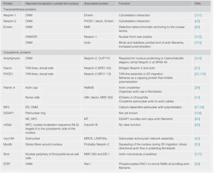
Table I. Actin-Binding Proteins around the NE
Protein Reported localization outside the nucleus Associated protein Function Refs Transmembrane proteins
Nesprin-1 ONM Emerin Cytoskeleton interaction [101]
Nesprin-2 ONM FHOD1, fascin, Emerin Cytoskeleton interaction [20] Emerin ONM NMII Defective heterochromatin anchoring to the nuclear
lamina
[60]
ONM/ER Nesprin-1 Nuclear front–rear polarity [102]
ONM Actin Binds and stabilizes pointed end of actinfilaments, increased polymerization
[103]
Cytoplasmic proteins
Amphiphysin ONM Nesprin-2, CLIP170 Required for nucleus positioning in Caenorhabditis elegans; binds Nesprin-2 at SR48-49
[104]
Fascin TAN lines, dorsal side Nesprin-2 (SR51-53) Bridges Nesprin-2 and actin [21] FHOD1 TAN lines, dorsal side Nesprin-2 (SR11-13) TAN line assembly in 2D migration
Behaves as a capping protein that inhibits polymerization
[20,105]
Filamin A Actin cap RefilinB Actin crosslinker
Organizes actin cap infibroblasts
[36]
Nurse cells Villin, fascin, MSP-300 (Cheerio in Drosophila)
Crosslinks perinuclear actin to actin cables
[14]
INF2 ER, ONM Calcium-dependent perinuclear actin polymerization [57,58]
IQGAP1 Perinuclear ring Not yet known [106]
NE, NPC MT IQGAP1 bundles and caps actinfilaments [80] mDia2 NPC, nuclear localization sequence (NLS)
targets it to the cytoplasmic side of the nucleus
Importin B No clear function [92]
myo18A Subnuclear MRCK, LRAP35a Subnuclear actomyosin network assembly [40] MyoIIb Stressfibers around nucleus Probably Nesprin-2 Squeezing of the nucleus during 3D migration; drives
directional actinflow in polarizing fibroblasts
[53]
Shot Nuclear periphery of Drosophila larval wall cells
MSP-300 and EB-1 Actin–microtubule crosslinker [107]
STEF ONM Rac1 Phosphorylates RAC1 to recruit Refilin at bundling actin filaments
as abolishing the actin-binding domain of ANC-1 is not necessary for nucleus anchorage [13]. It now remains to be determined how these giant actin-binding KASH proteins anchor the nucleus and the rest of the cellular organelles in place. In Drosophila nurse cells, nuclei are maintained in place by actin. This prevents the nuclei from clogging ring canals when nurse cells contract and expel their nutrient-rich cytoplasm to feed the growing oocyte. Actinfilaments emerge from the ring canals and crosslink perinuclear actin via Cheerio, the Drosophila ortholog offilamin A that appears at the NE before dumping [14]. This mechanism is independent of LINC complex proteins. Mesenchymal stem cells and epithelial cells position their nucleus in concave areas below the cell, releasing strain on the nucleus in a mechanism that is dependent on cell division cycle 42 (Cdc42), the actin-related protein 2/3 complex (Arp2/3), A-type lamins, and LINC complex proteins [15].
Cell polarization prior to migration is characterized by the positioning of the nucleus behind the centrosome respective to the direction of migration. In a tissue-culture cell-based assay in which actin stressfibers are abolished and allowed to form again, actomyosin contraction at the cell front is transmitted toward the nucleus through actin retrogradeflow [16,17]. Steered by a Cdc42/myotonic dystrophy kinase-related Cdc42-binding kinase (MRCK) pathway, nuclear movement is then driven by transverse actin cables bound to Nesprin-2, forming transmembrane actin-associated nuclear (TAN) lines, which undergo retrogradeflow. These rearward-traveling actinfibers drag the nucleus toward the back of the cell, behind the centrosome [18], in concert with TorsinA, Emerin, Samp1, SUNs, lamins, lamin-associated polypeptide 1 (LAP1), formin homology 2 domain-containing 1 (FHOD1), fascin, and nuclear pore complexes (NPCs) (Figure 2) [19–24]. TorsinA, in the perinuclear space, interacts with LAP1, which spans the inner nuclear membrane (INM), to assemble TAN lines. Emerin, a NE-associated protein known to be present on both nuclear membranes, is involved in the direc-tionality of the retrogradeflow [24]. Samp1, a component of TAN lines, is crucial for rearward nu-clear movement [19]. FHOD1 and fascin are localized at the cytoplasmic side of the NE and crosslink Nesprin-2 to actin [20,21]. This TAN line assay, coupled with centrifugation to displace the nucleus away from its natural position in the cell, was used to demonstrate that nucleus movement toward the front of the cell is SUN1 and microtubule dependent, while rearward move-ment is actin and SUN2 dependent [25]. It is unclear how cells differentiate between these two mechanisms, as both use Nesprin-2G as the central element linking SUN1 to microtubules and SUN2 to actin, respectively. However, this mechanism has potential implications in a disease context: the mutant form of lamin A that causes progeria, an extremely rare disorder
Box 2. The NE
One common feature among eukaryotes is the presence of a double lipid bilayer encapsulating the genome. Continuous with the ER, the NE is evenly punctured with large protein assemblies known as NPCs, allowing the nucleocytoplasmic shuttling of proteins. Close to the inner surface of the NE, a meshwork of intermediatefilaments, the lamins, bind to chromatin and NE proteins and provide a physical scaffold that is partially responsible for nuclear shape and stiffness. While its role was initially thought to be solely as a separation between DNA and the cytoplasm, several embedded proteins confer additional roles on the NE. A complex of proteins bridges the NE: the LINC complex, comprising two transmembrane proteins, SUN proteins at the INM and KASH proteins at the ONM. SUN proteins interact with lamins and chromatin-binding proteins inside the nucleus and connect to KASH proteins in the luminal space. These in turn bind cytoskeletalfilaments. This complex is decisive for the transfer of mechanical cues from the exterior of the cell to the nucleus interior. The SUN and SYNE (encoding KASH proteins) genes are able to generate different splicing variants in a cell-type-dependent manner, thus fine-tuning their ability to transmit forces to the nuclear interior. The KASH proteins Nesprin-1 and -2, can bind actinfilaments through calponin homology domains or through other intermediary proteins. Nesprins can indirectly bind to microtubules, while plectin links Nesprin-3 to intermediatefilaments. Nesprins contain multiple spectrin repeats that may act as springs to buffer mechanical forces. This diversity in Nesprin isoforms contributes to the differences observed in the response to mechanical stress by cells and tissues.
characterized by premature aging symptoms, induces accumulation of SUN1 at the NE, reducing the potential for actin-based nuclear movements [26] (Figure 3B).
Actin around the Nucleus during Cell Migration
In vivo, nuclear movement is particularly important during the formation of the brain, when neuro-nal progenitors migrate across long distances. This movement is saltatory due to decoupling of the growth cone and the nucleus: extension of the leading process occurs through actin polymer-ization in the growth cone followed by nuclear movement, often initiated by myosin IIB-dependent actomyosin contraction at the nucleus rear [27]. Although lack of Nesprin-1 and -2 results in morphological defects in the mouse brain, it remains to be established whether this intermittent nuclear movement is dependent on a direct connection between actin and the NE [28]. It is, however, likely to involve drebrin, a microtubule–actin crosslinking protein [29].
Trends
Trends inin Cell BiologyCell Biology
Figure 3. Nuclear Envelope–Actin Interactions Have Been Observed in Several Important Cellular Processes.(A) Actin-dependent nuclear migration in neurons is decisive during development for cell division and cell migration. The interkinetic nuclear movement preceding cell division is driven by Rho and formin-2 (FMN2) and actin polymerization at the nucleus rear. For neuronal migration, saltatory nuclear movement is driven by myosin IIB (MYOIIB) and actin polymerization at the nucleus rear. (B) During cell polarization, using actin retrogradeflow, transmembrane actin-associated nuclear lines move the nucleus toward the back of the cell. (C) Actin polymerization is required to displace the nucleus through a constriction infibroblasts and dendritic cells. In fibroblasts, nuclei are pulled through constrictions as Nesprin-2 accumulates at the front of the nucleus (orange). Actin is organized in bundles around the nucleus parallel to the direction of migration. In dendritic cells, actin-related protein 2/3 complex (Arp2/3) along the nucleus (purple) creates an actin sleeve to squeeze the nucleus through the constriction. (D) On mechanical stress, calcium (yellow) release into the cytoplasm [from the cells’ exterior or endoplasmic reticulum (ER) stores] induces actin polymerization around the nucleus in an inverted formin-2- (INF2) and Emerin-dependent manner. (E) Immediately after nuclear envelope breakdown (NEBD), actin surrounds the chromosomes and contracts, preventing them from dispersing and thus reducing mitotic errors. (F) Actomyosin contraction induces nuclear deformation and chromatin organization changes in the nucleus.
Infibroblasts, actin filaments above the nucleus, termed the perinuclear actin cap, orient the nu-cleus in the direction of migration [30,31]. These dynamic structures align with the long axis of the cell, contain phosphorylated myosin II andα-actinin-1, and terminate at vinculin-containing focal adhesions [30]. Unlike ventral actinfibers, the actin cap is connected to the nucleus through Nesprin-2, is involved in nuclear shape, and controls mechanotransduction [30,32]. Actinfibers of the perinuclear actin cap connect to 30% of focal adhesions and can thus exert higher tension than other actin structures, potentially increasing their responsiveness to mechanical stress [33,34]. In a similar manner, actin connects cell adhesion complexes with the NE of human breast epithelial cells in 3D culture [35].
Many proteins participate in linking the nucleus to actin and help to organize the actin cap. Cells that do not assemble perinuclear actin caps, such as cancer cells or cells lackingα-actinin-1, do not orient their nucleus in the direction of migration even if dorsal and transverse actinfibers are intact. At the nuclear periphery, the actin branching proteinfilamin A is converted into an actin bundler by RefilinB to organize the actin structures above the nucleus [36] and is also involved in mechanotransduction [37]. Interestingly, depletion of the Rac1-selective guanine exchange factor (GEF) Sif and Tiam1-like exchange factor (STEF), localized at the NE and colocalizing with Nesprin-2, leads to a decrease of perinuclear actin caps [38]. Lamin A/C is also required for the formation of the actin cap, suggesting feedback from the nuclear interior that dictates the ar-chitecture of the actin network [39]. The actin structures that exist below the nucleus in HeLa and Cos7 cells are not organized in the same direction as the actin cap and colocalize with MRCK and leucine repeat adaptor protein 35 (LRAP35) [40]. The role of these subnuclear structures is not clear yet, but they may be involved in contact guidance, characterized by cellular adaptation and cytoskeletal organization along topographical cues [41].
TAN lines and the perinuclear cap share many similarities (Figure 2), although TAN lines have not yet been shown to connect to focal adhesions nor has their role in mechanotransduction been demonstrated [18]. Recently, Hoffman et al. [23] described TAN lines formation upon cyclic stretching of cells. However, in a previous report Kim et al., using the same cell-based assay, showed that the actin lines formed upon stretch are perinuclear actin caps instead of TAN lines, as they are connected to focal adhesions [32]. Several partners of TAN lines have been as-sociated with multiple diseases where mechanical load is known to occur, such as laminopathies [42–44], Emery–Dreifuss muscular dystrophy [45–47], and cancer [48,49], thus indicating a po-tential role of this structure in transmitting mechanical cues to the nucleus interior. Further studies are required to determine whether TAN lines and the perinuclear cap could be specialized struc-tures emanating from the same mechanism and whether they share partner proteins. Further-more, it remains unclear whether these structures are found in vivo.
Actin around the Mechanically Deformed Nucleus
The deformability of the cell nucleus is a limiting factor for cell migration through narrow constrictions. In some cell types, actin-based mechanisms facilitate nuclear deformation. Dendritic cells squeeze their nuclei during entry into a narrow channel using an Arp2/3-nucleated actin sleeve to facilitate in-gression [50]. This LINC complex-independent mechanism relies on the size of the object to be pushed through the constriction, as demonstrated using beads of different sizes injected in the cy-toplasm: beads larger than the constriction are surrounded by actin when the cell attempts to trans-fer them through the constriction, but smaller beads are not. Actomyosin contractility, observed at the back of the cell indicating that the nuclei are pushed through the constriction from the rear, is not necessary for the formation of this actin sleeve. In a contrasting mechanism, murinefibroblasts deform their nucleus through constrictions via actomyosin contractility at the front of the cell [51], reminiscent of the nuclear movement observed when actomyosin contraction is induced at the
cell front on 2D substrates [52]. Fibroblasts lacking Nesprin-2G and fascin have defects in nu-clear translocation through constrictions [21,53]. In these cells, actinfilaments run parallel to the axis of migration, myosin IIB accumulates along thesefilaments [53], and Nesprin-2G accu-mulates at the front of the nucleus in an actin-dependent manner, implying that Nesprin-2G participates in pulling the nucleus through the constriction [51]. Primary humanfibroblasts un-dergoing lobopodial migration through the extracellular matrix display a similar mechanism. (Lobopodia are blunt, cylindrical protrusions forming whenfibroblasts migrate in 3D matrices [54].) In these cells, the confined nucleus is pulled forward via actin, vimentin, Nesprin-3, and myosin II at the cell front, resulting in increased pressure at the front of the cell and the formation of lobopodia in a piston-like mechanism [55]. It remains to be determined whether these similar mechanisms are related (Figure 3C).
Actin around the Nucleus in Response to Mechanical Stimuli
Actin structures around the nucleus can protect it from mechanical damage. The nuclei of stretched cells devoid of lamins A/C (that do not assemble a perinuclear actin cap) are more deformed than their wild-type counterparts, suggesting that the actin cap protects the nucleus from mechanical deformation [32]. Conversely, mechanical confinement exerted by actin on the nucleus is the force behind spontaneous NE rupture on 2D substrates [56].
Application of force to the cell, by direct probing, cell stretching, or shear, triggers an accumula-tion of actin in a ring around the nucleus that is dependent on calcium and inverted formin 2 (INF2) [57,58]. This rapid response (under 2 min), termed‘calcium-mediated actin reset’ (CaAR), is observed in epithelial, mesenchymal, endothelial, and immune cells. It can be triggered by the import of extracellular calcium into the cytoplasm or the release of calcium from the endoplasmic reticulum (ER) in a Piezo-1-dependent mechanism [58,59]. In endothelial cells, actin accumulation around the nucleus in response to stretch is Emerin dependent and triggers the displacement of Emerin from the INM to the ONM [60]. This rapid, force-dependent response induces nuclear softening by reducing heterochromatin levels, thereby protecting the genome [59]. In mesenchymal stem cells, high-frequency cell stretching (above 5 Hz) results in decoupling of the nucleus from the cytoskeleton in a SUN2-dependent manner [61]. This is consistent with nucleus rounding observed in response to high-amplitude stretch in endothelial cells [59] and calcium/Piezo-1-dependent nuclear shrinkage in sheared epithelial cells [62]. Sustained force application leads to tissue-level reorganization into a cobblestone pattern [59], but changing the direction of the force exertion rapidly triggers a new actin ring around the nucleus. It is not yet clear how this calcium-mediated mechanism triggers changes inside the nucleus, although the pivotal role of the formin INF2 is a strong indicator that actin polymerization is involved (Figure 3D).
Actin around the Nucleus during Mitosis and Meiosis
During meiosis, actin plays roles immediately before and during NEBD and moves the nucleus and the spindle in mouse oocytes. Actin also formsfilaments at centrosomes and in the spindle during mitosis and contributes to NE reassembly [63].
While microtubules are responsible for tearing down the NE in many systems [64], they are dispensable in some systems, where actin may play the starring role instead. Transient actin shells appear immediately before NEBD in echinoderms, cnidaria, and polychaetes [65]. In starfish oocytes, actin spikes, nucleated by Arp2/3 at the nuclear lamina, tear the INM between NPCs [65]. This mechanism may allow NPCs to remain intact in nucleoplasmic bod-ies throughout meiosis, thereby reducing the energy necessary to reform these NPCs when the NE is reassembled.
In mouse oocytes, nuclear movements during meiosis to the center of the cell and back to the cortex are actin dependent. Before meiosis I, actin polymerization creates a pressure gradient that nonspecifically centers large objects, thereby moving the largest organelle, the nucleus, to the cell center [66–69]. After this nucleus-centering step, the spindle is formed and it is then moved back to the cortex. Both of these movements depend on actin and its nucleator formin-2 [66,70].
At the start of mitosis, a contractile actin network reduces the volume taken up by chromosomes to ensure efficient chromosome capture by spindle microtubules. This serves to reduce the dispersion of individual chromosomes after NEBD. Removing LINC complexes from the NE or re-ducing myosin II contractility increases chromosome congression time and chromosome mislocalization, resulting in inefficient mitosis [71]. This actin network was not found in HeLa cells, suggesting that loss of this nonessential mechanism could be a source of mitotic errors in malignant cells (Figure 3E).
Actin interacts with centrosomes and the spindle in vitro and during mitosis [72–74]. Inhibition of Arp2/3-dependent branched actin nucleation reduces this spindle-associated pool of actin and results in disorganized chromosome congression and mitotic defects [75]. Similarly, centrosome separation and positioning around the cell centroid are orchestrated by transient perinuclear actin and the LINC complex [76]. Actinfilaments nucleated by formin-2 can even be found in the spin-dle itself during mitosis, working with microtubules to ensure faithful chromosome segregation [77]. Actomyosin plays indirect roles during cell mitosis by exerting forces thatflatten the cell. In HeLa cells, compressing the cells is sufficient to rescue mitosis when myosin II contractility is inhibited [78]. After mitosis, actin contributes to the reassembly of the NE; transient perinuclear F-actin rings have been reported in reforming nuclei (late telophase) in murinefibroblast and epidermal cell cultures, as well as insect epidermis [79]. The actin-regulator IQ motif-containing GTPase activating protein 1 (IQGAP1) coordinates NPC reassembly after mitosis [80]. Taken together, these results indicate that actin plays important roles during mitosis and meiosis, alongside microtubules. Actin plays important roles in setting up the nucleus for mitosis, reducing chromosomal segregation errors, and NE reassembly after mitosis. Whether these two cytoskeletal components work in parallel or together remains to be investigated.
Actin around the Nucleus and Gene Expression
Actomyosin contraction around the nucleus can affect the organization of chromatin. One striking example is the inversion of genomic architecture in rod photoreceptor cells caused by actomyosin-mediated nuclear deformation [81,82]. Actin contraction around the nucleus reduces nuclear volume and chromatin accessibility, resulting in poor reprogramming offibroblasts to plurip-otent stem cells [83]. It can also reduce telomere and heterochromatin dynamics [81] in a mecha-nism that is dependent on SUN2 [83], Nesprin-2G and lamin A/C [84]. Similarly, pulling on the LINC complex results in nucleus stiffening and altered gene expression [85,86]. This modulation of gene expression is well described in cells harboring high actomyosin contractility, such as cardiac and skeletal muscle cells [87]. In agreement with this, mutations in proteins associated with the LINC complex affect tissues subject to high mechanical forces. Actomyosin contractility increases SUN2’s association with lamins in vascular smooth muscle cells, thus remodeling interactions at the INM and altering the balance between actin and microtubule associations with the ONM [88]. Downregulation of Nesprin-2 or non-muscle myosin II, both involved in the formation and mainte-nance of TAN lines and actin perinuclear caps, results in similar alterations of the expression of genes associated with the epithelial-to-mesenchymal transition [18,24,89,90]. Overall, actomyosin contractility around the nucleus results in alteration of gene expression by reducing the nuclear
volume and via LINC complex-dependent mechanisms (i.e., mechanotransduction). These mecha-nisms may explain in part the disease phenotypes observed in nuclear envelopathies [91] (Figure 3F). Preventing monomeric actin from entering the nucleus is a promising strategy to modulate the ex-pression of genes that are regulated by nuclear actin or to reduce the import of gene-regulating proteins that require actin to enter the nucleus [e.g., myocardin-related transcription factor A (MRTF-A)]. Actin polymerization at the NE reduces stores of actin at the cell periphery [58] and depletes nuclear actin [60]. The formin mDia2 shuttles between the nucleoplasm and cyto-plasm and accumulates at the outer NE due to importin beta, colocalizing with NPCs [92]. These mechanisms could explain how actin polymerization around the NE (e.g., during CaAR) can result in downstream signaling.
In Vitro Reconstitution of Actin around the Nucleus
To better understand the factors that regulate actin assembly around the nucleus, the interaction between actin and the ONM is investigated by injectingfluorescent actin into cells, extracting nu-clei out of the cell to observe actin structures present on the ONM, or observing the interaction of isolated nuclei with monomeric or polymerized actin. Injection or transient expression of fluores-cent actin in HeLa cells results in accumulation of actin at the nuclear periphery, while isolated he-patocyte nuclei incubated in the presence of fluorescent actin monomers accumulate polymerized actin [93]. A similar accumulation of actin at the nuclear periphery was observed in nuclei isolated using non-ionic detergents [94]. Antibodies against NE proteins (Nesprin-2 and a NPC protein) reduce the accumulation of actin around the nucleus, but it is unclear whether this is due to a specific interaction with these specific proteins or whether the bulky antibodies mask the surface of the NE from actin. Precise studies are thus still required to determine whether actinfilament accumulation around nuclei is due to the binding of one or several specific proteins. Nevertheless, this system presents exciting opportunities to study actin assembly at the NE. To study force transmission between the cytoskeleton and the nucleus, beads coated with actin are brought into contact with nuclei using optical tweezers [95]. Jumps of a few nanometers were recorded upon retraction of the beads using forces up to 50 pN, likely indicating protein unfolding. The authors hypothesize that they are probing Nesprin-1 or -2, and that their transitions correspond to the unfolding of a helix (5–10 nm) or of a Nesprin spectrin repeat (15 nm) [95]. Further study is necessary to confirm that actin-coated beads are specifically probing Nesprins at the NE. Nevertheless, applying forces to Nesprins using antibody-coated beads triggers nu-clear stiffening that is dependent on the nunu-clear lamina and Emerin [85].
Concluding Remarks
The diversity of architectures that actin can adopt and its wide variety of target proteins make it a versatile actor in the cell (Figure 3). Whereas actin was historically depicted as the cytoskeletal network in charge of cell shape and contractility, we now know that it has leading and supportive roles in many other cell processes. Careful examination of actin structures and associated pro-teins in vivo is likely to provide evidence for many more extraordinary ways that actin orchestrates functions in the cell.
Much work remains on many fronts to understand the roles that NE proteins play with actin. Many NE transmembrane (NET) proteins have yet to be described fully and overexpression or knock-down of some of these leads to cytoskeletal disruption. The results obtained in vitro or in cell-based assays to decipher the actin–nucleus connection are not always readily transferable in vivo. For example, the existence of TAN lines in tissues or even in 3D culture remains confirmed. The recent development of organoids, recapitulating organ structure and function, combined
Outstanding Questions How is actin selectively engaged at the NE (knowing that actin-binding Nesprins can also bind microtubules)? Are the actinfibers under the nucleus connected to the NE?
Do TAN lines and the perinuclear actin cap exist in vivo? Are they related to the actin structures observed in 3D migration?
What are the nuclear envelope proteins involved in forming TAN lines and actin caps and how are they similar or differ-ent between these two structures? How does the CaAR response regulate gene expression and nucleus deformability?
Are there other NET proteins that bind and regulate actinfilaments? What is the organization of Nesprins and Nesprin-binding proteins at the surface of the nucleus? Does actin contribute to organizing Nesprins at the surface of the nucleus? Is there a relation between the viscoelasticity of the nucleus and perinuclear actin?
with the latest improvements in super-resolution imaging could allow better visualization and comprehension of the mechanisms involved. Optogenetics to locally perturb protein conforma-tion or localizaconforma-tion, Förster resonance energy transfer (FRET) sensors inserted in proteins under mechanical stress, and microfluidics are some of the techniques to be applied to gain more in-sight into the nucleus–actin connection. A recent approach using mutually attracted magnetic beads on both sides of the plasma membrane has revealed thefluctuations of actomyosin con-tractions [96]. A similar approach using a single bead inside the cytoplasm revealed the elastic component of the NE [97]. In addition, exciting new tools are being developed that could be adapted to expand our understanding of actin around the NE: a technique to reveal actin that is in contact with the cytoplasmic membrane could be adapted to similarly reveal actin structures in contact with the NE [98]. In vitro models could help to elucidate the structures we observe in cells and in vivo: a recent publication, studying the interaction of actin with lipid membranes, strik-ingly recapitulated the membrane protrusions observed during NEBD in starfish oocytes [65,99]. We discussed previously the potential impact that the balance between perinuclear actin polymer-ization and the shuttling of monomeric actin across the nuclear membrane can have on gene ex-pression and cell fate. Further research is necessary to determine whether this mechanism is used by the cell and whether it can explain the impact of actin accumulation around the nucleus on gene expression (e.g., during CaAR) (see Outstanding Questions). Many mechanisms discussed here, from mitosis to nuclear displacement during 3D migration, could play a particularly important role in tumor cell proliferation and metastatic dissemination, implying that actin binding partners could provide new therapeutic partners to investigate in thefight against cancer and metastatic disease. The link between actin and the NE may also be decisive in the mechanisms of nuclear envelopathies.
Acknowledgments
We thank the Cadot and Bitoun Laboratories for discussions. P.M.D. and B.C. were supported by Association Institut de Myologie. We thank Gautam Dey for proofreading the manuscript.
References
1. Crisp, M. et al. (2006) Coupling of the nucleus and cytoplasm: role of the LINC complex. J. Cell Biol. 172, 41–53 2. Sauer, F. (1935) Mitosis in the neural tube. J. Comp. Neurol.
62, 377–405
3. Strzyz, P.J. et al. (2016) Heterogeneity, cell biology and tissue mechanics of pseudostratified epithelia: coordination of cell divisions and growth in tightly packed tissues. Int. Rev. Cell Mol. Biol. 325, 89–118
4. Norden, C. et al. (2009) Actomyosin is the main driver of interkinetic nuclear migration in the retina. Cell 138, 1195–1208 5. Strzyz, P.J. et al. (2015) Interkinetic nuclear migration is centro-some independent and ensures apical cell division to maintain tissue integrity. Dev. Cell 32, 203–219
6. Yu, J. et al. (2011) KASH protein Syne-2/Nesprin-2 and SUN proteins SUN1/2 mediate nuclear migration during mammalian retinal development. Hum. Mol. Genet. 20, 1061–1073 7. Falk, N. et al. (2019) Lack of a retinal phenotype in a Syne-2/
Nesprin-2 knockout mouse model. Cells 8, 1238 8. Yanakieva, I. et al. (2019) Cell and tissue morphology
deter-mine actin-dependent nuclear migration mechanisms in neuroepithelia. J. Cell Biol. 218, 3272–3289
9. Lahne, M. et al. (2015) Actin–cytoskeleton- and Rock-mediated INM are required for photoreceptor regeneration in the adult zebrafish retina. J. Neurosci. 35, 15612–15634 10. Kirkland, N.J. et al. (2020) Tissue mechanics regulate mitotic
nuclear dynamics during epithelial development. Curr. Biol. 30, 2419–2432.e4
11. Roman, W. et al. (2017) Myofibril contraction and crosslinking drive nuclear movement to the periphery of skeletal muscle. Nat. Cell Biol. 19, 1189–1201
12. Starr, D.A. and Han, M. (2002) Role of ANC-1 in tethering nuclei to the actin cytoskeleton. Science 298, 406–409 13. Hao, H. et al. (2020) The Nesprin-1/-2 ortholog ANC-1 regulates
organelle positioning in C. elegans without its KASH or actin-binding domains. bioRxiv Published online July 15, 2020. https://doi.org/10.1101/2020.07.14.202838
14. Huelsmann, S. et al. (2013) Filopodia-like actin cables position nuclei in association with perinuclear actin in Drosophila nurse cells. Dev. Cell 26, 604–615
15. Pieuchot, L. et al. (2018) Curvotaxis directs cell migration through cell-scale curvature landscapes. Nat. Commun. 9, 3995 16. Palazzo, A.F. et al. (2001) Cdc42, dynein, and dynactin regulate
MTOC reorientation independent of Rho-regulated microtubule stabilization. Curr. Biol. 11, 1536–1541
17. Gomes, E.R. et al. (2005) Nuclear movement regulated by Cdc42, MRCK, myosin, and actinflow establishes MTOC polarization in migrating cells. Cell 121, 451–463
18. Luxton, G.W.G. et al. (2010) Linear arrays of nuclear envelope proteins harness retrograde actinflow for nuclear movement. Science 329, 956–959
19. Borrego-Pinto, J. et al. (2012) Samp1 is a component of TAN lines and is required for nuclear movement. J. Cell Sci. 125, 1099–1105 20. Kutscheidt, S. et al. (2014) FHOD1 interaction with Nesprin-2G mediates TAN line formation and nuclear movement. Nat. Cell Biol. 16, 708–715
21. Jayo, A. et al. (2016) Fascin regulates nuclear movement and deformation in migrating cells. Dev. Cell 38, 371–383 22. Saunders, C.A. et al. (2017) TorsinA controls TAN line assembly
and the retrogradeflow of dorsal perinuclear actin cables during rearward nuclear movement. J. Cell Biol. 216, 657–674
23. Hoffman, L.M. et al. (2020) Mechanical stress triggers nuclear remodeling and the formation of transmembrane actin nuclear lines with associated nuclear pore complexes. Mol. Biol. Cell 31, 1774–1784
24. Chang, W. et al. (2013) Emerin organizes actinflow for nuclear movement and centrosome orientation in migratingfibroblasts. Mol. Biol. Cell 24, 3869–3880
25. Zhu, R. et al. (2017) Centrifugal displacement of nuclei reveals multiple LINC complex mechanisms for homeostatic nuclear positioning. Curr. Biol. 27, 3097–3110.e5
26. Chang, W. et al. (2019) Imbalanced nucleocytoskeletal con-nections create common polarity defects in progeria and phys-iological aging. Proc. Natl. Acad. Sci. U. S. A. 116, 3578–3583 27. Nakazawa, N. and Kengaku, M. (2020) Mechanical regulation of nuclear translocation in migratory neurons. Front. Cell Dev. Biol. 8, 150
28. Zhang, X. et al. (2009) SUN1/2 and Syne/Nesprin-1/2 complexes connect centrosome to the nucleus during neurogenesis and neuronal migration in mice. Neuron 64, 173–187
29. Trivedi, N. et al. (2017) Drebrin-mediated microtubule– actomyosin coupling steers cerebellar granule neuron nucleokinesis and migration pathway selection. Nat. Commun. 8, 14484
30. Maninová, M. and Vomastek, T. (2016) Dorsal stressfibers, transverse actin arcs, and perinuclear actinfibers form an interconnected network that induces nuclear movement in polarizingfibroblasts. FEBS J. 283, 3676–3693
31. Khatau, S.B. et al. (2009) A perinuclear actin cap regulates nu-clear shape. Proc. Natl. Acad. Sci. U. S. A. 106, 19017–19022 32. Kim, J.-K. et al. (2017) Nuclear lamin A/C harnesses the perinuclear apical actin cables to protect nuclear morphology. Nat. Commun. 8, 2123
33. Kim, D.-H. et al. (2012) Actin cap associated focal adhesions and their distinct role in cellular mechanosensing. Sci. Rep. 2, 555
34. Kim, D.-H. et al. (2013) The multi-faceted role of the actin cap in cellular mechanosensation and mechanotransduction. Soft Matter 9, 5516–5523
35. Jorgens, D.M. et al. (2017) Deep nuclear invaginations are linked to cytoskeletalfilaments – integrated bioimaging of epithelial cells in 3D culture. J. Cell Sci. 130, 177–189 36. Gay, O. et al. (2011) Refilin holds the cap. Commun. Integr.
Biol. 4, 791–795
37. Baudier, J. et al. (2018) Thefilamin-B–refilin axis – spatiotemporal regulators of the actin–cytoskeleton in development and disease. J. Cell Sci. 131, jcs213959
38. Woroniuk, A. et al. (2018) STEF/TIAM2-mediated Rac1 activity at the nuclear envelope regulates the perinuclear actin cap. Nat. Commun. 9, 2124
39. Kim, H.Y. et al. (2017) On the role of mechanics in driving mesenchymal-to-epithelial transitions. Semin. Cell Dev. Biol. 67, 113–122
40. Tan, I. et al. (2008) A tripartite complex containing MRCK mod-ulates lamellar actomyosin retrogradeflow. Cell 135, 123–136 41. Tamiello, C. et al. (2015) Competition between cap and basal actinfiber orientation in cells subjected to contact guidance and cyclic strain. Sci. Rep. 5, 8752
42. Antoku, S. et al. (2019) ERK1/2 phosphorylation of FHOD con-nects signaling and nuclear positioning alternations in cardiac laminopathy. Dev. Cell 51, 602–616.e12
43. Berk, J.M. et al. (2013) The nuclear envelope LEM-domain protein Emerin. Nucleus 4, 298–314
44. Zwerger, M. et al. (2013) Myopathic lamin mutations impair nu-clear stability in cells and tissue and disrupt nucleo-cytoskeletal coupling. Hum. Mol. Genet. 22, 2335–2349
45. Meinke, P. et al. (2014) Muscular dystrophy-associated SUN1 and SUN2 variants disrupt nuclear-cytoskeletal connections and myonuclear organization. PLoS Genet. 10, e1004605 46. Taranum, S. et al. (2012) LINC complex alterations in DMD and
EDMD/CMTfibroblasts. Eur. J. Cell Biol. 91, 614–628 47. Puckelwartz, M.J. et al. (2009) Disruption of Nesprin-1
pro-duces an Emery Dreifuss muscular dystrophy-like phenotype in mice. Hum. Mol. Genet. 18, 607–620
48. Matsumoto, A. et al. (2015) Global loss of a nuclear lamina component, lamin A/C, and LINC complex components
SUN1, SUN2, and Nesprin-2 in breast cancer. Cancer Med. 4, 1547–1557
49. Reis-Sobreiro, M. et al. (2018) Emerin deregulation links nuclear shape instability to metastatic potential. Cancer Res. 78, 6086–6097
50. Thiam, H.-R. et al. (2016) Perinuclear Arp2/3-driven actin poly-merization enables nuclear deformation to facilitate cell migra-tion through complex environments. Nat. Commun. 7, 10997 51. Davidson, P.M. et al. (2020) Nesprin-2 accumulates at the front of the nucleus during confined cell migration. EMBO Rep. 21, e49910
52. Wu, J. et al. (2014) Actomyosin pulls to advance the nucleus in a migrating tissue cell. Biophys. J. 106, 7–15
53. Thomas, D.G. et al. (2015) Non-muscle myosin IIB is critical for nuclear translocation during 3D invasion. J. Cell Biol. 210, 583–594
54. Petrie, R.J. and Yamada, K.M. (2012) At the leading edge of three-dimensional cell migration. J. Cell Sci. 125, 5917–5926 55. Petrie, R.J. et al. (2014) Generation of compartmentalized
pressure by a nuclear piston governs cell motility in a 3D matrix. Science 345, 1062–1065
56. Hatch, E.M. and Hetzer, M.W. (2016) Nuclear envelope rupture is induced by actin-based nucleus confinement. J. Cell Biol. 215, 27–36
57. Shao, X. et al. (2015) Mechanical stimulation induces formin-dependent assembly of a perinuclear actin rim. Proc. Natl. Acad. Sci. U. S. A. 112, E2595–E2601
58. Wales, P. et al. (2016) Calcium-mediated actin reset (CaAR) mediates acute cell adaptations. Elife 5, e19850
59. Nava, M.M. et al. (2020) Heterochromatin-driven nuclear softening protects the genome against mechanical stress-induced damage. Cell 181, 800–817.e22
60. Le, H.Q. et al. (2016) Mechanical regulation of transcription controls Polycomb-mediated gene silencing during lineage commitment. Nat. Cell Biol. 18, 864–875
61. Gilbert, H.T.J. et al. (2019) Nuclear decoupling is part of a rapid protein-level cellular response to high-intensity mechanical loading. Nat. Commun. 10, 4149
62. Jetta, D. et al. (2019) Shear stress-induced nuclear shrinkage through activation of Piezo1 channels in epithelial cells. J. Cell Sci. 132, jcs226076
63. Kunda, P. and Baum, B. (2009) The actin cytoskeleton in spin-dle assembly and positioning. Trends Cell Biol. 19, 174–179 64. Beaudouin, J. et al. (2002) Nuclear envelope breakdown
proceeds by microtubule-induced tearing of the lamina. Cell 108, 83–96
65. Wesolowska, N. et al. (2020) Actin assembly ruptures the nu-clear envelope by prying the lamina away from nunu-clear pores and nuclear membranes in starfish oocytes. Elife 9, e49774 66. Almonacid, M. et al. (2015) Active diffusion positions the
nucleus in mouse oocytes. Nat. Cell Biol. 17, 470 67. Almonacid, M. et al. (2019) Activefluctuations of the nuclear
envelope shape the transcriptional dynamics in oocytes. Dev. Cell 51, 145–157.e10
68. Ahmed, W.W. et al. (2018) Active mechanics reveal molecular-scale force kinetics in living oocytes. Biophys. J. 114, 1667–1679
69. Colin, A. et al. (2020) Active diffusion in oocytes nonspecifically centers large objects during prophase I and meiosis I. J. Cell Biol. 219, e201908195
70. Duan, X. et al. (2020) Dynamic organelle distribution initiates actin-based spindle migration in mouse oocytes. Nat. Commun. 11, 277
71. Booth, A.J. et al. (2019) Contractile acto-myosin network on nuclear envelope remnants positions human chromosomes for mitosis. Elife 8, e46902
72. Blanchoin, L. et al. (2014) Actin dynamics, architecture, and mechanics in cell motility. Physiol. Rev. 94, 235–263 73. Inoue, D. et al. (2019) Actinfilaments regulate microtubule
growth at the centrosome. EMBO J. 38, e99630 74. Colin, A. et al. (2018) Actin-network architecture regulates
microtubule dynamics. Curr. Biol. 28, 2647–2656.e4 75. Plessner, M. et al. (2019) Centrosomal actin assembly is
re-quired for proper mitotic spindle formation and chromosome congression. iScience 15, 274–281
76. Stiff, T. et al. (2020) Prophase-specific perinuclear actin coordinates centrosome separation and positioning to ensure accurate chromosome segregation. Cell Rep. 31, 107681 77. Mogessie, B. and Schuh, M. (2017) Actin protects mammalian
eggs against chromosome segregation errors. Science 357, eaal1647
78. Aureille, J. et al. (2019) Nuclear envelope deformation controls cell cycle progression in response to mechanical force. EMBO Rep. 20, e48084
79. Clubb, B.H. and Locke, M. (1996) F-Actin forms transient perinuclear shells at the mitosis–interphase transition. Cell Motil. 33, 151–162
80. Lian, A.T.Y. et al. (2015) IQGAP1 is associated with nuclear envelope reformation and completion of abscission. Cell Cycle 14, 2058–2074
81. Makhija, E. et al. (2016) Nuclear deformability and telomere dynamics are regulated by cell geometric constraints. Proc. Natl. Acad. Sci. U. S. A. 113, E32–E40
82. Seirin-Lee, S. et al. (2019) Role of dynamic nuclear deformation on genomic architecture reorganization. PLoS Comput. Biol. 15, e1007289
83. Hu, X. et al. (2019) MKL1–actin pathway restricts chromatin accessibility and prevents mature pluripotency activation. Nat. Commun. 10, 1695
84. Jokhun, D.S. et al. (2018) Actin dynamics couples extracellular signals to the mobility and molecular stability of telomeres. Biophys. J. 115, 1166–1179
85. Guilluy, C. et al. (2014) Isolated nuclei adapt to force and reveal a mechanotransduction pathway in the nucleus. Nat. Cell Biol. 16, 376
86. Cho, S. et al. (2019) Mechanosensing by the lamina protects against nuclear rupture, DNA damage, and cell-cycle arrest. Dev. Cell 49, 920–935.e5
87. Piccus, R. and Brayson, D. (2020) The nuclear envelope: LINCing tissue mechanics to genome regulation in cardiac and skeletal muscle. Biol. Lett. 16, 20200302
88. Porter, L. et al. (2020) SUN1/2 are essential for RhoA/ROCK-regulated actomyosin activity in isolated vascular smooth muscle cells. Cells 9, 132
89. Halder, D. et al. (2019) Nonmuscle myosin IIA and IIB differen-tially modulate migration and alter gene expression in primary mouse tumorigenic cells. Mol. Biol. Cell 30, 1463–1476 90. Déjardin, T. et al. (2020) Nesprins are mechanotransducers
that discriminate epithelial–mesenchymal transition programs. J. Cell Biol. 219, e201908036
91. Janin, A. et al. (2017) Nuclear envelopathies: a complex LINC between nuclear envelope and pathology. Orphanet J. Rare Dis. 12, 147
92. Shao, X. et al. (2015) Novel localization of formin mDia2: importinβ-mediated delivery to and retention at the cytoplas-mic side of the nuclear envelope. Biol. Open 4, 1569–1575 93. Münter, S. et al. (2006) Actin polymerisation at the cytoplasmic
face of eukaryotic nuclei. BMC Cell Biol. 7, 23
94. Sardo, L. et al. (2017) Real-time visualization of chromatin modification in isolated nuclei. J. Cell Sci. 130, 2926–2940 95. Balikov, D.A. et al. (2017) The Nesprin–cytoskeleton interface
probed directly on single nuclei is a mechanically rich system. Nucleus 8, 534–547
96. Laplaud, V. et al. (2020) Pinching the cortex of live cells reveals thickness instabilities caused by myosin II motors. bioRxiv Published online September 29, 2020. https://doi.org/ 10.1101/2020.09.28.316729
97. Venturini, V. et al. (2020) The nucleus measures shape changes for cellular proprioception to control dynamic cell be-havior. Science 370, eaba2644
98. Bisaria, A. et al. (2020) Membrane-proximal F-actin restricts local membrane protrusions and directs cell migration. Science 368, 1205–1210
99. Simon, C. et al. (2019) Actin dynamics drive cell-like membrane deformation. Nat. Phys. 15, 602–609
100. Firat-Karalar, E.N. and Welch, M.D. (2011) New mechanisms and functions of actin nucleation. Curr. Opin. Cell Biol. 23, 4–13 101. Wheeler, M.A. et al. (2007) Distinct functional domains in Nesprin-1α and Nesprin-2β bind directly to Emerin and both interactions are disrupted in X-linked Emery–Dreifuss muscular dystrophy. Exp. Cell Res. 313, 2845–2857
102. Nastały, P. et al. (2020) Role of the nuclear membrane protein Emerin in front–rear polarity of the nucleus. Nat. Commun. 11, 2122
103. Holaska, J. et al. (2004) Emerin caps the pointed end of actin filaments: evidence for an actin cortical network at the nuclear inner membrane. PLoS Biol. 2, e231
104. D’Alessandro, M. et al. (2015) Amphiphysin 2 orchestrates nucleus positioning and shape by linking the nuclear envelope to the actin and microtubule cytoskeleton. Dev. Cell 35, 186–198
105. Schönichen, A. et al. (2013) FHOD1 is a combined actin fila-ment capping and bundling factor that selectively associates with actin arcs and stressfibers. J. Cell Sci. 126, 1891–1901 106. Johnson, M.A. and Henderson, B.R. (2012) The scaffolding
protein IQGAP1 co-localizes with actin at the cytoplasmic face of the nuclear envelope: implications for cytoskeletal regulation. Bioarchitecture 2, 138–142
107. Wang, S. et al. (2015) Nesprin provides elastic properties to muscle nuclei by cooperating with spectraplakin and EB1. J. Cell Biol. 209, 529–538