Control of Gene Expression by Cell Size
by Chia-Yung Wu B.A. Biochemistry Rutgers University, New Brunswick, NJ 2001Submitted to the Department of Biology in partial fulfillment of the requirements for the thesis degree of
Doctor of Philosophy in Biology at the
Massachusetts Institute of Technology May, 2010
© 2010 Chia-Yung Wu. All rights reserved.
The author hereby grants to MIT permission to reproduce and to distribute publicly paper and electronic copies of this thesis document in whole or in part in any medium now known or
hereafter created. Signature of Author: ____________________________________________________________ Department of Biology Date Certified by: ___________________________________________________________________ Gerald R. Fink, Ph.D.
Professor, Department of Biology Thesis Supervisor
Accepted by: __________________________________________________________________ Stephen P. Bell, Ph.D.
Professor, Department of Biology Chairman, Committee for Graduate Students
Table of Contents
Abstract ………. 3
Acknowledgements ………. 4
Chapter 1. Introduction ………. 5
Polyploidization/whole genome duplication as a mechanism of evolution … 5
Polyploidy in diploid multi-cellular organisms during development, stress response and tumorigenesis ………. 8
Ploidy and gene expression – examples from multi-cellular eukaryotes ….. 12
Budding yeast as a model system to study consequences of polyploidy …... 15
Control of cell size in yeast and metazoans ………. 19
Summary ………. 24
References ………. 25
Chapter 2. Control of gene expression by cell size Summary ………. 29
Introduction ………. 29
Results ………. 32
Discussion ………. 38
Materials and Methods ………. 42
References ………. 49
Figures ………. 52
Tables ………. 59
Chapter 3. Reduced binding of transcriptional activators of FLO11 in polyploids Summary ………. 65
Introduction ………. 66
Results ………. 79
Discussion …….……… 88
Materials and Methods ………. 91
References ………. 96
Figures and Table ………. 101
Chapter 4. Discussion and future directions ………. 109
3
Abstract
Polyploidy, increased copy number of whole chromosome sets in the genome, is a common cellular state in evolution, development and disease. Polyploidy enlarges cell size and alters gene expression, producing novel phenotypes and functions. Although many polyploid cell types have been discovered, it is not clear how polyploidy changes physiology. Specifically, whether the enlarged cell size of polyploids causes differential gene regulation has not been investigated. In this thesis, I present the evidence for a size-sensing mechanism that alters gene expression in yeast. My results indicate a causal relationship between cell size and gene expression. Ploidy-associated changes in the transcriptome therefore reflect transcriptional adjustment to a larger cell size. The causal and regulatory connection between cell size and transcription suggests that the physical features of a cell (such as size and shape) are a systematic factor in gene regulation. In addition, cell size homeostasis may have a critical function – maintenance of transcriptional homeostasis.
4
Acknowledgements
I sincerely thank my thesis advisor, Dr. Gerald R. Fink, for his professional guidance and for providing an excellent research environment. I am highly appreciative of the intellectual freedom entrusted to me during my pursuit of fundamental questions in biology. In the Fink lab, one not only learns to conduct rigorous scientific research but also to become a versatile, professional creator of knowledge.
I have benefited much from the wisdom and kindness of many former and current Fink lab members in my personal and professional development. In the Fink lab, our work atmosphere is always friendly and collaborative. Our conversations are fun and intellectually stimulating. It has been a great pleasure working daily with a group of talented and well-rounded individuals.
The Biology department and Whitehead Institute are both instrumental to my successful education at MIT. Many thanks to the faculty members and the Biology headquarter for nurturing us graduate students into competent and dependable professionals. I am deeply
indebted to the wholesome community at Whitehead, from whom I learned what a community is about, and without whom my experience at MIT would not have been fulfilling.
Special thanks to the following groups at MIT for their helpful discussions and generous support: Amon lab, Bell lab, Gifford lab, Hochwagen lab, Lindquist lab, Regev group, and Young lab.
Many thanks as well to members of my thesis committee: Dr. Frank Solomon and Dr. Stephen Bell for much helpful advice since the beginning of this thesis project, Dr. Angelika Amon for many insightful discussions and Dr. David Pellman for constructive feedback on this thesis.
Much appreciation to National Institute of Health, Massachusetts Institute of Technology and Whitehead Institute for Biomedical Research for funding and infrastructural support.
Last but not least, I sincerely thank my family and friends for enriching my life by making me a part of their lives with their love and friendship.
5
Chapter 1. Introduction
Polyploidization/whole genome duplication as a mechanism of evolution
Recent advances in genome sequencing and comparative genomics indicate that many living diploid organisms are paleopolyploids – ancient polyploids that underwent diploidization through sequence divergence between the duplicated chromosomes (Wolfe, 2001). Polyploidy is particularly common in the plant kingdom. Many diploid plants have been referred to as
paleopolyploids (Blanc and Wolfe, 2004). Species that are polyploid appear to be more prevalent in colder regions, usually establishing niches different from those of their diploid progenitors (Hegarty and Hiscock, 2008; Hijmans et al., 2007). The relationship between genome duplication and evolution was pioneered by Susumu Ohno, who proposed the idea that whole genome
duplication/polyploidization creates substrates for evolution (Ohno, 1970).
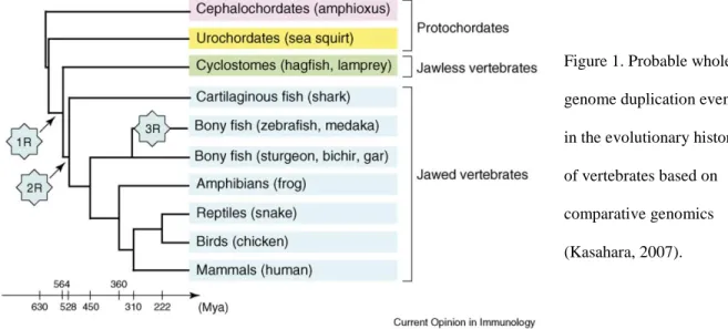
Ohno specifically hypothesized that two rounds (2R) of whole genome duplication shaped the evolutionary history of vertebrates. This hypothesis was largely based on his discovery of seemingly quadruplicated chromosomal segments in the human genome. A later study showed that the four large homeobox gene clusters in vertebrates arose from duplications of one ancestral cluster related to the cluster in Drosophila (Schughart et al., 1989). The high degree of conservation of structural organization between the vertebrate and insect clusters suggests that the duplications involved large chromosomal regions, or possibly entire chromosomes. Since duplications of individual chromosomes tend to be deleterious, whole genome duplication was proposed as a plausible mechanism.
Recently, four copies of large (>100 gene) chromosomal segments have been identified in at least 25% of the human genome, and these segments have corresponding, single-copy
6
paralogous regions in the primitive cordate sea squirt Ciona (Dehal and Boore, 2005). The conserved structural organization and the large number of such paralogous genomic segments favor whole genome duplication. Generation of these regions through a series of independent small gene amplification would be extremely unlikely at the same time. Furthermore, four-fold duplication is the dominant mode of redundancy for the paralogs in the human genome. The simplest and most likely mechanism of the large scale four-fold duplication is two rounds of whole genome duplication, a strong argument for the 2R hypothesis.
Figure 1. Probable whole genome duplication events in the evolutionary history of vertebrates based on comparative genomics (Kasahara, 2007).
More striking evidence for Ohno's hypothesis comes from the fungal kingdom. Species of the Saccharomyces genus appear to have evolved from a tetraploid ancestor that arose from whole genome duplication. A systematic search for sequence homology within the
Saccharomyces cerevisiae genome led to the discovery of 55 pairs of large (longer than 50Kb on
average) duplicated regions scattered throughout the genome. Within each pair of duplicated regions are homolgous genes in conserved gene order and orientation with respect to the
7
centromeres (Wolfe and Shields, 1997). A tetraploid origin for Saccharomyces is thus inferred. Whole genome duplication likely gave rise to the tetraploid ancestor around 100 million years ago, after Saccharomyces diverged from Kluyveromyces. Most of the functionally redundant gene pairs in the ancient tetraploid lost one member as the organism further evolved.
The emergence of fermentative yeast species is cited as one of the best documented example for the evolutionary consequences of whole genome duplication. In S. cerevisiae, many of the gene pairs retained after genome duplication participate in carbohydrate metabolism and show differential expression patterns in response to glucose or oxygen availability (Wolfe and Shields, 1997). The duplicated genes escaped deletion by gaining novel functions to cope with conditions related to fermentation. Independently, comparative genomics shows systematic transcriptional network rewiring in descendents of the tetraploid ancestor. The descendent species appear to have lost a cis-regulatory element upstream of dozens of ORFs encoding mitochondrial ribosomal proteins, whereasthe same cis element is retained in promoters of genes required for cytoplasmic ribosomal biogenesis (Ihmels et al., 2005). As a result, synthesis and assembly of cytoplasmic ribosomes are decoupled from mitochondrial ribosomes, enabling these organisms to thrive in low-oxygen conditions and to ferment. In contrast, species that did not evolve from the tetraploid ancestor, such as Candida albicans, have the cis-regulatory element in genes for both mitochondrial and cytoplasmic ribosome biogenesis. As expected, these species display a strong correlation between mitochondrial and cytoplasmic ribosomal biogenesis and do not grow anaerobically.
8
Figure 2. Phylogenetic tree of fermentative (rapid anaerobic growth) and non-fermentative (rapid aerobic growth) yeast species (Ihmels et al., 2005).
The apparent link between whole genome duplication and fermentative yeast speciation exemplifies the evolutionary potential of polyploidy. Gene duplication on the whole genome scale provides unparalleled evolutionary space, making rapid and coordinated extensive changes in fundamental cellular processes (such as metabolism) possible. It is worth noting that the whole genome duplication event occurred as fruiting angiosperms populated the earth’s flora (Wolfe and Shields, 1997). The enhanced evolutionary potential of polyploidy and the increasing availability of fruits likely fostered the emergence of fermentative yeast.
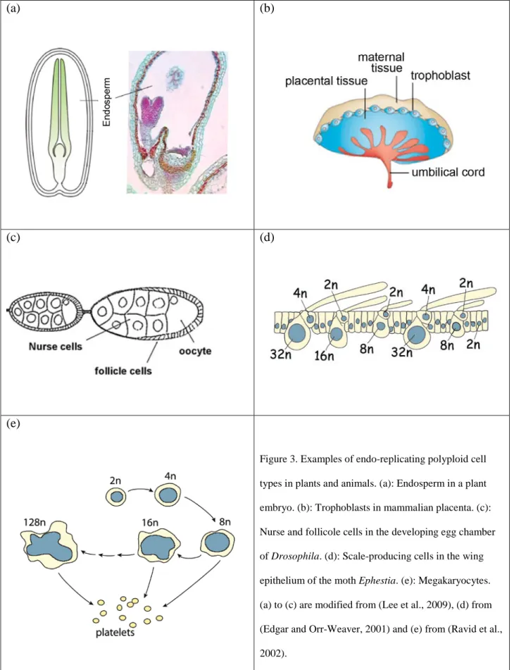
Polyploidy in diploid multi-cellular organisms during development, stress response and tumorigenesis
In a wide range of tissues in diploid organisms, polyploidy is achieved through endo-replication, a specialized cell cycle in which the genome content increases by DNA replication without subsequent cell division (Edgar and Orr-Weaver, 2001). Trophoblasts and
endo-9
replication (Edgar and Orr-Weaver, 2001; Ravid et al., 2002). Multiple tissues in Drosophila initiate endo-replication to become polyploid during embryogenesis, and endo-replication occurs again later in cells lining the larval gut track (Smith and Orr-Weaver, 1991). Various somatic tissues in seed plants are also known to become highly polyploid during development (Barow and Meister, 2003; Galbraith et al., 1991).
Polyploidy via endo-replication, typically associated with terminal differentiation, is considered a convenient mechanism to produce cells with high metabolic capability and
specialization in mass production/storage of macromolecules (Edgar and Orr-Weaver, 2001). In Drosophila adult females, polyploid nurse and follicle cells are crucial for oocyte development.
Specific disruption of endo-replication in these cells severely delays maturation of egg chambers and results in sterility (Maines et al., 2004). Trophoblasts in the mammalian placenta have a DNA content of >1000C. They facilitate embryo implantation and are needed to meet the high demand of molecular transport between mother and fetus (Zybina and Zybina, 2005).
Differentiation of megakaryocytes requires multiple rounds of endo-replication. High ploidy (up to 128n) facilitates mass production and release of platelets (Ravid et al., 2002). During plant seed development, endo-replication correlates with rapid biosynthesis of starch and overall increase of endosperm mass (Lee et al., 2009; Schweizer et al., 1995). Seed plants capable of endo-replication in somatic tissues tend to develop, flower, and produce seeds faster than species without endo-replication in the same geographical niche. Endo-replication/polyploidy in key organs seems to boost metabolism in these species to impart an advantage in growth (Barow and Meister, 2003).
10
(a) (b)
(c) (d)
(e)
Figure 3. Examples of endo-replicating polyploid cell types in plants and animals. (a): Endosperm in a plant embryo. (b): Trophoblasts in mammalian placenta. (c): Nurse and follicole cells in the developing egg chamber of Drosophila. (d): Scale-producing cells in the wing epithelium of the moth Ephestia. (e): Megakaryocytes. (a) to (c) are modified from (Lee et al., 2009), (d) from (Edgar and Orr-Weaver, 2001) and (e) from (Ravid et al., 2002).
11
Polyploidization has been observed in hepatocytes and cardiomyocytes as a proliferation-independent stress response. Hepatocytes with dysfunctional telomeres do not undergo apoptosis or senescence as do mitotically active cells. When challenged by the demand to regenerate liver after partial hepatectomy, these cells perform endo-replication to become polyploids, a process that accounts for the regeneration of liver mass and function (Lazzerini Denchi et al., 2006). Polyploid cardiomyocytes are found in human hearts after myocardial infraction (Herget et al., 1997). A switch to endo-replication may be a convenient means to cope with injuries when neither cell death nor proliferation is desirable.
Polyploidy in tissues can occur not only through programmed endo-replication but alsoas an undesirable result of failed cytokinesis, mitotic slippage from spindle checkpoint, or
pathogen-induced cell fusion (Ganem et al., 2007). The resultant polyploids exhibit greater chromosomal instability and increased tumorigenic potential. Tetraploid p53-null mouse mammary epithelial cells, but not the genetically matching diploid cells, produced tumors in vitro and in vivo with numerous gross chromosomal rearrangements (Fujiwara et al., 2005). A
study on clinical samples of cervical tissue revealed a strong correlation between tetraploidy and near-tetraploid aneuploidy, both of which were significantly over-represented in dysplastic samples compared with normal samples. The aneuploidy is largely dependent on the presence of tetraploidy in abnormal samples, indicating that aneuploidy developed from tetraploidy during disease progression (Olaharski et al., 2006). A number of other studies also indicate that
tetraploidy is an early, intermediate state in the development of cancer [summarized in (Ganem et al., 2007) and (Olaharski et al., 2006)]. Besides genome instability, dominant mutations in key oncogenes may accumulate more efficiently in polyploids and thereby raise tumorigenic
12
Ploidy and gene expression – examples from multi-cellular eukaryotes
Functional and morphological changes associated with polyploidy indicate that ploidy alters gene expression. Despite a plethora of known polyploid cell types in multi-cellular
organisms, few cell types have been transcriptionally characterized. Gene expression profiling of human and mouse cardiomyocytes and hepatocytes shows that a wide range of processes in stress response are induced by polyploidy, such as inhibition of apoptosis, response to hypoxia, DNA damage repair, glycolysis and protein turnover (Anatskaya and Vinogradov, 2007). This discovery is consistent with the known injury-coping capabilities of polyploid cardiomyocytes and hepatocytes (Herget et al., 1997; Lazzerini Denchi et al., 2006). However, it is unclear to what extent the observed differential gene expression was strictly caused by polyploidy. Cells at different ploidy states could reside in distinct micro-environments in the heart and liver tissues. Differential regulation of some of the genes could reflect changes in external factors.
Furthermore, it has been shown that the majority of polyploid hepatocytes in mice are
binucleated (Lu et al., 2007). Such binucleated cells could represent a different physiological state from endo-replication derived polyploidy. Whether or not the binucleated cells in heart and liver tissues were excluded from the transcriptome profiling was not specified. These technical concerns reflect the greater complexity of cellular biology in the context of tissue or organ development.
Transcriptional profiling of human megakaryocytes (2n to 16n) differentiated in vitro revealed down-regulation of genes involved in DNA replication and repair, whereas genes important for platelet production and secretion were up-regulated (Raslova et al., 2007). Ploidy-associated induction of platelet biogenesis genes is consistent with the physiology of polyploid megakaryocytes. It is unclear why DNA replication and repair factors were repressed in the
13
polyploids. The authors proposed two interpretations of the results, based on the increasing expression of platelet biogenesis genes with increasing ploidy. One interpretation is that
polyploidization is likely a part of the terminal differentiation program of megakaryocytes, rather than a pre-requisite of differentiation, as the platelet biogenesis genes were already induced during the polyploidization process. Alternatively, the results could reflect rising probability of terminal differentiation as ploidy increases. This study posed technical challenges that hindered a better understanding of the megakaryocyte ploidy series. Because of the low abundance of megakaryocytes in bone marrow, culturing the ploidy series in vitro was necessary. However the cultured megakaryocytes only could reach a much lower ploidy level and were mixed with progenitor cells. Consequently the results obtained in vitro may not reflect the physiology in vivo or the changes wholly unique to the megakaryoctye transcriptome.
Although many studies on gene expression in polyploid plants have been published, the literature is predominately based on allopolyploids, in which the “homologous” chromosome sets come from different parental species. Allopolyploidy is prominent in the plant kingdom, and many species important to agriculture and manufacture (wheat, oat, cotton, coffee, canola…etc) are allopolyploids (Osborn et al., 2003). Hybridization of different genomes, rather than
polyploidization per se, is known to cause large-scale, rapid changes in allopolyploids (Albertin et al., 2006; Albertin et al., 2005; Osborn et al., 2003; Otto, 2007). Some of the altered gene expression in allopolyploids results from deletion or rearrangement in the genome upon hybridization [mechanisms reviewed in (Osborn et al., 2003)]. Besides genetic changes, transcriptional regulators encoded by the different genomes may interact in a novel or unbalanced fashion, altering their molecular function. Factors encoded in one genome may differently regulate the chromatin on the other genome. Both mechanisms would lead to many
14
epigenetic changes in allopolyploids, which often display novel traits and thus evolutionary opportunities when compared with their parental species (Osborn et al., 2003; Wang et al., 2004).
Autopolyploid plants, in which the homologous chromosome sets come from a single species, have attracted much fewer molecular investigations than allopolyploids. The
autopolyploidy state may be more difficult to maintain. As illustrated in the genome evolution of yeast (Wolfe and Shields, 1997), duplicated homologous genes that are functionally redundant tend to face rapid elimination in the process of re-calibration of the genome to the diploid state. Nonetheless a few autopolyploid species have thrived and play important roles in agriculture, such as alfalfa, cassava and potato (Guo et al., 1996; Osborn et al., 2003).
One study compared expression levels of 18 genes in the leaves in a ploidy series of corn (Guo et al., 1996). One gene encoding a thiol protease was strongly repressed in the
autopolyploids. Another gene of unknown function was significantly induced by increasing ploidy. It was concluded that with few exceptions, autopolyploidy increases gene expression on a per cell basis, and the increase is proportional to the gene dosage at the given ploidy. Proteomics of leaf and stem tissues in autopolyploid cabbage agree with this notion (Albertin et al., 2006; Albertin et al., 2005).
The model plant species Arabidopsis thaliana has been examined in the context of autopolyploidy. A small number of genes among the ~2300 examined in the leaf tissue showed ploidy-associated changes in expression (Wang et al., 2004). Repressed in the autotetraploid were genes encoding a cellulose synthase subunit, a NAD-dependent malate dehydrogenase, a DNA-binding protein, and a putative protein of unknown function. Induced by ploidy were genes
15
encoding Rad54 for DNA repair, a protein for vacuolor sorting/transport, a nuclear matrix component, and a kinesin-related protein. Why these genes were differentially regulated in the autotetraploid was not explored, since allotetraploids were the focus of the study and the autotetraploid was included as a control for ploidy. Notably, RNAi inhibition of two DNA-methylases known to modify the chromatin of RAD54 derepressed its expression in
allotetraploids. It remains to be examined whether autotetraploidy alters the expression of RAD54 by affecting methylation of its chromatin.
In one study, polyploidy-dependent differential methylation in A. thaliana was observed in the transgene hydromycin phosphotransferase (HTP) (Mittelsten Scheid et al., 2003).
Hydromycin resistance conferred by HTP was stably inherited in diploids but subject to progressive loss in autotetraploids. Loss of hydromycin resistance was not due to mutations in the nucleotide sequence but was correlated with cytosine methylation levels in the gene. Interestingly, presence of the methylated allele of HTP caused progressive methylation and repression of unmethylated allele. The trans-methylation phenomenon between epi-alleles required meiosis in tetraploids, suggesting that polyploidy alters chromosomal interactions in meiosis and enables novel trans-regulation between cis-elements.
Budding yeast as a model system to study consequences of polyploidy
The unicellular S. cerevisiae is amenable to precise genetic engineering and can be cultured at multiple ploidy states. Recent studies have demonstrated that budding yeast is a useful model system to investigate the effects of polyploidy on cellular physiology. Similar to higher eukaryotes, yeast cells enlarge as ploidy increases (Andalis et al., 2004; Di Talia et al.,
16
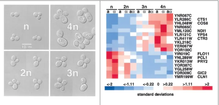
2007; Galitski et al., 1999; Storchova et al., 2006). Besides the enlarged cell size, polyploid yeast exhibits different gene regulation and phenotypes compared to haploids and diploids.
A microarray-based study identified a small number of ploidy-regulated genes in yeast (figure 4), i.e., genes whose transcript representation is altered proportionally to ploidy (Galitski et al., 1999). Because the employed yeast strains had identical genome sequence, this study unambiguously demonstrates an effect of ploidy on gene regulation. Although the genes
identifieddid not reveal how ploidy alters gene expression, differential regulation of some genes accounts for phenotypes unique to polyploids. For example, strong repression of the adhesin-encoding gene FLO11 severely diminishes agar adhesion of polyploids. The strongly ploidy-induced CTS1 encodes an endochitinase promoting mother-daughter separation at cytokinesis. Elevated expression of CTS1 likely contributes to the much reduced cell-cell association of polyploids.
Figure 4. An isogenic ploidy series and genes differentially regulated in them (Galitski et al., 1999). Left:
Ploidy-17
regulated genes identified by transcriptome profiling with microarrays. Expression levels of a gene in the ploidy series are normalized to the mean (set to 0) and the standard deviation (set to 1).
In stationary phase, polyploid but not haploid yeast cells fail to arrest their cell cycle and lose viability rapidly, although the transcriptional profile of polyploids resembled stationary phase (Andalis et al., 2004). The loss of viability is partially rescued by heterozygosity at the mating-type loci, which improves the efficiency of mitotic arrest of polyploids in stationary phase. When incubated in water instead of spent medium, post-diauxic polyploid cells arrest cell cycle and maintain viability like haploids. These observations of polyploid growth suggest abnormalities in signaling pathways transmitting nutrient availability to cell cycle regulation, so that depletion of nutrients is not sensed properly and that absolute deprivation from energy source is needed to arrest cell cycle. Consistent with this explanation, in the absence of the G1 cyclin Cln3, mitotic entry is diminished and viability is significantly restored in polyploids (figure 5). In WT haploids, Cln3 protein level is down-regulated to delay mitosis in response to nitrogen (Gallego et al., 1997) and carbon (Hall et al., 1998) depletion. Polyploids likely suffer from an inability to down-regulate CLN3 and thus aberrantly continue cell cycle progression, regardless how unfavorable mitosis is under the growth condition. Deletion of CLN3 could restore viability by enabling a switch from proliferation to quiescence (G0), even though CLN3 appears to be required for optimal metabolic adaption to stationary phase (figure 5).
Figure 5. Deletion of CLN3 restores
survivorship of polyploids in stationary phase (Andalis et al., 2004). Cells grown in
stationary phase for 12.5 days were serially diluted and spotted onto YPD to compare viability.
18
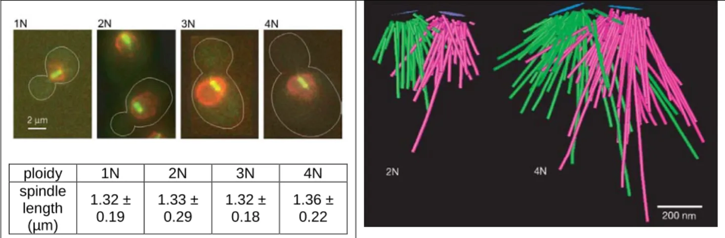
Polyploid yeast has also provided much insight on the origin of genome instability associated with increasing ploidy during tumorigenesis in metazoans. A systematic survey identified lethal mutations specific to tetraploids but not to haploids or diploids (Storchova et al., 2006). The mutations collectively reveal vulnerability in sister chromatid cohesion, homologous recombination and mitotic spindle function. Sensitivity to perturbations in these processes reflects the greater burden of maintaining a much larger and more complex genome. Because the probability of spontaneous DNA damage increases with increasing DNA content, polyploids endure a higher frequency of DNA lesion, rendering DNA damage repair an essential process for survival. The most pronounced genomic defect in polyploids is the much higher rate (>200x) of chromosome loss, and syntelic attachment of chromosomes (both sister chromatids are attached to the same spindle pole) occurs more frequently in polyploids. Fluorescence imaging and electron tomography show that while the spindle pole body expands its surface area
proportionally to ploidy and to the number of nuclear microtubules, the length of pre-anaphase spindle remains unchanged by ploidy (figure 6). The unequal scaling between spindle pole body and spindle length could favor syntelic attachment. Defects in sister chromatid cohesion, of which the basis in polyploids is unclear, could lead to increased synthelic attachment and chromosomal loss as well.
19 ploidy 1N 2N 3N 4N spindle length (µm) 1.32 ± 0.19 1.33 ± 0.29 1.32 ± 0.18 1.36 ± 0.22
Figure 6. Unequal scaling of pre-anaphase spindle length and spindle pole body surface area (Storchova et al., 2006). Left: Fluorescence microscopy images of cells expressing Nup116-YFP (red) to mark the nucleus envelope and Tub1-GFP (green) to label the mitotic spindle. The spindle length is maintained constant, independently of ploidy. Right: Models of the mitotic spindle in diploid and tetraploid cells constructed from electron tomography. Central plaques of spindle pole bodies are shown in blue. Green and magenta lines denote microtubules nucleating from different spindle pole bodies. Measured average surface area of spindle pole body is 0.022 µm2 in diploid and 0.043 µm2 in tetraploid.
Control of cell size in yeast and metazoans
One immediate effect and prominent feature of polyploidy is enlarged cell size (Otto, 2007), the threshold of which is established dynamically as a coordinated outcome of cell growth and cell cycle progression (Cook and Tyers, 2007). How polyploidy raises the cell size threshold is unknown, because the exact mechanism of cell size regulation in haploids or diploids is not yet well understood. Much of the molecular insight into control of cell size comes from elegant studies in budding and fission yeast.
In budding yeast, mutations that alter either cell growth or cell cycle progression change the size threshold. Cell cycle mutants that hasten or delay cell cycle progression lead to reduced or increased cell size, respectively (Cross, 1988; Jorgensen et al., 2002). A systematic screen for mutations altering cell size revealed a role of ribosome biogenesis in cell size control. Mutations
20
that impair ribosome assembly lower cell size threshold (Jorgensen et al., 2002), reminiscent of the slower ribosome synthesis and reduced cell size during nutrient starvation (Kief and Warner, 1981; Schneider et al., 2004). Details of the functional link between ribosome assembly/activity and cell size regulation await elucidation. Polyploidy may enlarge cell size by increasing the rate of ribosome biogenesis, since the cell division frequency is unchanged by ploidy (Di Talia et al., 2007). A recent discovery, likely a promising lead to a greater understanding of the connection between ribosome activity and size control, is chaperone-dependent release of G1 cyclin Cln3 from the rough ER periphery in late G1 (Verges et al., 2007). Cln3 is the most upstream activator of cell cycle progression (Bloom and Cross, 2007), and its mutant alleles alter the cell size threshold (Cross, 1988). Regulation of Cln3 localization by chaperones at the ribosome-associated ER could represent a simple device for coupling growth and cell cycle progression. The rate of ribosome biogenesis and activity of chaperones likely reflect a cell’s growth status and thus the fitness level for mitosis. Only when the capacity for mitosis exceeds a threshold level would the chaperons on the ER release a sufficient amount of Cln3 to initiate cell cycle progression.
Figure 7: Model for ER-tethering of Cln3 and its release for nuclear accumulation in late G1 (Verges et al., 2007). During most of the cell cycle, Cln3 is associated with the rough ER. Two domains in the Cln3 polypeptide mediate localization to the ER, likely through binding the adaptors Cdc28 and chaperone Ssa1. Ssa1 recognizes the J domain of Cln3 and is thought to maintain a catalytically inactive state when bound to Cln3 in vivo. In late G1, the J domain of another chaperone Ydj1 presumably displaces Cln3 from Ssa1. Localization of the CLN3 mRNA by Whi3 at the ER is based on findings in a previous study (Gari et al., 2001).
21
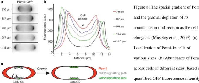
In fission yeast, a recently identified gradient of the kinase Pom1, coupled to a sensor of this gradient, the kinase Cdr2, is involved in the coordinated cell growth and cell cycle
progression (Moseley et al., 2009). Activation of Cdr2 is necessary for entry into mitosis, and Pom1 inhibits the activity of Cdr2. Anchoring of Pom1 at the two poles of the cell creates zones of mitotic inhibition. Confined localization of Cdr2 to the middle of the cell ensures mitotic entry does not occur until the cell elongates to a certain dimension so that Cdr2 is sufficiently
separated from the inhibition of Pom1.
Figure 8: The spatial gradient of Pom1 and the gradual depletion of its abundance in mid-section as the cell elongates (Moseley et al., 2009). (a) Localization of Pom1 in cells of various sizes. (b) Abundance of Pom1 across cells of different sizes, based on quantified GFP fluorescence intensity. (c) Model for spatial regulations of Pom1 and Cdr2 that enables size-dependent activation of Cdr2 to prevent premature mitotic entry of smaller cells.
Multiple proliferating cell types of vertebrates cultured in vitro have demonstrated the existence of a size-sensing mechanism in G1 that regulates timing of S phase onset (Dolznig et al., 2004). This mechanism coordinates cell growth and cell cycle progression, thereby
maintaining a size threshold. Human and chicken erythroblasts, as well as mouse fibroblasts, shorten the duration of G1 phase when they have been manipulated to enlarge prior to early G1
22
in the cell cycle. The result is an efficient reset of cell size in one doubling time. The size threshold is also adjusted accordingly to growth conditions. A faster growth rate promoted by stronger growth signaling raises the size threshold despite more frequent cell division. The cell size threshold, growth rate and doubling time all appear to adapt to changes in growth conditions and reach their defined set points rapidly and reversibly.
Figure 9: Cultured erythroblasts reversibly alter cell size in response to changing growth conditions (Dolznig et al., 2004). Large cells in a fast growing condition (black filled circles) are either maintained in the same condition or switched to a slower growing condition (open white squares) for 9 days (gray area). The latter population was then switched back to the fast growing condition (gray filled circles).
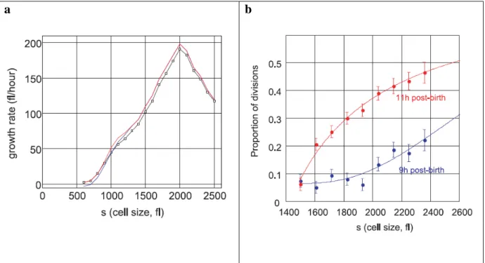
In cultured mouse lymphoblasts, the cell size appears to be maintained by regulated growth rate and cell division frequency (Tzur et al., 2009). The growth rate positively correlates with cell size until a critical size threshold, beyond which the growth rate declines with
increasing cell size. The frequency of cell division also correlates positively with cell size. Both observations reveal how growing cells efficiently reach a desirable size and divide in a regulated fashion. Collectively, the growth and division parameters of multiple cell types support the existence of an exquisite cell autonomous mechanism that maintains size homeostasis in metazoans.
23
a b
Figure 10: Maintenance of cell size by controlled growth rate and cell division frequency (Tzur et al., 2009). (a) Growth plot (black line with open squares) of isolated newborn lymphoblasts. Red and blue curves indicate two different approaches of extrapolating the data. Before reaching the critical cell size 2000 fL, 65% of the population would have undergone cell division. (b) Populations of cells of the same age at 9 hours and 11 hours after birth were tracked for the proportion of cells divided at a specified cell size.
24
Summary
Polyploidy is a prevalent physiological state achieved by multiple mechanisms in evolution, differentiation and disease. In the context of evolution, the duplicated copy of a genome is less functionally constrained and has greater capacity to evolve novel functions in a coordinated fashion on a genome-wide scale. In development, differentiation and stress response, polyploidy is part of an organ’s repertoire to rapidly increase biomass and metabolism when proliferation is either undesirable or incapable of achieving the intended physiology. Formation of aneuploid tumors likely requires a polyploid intermediate that is genomically unstable and thus increases the probability of transformation.
Although many polyploid cell types have been discovered, how increasing ploidy
enlarges cell size and alters cellular physiology remains enigmatic. Few polyploid cell types have been thoroughly analyzed and compared to their genetically matching haploid or diploid
progenitors. Detailed mechanistic investigations on isogenic ploidy series can provide the missing information. In particular, studies directly probing a relationship between cell size and altered physiology in polyploids are lacking. Such a relationship likely exists and plays a fundamental role in cellular biology, since maintenance of cell size homeostasis through
sophisticated coordination of cell growth and division is found in a wide range of organisms. The ease of genetic engineering and the extensive information about cell metabolism and growth make yeast an ideal organism for deciphering the intricate relationships among ploidy, size and function.
25
References
Albertin, W., Balliau, T., Brabant, P., Chevre, A.M., Eber, F., Malosse, C., and Thiellement, H. (2006). Numerous and rapid nonstochastic modifications of gene products in newly synthesized Brassica napus allotetraploids. Genetics 173, 1101-1113.
Albertin, W., Brabant, P., Catrice, O., Eber, F., Jenczewski, E., Chevre, A.M., and Thiellement, H. (2005). Autopolyploidy in cabbage (Brassica oleracea L.) does not alter significantly the proteomes of green tissues. Proteomics 5, 2131-2139.
Anatskaya, O.V., and Vinogradov, A.E. (2007). Genome multiplication as adaptation to tissue survival: evidence from gene expression in mammalian heart and liver. Genomics 89, 70-80. Andalis, A.A., Storchova, Z., Styles, C., Galitski, T., Pellman, D., and Fink, G.R. (2004). Defects arising from whole-genome duplications in Saccharomyces cerevisiae. Genetics 167, 1109-1121. Barow, M., and Meister, A. (2003). Endopolyploidy in seed plants is differently correlated to systematics, organ, life strategy and genome size. Plant, Cell and Environment 26, 571-584. Blanc, G., and Wolfe, K.H. (2004). Widespread paleopolyploidy in model plant species inferred from age distributions of duplicate genes. Plant Cell 16, 1667-1678.
Bloom, J., and Cross, F.R. (2007). Multiple levels of cyclin specificity in cell-cycle control. Nat Rev Mol Cell Biol 8, 149-160.
Cook, M., and Tyers, M. (2007). Size control goes global. Curr Opin Biotechnol 18, 341-350. Cross, F.R. (1988). DAF1, a mutant gene affecting size control, pheromone arrest, and cell cycle kinetics of Saccharomyces cerevisiae. Mol Cell Biol 8, 4675-4684.
Dehal, P., and Boore, J.L. (2005). Two rounds of whole genome duplication in the ancestral vertebrate. PLoS Biol 3, e314.
Di Talia, S., Skotheim, J.M., Bean, J.M., Siggia, E.D., and Cross, F.R. (2007). The effects of molecular noise and size control on variability in the budding yeast cell cycle. Nature 448, 947-951.
Dolznig, H., Grebien, F., Sauer, T., Beug, H., and Mullner, E.W. (2004). Evidence for a size-sensing mechanism in animal cells. Nat Cell Biol 6, 899-905.
Edgar, B.A., and Orr-Weaver, T.L. (2001). Endoreplication cell cycles: more for less. Cell 105, 297-306.
Fujiwara, T., Bandi, M., Nitta, M., Ivanova, E.V., Bronson, R.T., and Pellman, D. (2005). Cytokinesis failure generating tetraploids promotes tumorigenesis in p53-null cells. Nature 437, 1043-1047.
26
Galbraith, D.W., Harkins, K.R., and Knapp, S. (1991). Systemic Endopolyploidy in Arabidopsis thaliana. Plant Physiol 96, 985-989.
Galitski, T., Saldanha, A.J., Styles, C.A., Lander, E.S., and Fink, G.R. (1999). Ploidy regulation of gene expression. Science 285, 251-254.
Gallego, C., Gari, E., Colomina, N., Herrero, E., and Aldea, M. (1997). The Cln3 cyclin is down-regulated by translational repression and degradation during the G1 arrest caused by nitrogen deprivation in budding yeast. EMBO J 16, 7196-7206.
Ganem, N.J., Storchova, Z., and Pellman, D. (2007). Tetraploidy, aneuploidy and cancer. Curr Opin Genet Dev 17, 157-162.
Gari, E., Volpe, T., Wang, H., Gallego, C., Futcher, B., and Aldea, M. (2001). Whi3 binds the mRNA of the G1 cyclin CLN3 to modulate cell fate in budding yeast. Genes Dev 15, 2803-2808. Guo, M., Davis, D., and Birchler, J.A. (1996). Dosage effects on gene expression in a maize ploidy series. Genetics 142, 1349-1355.
Hall, D.D., Markwardt, D.D., Parviz, F., and Heideman, W. (1998). Regulation of the Cln3-Cdc28 kinase by cAMP in Saccharomyces cerevisiae. Embo J 17, 4370-4378.
Hegarty, M.J., and Hiscock, S.J. (2008). Genomic clues to the evolutionary success of polyploid plants. Curr Biol 18, R435-444.
Herget, G.W., Neuburger, M., Plagwitz, R., and Adler, C.P. (1997). DNA content, ploidy level and number of nuclei in the human heart after myocardial infarction. Cardiovasc Res 36, 45-51. Hijmans, R.J., Gavrilenko, T., Stephenson, S., Bamberg, J., Salas, A., and Spooner, D.M. (2007). Geographical and environmental range expansion through polyploidy in wild potatoes (Solanum section Petota). Global Ecology and Biogeography 16, 485-495.
Ihmels, J., Bergmann, S., Gerami-Nejad, M., Yanai, I., McClellan, M., Berman, J., and Barkai, N. (2005). Rewiring of the yeast transcriptional network through the evolution of motif usage. Science 309, 938-940.
Jorgensen, P., Nishikawa, J.L., Breitkreutz, B.J., and Tyers, M. (2002). Systematic identification of pathways that couple cell growth and division in yeast. Science 297, 395-400.
Kasahara, M. (2007). The 2R hypothesis: an update. Curr Opin Immunol 19, 547-552.
Kief, D.R., and Warner, J.R. (1981). Coordinate control of syntheses of ribosomal ribonucleic acid and ribosomal proteins during nutritional shift-up in Saccharomyces cerevisiae. Mol Cell Biol 1, 1007-1015.
27
Lazzerini Denchi, E., Celli, G., and de Lange, T. (2006). Hepatocytes with extensive telomere deprotection and fusion remain viable and regenerate liver mass through endoreduplication. Genes Dev 20, 2648-2653.
Lee, H.O., Davidson, J.M., and Duronio, R.J. (2009). Endoreplication: polyploidy with purpose. Genes Dev 23, 2461-2477.
Lu, P., Prost, S., Caldwell, H., Tugwood, J.D., Betton, G.R., and Harrison, D.J. (2007). Microarray analysis of gene expression of mouse hepatocytes of different ploidy. Mamm Genome 18, 617-626.
Maines, J.Z., Stevens, L.M., Tong, X., and Stein, D. (2004). Drosophila dMyc is required for ovary cell growth and endoreplication. Development 131, 775-786.
Mittelsten Scheid, O., Afsar, K., and Paszkowski, J. (2003). Formation of stable epialleles and their paramutation-like interaction in tetraploid Arabidopsis thaliana. Nat Genet 34, 450-454. Moseley, J.B., Mayeux, A., Paoletti, A., and Nurse, P. (2009). A spatial gradient coordinates cell size and mitotic entry in fission yeast. Nature 459, 857-860.
Ohno, S. (1970). Evolution by Gene Duplication (New York, Springer-Verlag).
Olaharski, A.J., Sotelo, R., Solorza-Luna, G., Gonsebatt, M.E., Guzman, P., Mohar, A., and Eastmond, D.A. (2006). Tetraploidy and chromosomal instability are early events during cervical carcinogenesis. Carcinogenesis 27, 337-343.
Osborn, T.C., Pires, J.C., Birchler, J.A., Auger, D.L., Chen, Z.J., Lee, H.S., Comai, L., Madlung, A., Doerge, R.W., Colot, V., et al. (2003). Understanding mechanisms of novel gene expression in polyploids. Trends Genet 19, 141-147.
Otto, S.P. (2007). The evolutionary consequences of polyploidy. Cell 131, 452-462.
Raslova, H., Kauffmann, A., Sekkai, D., Ripoche, H., Larbret, F., Robert, T., Le Roux, D.T., Kroemer, G., Debili, N., Dessen, P., et al. (2007). Interrelation between polyploidization and megakaryocyte differentiation: a gene profiling approach. Blood 109, 3225-3234.
Ravid, K., Lu, J., Zimmet, J.M., and Jones, M.R. (2002). Roads to polyploidy: the megakaryocyte example. J Cell Physiol 190, 7-20.
Schneider, B.L., Zhang, J., Markwardt, J., Tokiwa, G., Volpe, T., Honey, S., and Futcher, B. (2004). Growth rate and cell size modulate the synthesis of, and requirement for, G1-phase cyclins at start. Mol Cell Biol 24, 10802-10813.
Schughart, K., Kappen, C., and Ruddle, F.H. (1989). Duplication of large genomic regions during the evolution of vertebrate homeobox genes. Proc Natl Acad Sci U S A 86, 7067-7071.
28
Schweizer, L., Yerk-Davis, G.L., Phillips, R.L., Srienc, F., and Jones, R.J. (1995). Dynamics of maize endosperm development and DNA endoreduplication. Proc Natl Acad Sci U S A 92, 7070-7074.
Smith, A.V., and Orr-Weaver, T.L. (1991). The regulation of the cell cycle during Drosophila embryogenesis: the transition to polyteny. Development 112, 997-1008.
Storchova, Z., Breneman, A., Cande, J., Dunn, J., Burbank, K., O'Toole, E., and Pellman, D. (2006). Genome-wide genetic analysis of polyploidy in yeast. Nature 443, 541-547.
Tzur, A., Kafri, R., LeBleu, V.S., Lahav, G., and Kirschner, M.W. (2009). Cell growth and size homeostasis in proliferating animal cells. Science 325, 167-171.
Verges, E., Colomina, N., Gari, E., Gallego, C., and Aldea, M. (2007). Cyclin Cln3 is retained at the ER and released by the J chaperone Ydj1 in late G1 to trigger cell cycle entry. Mol Cell 26, 649-662.
Wang, J., Tian, L., Madlung, A., Lee, H.S., Chen, M., Lee, J.J., Watson, B., Kagochi, T., Comai, L., and Chen, Z.J. (2004). Stochastic and epigenetic changes of gene expression in Arabidopsis polyploids. Genetics 167, 1961-1973.
Wolfe, K.H. (2001). Yesterday's polyploids and the mystery of diploidization. Nat Rev Genet 2, 333-341.
Wolfe, K.H., and Shields, D.C. (1997). Molecular evidence for an ancient duplication of the entire yeast genome. Nature 387, 708-713.
Zybina, T.G., and Zybina, E.V. (2005). Cell reproduction and genome multiplication in the proliferative and invasive trophoblast cell populations of mammalian placenta. Cell Biol Int 29, 1071-1083.
29
Chapter 2. Control of gene expression by cell size
(contents of this chapter have been submitted to the journal PLoS Biology)
Summary
Polyploidy, increased genome content arising from whole genome duplication, plays an important role in evolution, development and tumorigenesis. Polyploids exhibit enlarged cell size and altered gene expression, but the basis for the relationship is not known. My data in this chapter show that ploidy-associated changes in gene expression reflect transcriptional adjustment to a larger cell size, implicating cellular geometry as a key parameter in gene regulation. Using RNA-seq, I identified genes whose regulation was altered in a tetraploid as compared with an isogenic haploid. Regulation of these genes was then examined in haploid genetic mutants that also produce increased cell size. In both contexts, cells with increased cell size share a
substantial number of identically differentially regulated genes. Analysis of the size-regulated genes identified a transcription factor that mediates size-dependent regulation. The result
suggests a causal relationship between cell size and transcription with a size-sensing mechanism that adjusts gene expression in response to changes in size. Transcriptional responses to enlarged cell size could underlie other cellular changes associated with polyploidy. Furthermore, the causal relationship between cell size and transcription suggests that cell size homeostasis serves a regulatory role in transcriptome maintenance.
Introduction
Mounting genomic evidence suggests that a wide range of diploid eukaryotic species have evolved from polyploid ancestors with duplicated genomes, as hypothesized by Ohno
30
(Kasahara, 2007; Semon and Wolfe, 2007). Polyploid organisms exist in multiple kingdoms and are especially prevalent among plants. During development, specific cell types in diploid
metazoan organisms perform endo-replication and differentiate into polyploid cells that manifest novel functions distinct from their diploid progenitors (Lee et al., 2009). Polyploidy also occurs as an intermediate state in aneuploid tumor formation (Ganem et al., 2007). From yeast to mammals, polyploidy is associated with enlarged cell size and altered cellular physiology (Andalis et al., 2004; Galitski et al., 1999; Lee et al., 2009; Storchova et al., 2006). Despite a plethora of known polyploid cell types, how ploidy changes cellular physiology remains elusive. Moreover, no causal relationship between enlarged cell size and altered physiology has been uncovered. Detailed molecular characterization of polyploid tissues in metazoans and plants has been limited by technical constrains. Methods for precise manipulation of the genome have yet to be fully developed for higher eukaryotes, and therefore it is difficult to access a large quantity of isogenic cells of different ploidies grown in native conditions.
The budding yeast presents an ideal system to decipher the relationship among ploidy, cell size and gene expression. Elegant molecular genetics enables convenient and precise
engineering of the yeast genome. Isogenic polyploids can be constructed from haploids and then stably maintained, greatly facilitating based experimentation. A small number of ploidy-regulated genes was identified by comparing transcriptomes of isogenic yeast strains in a ploidy series using microarrays (Galitski et al., 1999). Differential regulation of these genes explains some phenotypes displayed by polyploids. However, the small set of ploidy-regulated genes did not reveal any mechanistic relationship between ploidy and gene expression.
This earlier work was technically hampered in two aspects: First, the transcriptomes were isolated from the Sigma 1278b yeast strain, of which the genome sequence was unknown. Hence,
31
microarrays designed for the S288c strain background was employed. The Sigma 1278b genome has now been sequenced and annotated, and comparative genomics reveals substantial
differences in polymorphisms and genomic organizations between the two strain backgrounds (Dowell et al., 2010). The differences in genome sequences limited the power of detection by oligonucleotide hybridization in the earlier study. In addition, transcripts specific to the Sigma 1278b background would elude detection by the S288c-based microarray. Hence, genomic differences between the two strain backgrounds likely resulted in many false negatives. The second technical limitation was inherent to the use of microarrays. Undesirable cross-hybridization is always of concern, and the analog signal output diminishes the quantitation dynamic range. Shotgun transcriptome sequencing (RNA-seq) provides a more powerful
profiling platform than the microarray with a greater dynamic range, higher detection sensitivity, and better differentiation between paralogous sequences (Mortazavi et al., 2008; Shendure, 2008). The available Sigma 1278b genome sequence and the advent of RNA-seq now provide the
resolution necessary for the genome-wide determination of a functional connection among genes regulated by ploidy.
In this chapter, I present evidence for a size-sensing mechanism in yeast that alters gene expression. Ploidy-associated changes in gene expression thus reflect transcriptional adaptation to a larger cell size. Using RNA-seq, I identified genes differentially regulated in tetraploids when compared to haploids. Gene ontology (GO) analysis shows that ploidy-regulated genes are significantly enriched for those encoding cell surface components. I then found that haploid mutants with enlarged cell size, a prominent physical feature of polyploids, also show altered expression of ploidy regulated genes. These results indicate a causal, regulatory relationship between cell size and gene expression that has not been previously reported.
32
Results
Identification of ploidy regulated genes
Poly(A) RNA from 2 haploid and 2 tetraploid strains was extracted and processed for RNA-seq as previously described (Mortazavi et al., 2008) (figure 1a). Sequencing reads were mapped to the Sigma 1278b genome by my collaborator P. Alexander Rolfe in the Gifford group of Computer Science and Artificial Intelligence Laboratory. Approximately 9 million reads were reported for each sample, and more than 90% of reads mapped to the ORFs (table 1). A minimal expression threshold for transcripts was determined from read counts of genes known to be silenced under the growth condition, such as hypoxia response and sporulation. Overall, 5613 annotated transcripts were considered expressed in the experiment. The 5613 corresponding ORFs constituted the background gene list for GO term search. The comparison between haploid and tetraploid datasets shows that ploidy only affects the steady-state levels of a small number of transcripts (figure 1b), consistent with previous studies discussed in chapter 1.
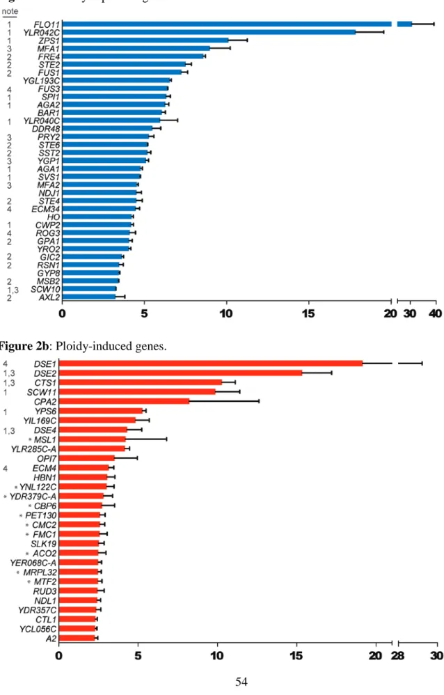
Additional steps were taken to identify regulated genes: (1) To enrich ploidy-regulated candidates, I eliminated genes whose expression was unaffected by ploidy (p > 0.001) in each haploid-tetraploid sample pair. (2) After ranking candidate genes by fold-change in each sample pair, I retained the overlapping, top-ranking candidates from both pairs (figure 1c). Using these criteria, 35 ploidy-repressed genes and 30 ploidy-induced genes were identified (figures 2a-b). Ploidy-associated differential regulation of these genes in this study is largely in agreement with the previous study using the same Sigma 1278b ploidy series (Galitski et al., 1999). Another study comparing the transcriptomes of MATa/α and MATaa/αα strains in the S288c background found an entirely different and much smaller set of genes that were
33
differentially expressed in the tetraploid (Storchova et al., 2006). Differences in strain backgrounds and mating types could account for the discrepancy between studies.
Ploidy regulated genes show significant bias for encoding cell surface components
The ploidy regulated genes are significantly enriched for those encoding proteins localized to the cell surface: cell wall, extracellular space and plasma membrane (table 2).
Several other ploidy-regulated genes encode regulators of cell surface components (figures 2a-b). Hence, polyploidy preferentially alters expression of cell surface components. Ploidy-repressed genes encode multiple factors important for mating and filamentation/adhesion, while enzymes promoting cytokinesis on the cell surface are predominant among ploidy-induced genes (tables 3-6). The cell cycle does not appear to play a role in ploidy-regulation of these genes (table 7).
A hypothesis on cell size and gene expression
The over-representation of genes encoding cell surface components could result from a geometric limitation: The surface area with respect to volume decreases as cells enlarge with increasing ploidy. For a spherical cell, a 4-fold increase in volume corresponds to only a ~2.5-fold increase in surface area. Reduction in surface area is likely to trigger differential regulation of components associated with the cell surface, where signaling and transport processes take place dynamically. Reduction in surface area could alter interactions between surface and cytoplasmic components in signaling pathways. It could also alter a cell’s ability to transport metabolites across the plasma membrane. Both types of alteration could change gene expression in the enlarged cell, and genes could be up- or down-regulated as a result depending on the net effect of cell size on their regulatory pathways.
34
Cell size affects expression of FLO11 independently of ploidy
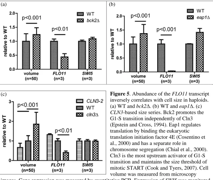
To test the hypothesis that enlarged cell size alters gene expression, I examined in
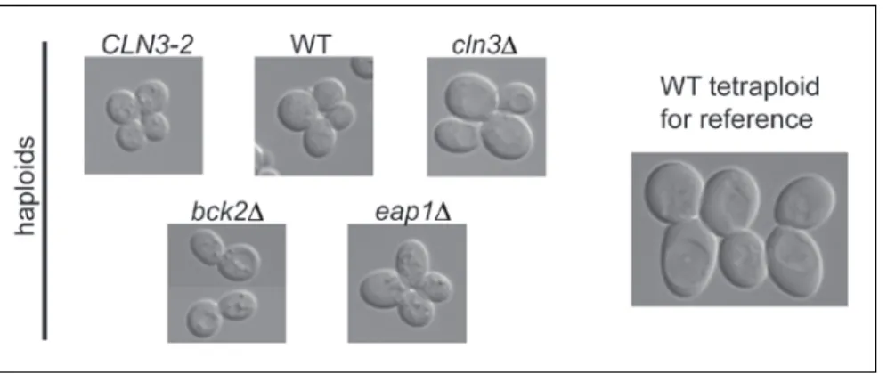
haploid size mutants the expression level of FLO11, a strongly ploidy-repressed gene encoding a cell wall protein (Lambrechts et al., 1996; Lo and Dranginis, 1996). Since cell size expansion and cell cycle progression are intimately linked processes (Cook and Tyers, 2007; Jorgensen et al., 2002), I investigated whether FLO11 is regulated by cell cycle before devising a method to manipulate cell size. In synchronized haploid cultures, FLO11 transcript abundance peaks during M-phase (figure 3). I thus focused on expression of FLO11 during M-phase in cell size mutants identified in a previous genome-wide study (Jorgensen et al., 2002): bck2Δ , eap1Δ and cln3Δ. These mutants were selected because they effectively enlarge cell size without significantly affecting fitness or cell shape (figure 4) and have no reported functional relationship with FLO11 expression. I also performed experiments using an ATP analog-sensitive allele of CDC28
(Bishop et al., 2000) that expands cell size upon inhibition by the analog (Goranov et al., 2009). Although this mutant showed an inverse correlation between FLO11 expression and cell size that was consistent with my hypothesis, concerns about aberrant cell cycle progression and cell shape prompted me to forgo further experiments with this mutant (Materials and Methods).
Expression of FLO11 is significantly reduced in the two mutants, bck2Δ and eap1Δ, that exhibit enlarged cell size (figures 5a & 5b), a trend that mimics the down-regulation of FLO11 in tetraploids (figure 2a). These results suggest a ploidy-independent inverse correlation between FLO11 expression and cell size. I employed two mutant alleles of the CLN3 gene to create a size
series. The CLN3-2 haploid arrested as small cells (66% of WT) in nocodazole, whereas the cln3Δ haploid mutant arrested as large cells (185% of WT) (figure 5c). In this size series, there is again an inverse correlation between FLO11 expression and cell size. The FLO11 transcript
35
abundance is highest in the small CLN3-2 haploid and lowest in the large cln3Δ haploid, the same relationship observed between FLO11 expression and cell size in haploid versus tetraploid cells.
Reduced activity of FLO11 promoter in both the cln3∆ haploid the WT tetraploid
To verify further that regulation of FLO11 is similar in the cln3∆ haploid and WT polyploids, I replaced the FLO11 ORF with a reporter gene to study the effect of cell size on FLO11 promoter activity. Transcriptional activation at FLO11 is repressed in the cln3∆ mutant as in the WT tetraploid (figure 6). The result further suggests that at least one transcription factor regulating the FLO11 promoter activity is sensitive to changes in cell size. In addition, the cln3∆ haploid could serve as a substitute for polyploids to facilitate the search for regulators sensing cell size and repressing FLO11.
Multiple ploidy-regulated genes are similarly regulated in the enlarged cln3∆ haploid
To identify the spectrum of genes influenced by cell size, I used quantitative PCR to compare expression of ploidy-regulated genes in WT and the cln3Δ mutant haploid. Among the size mutants I examined, the cln3Δ haploid displays the most pronounced increase in cell size and arrests in M-phase with efficiency most similar to WT. The set of genes that show
differential regulation due to cell size in the cln3∆ haploid (table 8) is very similar to those identified in the WT ploidy series (figure 2a-b). Thirty of the ploidy-regulated genes show the same regulatory trend in the cln3Δ haploid as was observed in tetraploids. Notably, the top-ranking genes that displayed the strongest ploidy-regulation in tetraploids (figures 2a-b) were significantly affected in the same direction in the cln3Δ haploid (table 8). This set of genes likely
36
represents those that respond most robustly to changes in cell size, since the cln3Δ haploid (185% of WT haploid in size) is still much smaller than the tetraploid (400% of WT haploid in size)(Di Talia et al., 2007).
Role of pheromone sensing and filamentation MAPK pathways in gene regulation by size
Differential transcription revealed by RNA-seq gave important clues as to the pathways that could affect size regulation. Thegenes repressed by large cell size are enriched for those regulated by the pheromone response and filamentation mitogen-activated protein kinase
(MAPK) pathways (table 4). Analysis of the promoter regions of size-repressed genes revealed a common binding motif for Dig1, a transcriptional repressor whose activity is repressed by the activated pheromone response or filamentation MAPK pathway (Cook et al., 1996; Tedford et al., 1997). The binding motif of Dig1 was detected ~9 fold more frequently in the promoters of size-regulated genes than the genome-wide average with a corresponding p-value of 4.28 e-9.
The over-representation of Dig1 binding motif suggests that disruption of the pathways that signal to this transcription factor or Dig1 itself should affect size regulation. The effect of such perturbations was examined in enlarged cells. Disruption of signaling in either the pheromone response or the filamentation MAPK pathway reduces the effect of cell size on FLO11 expression (figure 7a-b). Moreover, loss of Dig1 function renders several size-repressed
genes insensitive or much less responsive to the larger size (figure 7c). These results support our observation in WT tetraploid and cln3∆ haploid that reduced activities in pheromone response and filamentation pathways contribute to differential gene regulation in large cells.
37
Increasing ploidy raises basal cell wall stress
The down-regulation of multiple genes involved in mating and filamentation pathways (table 4) prompted me to examine genes regulated by other mitogen-activated protein kinase (MAPK) pathways. In multiple ploidy series, the cell wall integrity (CWI) pathway appears consistently induced in polyploids (figure 8a), indicating a higher basal level of cell wall stress. Polyploidy may also induce the high osmolarity glycerol (HOG) pathway (figure 8b). The RNA-seq data are consistent with the notion that CWI pathway is more active in polyploids. Multiple genes downstream of CWI pathway appear to be similarly regulated in the WT polyploid and a haploid strain over-expressing a gain-of-function allele of MKK1, the MAPKK of CWI pathway (Jung and Levin, 1999) (table 9).
38
Discussion
In this study, I identified transcripts whose relative level of expression in the transcriptome is altered in the polyploid yeast. Proteins encoded by these transcripts are predominantly cell surface components localized to the cell wall, plasma membrane and extracellular space. This localization bias led me to hypothesize an effect of cell size on
expression of these genes. First, increasing ploidy enlarges cell size. Second, the relative surface area of a cell with respect to its volume decreases as the cell enlarges. For a spherical or ellipsoid entity, the volume increases proportionally to the cube of radius but the area only increases proportionally to the square of radius. The significantly larger polyploid therefore is challenged by an intrinsic geometric imbalance: reduced cell surface area per cell volume. This imbalance could alter signaling pathways that involve interactions between cell surface components and their cytoplasmic counterparts. The reduced surface area could also affect transport of molecules across the plasma membrane. Both perturbations could trigger changes in gene expression. To test my hypothesis, I changed cell size in haploids and examined expression of ploidy-regulated genes in them. The strongly ploidy-repressed gene FLO11 is significantly repressed in enlarged haploids, consistent with the idea that increasing cell size down-regulates FLO11. I then found that the change in expression identified in a significant number of ploidy-regulated genes is paralleled in the in the enlarged cln3∆ haploid.
To illustrate further that cell size alters transcription via signaling pathways, I focused on the pheromone response and the filamentation MAPK pathways. Genes regulated by these pathways were preferentially down-regulated in large cells and composed the most significant category in Gene Ontology analysis. Genetic disruption in either one of the MAPKs as well as their common downstream transcriptional repressor Dig1 reduced the effect of cell size on target
39
gene expression. These results indicate that the pheromone sensing and filamentation MAPK cascades participate in downstream gene repression in the tetraploid and the cln3∆ haploid. Both the mating pheromone and the filamentation MAPKs are known to serve dual functions – as inhibitors of transcription in their kinase-inactive state and as activators of downstream transcription in their kinase-active state (Cook et al., 1997; Madhani et al., 1997).Hence, reduced activation of either kinase causes stronger inhibition, rather than a mere lack of activation, of downstream gene expression. In addition, transcriptional activators functioning downstream of both MAPK pathways perform complex positive auto- and cross-regulation (Borneman et al., 2006; Harbison et al., 2004; Kohler et al., 2002; Zeitlinger et al., 2003). The switch-like dual function of MAPKs and the positive feedback loops in these pathways likely exacerbate differences in pathway activity between the active state (in small cells) and the inactive state (in large cells). These attributes of the pathways may account for their pronounced transcriptional response to changes in cell size. Binding motif analysis did not reveal a common transcriptional regulator for genes up-regulated in large cells. Molecular pathways responsible for disproportionally higher expression of these genes will require more detailed computational and genetic examination.
The cause of a higher basal level of cell wall stress in polyploid yeast remains to be elucidated. Polyploids likely have a different cell wall composition because multiple genes encoding cell wall components are strongly differentially regulated (figure 2). Polyploids must also expand the cell wall at a much faster rate in order to reach a larger cell size without
increasing the doubling time (Di Talia et al., 2007, figures 2 & 3 in Appendix). The combination of an altered cell wall composition and a faster cell wall expansion rate could generate a higher basal level of stress.
40
I examined the activity of CWI pathway in cln3∆ haploid and did not observe a
difference when compared with WT haploid (data not shown). Two factors might explain why the CWI pathway is not more active in the cln3∆ haploid. First, the cln3∆ haploid mutant is similar to the WT diploid in size, about 2-fold larger than WT haploid. As seen in figure 6a, reporter genes of the CWI pathway are significantly induced in tetraploids but not in diploids. It is possible that a 2-fold increase in cell size is not sufficient to elevate cell wall stress. Second, the WT and cln3∆ haploids were treated with nocodazole for cell cycle arrest, whereas cells of the WT ploidy series were cultured without cell cycle inhibitors. The difference in growth conditions could influence the response of CWI pathway to cell size. Regardless of the underlying reason, results in the cln3∆ mutant indicate that induction of CWI pathway is not necessary for differential expression of the size-regulated genes in enlarged cells. Hence,
elevated cell wall stress in polyploids likely occurs independently of, or as a result of, differential regulation of the set of genes identified in this study.
Collectively my data show a causal relationship between cell size and gene regulation, suggesting the existence of a size-sensing mechanism that can alter transcription. Also
considered was an alternative model, in which polyploidy and the aforementioned mutations change transcription of size-regulated genes, whose altered expression then enlarges cell size. I found this model to be improbable on two bases. First, a previous genome-wide screen for size mutants in yeast identified a large set of genes distinct from the set of size-regulated genes in this study (Jorgensen et al., 2002). Second, the WT polyploid and the haploid size mutants are
physiologically discrete in growth, and they likely achieve the enlarged size via different modes (Cook and Tyers, 2007; Di Talia et al., 2007). Polyploids seem to achieve a larger size by
41
increasing growth rate. Mutants of cell cycle regulation such as cln3∆ and bck2∆ are thought to enlarge size by delaying the onset of START.
Control of gene expression by cell size may occur in a wide range of organisms and could partake in higher level physiological consequences of polyploidy such as differentiation and tumorigenesis. In fungi and mammals, cell size thresholds are actively maintained by regulated growth rates and cell division frequencies (Cook and Tyers, 2007; Tzur et al., 2009). The regulatory relationship between cell size and gene expression uncovered here suggests that the uniformity of cell size in unicellular organisms and within a tissue in multi-cellular organisms could be necessary to maintain the homeostasis of transcription.
42
Materials and Methods
Yeast strains and growth conditions
Strains are listed at the end of this section. Prior to analysis, cultures were inoculated from diluted overnight pre-cultures and grown until mid-log phase in the SC medium
supplemented with 2% glucose on a rotary shaker at 30°C. Synchronization of the mitotic cell cycle with α-factor was performed as described in (Amon, 2002).
Technical observations and concerns about the cdc28-as1 allele:
I treated a mid-log culture of asynchronously grown cdc28-as1 mutant with 50µM of the 1-NM-PP1(9) inhibitor (Bishop et al., 2000) and split the culture four ways: (1) no additional drug treatment (2) 0.1% azide (3) 10µM rapamycin (4) 100µg/mL cycloheximide. The PP1 treatment was intended to inhibit cell cycle progression while permitting cell growth, thereby enabling cells to enlarge. Azide, rapamycin and cycloheximide were used to inhibit cell growth in addition to PP1 treatment. Samples were taken hourly in a 4-hour time course to track FLO11 expression and cell size. Over time, cells treated with PP1 alone had significantly enlarged cell size and reduced FLO11 expression compared to cells treated with additional growth inhibitors. However, I also found increasing expression of CLN1 concurrent with the decreasing FLO11 expression in the PP1 treated cells. Since expression of FLO11 is repressed in the G1-stage of the cell cycle, I reasoned that the G1-like state in PP1 treated cells might repress FLO11 independently of cell size.
I considered using PP1 at a lower concentration (500nM) to arrest cells in G2/M in the aforementioned experiment to avoid complications associated with G1. However, cells became