HAL Id: tel-01236633
https://tel.archives-ouvertes.fr/tel-01236633
Submitted on 2 Dec 2015HAL is a multi-disciplinary open access archive for the deposit and dissemination of sci-entific research documents, whether they are pub-lished or not. The documents may come from teaching and research institutions in France or abroad, or from public or private research centers.
L’archive ouverte pluridisciplinaire HAL, est destinée au dépôt et à la diffusion de documents scientifiques de niveau recherche, publiés ou non, émanant des établissements d’enseignement et de recherche français ou étrangers, des laboratoires publics ou privés.
Hnf4α and Choline Metabolism Role in β-catenin
Activated Liver Carcinogenesis
Chiara Sartor
To cite this version:
Chiara Sartor. Hnf4α and Choline Metabolism Role in β-catenin Activated Liver Carcinogenesis. Molecular biology. Université Paris Sud - Paris XI, 2015. English. �NNT : 2015PA11T046�. �tel-01236633�
1
UNIVERSITÉ PARIS-SUD
ÉCOLE DOCTORALE 418 : DE CANCÉROLOGIE
Laboratoire : Inserm U1016-Institut Cochin
THÈSE DE DOCTORAT
ASPECTS MOLÉCULAIRES ET CELLULAIRES DE LA BIOLOGIE
par
Chiara SARTOR
Hnf4α and choline metabolism role in β-catenin activated
liver carcinogenesis
Date de soutenance : 24/09/2015
Composition du jury :
Président du jury : Chrstian AUCLAIR Directeur de thèse : Sabine COLNOT Rapporteur : Agnès RIBEIRO Rapporteur : Yu WEI
Examinateur : Olivier ROSMORDUC Examinateur : Thierry TORDJMANN
3 ACKNOWLEDGEMENTS
I would like to thank my committee members, who have accepted to evaluate and discuss my work, in particular Agnès Ribeiro and Yu Wei for their comments and feedback on my manuscript.
I would like to express my special appreciation and gratitude to my advisor, Sabine. I would like to thank you for encouraging my work and for allowing me to grow as a scientist.
I would like to thank Christine for welcoming me in her team and all its members for the work environment they create.
A special thanks is for the people of our little “sub-group”. I want to thank Angélique and Cécile for being there when I arrived, and for teaching me a lot of secrets and tricks in practical lab life. Angélique, I am so happy that you have obtained your surely deserved CR1 position. Your scientific brilliance is a model. Cécile, your expertise and your enthusiastic way to approach new techniques always surprises me. Rozenn, I have to thank you mostly for being yourself. Don’t stop and don’t change. A special thanks also for being a wonderful cat-sitter. I’ll write you a reference letter if you need it. Anaïs, you have just started your scientific experience, be focused and you will obtain wonderful results, I wish you the best. I would like to warmly thank Pascale and Nadia that are a precious help in the metabolic field. I enjoyed working with you, not only for the results obtained, but also, and maybe mostly, for the atmosphere during the experiments. You are as good in science as in real life.
Béa and JP, my consideration of your scientific capability is unquestionable, but I want to thank you in particular for other practical advices that you gave me. From the beginning you have been really welcoming with me and I am very grateful for this.
I would like to thank the “youngs”, Angé, Rozenn, Nadia, Anaïs, Marie, Julie, Coralie, Séverine, Vanessa, Manon, Sandrine and Antoine for being always there when there is a surprise to prepare, a birthday (or whatever) party to join. It is nice to spend happy moments with nice people at work and to find not only colleagues but also friends.
I would like to express my gratitude also to all the members of the team that have already left, in particular to Johny, Fred, Medhi, PAX, Laura, Ingrid, Aline. We had a great time together.
Anne-Marie, Jocelyne and Jean Christophe, I have a high consideration for your work, but I appreciate you in particular for your jokes and your direct (and not always politically correct) opinions. I enjoyed talking with you at lunch time about cats, books, trips and a lot of other topics. I really appreciate your attentions (i.e. fried vegetables during the PhD redaction, merci!!). I would like to thank Carole’s team, past and present members for being always available and nice with their neighbors. A special thank to Sara Zum, friend in real and lab life: your presence was often very important for not
4
going crazy, and also to Sabine, my favorite yoga mate, and Mariangela: keep calm and sii brava!
I would also like to thank Christophe Ferreira for being my reference during the year of my “mission d’expertise”. Your professionalism and competence taught me a lot.
I would especially like to thank people of Cochin and Tenon facilities. The histology and genomic facilities. The animal care members, in particular the ones of the “animalérie FAC”, Isabelle, Claire and JB. The PIPA’s members, especially Gilles, Franck and Carmen. The Limp members, Aurélie and Claire. I thank you all for your kindness and availability. All of you have been there to help me during my PhD experiments, demonstrating huge competence. Je tiens particulièrement à remercie la Ligue contre le cancer pour avoir financé ma 4ème année de thèse et pour avoir donc permis de conclure ces travaux.
A PhD is mostly a work experience. But it wouldn’t be the same without my wonderful “non-biological” family.
In primis gli amici storici Zum ed Erigo, dagli spriz e pomeriggi di studio a Padova siamo finiti a ritrovarci per un apéro post lab lungo Senna! Dopo questi anni in cui le nostre esperienze hanno coinciso, sarà un grosso cambiamento non avervi vicini. Aspetto solo che diventiate ricercatori con posto fisso per farmi assumere.. :) Un grazie speciale anche a Silvio, alla Giacoma, a Pietro Gori e Alice, presenze anche loro storiche e sempre piacevoli!
I Pariggini DOC che si sono rivelati ottimi amici, nei momenti più festivi, quand’eravamo giovani erasmus, come nei momenti più buii: Fiask, Cigna, Eli Omodei, Matteo Cognome, Diana&Diana, Della Marina, Marchesini, Andrea Araldo, Sam, Anthony, Ag, Bizz, Danielina, François, Arianna, Lorenzo, Michele, Amedeo. Abbiamo condiviso tanti momenti, compleanni, feste a tema, cene, aperitivi, concerti, spettacoli, viaggi, matrimoni. Un grazie anche ai Pariggini a breve termine: Omar e Paolo, Cattaneo, Chiaretta, Ciuffo, Campo e perché no anche un po’ Iacopo.
Grazie ragazzi, credo che la fortuna di avervi incontrato e di aver creato questo gruppo sia stata eccezionale.
I would like to express all my gratitude to my parents and my sister.
Il vostro sostegno è stato elemento cardine per arrivare al punto in cui sono oggi. Siete sempre stati un modello di vita e un motivo per impegnarmi. Grazie di tutto l’appoggio che mi avete sempre dato, vi voglio bene.
Un grazie anche alla mia sorella ad honorem Vale: nonostante la distanza, continui ad essere un’ottima amica.
Last but not the least, thanks to my little “kind of” family, Banti e Loli. Per esserci stati anche nei momenti più complicati e per continuare a sopportarmi quando vi rubo la coperta. <3
7
“Per aspera [sic itur] ad astra”
9
INDEX
LIST OF FIGURES: ... 11 LIST OF ABBREVIATIONS: ... 13 ABSTRACT: ... 17 INTRODUCTION... 19 The liver ... 19 Liver structure ...19 Liver functions ...22 Carbohydrate metabolism...23Hepatic Glucose uptake ...23
Hepatic glucose production ...24
Lipid metabolism ...26
Lipogenesis ...26
VLDL production and secretion ...27
Fatty acid oxidation ...28
Bile production ...29
Liver embryonic development ...31
The hepatoblast differentiation ...31
Commitment to the hepatocyte lineage ...33
Cholangiocyte and the bile ducts ...33
Liver pathologies ...37
Cholangiocytes-linked ...37
Hepatocyte- linked ...39
WNT/β-catenin pathway ... 44
The WNT signalling ...44
WNT/ β-catenin canonical pathway ...46
β-catenin in cell adhesion ...47
WNT/β-catenin in development, maintenance and pathology ...48
WNT/β-catenin in liver ...49
Development and physiology ...49
β-catenin engagement in the adult zonal liver ...50
Bile production and β-catenin ...52
The nuclear receptor Hnf4α ... 53
Hnf4α liver functions ...53
10
Hnf4α and liver cancer... 56
Hnf4α functions in other organs ... 57
Cancer ... 58
General introduction ... 58
Tumour metabolism ... 59
Warburg effect ... 60
Other examples of metabolism adaptation ... 62
Choline physiological metabolism... 64
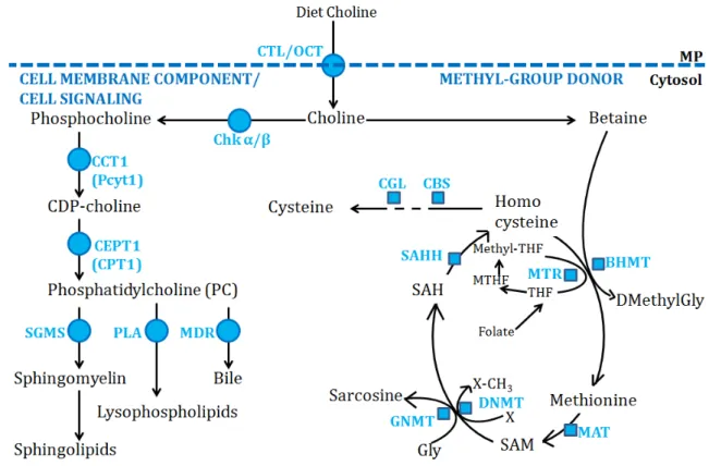
Choline as a methyl-group donor ... 66
Choline as a Kennedy pathway substrate ... 67
The different uses of phosphatidylcholine ... 69
Choline plays a role in different tumours ... 69
Choline-related cancer therapies ... 71
The hepatocellular carcinoma ... 72
Aetiologies ... 72
Classification ... 74
Peculiarity of CTNNB1-mutated HCCs ... 76
Diagnosis ... 77
HCC management: treatments ... 81
The aim of the work ... 83
Mouse models ... 83
Hnf4α conditional knock-out liver ... 83
APC conditional knock-out liver ... 84
RESULTS ... 87
DISCUSSION ... 145
BIBLIOGRAPHY ... 167
11
LIST OF FIGURES:
Figure 1. Liver structure. ...21
Figure 2. Glucose metabolism. ...25
Figure 3. Lipogenic pathway. ...27
Figure 4. VLDL structure. ...28
Figure 5. BAs synthesis. ...30
Figure 6. Bipotential hepatoblast ...32
Figure 7. From ductal plate to bile ducts. ...36
Figure 8. IPH phenotype and P-Smad2 staining. ...42
Figure 9. WNT canonical and non canonical pathways. ...45
Figure 10. Adherens junction structure ...47
Figure 11. Liver lobule and molecular zonation ...52
Figure 12. Cancer new hallmarks. ...59
Figure 13. Two distinct glycolytic ways in differentiated versus proliferating/ tumoral tissues. ...61
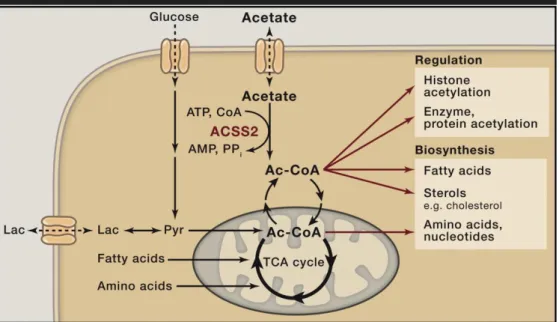
Figure 14. Acetate is the hub of metabolism. ...62
Figure 15. Specific metabolism for different type of tumours. ...63
Figure 16. Summary of know choline transporters. ...65
Figure 17. Choline pathway in hepatocytes. ...66
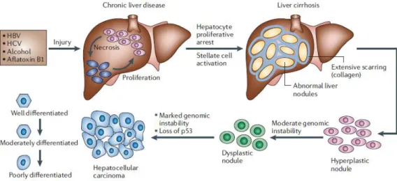
Figure 18. HCC histopathological progression. ...73
Figure 19. Classification of HCCs. ...74
Figure 20. Major pathways involved in HCC. ...76
Figure 21. HCC different phenotypes. ...77
Figure 22. Fluorocholine and Fluorodeoxyglucose PET. ...79
Figure 23. Principal histological markers in HCCs. ...80
Figure 24. Hnf4α knock-out strategy. ...84
Figure 25. APC knock-out strategy. ...84
Figure 26. APC knock-out mouse model and the two different protocols. ...85
Figure 27. β-catenin in regulating Tcf4 chromatin occupancy determines metabolic zonation. ... 147
13
LIST of ABBREVIATIONS:
A
α-SMA : α-smooth muscular actin ACC : Acetyl-CoA carboxylase ACh : acetylcholine
ACSS2 : acetyl-CoA synthetase enzyme
ADPKD : autosomal dominant polycystic kidney disease ARPKD : autosomal recessive polycystic disease
AFP : alpha fetoprotein ALB : albumin
ALD : alcohol liver disease
APC : adenomatous polyposis coli ATP : adenosine triphosphate B
BAs : bile acids
BHMT : betaine homocysteine methyltransferase
BMP : bone morphogenetic protein C
CAR : Constitutive Androstane Receptor
CC : cholangiocarcinoma CCK : cholecystokinin
ChoRE : glucose or carbohydrate responsive element
CHKA, CHKB : choline kinase α and β
ChREBP : carbohydrate responsive element binding protein
CHT : Choline transporter CK1 : Casein kinase 1 CK19 : citokeratin 19
CTL : Choline transporter like CT : computed tomography
CTNNB1 : catenin (cadherin-associated protein) beta 1 CTP (or Pcyt) : phosphocholine cytidyltransferase CTP : cytosine-5’triphosphate C/EBPα: CCAAT-enhancer-binding protein α D DAG : Diacylglycerol DEN : N-diethylinitrosamine DCP : des-carboxyprothrombin DMBA : 7,12-dimethylbenz[a]anthracene DNMT1 and 3 : DNA methyltransferase 1 and 3 DPM : Ductal plate malformation E
ECM : extracellular matrix EGF : epidermal growth factor (EGF)
EHBD : extrahepatic bile duct EMT :
epithelial-to-mesenchymal transition ER : endoplasmic reticulum F
FAP : familial adenomatous polyposis
FAS : fatty acids synthase FBPase : fructose 2,6 biphosphatase FDA : food and drug administration 18FDG : fluorine-18 fluorodeoxyglucose
14
FNH : nocal nodular hyperplasia FOXA1 : Forkhead box protein A1 G
GK : glucokinase
GLUT-1 : glucose transport 1 GS or GLUL : Glutamine synthetase GSDs : glycogen storage diseases GSK-3 : Glycogen synthase kinase 3 H
HAV/HBV/HCV : hepatitis A, B, C virus
HB : hepatoblastoma
HCA : hepatocellular adenomas HCC : hepatocellular Carcinoma HDL : high-density lipoprotein HE : eosin hematoxylin
HGF : hepatocyte growth factor HGFR : hepatocyte growth factor receptor
HNF 1α/4α/6 : hepatic nuclear factor 1alpha/4alpha/6
HHEX : Hematopoietically Expressed Homeobox
HIF : hypoxia-inducible factor 1 HRE : Hnf4α Responsive Element I
IL-6 : interleukin-6 INF : interferon
IHBD : intra hepatic bile duct IHCC : inrahepatic
cholangiocarcinoma
IHD1/2 : isocitrate dehydrogenase 1/2
IKK/NFkB : IKK/nuclear factor-kappa B
IPH : idiopathic portal hypertension
J
JAK : Janus kinase
JNK : jun N-terminal kinase L
LDHD : lactate dehydrogenase D LRP 5/6 : Low-density lipoprotein receptor-related protein 5 and 6 M
MAPK : MAP kinase
MCD : methionine choline deficient MDR2 : multi-drug resistance 2 MODY1/2 : Maturity Onset Diabetes of the Young 1 / 2
MRI : magnetic resonance imaging MRS : magnetic resonance
spectroscopy N
NAFL : non-alcoholic fatty liver NAS : National Academy of Sciences
NASH : non-alcoholic steatohepatis NSCLC : non-small cell lung cancer O
OAT : ornitine aminotranferase OCT/slc22a1 : Organic cation transporters OSM : oncostatin M OXPHOS : oxidative phosphorylation P PA : phosphatidic acid PAS : Periodic Acid Schiff PC : phosphatidylcholine PCK1 : phosphoenolpyruvate carboxykinase 1
PCP : planar cell polarity PC-PLC/D : PC-specific phospholipase C / D
15 PDX1 : Pancreatic and duodenal
homeobox 1
PE : phosphatidylethanolamine PEMT : phosphatidyl ethanolamine methyltransferase or
Phosphatidylethanolamine N-methyltransferase
PEPCK : Phosphoenolpyruvate carboxykinase
PI3K : phosphoinositide 3-kinase PKB : protein kinase B (AKT) PKC : protein kinase C
PSA : prostate- specific antigen PTEN: phosphatidylinositol-3,4,5-trisphosphate 3-phosphatase R
RAF : Radiofrequency ablation ROS : reactive oxygen species S SAM : S-adenosyl-L-methionine SAH : S-adenosyl-L-homocysteine SMP30 : senescence marker protein SNP : single nucleotide polymorphism
SOX7-9 : SRY-related HMG-box 7-9 SREB-1c : sterol regulatory
element binding protein 1c STAT : Signal Transducer and Activator of Transcription T
TAA : thioacetamide TACE : transarterial chemoembolization TCA : tricarboxylic acid
TGF-β : transforming growth factor β
TLC : thin layer chromatography TNFα : tumor necrosis factor alpha TRβ : thyroid hormone receptor β V
VEGF : vascular endothelial growth factor
VLDL : very low density lipoprotein
W
17
Abstract:
WNT/β-catenin is a pillar during development and in adult physiology. In particular in the adult liver it is a double-edged sword: it is necessary to establish the metabolic zonation, requirement for having a functional organ, but it is also involved in the onset of 11-32% of hepatocellular carcinoma (HCC).
My thesis work has been based on the team previous results and it is focused on two main subjects: (1) the first aim was to decipher the role of Hnf4α both in physiology and in HCC development and its relationship with WNT/β-catenin signalling and (2) the second part explores the possible use of Fluoro-choline (FCh) positron emission tomography (PET) in the diagnosis of β-catenin-activated liver tumours.
In this study I used cohorts of patients having HCC, but also inducible and hepatospecific knock-out mice for adenomatous polyposis coli (APC) gene (thereafter called ApcKO mice). Apc is the most important negative regulator of β-catenin, and it hepatic loss leads to aberrant activation of β-catenin, disrupting liver zonation and initiating long-term liver cancers.
I generated also inducible hepatospecific Hnf4α knock-out mice and I demonstrated an increased proliferation, lipids accumulation and disorganization in the portal triad architecture, together with a mild distruption of liver zonation. Then, looking at cancer onset, I demonstrated that Hnf4α loss is not able per se to initiate liver cancer, and has no tumour suppressor role in β-catenin activated tumours onset and progression.
We performed a metabolic analysis of ApcKO livers, showing that β-catenin is able to deregulate lipids metabolism, in particular that of phospholipids derived from choline. In collaboration with clinicians, I studied human patients who underwent FCh/PET, showing that β-catenin-mutated tumours had an increased uptaken of F-Choline whereas non-mutated β-catenin human HCC had not. Similar results were obtained with mice, either ApcKO β-catenin-activated HCC or β-catenin-independent mice HCC, obtained through a N-diethylinitrosamine (DEN) injection.
Choline in cells splits in two main pathways: it is both a methyl-group donor and a precursor for phospholipids production. I tested this through radiolabeled fluxes in in vitro experiments. In β-catenin activated hepatocytes and tumours there are more phospholipids and more methyl groups in DNA derived from choline than in control mice. Moreover in ApcKO DNA is hypermethylated, and it is dependent on choline supply from diet.
All these results together show the importance for β-catenin activated tumours to have a supply in choline, and so open a way not only in PET exploitation for having a precise diagnosis, but also in deciphering the importance of choline pathway, to possibly develop a targeted therapy.
19
Introduction
The liver
Liver structure
The adult liver is the second largest organ of the human body (the first is the skin) and it is also second in complexity (after the brain). Its architecture is critical to be functional (Yu et al., 2010), and reflects the wide range of functions that the liver has to fullfitt in the body.
Different cell types compose the liver: the hepatocytes are the most abundant ones (70-80%). They are the parenchymal cells that manage lipids, glucides and aminoacids metabolism, bile production, liver regeneration after damage and drug detoxification. This is possible thanks to the cell organelles organization: around 15% of their volume is endoplasmic reticulum (ER), and there is a high amount of peroxisomes, lysosomes and mitochondria. Other parenchymal cells are the cholangiocytes that derive from the same precursor, the hepatoblast, and are the epithelial cells of the bile ducts (as explained in more details in next section). Non-parenchymal cells include Kuppfer cells, the liver macrophages, having a role in immune response, stellate cells (involved in liver regeneration, fibrosis and cancer, due to their function of vitamin A storage), and different endothelial cells including sinusoidal endothelial cells (those limiting the sinusoids).
Each cell type has a proper role, but also the communication between hepatocytes and non-parenchymal cells is essential for normal liver function (Duncan, 2003).
The liver epithelium is a unique case among the vertebrate epithelia, as it is very different from the classical monolayer. The monolayer is generally composed by cells with a single basal surface at the opposite site of a single luminal surface, whereas the lateral surfaces are dedicated to cell-cell contact sides. In the liver bile ducts show this classical columnar epithelium organization. On the other hand, the hepatic epithelium is organised in plates one or two cells thick, with multiple luminal and basal domains, positioned in a non-perpendicular way to the later surfaces. The presence of multiple
20
surfaces and moreover the properties of the extracellular matrix (ECM) of the liver which is less dense and crosslinked than usual (due to the absence of laminin and nidogen) are able to maximise the molecules bidirectional exchanges between cells and blood (Musch, 2014).
The hepatocytes constitute the liver parenchyma and they do accommodate a huge macromolecules flow linked to both endocrine and exocrine functions (further detailed in the next section) thanks to the liver position in the body, its connection with the blood supply and the characteristic polarity of the hepatocytes.
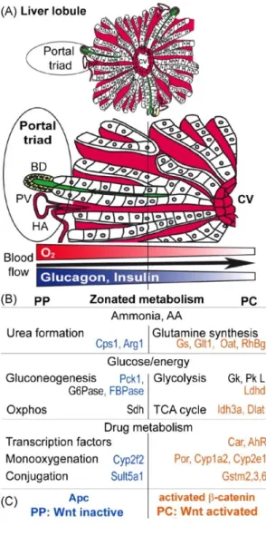
The liver unit is the lobule; the lobule is organised along an axis that goes from the portal space to the central vein. In the portal space is located the so-called portal triad, constituted of a bile duct, the hepatic artery branches and the portal vein, which run in parallel. The blood arrives from the hepatic artery (oxygen-rich) and from the portal vein (nutrients-rich) and flows within the lobule through the sinusoids (small capillaries in the spaces between the hepatocytes). This gradient of oxygen, nutrients and hormones creates a specification in molecular functions at the two sides, also called the liver zonation (Jungermann and Kietzmann, 1996). On the contrary the bile flows on the other sense, from the pericentral area to the periportal one (Figure 1).
21
Figure 1. Liver structure.
The liver unit is the lobule with the central vein on one side and the portal triad (hepatic artery, bile duct and portal vein) on the other. Blood flows from the portal space to the central vein, whereas bile does the inverse journey. Hepatocytes are liver cells rich in organelles and polarised: at the apical side bile canaliculi collect bile and at the other side the space of Disse allows the contact of hepatocytes with blood.
22
Liver functions
The liver has a range of different roles. It is involved in the digestive process, a function strictly correlated with body metabolism control and also with the xenobiotics (drugs) detoxification. It is necessary for retinols activation, catabolism and excretion and during the embryonic life it is the site of hematopoiesis, producing red blood cells, and moreover various plasma proteins. Liver pathologies cause a variety of digestive and metabolic disorders (Yu et al., 2010).
Liver functions are both endocrine and exocrine:
1. The endocrine function is linked to the blood that flows from the portal space. The blood components are uptaken and processed (blood is conditioned through detoxification and serum factors secretion). This is possible thanks to the connections between the basal surface of hepatocytes and the blood flowing through the sinusoidal capillaries into the space of Disse.
Some examples of endocrine function, such as the carbohydrate and lipid metabolism will be described in the next sections.
2. The exocrine activity consists in bile production. The bile synthesized by the hepatocyte is recovered in canaliculi, which form a network connected by the canals of Hering to the bile ducts (at the hepatocytes apical side), to converge in the hepatic duct that brings them to be finally stored in the gallbladder.
23
Carbohydrate metabolism
The liver is a key organ in blood glucose homeostasis. If glycemia is too high, insulin is secreted by the pancreatic β-cells and released in the blood circulation. Insulin is a peptide hormone able to stimulate hepatic glucose uptake and storage, while it inhibits hepatic glucose production. On the contrary if glycemia is low, glucagon is produced by pancreatic α-cells. It is another peptide hormone, amd it is able to increase liver glycogenolysis and, when glycogen stored is depleted, gluconeogenesis, to finally raise glycemia. (Aronoff et al., 2004; Quesada et al., 2008).
In this context, the role of the liver is clearly fundamental to maintain the homeostasis of glucose level in blood. When insulin is secreted in portal blood, it arrives at the liver at the same time than glucose, which has been absorbed by intestine after a meal. Hepatic glucose uptake by the liver is stimulated by the portal insulin, and the liver sequestrates 30-40% of the glucose passing through (Pagliassotti and Cherrington, 1992) and at the same time an important amount of the portal insulin (almost 70%) (Toffolo et al., 2006).
Insulin has a direct effect on the liver, it represses gluconeogenesis enzyme gene expression, and consequently hepatic glucose production (Duong et al., 2002; Ferrannini et al., 1999; Hall et al., 2000; O’Brien and Granner, 1996).
Hepatic Glucose uptake
In hepatocytes, glucose is uptaken mainly by the glucose transporter Glut-2 in a concentration-dependent manner. A hepato-specific Glut-2 deletion totally impairs liver glucose transport (Seyer et al., 2013). The rate-limiting enzyme for glucose metabolism is the glucokinase, whose expression is positively controlled by insulin (Girard et al., 1997, for review ). Glucokinase is the enzyme responsible for the phosphorylation of glucose in glucose-6-phosphate. This phosphorylation not only activates glucose for further transformations, but it will also decrease intracellular glucose concentration, promoting further entry. Mice overexpressing glucokinase or mice carrying
24
additional glucokinase copies in their genome have an increased rate of glucose uptake, and glycolysis and glycogen synthesis are higher than in control mice (Girard et al., 1997, for review). Consequently the transgenic mice glycemia is decreased, and therefore glucokinase was first identified as a possible target to treat diabetes. Moreover, identified polymorphisms on the glucokinase gene are linked to Mature Onset Diabetes of the Young 2 (MODY-2) diabetes. (Ferre et al., 1996a, 1996b; Froguel et al., 1993; Stoffel et al., 1992) (Figure 2).
Hepatic glucose production
During the fasting period, the liver is able to supply the glucose need of the whole body, first by breaking its glycogen stores and then by producing glucose de novo (gluconeogenesis). The two processes do not occur at the same time: in the early fasting (2-6h after a meal) glycogenolysis is predominant, whereas in prolonged fasting gluconeogenesis becomes fundamental (Rui, 2014). As demonstrated by Roden et al. in humans during the early fasting period, hepatic glycogen level decreases linearly (Roden et al., 2001).
Gluconeogenesis is a tightly regulated process: enzymes expression changes in relation to hormones. Insulin represses PEPCK, fructose-1,6-biphosphatase (FP2ase) and glucose-6-phosphatase (G6Pase) gene expression, whereas glucagon induces their expression. Another way to control the process is by regulating the availability of gluconeogenesis substrates, that could be either produced directly by the hepatocytes or by peripherical tissues and delivered by blood circulation (Figure 2) (Rui, 2014).
25
Figure 2. Glucose metabolism.
Intracellular glucose can be catabolised in order to provide energy supply through its oxidation in tricarboxylic acid (TCA) cycle or it can be stored as glycogen or fatty acids. The gluconeogenesis pathway is in blue, and the pentose phosphate pathway which generates NADPH (for lipogenesis and detoxification reactions) and nucleotides synthesis is in brown (Rui, 2014).
26
Lipid metabolism
In hepatocytes, fatty acids can be produced, stored in lipid droplets (as triglycerides), exported in lipoproteins, or oxidixed for energy producing. Depending on the nutritional status, fatty acids can be produced, through lipogenesis, but they can also be obtained through the hydrolysis of the triglycerids stored in liver droplets or uptaken from blood circulation, in the liver mainly enter non-esterified fatty acids (Rui, 2014).
Lipogenesis
In the liver, glucose catabolism is not only a metabolic source for energy production, but mainly provides the citrate, through the TCA cycle. Once in the cytoplasm citrate will be converted into acetyl-CoA, precursor of the fatty acids synthesis (Figure 3). The cytoplasmic acetyl-CoA is then converted into malonyl-CoA by the acetyl-CoA carboxylase (ACC).
Malonyl-CoA and acetyl-CoA are then transformed into palmitic acid by the fatty acid synthase (FAS) (Wakil and Abu-Elheiga, 2009). The palmitic acid will be elongated into stearate then desaturated to produce oleyl-CoA, one of the most abundant fatty acid with palmitoyl-CoA (Gutiérrez-Juárez et al., 2006) (Figure 3).
Insulin stimulates the expression of two lipogenic enzymes: ACC and FAS, through the activation of sterol responsive element binding protein (SREB-1c), a transcription factor able to trigger the transcription of both glycolytic (as the glucokinase named before) and lipogenic genes (Foretz et al., 1999). Therefore glucose is able to indirectly regulate lipogenic gene expression through insulin, but glucose also directly activates a transcription factor, the carbohydrate responsive element binding protein (ChREBP), that binds the carbohydrate responsive element (ChoRE) on the lipogenic genes, promoting their expression (Dentin et al., 2004, 2006).
27
Figure 3. Lipogenic pathway.
Glucose intermediates are in black, enzymes in blue. Glucose supplies acetyl-CoA, which is essential for fatty acids formation (Rui, 2014).
VLDL production and secretion
The liver produces very low density lipoprotein (VLDL). Excessive fatty acids (that are not used as fuel though their oxidation) can be transformed, being esterified, and then collected within lipid droplets in the hepatocytes cytoplasm, to be further secreted as VLDLs in blood. In healthy conditions triglycerides cannot persist in hepatocytes but have to be secreted, otherwise their accumulation in cytoplasm will trigger steatosis and consequent pathologies. VLDLs are carriers in the blood circulation of triglycerides and cholesterol esters; the scaffold elements are a phospholipids membrane, apolipoproteins, and cholesterols (Figure 4) (Li and Vance, 2008; Noga and Vance, 2003; Stein and Shapiro, 1960).
28
Figure 4. VLDL structure.
VLDLs are the triglycerides transporters in blood circulation, their membrane is composed of apolipoproteins, cholesterols and phospholipids (Li et al., 2005; Noga and Vance, 2003).
VLDLs are formed in two steps. In the first one a small dense VLDL precursor is generated. The essential component, apolipoprotein B, is translocated in the ER during its translation, and it starts to be conjugated with a small amount of triglycerides, phospholipids and cholesterol esters. If something in its structure is wrong, the apolipoprotein B and its conjugated elements are rapidly degraded. The second step consists in the VLDL precursor maturation, acquiring further triglycerides (Shelness and Sellers, 2001).
Fatty acid oxidation
In the liver there are two kinds of oxidation: β-oxidation and ω-oxidation. The first is localised in mitochondria and peroxisomes, the second in the ER. The β-oxidation site depends on the fatty acids chain length. Short (<C8), medium (C8-C12) and long (C14-C20) are principally oxidised in mitochondria, and very long chain (>C20) in peroxisomes; after the first degradation steps shortened chains are sent to mitochondria (Bartlett and Eaton, 2004; Reddy and Hashimoto, 2001). Fatty acids are first activated in acyl-CoA esters before being sent to mitochondria or peroxisomes. β-oxidation is a redundant process shortening at each step acyl-CoAs, with the loss of the two terminal carbons as acetyl-CoA subunits. In the liver, these acetyl-CoA can either be condensed in ketone bodies or enter in the TCA cycle (Laffel, 1999).
29 The ω-oxidation is able to use fatty acids chain from C10 to C26. It is an alternative pathway that is used in particular when the β-oxidation process is not functional. The first step consists in changing of the ω-methyl group of fatty acids in a ω-hydroxyl group. The second step is the dehydrogenation of this group into a dicarboxylic acid. The dicarboxylic acid is then sent to mitochondria or peroxisomes to be shortened (Sanders et al., 2006).
Bile production
The bile is mainly synthesized by hepatocytes. Bile is subsequently released and stored in gallbladder. This step needs to be coordinated with food arriving in the intestine, and is necessary because bile in gallbladder is concentrated (by removing water) to reach the functional concentration. Bile is then thrown in the duodenum where bile acids (BAs) are able to facilitate the digestion and absorption of dietary lipids and other lipophilic nutrients. This is a finely regulated process: in fact when in postprandial phase food enters in the duodenum, a peptide hormone, cholecystokinin (CCK), is produced and released. CCK stimulates the gallbladder contraction and so bile is released into the intestine. Bile acids are finally reabsorbed by passive diffusion and active transport in the terminal ileum and get back to the liver (Houten, 2006). The enterohepatic circulation, which is a continuous process in liver, allows this recycling.
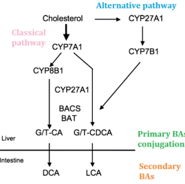
Cholesterol is the biological precursor of bile acids, and bile primary biosynthesis takes place in the liver. Two different pathways exist: the classical (neutral) pathway and the alternative (acidic) one.
The classical pathway is a multi-step process and the seventeen enzymes involved are localised in cytoplasm and in different cellular organelles (microsomes, mitochondria and peroxisomes); it is responsible for the production of 75% of BAs in mice and 90% in human. It starts with the conversion of cholesterol in hydroxycholesterol by cholesterol 7α-hydroxylase (CYP7A1), also called cytochrome P450 7A1 (enzyme specifically expressed in the liver). The cholesterol hydroxylation by CYP7A1
30
is the rate-limiting step of the process, as in knock-out mice for this enzyme the amount of BAs produced is 75% less than in control ones (Schwarz et al., 1996).
The alternative pathway first step consists in a cholesterol oxidation by mitochondrial sterol 27-hydroxylase CYP27A1. Enzymes involved in this alternative pathway are not only specifically expressed in liver but also in other tissues.
Both BAs produced by the two parallel ways are called primary BAs. The composition of BAs pool is different among species. For instance in humans and rats the major components of BAs are the cholic acid and the chenodeoxycholic acid, whereas in mice they are the cholic acid and the muricholic acid. The primary BAs are conjugated in the liver with an aminoacid, which is taurine for rodents and glycine for humans. Thereafter, at the intestinal level, the conjugated BAs are transformed by the intestinal microbiota to give rise to secondary BAs (Figure 5) (Chiang, 1998, 2004; Goodwin et al., 2000; Russell, 2003).
Figure 5. BAs synthesis.
BAs can be produced by the classical or alternative pathway. Primary BAs are conjugated in liver and then modified by the intestinal microbiota to give rise to secondary BAs (adapted from Chiang, 1998).
31
Liver embryonic development
The foregut endoderm is the origin of liver tissue, appearing at 8.5 days post-coïtum (E8.5) in mice. At this stage, GATA4 and FoxA transcription factors are expressed in the foregut and are able to bind to the enhancer controlling albumin (Alb) gene transcription to promote its expression (Si-Tayeb et al., 2010). Shortly after that step, at E8.5 - E9, the liver bud formed. Specific genes such as Alb, alpha fetoprotein (AFP) and hepatic nuclear factor4 alpha (Hnf4α) start to be expressed and the epithelial cuboidal cells become pseudostratified columnar cells, to form the liver diverticulum (Bort et al., 2006). A complex transcription factors network then controls the breakdown of the laminin-rich basal layer around endoderm to let the hepatoblasts delaminate and migrate to form the liver bud (Bort et al., 2006; Margagliotti et al., 2008). The liver bud then grows in a huge way and becomes the principal site of haematopoiesis, controlled by hepatoblast genes and paracrine signals from hepatic mesenchyme (Si-Tayeb et al., 2010).
The hepatoblast differentiation
The hepatoblasts are endodermal progenitors of the parenchymal lineages of the liver. They have an irregular shape, a big nuclear to cytoplasmic ratio and few organelles (in comparison to mature hepatocytes). They are bipotential as they are capable to give rise to both hepatocytes and cholangiocytes. The hepatoblasts start to migrate and associate with primitive endothelial cells that form the capillary-like structure between the migrating hepatic cords; the sinusoidal structures are also forming (Duncan, 2003). The differentiation of the hepatoblast is gradually determined by different transcription factors. This bipotential cell could become a cholangiocyte or a hepatocyte in relation to molecular pathways and transcription factors differentially activated (Figure 6).
32
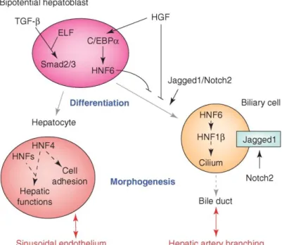
Figure 6. Bipotential hepatoblast
The fate of the bipotent hepatoblast is influenced by different transcription factors and signalling pathways, to differentiate into one or another cell type (Lemaigre and Zaret, 2004).
At the beginning, the hepatoblast expresses genes that characterise both adult hepatocytes (Hnf4α and alb) and adult cholangiocytes (cytokeratin 19, CK19) and Afp, typical of embryonic liver. Hepatocyte growth factor (HGF) promotes the establishment of bipotency in the hepatoblast, through the induction of CCAAT-enhancer-binding protein α (C/EBPα) expression and consequently HNF6 (also called onecut-1), which avoids an early differentiation into biliary lineage. Indeed HNF6 knock-out mice present a premature and exaggerated biliary differentiation (Clotman et al., 2002; Lemaigre and Zaret, 2004; Si-Tayeb et al., 2010). Another involved pathway that collaborates with HGF is the transforming growth factor β (TGF-β) and its effectors, the smad2 and smad3 proteins. Mutants for the smads show hepatocytes organized in clusters, instead of cords (Lemaigre and Zaret, 2004; Weinstein et al., 2001). During differentiation TGF-β forms a gradient to help the commitment, being higher near the portal vein, where cholangiocytes form, and lower at the other side, where hepatocytes differentiate (Clotman and Lemaigre, 2006).
33
Commitment to the hepatocyte lineage
As anticipated, the TGF-β, WNT, FGF and NOTCH secreted factors form a gradient, being higher in the portal region. This gradient is necessary to stimulate the hepatoblast to differentiate; in the periportal area where these signalling pathways are higher, the hepatoblast will differentiate into a cholangiocyte. On the other side is not only the fact that hepatoblast is less exposed to these signals, but also the presence of HGF that will make the hepatoblast to adopt the hepatocytes fate (Suzuki et al., 2003). Another fundamental transcription factor for the hepatocytes lineage commitment is C/EBPa. In particular it is able to repress the expression of TGF-β receptor 2 in the hepatoblast, inducing its commitment into the hepatocyte lineage. On the contrary C/EBPb is able to induce its expression and so to stimulate the cholangiocyte fate. Both influences of C/EBPa and C/EBPb were proved also in in vitro experiments (Takayama et al., 2014). The liver during the embryonic life is the hematopoiesis site. The hematopoietic cells indeed migrates around E10 into the liver and are able to secrete interleukin-6-related cytokines. One of these is oncostatin M (OSM), which is able to induce the expression of differentiation markers, in particular markers of the hepatocyte fate (Ito et al., 2000; Kamiya et al., 1999; Santamaria et al., 2013). The hepatocytes fate is also controlled by another transcription factor, TBX3. The used of knock-out embryos indeed demonstrated that its loss allows the cholangiocyte fate, instead of the hepatocyte one. It acts both by allowing the expression of hepatocytes markers, C/EBPa and Hnf4α, and in blocking the expression of Hnf6, necessary for the biliary lineage commitment (Suzuki et al., 2008).
To establish the hepatocytes adult phenotype, HNF4α is one fundamental transcription factor involved and its roles will be addressed in a specific chapter.
Cholangiocyte and the bile ducts
The cholangiocytes are also called biliary epithelial cells and they constitute the bile ducts, responsible for carrying the bile. The cholangiocytes constitute
34
3-5% of total cells amount in the liver. The 3-dimensional structure formed by biliary cells is called the biliary tree, which develops in parallel with the hepatic artery network (Figure 7). It can be divided into intrahepatic biliary ducts (IHBDs) and extrahepatic biliary ducts (EHBDs). IHBDs are composed of bile canaliculi, canals of Hering and interlobular bile ducts. EHBDs are composed by the gallbladder, the common hepatic duct, the common bile duct and the cystic duct (Nakanuma et al., 1997).
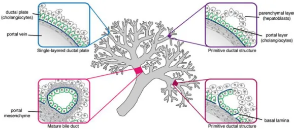
From a physical point of view, the development of bile ducts begins from E13.5. Hepatoblasts around the mesechyme near the portal vein first form a monolayer, which is called the ductal plate that then evolves in a bi-layer of cuboidal precursor cells expressing CK19 (Figure 7). Around E17 the ductal plate remodels itself: focal dilatations start to form in the bi-layer and portal mesenchyme starts to surround it, whereas at the same time the remaining bi-layer regresses. The fate of remaining ductal cells was long discussed. For instance, Terada and Nakuma in 1995 hypothesised apoptosis mechanisms as a cause of elimination of the excess cells, even if Carpentier et al. showed that a part of ductal cells can be commited into periportal (but not pericentral) hepatocytes (Figure 7) (Carpentier et al., 2011; Si-Tayeb et al., 2010; Terada and Nakanuma, 1995). The other hepatoblasts differentiate into hepatocytes and organise in chords, with bile canaliculi at the apical surface (Lemaigre, 2003; Sergi et al., 2000).
The differentiation of IHBDs thus derives from liver precursors of the hepatic bud: it starts from the liver hilum and continues until the peripherical areas, to be after anastomosed with EHBDs. Extrahepatic biliary tract (gallbladder, hepatic, cystic and common bile ducts) and IHBDs developments are indeed distinct: IHBDs derive directly from intrahepatic progenitors emerging from the primitive gut, and only after become connected to allow bile flow from hepatocytes to gallbladder (Clotman et al., 2002; Lemaigre, 2010; Lemaigre and Zaret, 2004). Extrahepatic biliary tract originates from pancreatobiliary precursors coexpressing pancreatic and duodenal homeobox 1 (PDX1) and SRY-related HMG-box 7 (SOX7), as cells expressing only PDX1 differentiate into pancreatic precursors.
35 Looking from a molecular point of view, HNF6 is a key transcription factor for cholangiocytes commitment from bipotential hepatoblast, as it is able to control HNF1β transcription and so the cilium associated proteins expression. HNF1β null mice show a dysplasia of big intra hepatic biliary ducts (IHBDs) and a lower amount of small IHBDs in comparison to controls; as an indirect consequence also interlobular arteries formation is lacking, because the biliary lineage differentiation and the artery branching are tightly linked (Clotman et al., 2002; Coffinier et al., 2002; Lemaigre and Zaret, 2004). For instance, an excess of artery branches correlates with an excess of bile ducts, and conversely a new proliferation of bile ducts due to external causes, is followed by new artery branches formation. Near the forming biliary epithelium the presence of a capillary network originated from the hepatic artery, called peribiliary plexus, is necessary to feed and oxygenate biliary cells. At a molecular level, the requested molecules for angiogenesis in
loco are vascular endothelial growth factor (VEGF) and angiopoietin-1. VEGF
is expressed in both cholangiocytes and hepatocytes whereas angiopoietin-1 is produced only by hepatocytes. At the beginning both cholangiocytes and α-smooth muscular actin (α-SMA) positive-cells (branch precursors) express the receptor for angiopoietin-1, but the cholangiocytes lose it while differentiating. The VEGF receptor is expressed in ductal plate cells and in portal endothelial precursors. A synergistic model has been hypothesised in which VEGF activates endothelial adjacent precursors and angiopoietin-1 is able to recruit myofibroblasts; the association between endothelial and fibroblastic cells forms the ducts. The final maturation of the hepatic artery will require the synergic action of VEGF and angiopoietin-1, thanks to the coexpression of both receptors in the same cells (Raynaud et al., 2011). Interestingly HNF6 knock-out results in the absence of gallbladder and EHBDs are structurally different: normally each lobule is drained by hepatic ducts and all ducts converge into the cystic duct, which enter into the duodenum; in mutants, hepatic ducts are substituted by a big structure that connect liver to duodenum (Clotman et al., 2002).
The biliary cells are also characterised by the Notch2/Jagged1 pathway. Heterozygous mutants for both Jagged1 and Notch2 are lacking IHBDs in
36
mice, while in humans mutations in Jagged1 gene are responsible for the Alagille syndrome. One main aspect of this syndrome is a jaundice developed by the babies affected, due to a similar paucity in bile ducts (Lemaigre and Zaret, 2004; McCright et al., 2002). As previously anticipated, the localisation of IHBDs is linked to different signals: TGF-β gradient, whose presence is maximal in the portal area, and Notch2/Jagged1 signalling. One characteristic transcription factor for nascent IHBDs is SOX9 (Si-Tayeb et al., 2010).
Figure 7. From ductal plate to bile ducts.
The ductal plate forms near the portal space and evolves forming PP hepatocytes and cholangiocytes, to finally create a duct able to transport the bile (Raynaud et al., 2011).
37
Liver pathologies
Cholangiocytes-linked
1. Ductopenia is a pathological condition caused mostly by Alagille syndrome, due to mutations in Jagged1 gene, modelized in mice by the invalidation of Jagged1 and Notch2. No other ductopenia is associated to bile ducts development regulators. For instance mutants for SOX9 gene have a mild and transient paucity of bile ducts (but show other problems like campomelic dysplasia and autosomal sex reversal) (Lemaigre and Zaret, 2004; McCright et al., 2002).
2. Ciliopathies are pathologies in which bile ducts are present but defective and consist in hepatorenal fibrocystic syndromes. An example is autosomal dominant polycystic kidney disease (ADPKD). The formation of renal cysts is parallel with hepatic biliary cysts. The biliary cysts are characterised by cholangiocytes hyperproliferation, linked to increased sensitivity to insulin-like growth factor-1 and estrogen, which stimulates VEGF production. The cilia are normally localised at the apical side of cells and are involved in osmo-, mechano- and chemosensing, while in ADPKD cilia are smaller or absent. Having non functional cilia is tightly linked with dysgenesis of bile ducts. The two genes associated with this pathology are polycystin-1 and -2 (PKD-1/2). Another example is autosomal recessive polycystic disease (ARPKD) in which multifocal bile ducts dilatations are present in liver, associated to polyductin or fibrocystin (PKHD1) mutations, which is a transmembranous protein located also in cilia (Lemaigre, 2011; Raynaud et al., 2011).
3. Ductal plate malformations (DPMs) are lesions present in different pathologies: the previously mentioned ARPKD, the later described embryonic atresia, and congenital hepatic fibrosis. DPMs consist in the persistence of the embryonic biliary structures, due to mutations in genes controlling the ductal plate remodelling (in particular the phase of ductal plate regression). As underlined by their association with ARPKD, the
38
DPMs are also a kind of ciliopathy, meaning that in this case the cause of the persistent embryonic biliary structures is the cilium defective role (Bezerra, 2011; Desmet, 1992; Lemaigre, 2011).
4. EHBDs pathologies: extrahepatic biliary atresia is a rare disease, more frequent in neonates. There are both embryonic and postnatal forms (the most common one) are chacterised. The pathogenesis of this disease consists in cycles of inflammatory and sclerosis processes that could provoke biliary cirrhosis (with the consequent occlusion of bile ducts). The aetiology is still not clear and many hypotheses have been made, like viral infection, autoimmunity and exposure to toxins and others. Choledochal cysts are another type of EHBDs pathologies, in particular in a context of congenital malformations. In this case, one or more biliary tract segments have cystic dilations, which could be focal lesions or a dilation of the entire common bile duct. The causes are unknown, but familial cases suggest that they could have a genetic component (Bezerra, 2011; Lemaigre, 2011; Raynaud et al., 2011).
5. Biliary cirrhosis is characterised by fibrous scar accumulation starting from peribiliary area and progressing until the portal area in the liver. Not only biliary cells are involved in the disease but also the portal fibroblasts. The biliary cirrhosis could be primary (an immunodeficiency disorder more likely to affect women than men, ratio 10:1), primary sclerosing (fibrosis inflammation able to destroy biliary ducts), biliary atresia (as explained in subsection 4) and cystic fibrosis hepatopathy (Hirschfield and Gershwin, 2013).
6. Cholangiocarcinoma (CC) is a cancer spreading from the cholangiocytes. It is classified as intra or extrahepatic, depending whether either the intra or extrahepatic tree branch is involved. The 50% of CCs originates from liver hilum, 42% from EHBDs and 8% from IHBDs. CC has a poor outcome due to the lack of both early diagnosis tools and treatments. The only therapy is surgery, even if often CC is diagnosed too late for this medical
39 approach. As the general cancer onset, CC is a multi step process and it is characterised by an uncontrolled cell proliferation, high invasion potential and so a high metastasis rate. A small portion of CCs can develop in a normal liver, but for the major part the onset is in a context of chronic inflammation of bile ducts. Risk factors are for instance virus infection, metabolic disorders and cirrhosis (Khan et al., 2005; Lazaridis and Gores, 2005; Patel, 2002).
A carcinoma can develop also in the gallbladder, mostly a women pathology, and known risk factors are gallstones, infection, metabolic disorders, environmental factors (Medina and Kaempffer, 2001; Randi et al., 2006).
Hepatocyte- linked
1. Steatohepatitis can be caused by alcohol consumption, namely alcohol liver disease (ALD) or come after non-alcoholic fatty liver disease (NAFLD). In the first case the elevated alcohol consumption is able to trigger a steatosis. NAFLD disease is linked to metabolic disorder like obesity and insulin resistance that also trigger steatosis. Both pathologies do lead to lipid accumulation in hepatocytes, which cause an inflammation in liver tissue (oxidative stress and mitochondria dysfunction). This induces a fibrotic state of liver, which often degenerates in cirrhosis (Browning et al., 2004; Ekstedt et al., 2006; French et al., 1993; Kleiner et al., Nonalcoholic Steatohepatitis Clinical Research Network, 2005; Paradis et al., 2009; Zakhari and Li, 2007).
2. Hepatitis: the most common cause of hepatitis is a viral infection. The main ones involved are the hepatitis B and C (HBV and HCV) viruses. On the contrary the hepatitis A virus (HAV) can only cause acute hepatitis, but never chronicle diseases (Sarin and Kumar, 2011). HBV has been well characterised and it has been associated with HCC. Chronically infected people indeed have a higher HCC onset, but the vaccine is effective and it is able not only to prevent HBV infection, but also HCC onset (Tiollais et al.,
40
1985; Yang et al., 2002). HCV is the most common cause of liver pathologies. HCV infected patients have fibrotic livers, associated in 30% of the cases with cirrhosis onset and after several years of chronic inflammation, cirrhosis can progress and causes HCC development (Poynard et al., 2001). At present time, no functional vaccine is available to prevent HCV infection, so it is necessary to develop antiviral drugs to stop its infection in the liver. Interferon therapy was tested in clinical trials, alone or in combination with other antiviral substances, like ribavirin, telaprevir and boceprevir. Recently, a new antiviral drug has been developed, which is a RNA polymerase inhibitor, the sofosbuvir. Clinical trials have reported very encouraging results but, due to the huge cost of the therapy, accessibility for patients is still a problem (Cortez and Kottilil, 2015; Sun et al., 2011).
Hepatitis can also develop in a context of liver inflammation that can originate from repetitive infections and/or toxic agents exposure (Czaja, 2001; Manns and Vogel, 2006; Vergani et al., 2002).
3. Cirrhosis is a culminant pathological state of the liver, characterised by hepatic architectural disorganization and loss of liver functionality. It occurs after cycles of reparation in case of chronic liver damages, involving inflammation, tissue remodelling and fibrosis. It can develop after steatohepatitis and viral or autoimmune hepatitis (Wynn, 2008).
4. Liver reaction against substances: liver injury can also be caused by chemical and drug absorption. The liver damages are often linked to drugs processing by cytochrome 450 system and require different processes like lipid peroxidation, covalent binding of proteins or production of reactive oxygen species (ROS) (Park et al., 2005).
Moreover metal accumulation can cause liver injury. For example, Wilson’s disease consists in an inherited abnormal copper accumulation in liver parenchyma. Copper accumulation stimulates free and hydroxyl radicals formation, causing oxidative injury to the cell membrane and DNA, impacting also protein synthesis (Schilsky et al., 1989).
41 Hemochromatosis consists in an iron accumulation in a variety of organs, including the liver, and is provoked by increased iron absorption from diet. Iron overload in liver involves an oxidative stress, inflammation and a consequent fibrosis. Hemochromatotic livers develop cirrhosis and so present an increased risk of developing HCC (Fix and Kowdley, 2008). Another example of accumulation that can cause liver problems is a heterogeneous group of disorders called glycogen storage diseases (GSDs), which consist in defective glycogen storage in the liver. GSDs are either genetic or acquired (by alkaloids intoxications) and can involve liver, muscle and brain; different enzymes deficiencies can be the cause of GSDs (Matern et al., 1999).
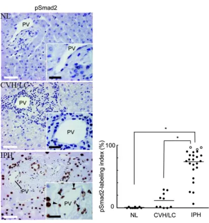
5. Impaired blood flow: the blood supply in the liver is guaranteed by portal vein for the 65-75% and by hepatic artery for 25-35%, while the oxygen is delivered in liver mostly by the hepatic artery, but also by portal blood. The blood supply has to be constant, so it is strictly regulated between the two sources. Pathologies involving an impaired blood flow could involve arterial as well as venous blood flow. For instance hepatic artery can suffer of thrombosis, which is generally a consequence of liver transplantation and aneurysms that are mostly extra- and not intra-hepatic aneurysms. On the other hand, the portal outflow can also be impaired, and this is mainly the case in cirrhosis, in which hypertension is due to fibrosis altering tissue elasticity and vein dilatation ability. Other pathologies present blood flow obstruction, even if their aetiology is different (Lautt and Greenway, 1987; Shah and Singla, 2011; TYGSTRUP et al., 1962). An interesting example is the idiopathic portal hypertension (IPH), a non-cirrhotic hypertension at the portal side. Aetiology is not known and the hypertension is due to the stenosis of the portal vein, whose lumen is dramatically reduced. Knowing that TGF-β signalling is involved in fibrotic process, the study of Kitao et al. focused on this signalling pathway. In particular a staining of nuclear phospho-Smad2 (one effector downstream TGF-β signalling) was found in fibrotic areas of patients. Furthermore the patients had a high circulating TGF-β level, demonstrating an implication
42
of this pathway in the IPH pathology (Figure 8) (Kitao et al., 2009; Nakanuma et al., 2001).
Figure 8. IPH phenotype and P-Smad2 staining.
The IPH consists in an increase portal tension due to the stenosis of the portal vein. These areas stain positive for phospho-Smad2 protein, showing an activation of TGF-β pathway, whereas normal liver (NL) and cirrhotic liver (CVH/LC) do not (Kitao et al., 2009).
6. Neoplasies: in liver different neoplasies may occur, being either benign or malignant.
The benign ones are characterised by hepatocytes proliferation, and the difficulty is the differential diagnosis between them and well-differentiated liver cancers (better described in next chapter, section hepatocellular carcinoma). The two most common benign neoplasies are focal nodular hyperplasia (FNH) and hepatocellular adenomas (HCA) (Bioulac-Sage et al., 2001, 2007). FNH generally onsets in women of 20-50 years old and a correlation between nodules growth and estrogens has
43 been described in literature (Scalori et al., 2002). Morphologically they have a fibrotic central core with malformed vascular structures. FNH is due to a response of the liver to an increased artery blood flow, and it has a low risk of side effects and to develop carcinoma (Cherqui et al., 1995; Wanless et al., 1985). HCA is a monoclonal, soft, well-demarcated tumour, with no or little fibrous capsule. Different subgroups exist, with specific molecular and phenotypic characteristics (Bioulac-Sage et al., 2007). The incidence is higher in women (85% of total patients affected) and in this case there is a correlation with the oral contraceptive intake (Bioulac-Sage et al., 2007, 2009; Edmondson et al., 1976; Vana et al., 1977). There are also other risk factors as androgen-anabolic steroids intake. The major risk in carrying a HCA is associated to the possibility of bleeding, and in some cases HCA have a risk to become malignant, in particular the ones associated to a mutation in β-catenin gene (Baum et al., 1973; Bioulac-Sage et al., 2010; Kerlin et al., 1983).
One malignant liver cancer is the hepatoblastoma (HB); it is the most common liver cancer in infants and young children (Perilongo and Shafford, 1999; Schnater et al., 2003). Generally it develops between 6 months and 3 years after birth, as only 10% of HBs appear in children of more than 4 years (Darbari et al., 2003). It is characterised by a high level of alpha fetoprotein (AFP) in the blood serum. Only 5% of HBs have a familial origin whereas the others are sporadic. There are two morphological types: pure epithelial HBs (56%) or mixed epithelial and mesenchymal ones (44%) (Okuda K, Tabor E, 1997). An implication of the Wnt/β-catenin pathway has been demonstrated in a high number of HBs, as 50-90% have a mutated β-catenin gene, at the level of residues phosphorylated by GSK3β, while 5-10% have a mutated Axin1 protein (Koch et al., 2004; Taniguchi et al., 2002).
The hepatocellular carcinoma (HCC) will be discussed more precisely in the next chapter.
44
WNT/β-catenin pathway
The WNT signalling
WNT proteins belong to a family of lipid-modified glycoproteins; their length varies between 350-400 aminoacids. They are produced in cells and then secreted in extracellular space, thanks to a palmitoylation, a lipid modification, necessary to allow the secretion by targeting them to the cell membrane, and moreover for the binding (by a covalent attachment) to the receptor on the other cells surface (Logan and Nusse, 2004).
The WNT proteins are highly conserved in species (Nusse and Varmus, 2012) and their signalling pathways are conserved in various organisms from worms to mammals and play important roles in development, cellular proliferation, and differentiation (Piedra et al., 2001). WNT proteins have also a range of various functions, indeed different Wnt pathways have been described: the non canonical ones, both independent of β-catenin, and the WNT/β-catenin pathway (Figure 9) (Rao and Kuhl, 2010).
In the organisms are present a variety of WNT proteins, but also of receptors that can modulate this signal in the cell.
45
Figure 9. WNT canonical and non canonical pathways.
Three different WNT signalling pathways have been characterised. The canonical is the one involving β-catenin as effector (Hitt, 2013).
The non canonical pathways involve different intracellular mediators. First the WNT/Jun N- terminal kinase (JNK) signalling is able to finely regulate cell polarity. This planar cell polarity (PCP) pathway acts both in epithelia polarity and polarised cell movements, such as, respectively, wing hair development in Drosophila and egg gastrulation. Second, the WNT/calcium pathway has initially been characterised in Danio rerio and Xenopus laevis and is involved for instance in cardiac development and differentiation (Koyanagi et al., 2009). It has been demonstrated that WNT5a activates this pathway, based on heterotrimeric G proteins action. This process is very fast and the calcium released from the endoplasmic reticulum (ER) modulates the action of Ca2+-dependent enzymes (Ma and Wang, 2006; Rao and Kuhl, 2010).
46
WNT/ β-catenin canonical pathway
The presented thesis work is focused on the canonical WNT pathway: it is based on the WNT-dependent accumulation of the β-catenin protein in the cytoplasm, its translocation in the nucleus and its action in synergy with LEF/TCF factors in activating gene expression.
More in details when WNT signal is not present at the membrane, β-catenin is first phosphorylated, then ubiquitinated to trigger it to proteosomal degradation, thanks to the destruction complex activity. Different proteins composed this complex: the tumour suppressors Axin and adenomatous polyposis coli (APC), and the kinases casein kinase 1 (CK1) and glycogen synthase kinase-3 (GSK-3). Axin acts as a scaffold protein recruiting APC and so permitting the phosphorylation of β-catenin in two steps: a priming phosphorylation of serine 45 (S45) residue by CK1, allowing subsequent phosphorylation on residues serines 33, 37 and threonin 41 (S33, S37, T41) by GSK-3. These phosphorylations generate a recognition site for the E3 ubiquitin ligase β-TrCP, needed for β-catenin ubiquitination and proteasomal degradation (MacDonald et al., 2009; Stamos and Weis, 2013).
On the contrary when WNT signals bind Frizzled receptors and low-density lipoprotein receptor-related protein 5 and 6 (LRP 5/6) co-receptors the destruction complex cannot be in place, because of its recruitment at the cell membrane, thanks to the interaction between Axin and Dishevelled (MacDonald et al., 2009). Unphosphorylated β-catenin is so free to enter in the nucleus where it acts as a transcription activactor for inducing the expression of its target genes. β-catenin is not able to directly bind the DNA but a transcriptionally active complex is formed by its interaction with its coactivator factors, the LEF/TCF proteins, which have a DNA binding domain that let occupy the promoter/enhancer region of β-catenin positive target genes. In the absence of β-catenin, LEF/TCF proteins could otherwise interact with Groucho proteins, a family of non DNA-binding corepressors, able to stop transcription via a conserved intrinsic repression domain (Brantjes et al., 2002; Fisher and Caudy, 1998).
In different tissues, a role of WNT/β–catenin has been demonstrated in cancer onset. In particular β-catenin is a proto-oncogene, so when an
47 activating mutation is present in the residues targets for the phosphorylations, or when a negative regulator has a loss of function mutation, β-catenin is aberrantly activated and becomes an oncogene responsible for example for hepatocellular carcinoma, colon cancer and ovarian cancer onset (Anastas and Moon, 2013).
β-catenin in cell adhesion
β-catenin is a 781 aminoacids protein (92 kDa), involved in cellular proliferation and cell fate, not only thanks to its control of transcription activation, but also because it is a structural protein. Indeed in cells there are three β-catenin pools: cytoplasmic, nuclear and membranous. β-catenin is mainly localised at the cell membrane because it is part of E-cadherin/catenin adhesion complexes (adherens junctions) which couple cadherins to the actin cytoskeleton. In this way it regulates cell to cell adhesion (Figure 10) (Piedra et al., 2001). β-catenin constitutive knock-out produces embryonic lethality, like E-cadherin mutants, but not for the same reason: E-cadherin knock-out is lethal for its protein structural function (lack of functional junctions), whereas β-catenin knock-out is lethal because it is defective in WNT canonical signalling. In these mice, plakoglobin or γ-catenin is able to established a compensatory mechanism to replace β-catenin at adherent junctions (Haegel et al., 1995).
Figure 10. Adherens junction structure
The adherens junction are composed by cadherins extracellular domains linked one to the other in extracellular space and linked to β-catenin by their intracellular domain. The catenin α and β are the structural connection with vinculin and actin (cytoskeleton protein).