REVIEW
Needle breakage during local anesthesia in the oral
cavity
—a retrospective of the last 50 years with guidelines
for treatment and prevention
Marcello Augello&Jeannette von Jackowski&
Klaus Wilhelm Grätz&Christine Jacobsen
Received: 22 February 2010 / Accepted: 29 June 2010 / Published online: 13 July 2010 # Springer-Verlag 2010
Abstract Needle breakage in the oral cavity after local anesthesia is a common complication with possible serious complications of injuring vital structures. There are differ-ent possible reasons for needle breakage, with a main focus on preventable mistakes in treatment. In this study, an analysis of literature of the last 50 years as well as own cases has been performed to renew knowledge and prevention and therapy strategies for this serious complica-tion. A systematic, multilingual review of medical literature from 1900 until today was conducted and information was evaluated systematically. In the majority of cases needle fracture happened during inferior alveolar nerve block. It is mainly a problem due to inadequate technique or the use of too thin needles for the performance of inferior alveolar nerve block. Different arguments about possible therapy strategies and methods exist. Basically, if a hypodermic needle fractures, it should be removed surgically under general anesthesia. To localize the fragment, use of either multi-plane X-rays or fluoroscopy with at least two reference needles in place or, if possible, of three-dimensional CT scans is recommended. This article shows, that despite progression in material, needle fracture is still an existing, preventable problem, if some basic rules are followed.
Keywords Broken hypodermic needle . Local anesthesia . Literature analysis . Needle dimension . Prevention
Introduction
Today, needle breakage during local anesthesia in the intraoral region is a rare complication; however, if it occurs, patient and dentist have reason to be afraid of such possible serious complications as needle migration or injury of surrounding anatomical structures, including blood vessels or nerves [1, 2]. Since the certification of standardized, disposable stainless steel needles in the 1960s by the International Organization for Standardization, a significant reduction of needle fractures has been achieved. La Crouse et al. attributed it to the invention of disposable needles, scientific advances in metallurgy, and better training in anesthesia [3,4]. Today, needle breakage is in most cases not due to a material defect, but to preventable reasons such as inappropriate injection techniques or choosing the wrong type of needle. Dealing with a fractured needle usually turns out to be a challenge for both dentist and patient. Different methods of locating and removing such a thin foreign body have been reported in the literature.
Six months ago, a young patient was referred to our department with a broken-needle fragment in an uncommon location in the pterygomandibular area after inferior alveolar nerve block [5]. After evaluation of the existing literature, it was noticed that, although it is an infrequent complication, neither a systematic analysis of existing cases, nor a summary of possible diagnostic measures or treatment methods exists. Therefore, a systematic evalua-tion of the literature from 1965 until today was performed. Interestingly, analysis of the literature produced some facts that show that the old lessons of how to perform a local
M. Augello (*)
:
J. von Jackowski:
K. W. Grätz:
C. Jacobsen Department of Oral and Craniomaxillofacial Surgery,University Hospital of Zurich, Frauenklinikstrasse 24, 8091 Zurich, Switzerland e-mail: [email protected]
M. Augello
:
J. von Jackowski:
K. W. Grätz:
C. Jacobsen Dental Institute, University of Zurich,anesthesia in the oral cavity are still important. Therefore, an overview and analysis of broken hypodermic needle cases of the last 50 years with guidelines for treatment and prevention will be presented in this article.
Material and methods
A systematic, multilingual review of medical literature from 1900 until today was conducted, using PUBMED, OVID, MEDLINE, NLM Gateway, Excerpta Medica Database (EMBASE), TRIP Database, Unboundmedicine.com, and the Cochrane Database of Systemic Reviews (COCH). The search was performed with a variety of key words, including“dental needles,” “needle fracture,” “needle breakage in dentistry,” “broken dental needle,” “dental needle fracture,” and “needle injuries in dentistry.” Studies in languages other than English, French, Dutch, and German, and case reports describing needle fractures resulting from instruments other than dental injection needles were excluded. Additionally, studies or case reports about needle fractures during local anesthesia outside the oral cavity were excluded. Other inclusion criteria were information about needle size, exact localization of the needle fracture, and method of localization of fractured needle, therapeutic meas-ures, and complications, as for example injury of vital structures as vessels or nerve damage during the removal procedure.
In 1955, Roehr Products Co. (Waterbury, USA) launched the first disposable plastic needle with moderate success, called Monoject®. In 1956, a plastic disposable syringe was invented by Colin Murdoch, a pharmacist from New Zealand to replace the glass syringe [6]. Since 1965, 34 articles were found that deal with fractured dental needles during local anesthesia. Of those 34 studies, eight have been excluded either because they were published in different languages, or offered insufficient data. All articles were case reports. Of the remaining 26 studies, which document a total of 64 cases, five authors presented studies of more than two patients [3, 4, 7-30]. Twenty-one reports each described one case. Fitzpatrick et al. and Progrel et al. were the only authors adding 13 and 16 cases over a period of 10 years drawn from questionnaires or retrospective analysis that they sent to residential dentists (Table1) [4,15].
Data was collected and analyzed using Microsoft Office Excel 2007© (Microsoft Cooperation, München, Ger-many).
Results
In total, 26 reports were found that documented 64 incidents of needle breakage in the oral cavity during local anesthesia from 1966 until today. Of 61 patients, 23 were younger than 16 years
old, while 40 patients were older than 16 years. The average age was 28 years, with a range from 3 to 71 years of age. Unfortunately, in three case presentations, the age of the patient was not mentioned.
Localization and cause
In 45 patients (70%), the needle broke during inferior alveolar nerve block. The needle fragment therefore was localized in the pterygomandibular area. In 12 patients, needle fracture occurred during local anesthesia in the buccal area, either in the maxilla or in the mandible. One case report described a needle fracture during local anesthesia in the lingual mandib-ular area and five patients were described as suffering from fractured needles during intraosseous anesthesia of the jaw (Fig.1).
Information about the cause of needle fracture was declared in the cases of seven patients. In six of those cases,
Table 1 Display of authors and published broken-needle cases from 1965 until today
Year Author n of cases
1967 Fitzpatrick et al. [15] 18 1969 Pratt et al. [27] 1 1970 Crouse et al. [3] 1 1971 Dudani et al. [12] 2 1972 Kenett et al. [18] 1 1973 Bump et al. [10] 1 1983 Hai et al. [17] 1 1983 Orr et al. [25] 1 1984 Marks et al. [20] 1 1986 Burke et al. [11] 1 1986 Fox et al. [16] 1 1986 Pietruszka et al. [26] 1 1989 Mima et al. [22] 1 1993 Moore et al. [23] 1 1996 Mc Donough et al. [21] 1 1998 Bhatia et al. [9] 1 1999 Bedrock et al. [8] 1 1999 Faura-Solé et al. [14] 5 2002 Zeltser et al. [30] 1 2003 Thompson et al. [29] 2 2006 Baart et al. [7] 1 2007 Ethunandan et al. [13] 1 2008 Augello et al. [5] 1 2008 Nezafati et al. [24] 1 2009 Progrel et al. [4] 16 2009 Shah et al. [28] 1 Total=64
the patients’ unexpected movements were the reason for the fracture of the needle. Of those seven patients, four were under 10 years of age. Two were 5 years old, one was 4, and one was 8 years of age. In one case, the dentist made an abrupt movement and caused the needle fracture.
Type of needle
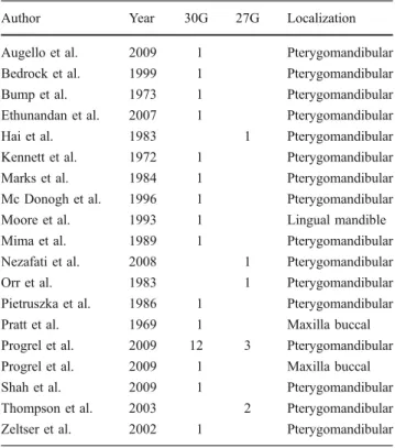
In 18 published papers reporting on the cases of 34 patients, information about the needle that had been fractured was found. In 23 patients (68%), the dentists used 30G needles for local nerve block of the inferior alveolar nerve. In three patients, infiltration in the maxillary buccal and lingual mandibular region was performed with a 30G needle. In eight patients, 27G needles were used for inferior alveolar nerve block (Table2).
Diagnostic measures
Different measures for localization of needle fragments in the oral cavity were described. Preoperative localization was carried out with conventional X-rays, or, since 2002, with CT scans. For localization in the operating room, different measures were recommended, without any stan-dardized protocol. These were magnets, metal detector, C-arm, and X-rays with at least two reference needles. In 18 of the 26 reports, authors used reference needles; two authors described the use of a metal detector.
Timing
Of all 64 reported patients, 47 (73%) received surgical removal of the needle fragment within 2 days. In 15 patients, surgical removal of the fragment was performed after an average of 134 days (with a range from 3 to 1,080 days). In two of the 64 patients, the surgeons decided to leave the needle fragment in place, because of an asymptomatic course. In one case, the dentist did not inform the patient about the needle fracture, therefore, the
needle had been left in place for an unknown time and was found due to an infection in this area. Overall, complica-tions did not occur frequently, apart from mild trismus and dysphagia. One patient suffered of facial palsy after surgery.
Discussion
Needle breakage during local anesthesia is rare, but one can guess that the number of unreported cases is higher than the number that is reported. Progrel et al., for example, just recently published a case study of 16 cases of fractured needles in a 25-year period in one single center [4].
In the majority of cases (70%), needle fracture happened during inferior alveolar nerve block. Block anesthesia in the area of the posterior mandible therefore seems still to be somewhat difficult. The high percentage of needle breakage during inferior alveolar nerve block anesthesia is caused either by false technique of injection, incorrect choice of hypodermic needle dimension, or abrupt movement of the patient or staff. In analyzing the literature, there is not one single report of material defect during the last 50 years.
Some authors reported unusual localization of the fractured fragment. They mentioned either an abnormal administration of the nerve block, or a migration of the fragment [4, 5], which supports the hypothesis of either
Fig. 1 Overview over localization of fractured needle fragments
Table 2 Display of Information about needle size in correlation to the localization of local anesthesia
Author Year 30G 27G Localization
Augello et al. 2009 1 Pterygomandibular Bedrock et al. 1999 1 Pterygomandibular Bump et al. 1973 1 Pterygomandibular Ethunandan et al. 2007 1 Pterygomandibular
Hai et al. 1983 1 Pterygomandibular
Kennett et al. 1972 1 Pterygomandibular Marks et al. 1984 1 Pterygomandibular Mc Donogh et al. 1996 1 Pterygomandibular Moore et al. 1993 1 Lingual mandible Mima et al. 1989 1 Pterygomandibular Nezafati et al. 2008 1 Pterygomandibular
Orr et al. 1983 1 Pterygomandibular
Pietruszka et al. 1986 1 Pterygomandibular Pratt et al. 1969 1 Maxilla buccal Progrel et al. 2009 12 3 Pterygomandibular Progrel et al. 2009 1 Maxilla buccal Shah et al. 2009 1 Pterygomandibular Thompson et al. 2003 2 Pterygomandibular Zeltser et al. 2002 1 Pterygomandibular
insufficient teaching of technique or“forgotten lessons” by dentists.
Kronman et al. showed in their cadaver study in 1994 that the needle has to penetrate about 21 mm deep into the soft tissue to reach the mandibular foramen for adequate anesthesia of the inferior alveolar and lingual nerve [31]. In looking at the anatomical situation of this area in more detail, Okamoto et al. showed in their radiographic study in 2000 that the best possible anesthetic effect is achieved by inserting the needle between the muscle tendon of the temporalis muscle and the medial pterygoid muscle directly into the pterygomandibular space [32,33]. However, in the majority of cases, the needle penetrates dense, bulky structures as the medial pterygoid muscle or the tendon of the temporalis muscle. Therefore, tissue resistance is high in this area, and increases when the mouth is open. During deep penetration of the needle in these tight structures, the risk of needle fracture increases. If the dentist changes syringe angulation during the injection, the risk for needle breakage in the hub area is high. An additional reason might be the dentists’ efforts to achieve needle–bone contact to reassure correct depth and placement of the needle. Furthermore, this review shows that, particularly in children, the risk of unexpected patient movement is high. Of seven reports that described patient movement during injection, four patients were under 10 years of age.
One important cause for a high percentage of needle fractures is the fact that most dentists (76%) used thin and short needles with a dimension of 30G, which equals to 0.305 mm outer diameter, probably due to the mistaken belief that the thickness of the needle correlates to the degree of pain. But pain perception is individual and is not only dependent on needle size, as was shown in the studies of Fuller et al. and Mollen et al. [34, 35]. A thin needle increases injection pressure and therefore causes an increase of pain [30]. The appropriate needle length and dimension is determined by a number of factors, including the target tissue, injection formulation, and patient population. For the administration of vaccines, for example with penetration into muscular tissue and administration of less than 1 ml of fluid, needles with a dimension of 25 to 22 gauge with a length of 16 to 38 mm are recommended [36]. So, as they have been thoroughly
instructed in the past, dentists should still use an adequately sized needle for nerve block anesthesia in the posterior mandibular area, despite improvements in needle material. Another option would be using another injection technique, as Okamoto et al. described in 1998, for example. With their technique, the hypodermic needle has to penetrate only about 10 mm deep into the soft tissue [9,26,32].
Another possible factor for needle breakage might be needle bending before penetrating the tissue, especially in the hub area. Usually with bending, a predetermined breaking point is activated. Progrel et al. documented in their study that nine of 16 dentists admitted to having bent the needle before insertion [4,37]. As mentioned before, the needle hub is the weakest part and has the highest risk of fracture, and therefore should not be pre-bent [37].
Some authors suggest leaving the needle fragment in place as long as the patient has no symptoms [23, 37]. Other authors advocate immediate removal [8,15,26,29]. Patients with needle fragments in place might develop severe pain, trismus, or infection. The needle fragment might migrate and develop severe complications such as life-threatening bleeding out of the main cervical blood vessels, although we did not find a published case of such a complication during the last 50 years. This might be due to the fact that in 97% of the published cases, the needle had been surgically removed. After mandibular nerve block, the needle fragment might migrate into the lateral pharyngeal space with development of severe dysphagia [9,14,20,38]. Another important reason for prompt removal of this foreign body is psychological. For patients, having a spiky foreign body in the facial soft tissue is frightening [20]. Therefore, removal of the fragment as soon as possible under general anesthesia is recommended [13, 24]. We specifically advise against removal using local anesthesia. Trying to fish the fragment out might push it even deeper into the soft tissue. In any case, the patient should be informed about possible migration during chewing and swallowing, which should be kept to a minimum [39]. The other part of the needle should be kept and the patient referred with the needle and packaging to a maxillofacial unit. Removal of the fragment should be done under general anesthesia with muscle relaxation.
Measures for prevention of needle fracture Check the needle before using it
Do not pre-bend the needle in the hub area
Take a sufficient needle dimension (25–27G and 35 mm length for inferior alveolar nerve block) Inform the patient about possible abrupt pain
Inform the patient before puncturing the mucosa
Leave at least 5 mm needle outside the tissue during penetration
If changing needle angulation during injection, take the needle off the tissue Table 3 Important preventive
measures of needle fracture dur-ing local anesthesia in the oral cavity
For adequate removal, a detailed knowledge about the anatomical site of the needle fragment is essential. Several methods of localizing the thin metal piece were described. Most authors used conventional radiographs. Since 2002, five authors have used CT scans for better three-dimensional localization. Zeltser et al. were the first to describe the use of a CT scan to localize surrounding structures such as vessels, the parotid gland, muscular tissue, and to analyze the exact, three-dimensional location of the fragment [30]. Surgical access and proceedings can be planned properly. In the literature, several methods for fragment localization in the operation theater were de-scribed. Special options included using a metal detector or a magnet [21,23,40]. Ho et al. described in 1988 the use of a prefabricated localizer with an embedded needle [38]. Other techniques were the Kazanijan technique with an individ-ualized wire locator cemented to a molar or the angiocath-eter method [16]. In 69% of published cases, authors used intraoperative imaging with previous placement of at least two reference needles, as it was described in 1948 by McIntyre [41]. Imaging was performed either with fluoro-scopic detection with a C-arm or intraoperative radiographs [5,24,29,42]. Another possible option in the future might be computer-navigated fragment extraction, which has not been described yet. In contrast to magnets, which are said not to be useful because of decreased ferrous compounds in the needle material [14, 40], using a metal detector was described as inefficient and circumstantial.
To conclude, it can be stated that needle fracture mainly is a problem due to inadequate technique or the use of too thin needles for the performance of inferior alveolar nerve block. If a hypodermic needle fractures, it should be removed surgically under general anesthesia as soon as possible. To localize the fragment, use of either multi-plane X-rays or fluoroscopy with at least two reference needles in place or of three-dimensional CT scans is recommended, depending on availability. But overall, needle fracture is preventable, if some old rules are followed (Table3).
Conflict of interest There was no conflict of interest of any of the authors.
References
1. Amies AB (1951) Broken needles. Aust J Dent 55(6):403–406 2. Fraser-Moodie W (1966) Location and localisation of metal in the
tissues. Br J Oral Surg 4(2):99–105
3. Crouse VL (1970) Migration of a broken anesthetic needle: report of a case. S C Dent J 28(9):16–19
4. Progrel A (2009) Broken local anesthetic needles. A case series of 16 patients, with recommendations. J Am Dent Assoc 140:1517– 1522
5. Augello MVJJ, Jacobsen C (2009) Nadelbruch als komplikation bei der intraoralen leitungsanästhesie im unterkiefer. Quintessence Germany 60:1263–1267
6. Gill HS, Prausnitz MR (2007) Does needle size matter? J Diabetes Sci Technol 1(5):725–729
7. Baart JA, van Amerongen WE, de Jong KJ, Allard RH (2006) needle breakage during mandibular block anaesthesia: prevention and retrieval. Ned Tijdschr Tandheelkd 113(12):520–523 8. Bedrock RD, Skigen A, Dolwick MF (1999) Retrieval of a broken
needle in the pterygomandibular space. J Am Dent Assoc 130 (5):685–687
9. Bhatia S, Bounds G (1998) A broken needle in the pterygoman-dibular space: report of a case and review of the literature. Dent Update 25(1):35–37
10. Bump RL, Roche WC (1973) A broken needle in the pterygo-mandibular space. Report of a case. Oral Surg Oral Med Oral Pathol 36(5):750–752
11. Burke RH (1986) Management of a broken anesthetic injection needle in the maxilla. J Am Dent Assoc 112(2):209–210 12. Dudani IC (1971) Broken needles following mandibular
injec-tions. J Indian Dent Assoc 43(1):14–17
13. Ethunandan M, Tran AL, Anand R, Bowden J, Seal MT, Brennan PA (2007) Needle breakage following inferior alveolar nerve block: implications and management. Br Dent J 202(7):395–397 14. Faura-Sole M, Sanchez-Garces MA, Berini-Aytes L, Gay-Escoda
C (1999) Broken anesthetic injection needles: report of 5 cases. Quintessence Int 30(7):461–465
15. Fitzpatrick B (1967) The broken dental needle. Aust Dent J 12 (3):243–245
16. Fox LJ, Belfiglio EJ (1986) Report of a broken needle. Gen Dent 34(2):102–106
17. Hai HK (1983) Retrieval of a broken hypodermic needle. A new technique of localising. Singap Dent J 8(1):27–29
18. Kennett S, Curran JB, Jenkins GR (1972) Management of a broken hypodermic needle: report of a case. J Can Dent Assoc (Tor) 38(11):414–416
19. Kennett S, Curran JB, Jenkins GR (1973) Management of a broken hypodermic needle: report of a case. Anesth Prog 20 (2):48–50
20. Marks RB, Carlton DM, McDonald S (1984) Management of a broken needle in the pterygomandibular space: report of case. J Am Dent Assoc 109(2):263–264
21. McDonogh T (1996) An unusual case of trismus and dysphagia. Br Dent J 180(12):465–466
22. Mima T, Shirasuna K, Morioka S, Sugiyama M, Matsuya T (1989) a broken needle in the pterygomandibular space. Osaka Daigaku Shigaku Zasshi 34(2):418–422
23. Moore UJ, Fanibunda K, Gross MJ (1993) The use of a metal detector for localisation of a metallic foreign body in the floor of the mouth. Br J Oral Maxillofac Surg 31(3):191–192
24. Nezafati S, Shahi S (2008) Removal of broken dental needle using mobile digital c-arm. J Oral Sci 50(3):351–353
25. Orr DL 2nd (1983) The broken needle: report of case. J Am Dent Assoc 107(4):603–604
26. Pietruszka JF, Hoffman D, McGivern BE Jr (1986) A broken dental needle and its surgical removal: a case report. NY State Dent J 52(7):28–31
27. Pratt GJ (1969) Broken needle: case report. Alaska Med 11(2):67 28. Shah A, Mehta N, Von Arx DP (2009) Fracture of a dental needle during administration of an inferior alveolar nerve block. Dent Update 36(1):20–22, 25
29. Thompson M, Wright S, Cheng LH, Starr D (2003) Locating broken dental needles. Int J Oral Maxillofac Surg 32(6):642–644 30. Zeltser R, Cohen C, Casap N (2002) The implications of a broken needle in the pterygomandibular space: clinical guidelines for prevention and retrieval. Pediatr Dent 24(2):153–156
31. E-BA KJH, Wongwatana S, Kumar A (1994) Preferred needle lengths for inferior alveolar anesthesia. Gen Dent 42:74–76 32. Okamoto Y, Takasugi Y, Moriya K, Furuya H (2000) Inferior
alveolar nerve block by injection into the pterygomandibular space anterior to the mandibular foramen: radiographic study of local anesthetic spread in the pterygomandibular space. Anesth Prog 47(4):130–133
33. Takasugi Y, Furuya H, Moriya K, Okamoto Y (2000) Clinical evaluation of inferior alveolar nerve block by injection into the pterygomandibular space anterior to the mandibular foramen. Anesth Prog 47(4):125–129
34. Mollen AJ, Ficara AJ, Provant DR (1981) Needles—25 gauge versus 27 gauge—can patients really tell? Gen Dent 29(5):417– 418
35. Fuller NP, Menke RA, Meyers WJ (1979) Perception of pain to three different intraoral penetrations of needles. J Am Dent Assoc 99(5):822–824
36. Kroger AAW, Marcuse E, Pickering L (2006) General recom-mendations on immunization: Reccorecom-mendations of the advisory
committee on immunization practices (acip). MMWR Recomm Rep 55(RR-15) (Centers for Disease Control and Prevention ) 37. Malamed SF (2005) Local complications. In: Handbook of local
anesthesia, 5th edn. Mosby, USA pp 285–300
38. Ho KH (1988) A simple technique for localizing a broken dental needle in the pterygomandibular region. Aust Dent J 33(4):308– 309
39. Dojcinovic I, Hugentobler M, Richter M (2007) needle breakage: a rare and potentially dangerous complication during local anaesthesia. Rev Stomatol Chir Maxillofac 108(3):222–224 40. Johansson B, Krekmanov L (1987) Fragment of broken
instru-ment removed from field of operation by an electromagnet. Br J Oral Maxillofac Surg 25(3):265–266
41. Mc Intyre Ad (1948) The broken needle in inferior dental injections. Br Dent J 85(12):271–274
42. Cohen DM, Garcia CT, Dietrich AM, Hickey RW Jr (1997) Miniature c-arm imaging: an in vitro study of detecting foreign bodies in the emergency department. Pediatr Emerg Care 13 (4):247–249