JON 3092 David H. Benninger Sebastian Thees Spyros S. Kollias Claudio L. Bassetti Daniel Waldvogel
Morphological differences in Parkinson’s
disease with and without rest tremor
Introduction
Rest tremor is a hallmark of Parkinson’s disease (PD), yet its pathogenesis is incompletely understood. A spe-cific pattern of neuronal loss in the substantia nigra in PD patients with tremor has been described [38–40, 53], but does not translate into a different pattern of striatal dopamine deficiency or postsynaptic dopamine recep-tor density compared to akinetic-rigid PD [19, 25, 51]. Nigro-striatal dopamine deficiency correlates best with
bradykinesia, but not with tremor [65]. Oscillating neu-rons in one or multiple localizations within the basal ganglia-thalamo-cortical loop may cause rest tremor, and an active contribution of the cerebellum and the cerebello-thalamo-cortical projections has been postu-lated [16, 42, 61].
Clinical manifestation and disease progression are variable and highlight the heterogeneity of PD. The clin-ical heterogeneity reflects the extent and topography of the pathological process and may indicate different causes of PD as also suggested by specific phenotypic Received: 7 April 2008
Received in revised form: 8 July 2008 Accepted: 21 August 2008
Published online: 4 February 2009
D. H. Benninger, MD (쾷) Human Motor Control Section Medical Neurology Branch
National Institute of Neurological Disorders and Stroke
National Institutes of Health Building 10 Room 5N240 (MSC1428) Center Drive Bethesda, MD 20892 Tel.: 301-402-6231 Fax: 301-480-2286 E-Mail: [email protected] D. H. Benninger, MD · C. L. Bassetti, MD · D. Waldvogel, MD Dept. of Neurology
University Hospital of Zurich Zurich, Switzerland
S. Thees, MD, MSc · S. S. Kollias, MD Dept. of Neuroradiology
University Hospital of Zurich Zurich, Switzerland
S. Thees, MD, MSc
Dept. of Diagnostic and Interventional Radiology
University Hospital of Leipzig Leipzig, Germany
■ Abstract Background Rest tremor is a hallmark of Parkinson’s disease (PD), but its pathogenesis remains incompletely understood. Nigro-striatal dopamine deficiency correlates best with bradykinesia, but not with tremor. Oscillating neurons in one or multiple local-izations within the basal ganglia-thalamo-cortical loop may cause rest tremor, and an active contribu-tion of the cerebellum and the cer-ebello-thalamo-cortical projections has been postulated. Objective To compare the pattern of grey matter volume in PD patients with and without tremor to identify struc-tural correlates of rest tremor. Methods Voxel-based morphome-try (VBM) of a high-resolution 3
Tesla, T1-weighted MR images, pre-processed according to an opti-mized protocol using SPM2, was performed in 24 patients with mild to moderate PD comparing local grey matter volume in patients with (n = 14) and without rest tremor (n = 10). Results Grey matter volume is decreased in the right quadrangular lobe and declive of the cerebellum in PD with tremor compared to those without (PFDR < 0.05). Conclusions These
results demonstrate for the first time morphological changes in the cerebellum in PD patients with rest tremor and highlight the involve-ment of the cerebellum and cere-bello-thalamo-cortical circuit in the pathogenesis of parkinsonian rest tremor.
■ Key words Parkinson’s disease · voxel-based morphometry · tremor akinetic-rigid syndrome
presentations of patients with defined genetic forms of PD. Approaches to classify PD into subgroups have prin-cipally been based on the clinical phenomenology, in particular, discriminating tremor-dominant, akineto-rigid and also “mixed” PD. Clinical evidence suggests that the motor phenotype may be predictive of clinical course and prognosis [21]. Compared to patients with tremor-dominant PD, “akinetic-rigid” patients show a more rapid clinical progression and have an increased risk to develop disability [27, 30, 31, 33, 35, 48] and de-mentia [1].
Brain imaging may allow identification of anatomical correlates of phenotypic differences such as a distinct pattern of grey matter loss in PD patients with and with-out tremor. MRI-based voxel-based morphometry (VBM) offers an operator-independent, unbiased and comprehensive morphological analysis of the brain. VBM has shown more widespread loss of grey matter in PD with dementia (PDD) [8, 9, 50, 56, 62]. VBM may also allow to distinguish PD from atypical Parkinsonian dis-orders [7, 55].
We intended to study the differences in the pattern of grey matter volume in PD patients with and without tremor to identify possible structural correlates of rest tremor, and also to test the hypothesis of a more ad-vanced cortical grey matter loss in PD patients without tremor.
Methodology
■ Study population
Patients with idiopathic PD were prospectively re-cruited in the Department of Neurology, University Hospital of Zurich, Switzerland. Inclusion criteria were mild to moderate PD corresponding to Hoehn and Yahr stages (HY) 2–3 [33]. Diagnosis of PD was based on UK PD Brain Bank criteria [26] with a minimum disease duration of three years. We chose to investigate only men of a certain age span of 55–70 years due to sex and age differences in brain size [13, 29]. Exclusion criteria were advanced PD stages equivalent to HY 4–5, second-ary parkinsonism, atypical parkinsonian diseases and other concomitant neurodegenerative diseases, tremor of non-parkinsonian origin, dementia as defined by a score < 24 in the Mini-Mental State Examination [20], any structural pathology in brain MR and CT imaging except for mild vascular changes within age norms, pre-vious stereotactic surgery, and medical or psychiatric conditions precluding study participation and/or per-formance of MRI. Patients were dichotomized in a group with and without tremor. Patients with tremor were required to have classical parkinsonian rest tremor with or without postural or action tremor of similar frequency, and in PD patients without tremor, rest as
well as postural or action tremor must have been ex-cluded, also by meticulous review of all medical docu-ments.
All patients gave written informed consent and the local ethics committee had approved the study.
■ Study protocol
All patients underwent a detailed clinical assessment in-cluding history, physical and neurological examination, and neuropsychological and -psychiatric evaluation to control for any differences between PD patients with and those without tremor which could confound mor-phological analysis. We presumed that a distinct pattern of grey matter volume distribution related to tremor would show up when comparing patient groups with similar clinical deficits except for tremor as their corre-sponding grey matter volume changes would match. For this reason, a comparison of PD patients with healthy controls would have been confounded and was, there-fore, not performed.
All patients were examined on medication and in the “off ” condition when medication effect was wearing off or during an (unpredictable) “off ”. A practically “de-fined off ” condition, which would have required a tem-porary withdrawal of dopaminergic and also anti-cho-linergic medication was not undertaken. To assess the severity of rest tremor with and without postural or ac-tion tremor more accurately considering its variability, an additional composite tremor score was based on clin-ical and historclin-ical data of the last 6 months using a sim-ilar scale as in UPDRS (0 = absent; 1 = mild and infre-quent, not bothersome; 2 = moderate and bothersome; 3 = severe and interfering with many activities and 4 = marked and interfering with most activities).
Detailed neuropsychological evaluation included Mini-Mental State Examination [20], verbal and figural (five-point test) fluency, Rey Auditory Verbal Learning Test (RAVLT), Visual Design Learning Test (RVDLT) and Complex Figure Test, Stroop Interference Test [60], Goldenberg Association Learning [28] and screening for other cognitive deficits such as apraxia, aphasia and ag-nosia. A handedness questionnaire [12] was applied. Pa-tients were also screened for hallucinations, delusions and other psychiatric disorders, and evaluated by a psy-chiatrist if deemed necessary. Depression was consid-ered present if either one or both cut-offs of 14/15 [66] and 12/13 [17] in the Beck Depression Inventory (BDI) [4] and the Hamilton Depression Rating Scale (HAMD) [32], respectively, was/were exceeded.
Medical records including history and examination, and anatomical and, if performed nuclear brain imaging were reviewed.
■ Statistical analysis
Statistical analysis of clinical data was carried out with SPSS Version 12.0.1 for Windows. Normality of distribu-tion was checked with skewness and kurtosis tests. Un-paired t-tests were used to compare parametric data between groups, and Mann-Whitney U test for non-parametric data. Chi-square tests or Fisher exact test were used to compare nominal data.
■ Brain MR scanning procedure
MR imaging using a standard head coil was performed at a 3T MR-scanner (PHILIPS, Achieva). For T1-weighted high resolution structural MRI, a T1-weighted, 3D-MP-RAGE sequence technique (TR = 8.7 ms, TE 2.3 ms, flip angle 8.0°, voxel-size 0.86 × 0.86 × 1.0 mm, axial slice ori-entation, matrix size 256 × 256) covering the anterior and posterior commissure within the same trans-axial plane was acquired in each patient. Images were re-viewed for motion artifacts before pre-processing and redone if deemed necessary.
■ Voxel-based morphometry
Image pre-processing followed the optimized protocol as described by Good et al. [29] using SPM2 (http://www. fil.ion.ucl.ac.uk/spm): first an automatic segmentation was performed using age-matched template and priors (mean age: 56.6 ± 18.6), second the deformation field was determined by normalization of the segmented gray and white matter image using the respective prior, third the initial image was normalized using the determined deformation field followed by an automatic segmenta-tion of the normalized image again using age-matched template and priors. Finally, in order to preserve the to-tal amount of brain tissue, grey and white matter voxel values were multiplied by the Jacobian determinants de-rived from the spatial normalization step, respectively [29].
For comparison of the two groups of patients with and without tremor, a voxel-wise ANCOVA of these maps was performed. To compensate for effects result-ing from general brain atrophy, a measure for the rela-tive total brain volume Vbrain = (Vgrey matter + Vwhite matter)/
Vliquor was included as nuisance variable into the
statisti-cal model. Two orthogonal contrasts: patients with ver-sus without tremor and patients without verver-sus with tremor were calculated (puncorrected < 0.001) to test for re-gions of lower amount of grey matter volume within the non tremor group (CT-NT) and tremor group (CNT-T) group, respectively.
Additional voxel-wise ANCOVAs were performed to test for effects of impaired balance and fluctuations,
re-spectively, by comparing groups of patients with and without impaired balance and groups of patients with and without fluctuations as well as a regression analysis using the UPDRS III Balance and Gait score as explana-tory variable to exclude possible confounding effects of clinical differences between patients with and those without tremor.
Based on current understanding of tremor pathogen-esis [16, 42, 61], we predicted structural changes in the basal ganglia, the thalamus, the brainstem and/or the cerebellum (RBG-thalamus-brainstem-cerebellum), and, therefore, restricted the statistical analysis to these regions apply-ing a mask of these regions to the grey matter volume distribution maps prior to statistical analysis.
To control for additional differences outside these gions, a second statistical analysis was performed re-stricted to the cerebral cortex (RCerebrum). Data were cor-rected for multiple comparisons across the respective region using the false-discovery rate (FDR) method [24].
Results
All patients with PD seen in the Department of Neurol-ogy in the years 2002–2004 were screened, and 24 of 27 men who qualified for the study could be included. Two had declined participation and one could not be in-cluded due to MR-incompatibility (pacemaker). Four-teen of the 24 (58 %) had classical parkinsonian rest tremor including four who fulfilled criteria of tremor-dominant PD [36, 67] and ten who could be considered “mixed” PD. In the ten remaining PD patients without tremor, rest tremor as well as postural and/or action tremor was firmly excluded. PD diagnosis was con-firmed and ET excluded in all patients in a follow-up assessment > 2 years (in 2007) after enrollment in this study.
Demographic and clinical characteristics are shown in Table 1. Motor complications and impaired balance were more prevalent in patients without tremor, who had a higher score of balance and gait (UPDRS items 27–30). No other differences were found, neither in scores of bradykinesia (items 18–19, 23–26, 31), rigidity (items 22), single and combined UPDRS parts (I, II, III on and IV; sparse III “off ” data precluded comparison), nor in regard to the degree of asymmetry and to the lat-erality of disease preponderance.
Rest tremor was bilateral in ten and unilateral in four patients (right, n = 3; UPDRS item 20 on, median 2, range 0–10). Ten of those fourteen had an additional postural and/or action tremor at a similar frequency range as the ipsilateral rest tremor (bilateral, n = 8; unilateral right, n = 2; UPDRS item 21 on, median 1.5, range 0–3), which lessened transiently during action and re-emerged dur-ing posture after a delay of some seconds corresponddur-ing
to postural and action tremor related to PD [37]. Pos-tural and/or action tremor was milder and less persis-tent than rest tremor in all patients and none had an isolated postural and/or action tremor. Concomitant es-sential tremor was excluded in all. Composite rest ± pos-tural and/or action tremor scores were similar in most patients when off medication (moderate, n = 6; severe, n = 7; marked, n = 1). The response of rest tremor and also of postural and/or action tremor to dopaminergic (in all) and anti-cholinergic (n = 3) medication was moderate to good. Rest with and without action and/or postural tremor in the best “on” condition was either mild (n = 7) or even absent (n = 4), but remained moder-ate and bothersome in three without interfering in daily activities. Neurological examination was otherwise nor-mal in all patients.
Compared to age- and sex-matched healthy controls (unpublished data), neuropsychological assessment of the patients yielded mild impairments in executive functions (figural fluency, and concept learning and shifting) and in memory (verbal and figural learning and recall). No significant differences in the neuropsy-chological test results were found when comparing pa-tients with and those without tremor. Depression was prevalent in both groups, but a short neuro-psychiatric screening did not identify significant psychiatric co-morbidities requiring further evaluation or therapeutic intervention. Four (two with tremor) reported single episodes of visual hallucinations, but no confusion or delusions. All patients were right-handed.
■ Voxel-based morphometry
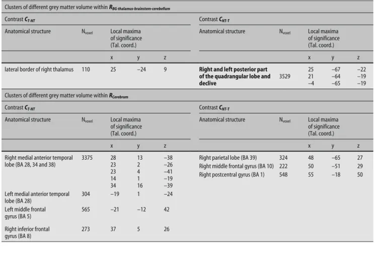
Within the region RBG-thalamus-brainstem-cerebellum at a level of statistical significant puncorrected < 0.001, the contrast CTN-T revealed a lower amount of grey matter volume for the tremor group in the posterior part of the cerebellar quadrangular lobe mainly right, but also paramedian on the left side, and in the declive of the vermis (Table 2). The contrast CT-NT revealed a small cluster of lower amount of grey matter volume for the non-tremor group along the lateral border of the right thalamus (Table 2).
Within the region RCerebrum, four clusters of higher amount of grey matter volume were found for the con-trast CT-NT (Table 2) located in the medial anterior tem-poral lobes (right: Brodmann area [BA] 38, 34 and 28; left: BA 28), the left middle frontal gyrus and the right inferior frontal gyrus (BA 9). The contrast CTN-T revealed in total three small clusters of reduced grey matter vol-ume, located in the right frontal and parietal lobe (Ta-ble 2).
At statistical significance level pFDR < 0.05, only the cluster within the posterior part of the right quadrangu-lar lobe and the declive of the cerebellum was significant (Fig. 1).
No significant clusters of grey matter volume changes (pFDR < 0.05) were found neither for the region BG-thal-amus-brainstem-cerebellum nor the region Cerebrum in the additional analysis of the groups with and without impaired balance, the groups with and without fluctua-tions and the regression analysis of the UPDRS III Bal-ance and Gait score.
Tremor (n = 14) No tremor (n = 10) P Age [years; mean ± SD] 61.5 ± 3.5 62.3 ± 5.1 nsa Age at onset [years; mean ± SD] 53.1 ± 6 55 ± 7.6 nsb Hoehn-Yahr stages [median (range)] 2.3 (2–3) 2.5 (2.5–3) nsb Disease duration [years; median (range)] 6 (3–20) 7.5 (3–18.5) nsb Duration of therapy [years; median (range)] 6 (3–18) 6 (2.3–18.5) nsb LED [mg; median (range)] 980 (150–1790) 1140 (100–1540) nsb Motor score (UPDRS III) [score; median (range)] 25 (12–35) 28 (12–35) nsb Impaired balance (HY ≥ 2.5)c [% (n)] 50 % (7) 100 % (10) 0.008d Balance and Gait (UPDRS items 27–30) 3.4 ± 1.5 4.5 ± 0.9 0.041b Motor complicationse [% (n)] 21.4 % (3) 80 % (8) 0.005d MMSE [score; mean ± SD] 28.6 ± 1.2 28.0 ± 0.8 nsa PDQ39 [score; median (range)] 35 (9–86) 53 (9–90) nsb Depression (Beck/Hamilton) [% (n)] 42.9 % (6) 40 % (4) nsd LED Levodopa equivalent dosage; ns statistically non-significant; PDQ 39 Parkinson’s Disease Questionnaire (39 items); SD standard deviation; UPDRS III Unified Parkinson’s Disease Rating Scale, part III (motor score) a independent t-Test; b Mann-Whitney U-Test; c Patients with impaired balance have an increased prevalence of motor fluctuations (64.7 % vs 0 %, p = 0.004) and decreased prevalence of tremor (35.3 % vs 100 %, p = 0.004); d Pearson χ2-Test; e Patients with motor complications have a longer duration of disease (median 108, 60–240 months vs 60, 36–156 months; p = 0.005) and of dopaminergic therapy (median 104, 58–222 months vs 60, 27–144 months; p = 0.015) with higher LED (median 1355, 570–1540 mg vs 900, 100–1790 mg; p = 0.01) and younger age at onset (50.3 ± 5.9 vs 56.9 ± 5.7 years, p = 0.011)
Table 1 Demographic and clinical findings of patients with Parkinson’s disease with and without tremor
Fig. 1 The cerebellar region of lower amount of grey matter for the tremor group in comparison to the non-tremor group is shown. At a significance level corrected for false positive clusters due to multiple testing PFDR < 0.05, this region is located in the posterior part of the right quadrangular lobe and the declive (A). Without this correction at puncorrected < 0.001, this region extends over the midline to a small part in the contralateral left quadrangular lobe (B)
Table 2 Clusters of different grey matter volume for the two brain regions and the two different contrasts at a significance level puncorrected < 0.001 and in bold at a significance level pFDR < 0.05 corrected for false positive clusters due to multiple testing. Anatomical structure, size of the cluster and coordinates of local maxima of significance are given
Clusters of different grey matter volume within RBG-thalamus-brainstem-cerebellum
Contrast CT-NT Contrast CNT-T
Anatomical structure Nvoxel Local maxima of significance (Tal. coord.)
Anatomical structure Nvoxel Local maxima of significance (Tal. coord.)
x y z x y z
lateral border of right thalamus 110 25 –24 9 Right and left posterior part of the quadrangular lobe and declive 3529 25 21 –4 –67 –64 –65 –22 –19 –19 Clusters of different grey matter volume within RCerebrum
Contrast CT-NT Contrast CNT-T
Anatomical structure Nvoxel Local maxima of significance (Tal. coord.)
Anatomical structure Nvoxel Local maxima of significance (Tal. coord.)
x y z x y z
Right medial anterior temporal lobe (BA 28, 34 and 38)
3375 28 23 23 14 34 13 2 4 1 16 –38 –26 –41 –19 –39
Right parietal lobe (BA 39) 324 48 –65 27 Right middle frontal gyrus (BA 10) 222 50 –51 29 Right postcentral gyrus (BA 1) 548 55 –18 50 Left medial anterior temporal
lobe (BA 28)
304 –19 1 –24
Left middle frontal gyrus (BA 5)
565 –21 –12 42
Right inferior frontal gyrus (BA 8)
Discussion
The principal finding of this study is a reduction of the grey matter volume in the right quadrangular lobe and declive of the cerebellum in PD patients with rest tremor compared to PD patients without tremor. This prospec-tive study using high-resolution MRI VBM to compare brain morphology of the different motor phenotypes of PD demonstrates for the first time a structural abnor-mality in the cerebellum in PD with rest tremor and highlights the postulated cerebellar involvement in the pathogenesis of rest tremor [16, 42, 61]. Rest tremor has been shown to correlate with increased metabolic [3, 15, 22] and oscillatory activity [64] in the cerebellum, thala-mus and motor cortex. Those structures are connected by somatotopically organized cerebello-thalamo-corti-cal projections, and the posterior quadrangular lobule (lobule VI) of the cerebellar cortex has been shown to be linked with the hand area of the motor cortex [43]. This finding underscores previously reported grey matter volume changes in the ventral intermediate nucleus (Vim) in the lateral thalamus which relays the cerebel-lum with the motor cortex [42]. The grey matter vocerebel-lume change in our study bordering the lateral thalamus lacked statistical significance and also its small size pre-cludes further interpretation.
A strong argument in favor of a contribution of the cerebellum and its projections to the thalamus in the pathogenesis of rest tremor comes from experience with stereotactic surgery. The thalamic targets, Vim [5, 10, 44, 46, 47] and the cerebello-thalamic projections [11], have proven efficacious in the suppression of rest tremor and Vim was even postulated to a better target than the ven-tro-oralis posterior nucleus (Vop) receiving pallidal af-ferents [61]. The fact that Vim receives cerebellar affer-ents, but none from the basal ganglia [34], implies that rest tremor originates from either a cerebellar generator [61] or an interaction of the basal ganglia- and cerebello-thalamo-cortical circuits in the cerebral cortex, where those segregated circuits converge [16].
In monkeys, ablation of the interpositus nucleus manifests with tremor during reaching movements [63]. This nucleus receives projections from the intermediate zone of the cerebellum where the decrease of grey mat-ter volume is mainly found in our patients with tremor. The intermediate zone and its output via the interposi-tus nucleus have been postulated to modulate the activ-ity of agonist and antagonist muscles during movements [63], and also to control whether the muscle activity pat-tern is reciprocal or a co-contraction [58].
A possible explanation for the preponderance of the findings in the right cerebellum may be the disparity of tremor manifestation. All patients with tremor but one had tremor on the right, while three had no tremor on the left. Brain asymmetry due to lateralization [2, 23] and consistent right-handedness of all patients may
have also contributed to the asymmetry of findings. A structural asymmetry of the cerebellar hemispheres in association with handedness has been described, and seems to be more marked in right-handers [59]. On the contrary, the equal-handedness may have minimized a confounding effect of brain asymmetry.
These grey matter volume changes in VBM raise questions about their nature. In PD, α-synuclein aggre-gates in cerebellar glia [54] and Purkinje cells [49] have been shown, but their appearance in regard to disease stage and clinical correlate remain yet to be identified. The decrease of grey matter volume may reflect a loss of neurons and/or glial cells, but also changes on the syn-aptic or cellular level [18].
Also, large clusters of decreased grey matter volume are found in the right medial anterior temporal lobe and to a lesser degree in left homologous counterpart in pa-tients without tremor. These are the cortical regions which become earliest affected in the disease process after the brainstem nuclei [6], which is in line with the notion that “akinetic-rigid” patients without tremor ex-perience a more rapid clinical progression to disability [27, 30, 31, 33, 35, 48] and dementia [1]. However, clusters in these regions have not reached significance after cor-rection. A longitudinal analysis may provide more evi-dence to this preliminary observation.
Shortcomings of this study may be the available spa-tial resolution of MRI precluding the differentiation of small neuronal structures, particularly in the brainstem and thalamus. Changes in thalamic grey matter may have been missed, but the other VBM study had com-pared PD patients with unilateral rest tremor with healthy subjects [42]. Confounding factors had been minimized by exclusion of advanced PD, dementia and other co-morbidities. Both groups had a similar demo-graphic, clinical and neuropsychological profile, except for more prevalent balance impairment, and motor fluc-tuations and dyskinesias in the patients without tremor. These differences are not expected to have confounded our findings since no morphological correlates of either balance impairment or motor fluctuations and dyskine-sias were identified in additional VBM analyses, motor fluctuations and dyskinesias presumably originate from long-lasting functional alterations without known struc-tural abnormality, and impaired balance and eventually loss of postural stability in PD is primarily caused by degeneration of brainstem nuclei such as the tegmental pedunculopontine nucleus [45, 52, 57]. Essential tremor which could be a confounding factor, even though a re-cent VBM study had failed to detect cerebellar changes in essential tremor [14], was clinically excluded. Postural and action tremor related to PD differs from essential tremor and is considered a continuation of rest tremor during action and posture presumably originating from the same tremor generator [37].
based on the presence of tremor alone ensuring that all patients with tremor and, therefore, a presumed under-lying tremor generator were included. The deviating tremor-dominant PD criteria [36, 67] may allow the identification of a subgroup of patients with a compara-tively benign disease course [27, 30, 31, 33, 35, 41, 48], but the small number of tremor-dominant PD patients in this collective precludes a separate VBM analysis of this subgroup.
In line with physiological and functional imaging studies, this study provides good evidence of a struc-tural substrate for the involvement of the cerebellum in the pathogenesis of rest tremor.
■ Conflict of interest The authors declare no conflict of interest.
■ Acknowledgment We would like to thank Susanne G. Mueller, MD, for providing the template.
References
1. Aarsland D, Andersen K, Larsen JP, Lolk A, Kragh-Sorensen P (2003) Pre-valence and characteristics of demen-tia in Parkinson disease – An 8-year prospective study. Arch Neurol 60: 387–392
2. Amunts K, Schlaug G, Schleicher A, Steinmetz H, Dabringhaus A, Roland PE, Zilles K (1996) Asymmetry in the human motor cortex and handedness. Neuroimage 3:216–222
3. Antonini A, Moeller JR, Nakamura T, Spetsieris P, Dhawan V, Eidelberg D (1998) The metabolic anatomy of tremor in Parkinson’s disease. Neurol-ogy 51:803–810
4. Beck AT, Ward CH, Mendelsohn M, Mock J, Erbaugh J (1961) An inventory for measuring depression. Arch Gen Psychiatry 4:561–571
5. Benabid AL, Pollak P, Gervason C, Hoffmann D, Gao DM, Hommel M, Perret JE, Derougemont J (1991) Long-Term Suppression of Tremor by Chronic Stimulation of the Ventral Intermediate Thalamic Nucleus. Lancet 337:403–406
6. Braak H, Del Tredici K, Rub U, De Vos RAI, Steur ENHJ, Braak E (2003) Staging of brain pathology related to sporadic Parkinson’s disease. Neuro-biol Aging 24:197–211
7. Brenneis C, Seppi K, Schocke MF, Muller J, Luginger E, Bosch S, Loscher WN, Buchel C, Poewe W, Wenning GK (2003) Voxel-based morphometry detects cortical atrophy in the Parkin-son variant of multiple system atrophy. Mov Disord 18:1132–1138
8. Burton EJ, McKeith IG, Burn DJ, O’Brien JT (2005) Brain atrophy rates in Parkinson’s disease with and with-out dementia using serial magnetic resonance imaging. Mov Disord 20: 1571–1576
9. Burton EJ, McKeith IG, Burn DJ, Williams ED, O’Brien JT (2004) Cere-bral atrophy in Parkinson’s disease with and without dementia: a compar-ison with Alzheimer’s disease, demen-tia with Lewy bodies and controls. Brain 127:791–800
10. Caparros-Lefebvre D, Blond S, Vermer-sch P, Pecheux N, Guieu JD, Petit H (1993) Chronic Thalamic-Stimulation Improves Tremor and Levodopa Induced Dyskinesias in Parkinson Disease. J Neurol Neurosurg Psychiatry 56:268–273
11. Caparros-Lefebvre D, Ruchoux MM, Blond S, Petit H, Percheron G (1994) Long-Term Thalamic-Stimulation in Parkinsons-Disease – Postmortem Anatomoclinical Study. Neurology 44:1856–1860
12. Chapman LJ, Chapman JP (1987) The Measurement of Handedness. Brain and Cognition 6:175–183
13. Coffey CE, Lucke JF, Saxton JA, Ratcliff G, Unitas LJ, Billig B, Bryan RN (1998) Sex differences in brain aging – A quantitative magnetic resonance imag-ing study. Arch Neurol 55:169–179 14. Daniels C, Peller M, Wolff S, Alfke K,
Witt K, Gaser C, Jansen O, Siebner HR, Deuschl G (2006) Voxel-based mor-phometry shows no decreases in cere-bellar gray matter volume in essential tremor. Neurology 67:1452–1456 15. Deiber MP, Pollak P, Passingham R,
Landais P, Gervason C, Cinotti L, Friston K, Frackowiak R, Mauguiere F, Benabid AL (1993) Thalamic-Stimula-tion and Suppression of Parkinsonian Tremor – Evidence of A Cerebellar Deactivation Using Positron Emission Tomography. Brain 116:267–279 16. Deuschl G, Raethjen J, Baron R,
Linde-mann M, Wilms H, Krack P (2000) The pathophysiology of parkinsonian tremor: a review. J Neurol 247:33–48 17. Dissanayaka NN, Sellbach A, Matheson
S, Marsh R, Silburn PA, O’Sullivan JD, Byrne GJ, Mellick GD (2007) Validity of Hamilton Depression Inventory in Parkinson’s disease. Mov Disord 22: 399–403
18. Draganski B, Gaser C, Busch V, Schui-erer G, Bogdahn U, May A (2004) Neu-roplasticity: Changes in grey matter induced by training. Nature 427: 311–312
19. Eidelberg D, Moeller JR, Dhawan V, Sidtis JJ, Ginos JZ, Strother SC, Cedar-baum J, Greene P, Fahn S, Rottenberg DA (1990) The Metabolic Anatomy of Parkinsons-Disease – Complementary [F-18] Fluorodeoxyglucose and [F-18] Fluorodopa Positron Emission Tomo-graphic Studies. Mov Disord 5:203–213 20. Folstein MF, Folstein SE, McHugh PR
(1975) “Mini-mental state”. A practical method for grading the cognitive state of patients for the clinician. J Psychiatr Res 12(3):189–198
21. Foltynie T, Brayne C, Barker RA (2002) The heterogeneity of idiopathic Par-kinson’s disease. J Neurol 249:138–145 22. Fukuda M, Barnes A, Simon ES,
Holmes A, Dhawan V, Giladi N, Fodstad H, Ma YL, Eidelberg D (2004) Thalamic stimulation for parkinsonian tremor: correlation between regional cerebral blood flow and physiological tremor characteristics. NeuroImage 21:608–615
23. Galaburda AM, LeMay M, Kemper TL, Geschwind N (1978) Right-left asymmetrics in the brain. Science 199:852–856
24. Genovese CR, Lazar NA, Nichols T (2002) Thresholding of Statistical Maps in Functional Neuroimaging Using the False Discovery Rate. Neuro-Image 15:870–878
25. Ghaemi M, Raethjen J, Hilker R, Rudolf J, Sobesky J, Deuschl G, Heiss WD (2002) Monosymptomatic resting tremor and Parkinson’s disease: A multitracer positron emission tomo-graphic study. Mov Disord 17:782–788 26. Gibb WR, Lees AJ (1988) The relevance
of the Lewy body to the pathogenesis of idiopathic Parkinson’s disease. J Neurol Neurosurg Psychiatry 51: 745–752
27. Goetz CG, Tanner CM, Stebbins GT, Buchman AS (1988) Risk-Factors for Progression in Parkinsons-Disease. Neurology 38:1841–1844
28. Goldenberg G, Podreka I, Müller C, Deecke L (1989) The relationship between cognitive deficits and frontal lobe function in patients with Parkin-son’s disease. Behav Neurol 2:79–87 29. Good CD, Johnsrude IS, Ashburner J,
Henson RNA, Friston KJ, Frackowiak RSJ (2001) A voxel-based morphomet-ric study of ageing in 465 normal adult human brains. NeuroImage 14:21–36 30. Guillard A, Chastang C (1978) Long-Term Prognostic Factors in Parkin-sons-Disease. Rev Neurol 134:341–354 31. Guillard A, Chastang C, Fenelon G
(1986) Parkinsons-Disease – A Long-Term Study of 416 Patients – Prognos-tic Factors and Implications for Treat-ment. Rev Neurol 142:207–214 32. Hamilton M (1960) A Rating Scale for
Depression. J Neurol Neurosurg Psy-chiatry 23:56–62
33. Hoehn MM, Yahr MD (1967) Parkin-sonism: onset, progression and mortality. Neurology 17(5):427–442 34. Inase M, Tanji J (1995) Thalamic
Distribution of Projection Neurons to the Primary Motor Cortex Relative to Afferent Terminal Fields from the Globus-Pallidus in the Macaque Monkey. J Comp Neurol 353:415–426 35. Jankovic J, Kapadia AS (2001)
Func-tional decline in Parkinson disease. Arch Neurol 58:1611–1615 36. Jankovic J, Mcdermott M, Carter J,
Gauthier S, Goetz C, Golbe L, Huber S, Koller W, Olanow C, Shoulson I, Stern M, Tanner C, Weiner W (1990) Variable Expression of Parkinsons-Disease – A Base-Line Analysis of the Datatop Cohort. Neurology 40:1529–1534 37. Jankovic J, Schwartz KS, Ondo W
(1999) Re-emergent tremor of Parkin-son’s disease. J Neurol Neurosurg Psychiatry 67:646–650
38. Jellinger KA (1999) Post mortem studies in Parkinson’s disease – is it possible to detect brain areas for specific symptoms? J Neural Transm (Supplement):1–29
39. Jellinger KA (2002) Recent develop-ments in the pathology of Parkinson’s disease. J Neural Transm (Supple-ment):347–376
40. Jellinger KA, Paulus W (1992) Clinico-pathological correlations in Parkin-sons-dtsease. Clin Neurol Neurosurg 94:S86–S88
41. Josephs KA, Matsumoto JY, Ahlskog JE (2006) Benign Tremulous parkin-sonism. Arch Neurol 63:354–357 42. Kassubek J, Juengling FD, Hellwig B,
Spreer J, Lucking CH (2002) Thalamic gray matter changes in unilateral Parkinsonian resting tremor: a voxel-based morphometric analysis of 3- dimensional magnetic resonance imaging. Neurosci Lett 323:29–32
43. Kelly RM, Strick PL (2003) Cerebellar loops with motor cortex and prefrontal cortex of a nonhuman primate. J Neu-rosci 23:8432–8444
44. Koller W, Pahwa R, Busenbark K, Hubble J, Wilkinson S, Lang A, Tuite P, Sime E, Lazano A, Hauser R, Malapira T, Smith D, Tarsy D, Miyawaki E, Norregaard T, Kormos T, Olanow CW (1997) High-frequency unilateral thal-amic stimulation in the treatment of essential and parkinsonian tremor. Ann Neurol 42:292–299
45. Lee MS, Rinne JO, Marsden CD (2000) The pedunculopontine nucleus: Its role in the genesis of movement disorders. Yonsei Med J 41:167–184
46. Lenz FA, Kwan HC, Martin RL, Tasker RR, Dostrovsky JO, Lenz YE (1994) Single-unit analysis of the human ventral thalamic nuclear group – Tremor-related activity in functionally identified cells. Brain 117:531–543 47. Limousin P, Speelman JD, Gielen F,
Janssens M, study c (1999) Multicentre European study of thalamic stimula-tion in parkinsonian and essential tremor. J Neurol Neurosurg Psychiatry 66:289–296
48. Marras C, Rochon P, Lang AE (2002) Predicting motor decline and disabil-ity in Parkinson disease – A systematic review. Arch Neurol 59:1724–1728 49. Mori F, Piao YS, Hayashi S, Fujiwara H,
Hasegawa M, Yoshimoto M, Iwatsubo T, Takahashi H, Wakabayashi K (2003) alpha-Synuclein accumulates in Purkinje cells in Lewy body disease but not in multiple system atrophy. J Neuropathol Exp Neurol 62:812–819 50. Nagano-Saito A, Washimi Y, Arahata Y,
Kachi T, Lerch JP, Evans AC, Dagher A, Ito K (2005) Cerebral atrophy and its relation to cognitive impairment in Parkinson disease. Neurology 64: 224–229
51. Otsuka M, Ichiya Y, Kuwabara Y, Hosokawa S, Sasaki M, Yoshida T, Fukumura T, Masuda K, Kato M (1996) Differences in the reduced F-18-Dopa uptakes of the caudate and the puta-men in Parkinson’s disease: Correla-tions with the three main symptoms. J Neurol Sci 136:169–173
52. Pahapill PA, Lozano AM (2000) The pedunculopontine nucleus and Parkin-son’s disease. Brain 123:1767–1783 53. Paulus W, Jellinger K (1991) The
Neu-ropathologic Basis of Different Clinical Subgroups of Parkinsons-Disease. J Neuropathol Exp Neurol 50:743–755 54. Piao YS, Mori F, Hayashi S, Tanji K,
Yoshimoto M, Kakita A, Wakabayashi K, Takahashi H (2003) alpha-synuclein pathology affecting Bergmann glia of the cerebellum in patients with alpha-synucleinopathies. Acta Neuropathol 105:403–409
55. Price S, Paviour D, Scahill R, Stevens J, Rossor M, Lees A, Fox N (2004) Voxel-based morphometry detects patterns of atrophy that help differentiate pro-gressive supranuclear palsy and Par-kinson’s disease. NeuroImage 23: 663–669
56. Ramirez-Ruiz B, Marti MJ, Tolosa E, Bartres-Faz D, Summerfield C, Salgado-Pineda P, Gomez-Anson B, Junque C (2005) Longitudinal evalua-tion of cerebral morphological changes in Parkinson’s disease with and without dementia. J Neurol 252: 1345–1352
57. Rye DB (1997) Contributions of the pedunculopontine region to normal and altered REM sleep. Sleep 20(9): 757–788
58. Smith AM, Bourbonnais D (1981) Neuronal-Activity in Cerebellar Cortex Related to Control of Prehensile Force. J Neurophysiol 45:286–303
59. Snyder PJ, Bilder RM, Wu HW, Bogerts B, Lieberman JA (1995) Cerebellar volume asymmetries are related to handedness: a quantitative MRI study. Neuropsychologia 33:407–419 60. Spreen O, Strauss E (1998) A
compen-dium of neuropsychological tests. Oxford: University Press
61. Stein JF, Aziz TZ (1999) Does imbal-ance between basal ganglia and cere-bellar outputs cause movement disor-ders? Curr Opin Neurol 12:667–669 62. Summerfield C, Junque C, Tolosa E,
Salgado-Pineda P, Gomez-Anson B, Marti M, Ramirez-Ruiz B, Mercader J (2005) Structural brain changes in Parkinson disease with dementia. Arch Neurol 62:281–285
63. Thach WT, Goodkin HP, Keating JG (1992) The cerebellum and the adap-tive coordination of movement. Annu Rev Neurosci 15:403–442
64. Timmermann L, Gross J, Dirks M, Volkmann J, Freund HJ, Schnitzler A (2003) The cerebral oscillatory net-work of parkinsonian resting tremor. Brain 126:199–212
65. Vingerhoets FJ, Schulzer M, Calne DB, Snow BJ (1997) Which clinical sign of Parkinson’s disease best reflects the nigrostriatal lesion? Ann Neurol 41: 58–64
66. Visser M, Leentjens AFG, Marinus J, Stiggelbout AM, van Hilten JJ (2006) Reliability and validity of the Beck Depression Inventory in patients with Parkinson’s disease. Mov Disord 21: 668–672
67. Zetusky WJ, Jankovic J, Pirozzolo FJ (1985) The heterogeneity of Parkin-sons-disease – Clinical and prognostic implications. Neurology 35:522–526