ENSEIGNEMENT SUPÉRIEUR EN RÉANIMATION FONDAMENTAL MÉDECIN
Mitochondria
— key roles in sepsis*
Mitochondrie
— rôles clés dans le sepsis
S. Saeed · M. Singer
Revised: 10 November 2012, Accepted: 3 December 2012 © SRLF et Springer-Verlag France 2012
Abstract The pathophysiological mechanisms underpin-ning the development of, and recovery from, sepsis-induced organ failure require further delineation. Mitochondrial dys-function may well play a key role. This review will therefore consider mitochondria’s function in normal physiology, evi-dence linking bioenergetic alterations to organ dysfunction after severe and prolonged inflammation, and potential ther-apeutic strategies that may be applied.
Keywords Mitochondria · Sepsis · Multi-organ failure · Pathophysiology · Therapeutics
Résumé Au cours du sepsis, les mécanismes physiopatholo-giques sous-tendant le développement et la récupération des dysfonctions d’organe sont encore mal compris. La dysfonc-tion mitochondriale pourrait jouer un rôle majeur. Cette mise au point décrit la physiologie normale des mitochondries, les arguments reliant l’altération bioénergétique mitochondriale aux dysfonctions d’organes après des périodes d’inflamma-tion sévère et prolongée. Enfin, les perspectives thérapeu-tiques envisageables seront abordées.
Mots clés Mitochondrie · Sepsis · Défaillance multiviscérale · Physiopathologie · Traitement
Introduction
The systemic inflammatory response to infection and severe sepsis may progress to multi-organ failure and carries with it
a high morbidity and mortality [1,2]. Precise pathophysio-logical mechanisms remain elusive. The contribution of an impaired circulation leading to tissue hypoperfusion is well established, but an important role of bioenergetic dysfunc-tion is also emerging. An associadysfunc-tion is found between the degree of mitochondrial dysfunction and outcomes in patients with sepsis-induced multi-organ failure [3]. While this does not confirm cause-and-effect, it does nevertheless suggest a new route for therapeutic intervention focused on either protection or acceleration of the recovery process. This is particularly pertinent in light of the multiple trial fail-ures related to immunomodulatory therapies. This overview will provide an insight into mitochondrial biology, its rele-vance to sepsis, and possible therapeutic opportunities that emerge.
Mitochondria in health
The physiological roles of mitochondria
Virtually all cell types possess mitochondria, the notable exception being erythrocytes. Most cell types rely upon mitochondria to provide the bulk of the energy requirement [in the currency of adenosine triphosphate (ATP)] needed to enable normal cellular functioning, and to be able to respond to any intrinsic or extrinsic physiological or pathophysiolog-ical stress. Mitochondria utilize approximately 98% of total body oxygen consumption and generate 90% of human power by proton transfer through ATP [4,5]. Proton transfer occurs through a series of enzymatic steps that occur within the electron transfer chain located in the inner mitochondrial membrane, leading to oxidative phosphorylation of adeno-sine diphosphate (ADP).
Mitochondria also have roles in cell signaling and trigger-ing of cell death pathways. The co-ordinated release of cyto-chrome c from mitochondria activates intrinsic pathways of programmed cell death, apoptosis, whereas necrosis can be triggered when the ATP level falls below a certain threshold. Reactive oxygen species (ROS), produced as a‘by-product’
S. Saeed · M. Singer (*)
Bloomsbury Institute of Intensive Care Medicine, Department of Medicine, University College London, London, United Kingdom
e-mail : m.singer@ucl.ac.uk
* Cet article correspond à la conférence faite par l’auteur au con-grès de la SRLF 2013 dans la session : Voies de recherche dans le sepsis.
of oxidative phosphorylation, plays an important role in maintaining vascular tone, oxygen sensing and, possibly, glucose regulation during skeletal muscle contraction [6]. Indeed, mitochondria are the predominant source of ROS production within the body. The three endogenous gases— nitric oxide (NO), carbon monoxide, and hydrogen sulphide —are also important regulators of mitochondrial signaling in health. Their higher concentrations in disease states such as sepsis have progressively greater inhibitory effects on mito-chondrial respiration and ROS generation.
Other functions of the mitochondrion include the site of production (e.g. cortisol) or action (e.g. triiodothyronine) of many hormones, the biosynthesis of heme and iron-sulphur clusters, and heat generation.
Energy generation by mitochondria
In the cytosol, glucose is metabolized to pyruvate by gly-colysis. Pyruvate is transported into the mitochondria, through an antiporter with hydroxide ions, for conversion by pyruvate dehydrogenase to acetyl coenzyme A (acetyl CoA). Fatty acids are esterified to fatty acyl coA in the cytosol. Medium chain fatty acids (C8 to 10) can diffuse through the mitochondrial membrane, whereas long chain fatty acids rely on the carnitine pathway. Carnitine palmi-toyltransferase (CPT)-1 in the mitochondrial membrane exchanges carnitine for CoA attached to the fatty acid and a conjugate form. This conjugate is transported into the matrix where CPT-2 breaks the conjugate, allowing the fatty acid CoA to reform and undergo beta-oxidation within the mitochondrial matrix.
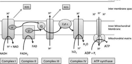
Acetyl-CoA feeds into the tricarboxylic acid (TCA) or Krebs’ cycle, generating nicotinamide and flavin adenine dinucleotide (NADH and FADH2). As summarized in the
Figure 1, NADH passes electrons to complex I (NADH dehydrogenase) of the electron transport chain, becoming oxidized to NAD+, while FADH2donates electrons to
com-plex II (succinate dehydrogenase). Electrons are then passed onto ubiquinone (coenzyme Q), before moving on to com-plex III (cytochrome bc1 comcom-plex), cytochrome c, and then complex IV (cytochrome a, a3, cytochrome c oxidase). Oxy-gen is the terminal electron acceptor of the chain at this enzyme complex, being reduced to water. If oxygen is pre-maturely or incompletely reduced, an increase in superoxide radical (ROS) production occurs, particularly at complexes III and I. The mitochondrion deals with ROS production through its large array of antioxidants, such as superoxide dismutase, catalase, glutathione peroxidase, and peroxire-doxins. This can, however, be overwhelmed in pathological processes generating large amounts of ROS.
As the electrons transfer down the chain, protons move across the inner mitochondrial membrane generating an elec-trochemical gradient. This‘chemiosmotic gradient’ provides the energy to drive ATP synthase (complex V) to produce ATP from ADP. ATP is transported out of the mitochondria and ADP moves back in via the adenine nucleotide translo-case (ANT).
The process is not 100% ‘efficient’ in terms of ATP production. Some of the proton gradient is dissipated before oxidative phosphorylation is complete. This‘uncoupling’ is due to a variety of mechanisms, including specialized uncou-pling proteins within the inner mitochondrial membrane.
Fig. 1 The mitochondrial electron transport chain. A number of redox reactions enable the generation of a proton (H+) gradient to gener-ate ATP. Electron (e-) transfer is shown along with main sites of reactive oxygen species (ROS) production. NADH: nicotinamide ade-nine dinucleotide; FADH1: flavin adeade-nine dinucleotide; Q: ubiquinone, or coenzyme Q; Cyt c: cytochrome c; ADP: adenosine diphos-phate; PI: Phosdiphos-phate; ATP: adenosine triphosphate
Uncoupling has particular importance in heat generation and hibernation.
The mitochondrial life cycle
Mitochondrial biogenesis is the production of new mito-chondria/mitochondrial protein occurring with, and indepen-dent of, cell mitosis. In the non-dividing cell, biogenesis improves the capacity for energy production if energy demands increase. The process involves production of mito-chondrial proteins encoded either by the cell nucleus with subsequent import and integration into the mitochondria, or via mitochondrial deoxyribonucleic acid (DNA) which encodes 13 proteins that are mainly situated within the oxidative phosphorylation pathway.
A key player that orchestrates mitochondrial biogenesis is the peroxisome proliferator-activated receptor gamma coac-tivator (PGC)-1alpha, a co-accoac-tivator of nuclear transcription factors such as nuclear respiratory factors 1 and 2 (NRF-1 and -2) that upregulate nuclear production of mitochondrial proteins [7–9]. NRF-1 also increases expression of Tfam (transcription factor A for the mitochondrion), which, once transported into the mitochondrion, stimulates transcription of mitochondrial DNA [10].
Numerous influences on PGC-1alpha occur in response to physiological (e.g. exercise) and pathophysiological (e.g. hypoxia) stimuli. AMP (adenosine monophosphate)/ ATP ratios are known inducers in brown adipose tissue and liver via beta-adrenergic/cyclic AMP pathways [8]. In skel-etal muscle, the calcineurin A, CaMK (Ca2+ /calmodulin-dependent kinase), p38 MAPK (mitogen-activated protein kinase), and AMPK (AMP-activated protein kinase) path-ways have been implicated [11–14], as well as sirtuins (enzyme deacylators) [15]. Negative regulators influence energy balance via endogenous RIP140 (nuclear receptor-interacting protein 1), the p160 myb-binding protein, and the GCN5 acetyltransferase complex [16–18]. Interaction also occurs with thyroid, glucocorticoid, estrogen, and estrogen-related receptors [19].
An association is also emerging between NO and mito-chondrial biogenesis. Endogenous NO upregulates PGC-1alpha mRNA expression [20]. NO donors can increase mitochondrial DNA in cell cultures, while mice deplete of endothelial nitric oxidase synthase show reduced mitochon-drial biogenesis, mitochonmitochon-drial mass, basal oxygen con-sumption, and ATP levels [21].
During their lifetime, mitochondria undergo numerous morphological changes during fusion and fission events. These mitochondrial dynamics are primarily affected by GTPases; this links with roles in cell division and prolifera-tion as well as self-directed removal of damaged or surplus mitochondria, a process known as mitophagy. Mitofusin-2 and OPA-1 (optic atrophy-1), proteins driving fusion
events, and DRP-1 (dynamin-related protein-1), a protein that influences fission, have been associated with altered mitochondrial membrane potential and reduced oxygen consumption [22].
Mitochondrial variation in tissue types
Mitochondria have an intricate, sophisticated, and complex purpose within the cell and in overall tissue physiology. In skeletal muscle, mitochondria exist as reticular networks located near the sarcolemma and also within muscle fibres [23]. Heart and skeletal muscles have more mitochondrial content, including respiratory chain subunits, than in liver, kidney, and brain tissue. Heart muscle mitochondria also have more cristae (folds) per surface area [24].
Morphological variation is seen in human hepatocytes, neuronal cells, and umbilical vein endothelial cells in terms of shape, number of cristae, and distribution within the cell [25]. Even within the same cell type, variable membrane potentials are seen, being higher in peripherally located mitochondria and possibly related to calcium sequestration. It is likely that mitochondrial antioxidant capacity also varies between cell types, making some more vulnerable to oxida-tive stress.
Mitochondrial dysfunction in sepsis and
multi-organ failure
The systemic inflammatory response syndrome is triggered by microbial antigens (sepsis) or other factors (e.g. trauma, hemorrhage, burn injury). Micro-organisms or their consti-tuents are recognized by specialized pattern recognition receptors (PRRs) situated either on or inside immune, endo-thelial, and epithelial cells. The best characterized set of PRRs is the toll-like receptors (TLRs). PRRs recognize both pathogen-associated molecular patterns (PAMPs) on invad-ing organisms, as well as host-derived danger-associated molecular patterns (DAMPs) released in response to stress, tissue injury, or cell death. Mitochondria released into the circulation due to tissue damage act as a DAMP.
The subsequent release of pro-inflammatory cytokines and other mediators leads to activation or suppression of multiple pathways involving cardiovascular, immunologi-cal, hormonal, coagulation, metabolic, and bioenergetic sys-tems. This leads to organ dysfunction that is manifest clini-cally as impaired physiological or biochemical activity of that organ. Severe dysfunction leads to a state of failure that may require significant levels of pharmacological or mechanical organ support to maintain an acceptable level of homeostasis compatible with continued survival.
Impaired perfusion early in the septic process (due to intrinsic and extrinsic fluid losses and decreased intake)
can lead to tissue hypoxia and amplification of the systemic inflammatory response. While early and aggressive correc-tion of the tissue oxygen debt may prove clinically beneficial [26], attempts to correct cellular hypoxia when organ dys-function was established proved fruitless, or even harmful [27,28]. This suggests an important temporal component to the pathophysiology of sepsis. Indeed, it appears that the condition shifts from hypoxia to dysoxia. In other words, there is availability yet an inability of cells and tissues to use oxygen. This was termed cytopathic hypoxia but, more accurately, should be labeled cytopathic dysoxia [29]. The data in support of this are the findings of (i) progressive reductions in oxygen consumption with increasing sepsis severity [30]; yet (ii) maintained, or even elevated levels of oxygen at the tissue level found in both humans and animal models [31,32]; and (iii) a remarkable lack (or minimal pres-ence) of cell death, apoptotic or necrotic, in the failed organs that supports the notion that oxygen lack and tissue hypoper-fusion is a continuing and pathophysiologically significant problem. The parallel findings of oxygen availability yet decreased utilization infer a new metabolic steady state has been achieved. As mitochondria are the predominant utili-zers of oxygen, their likely importance in septic multi-organ failure should be considered. Although there may be an as yet unrecognized mechanism invoked by prolonged and severe inflammation that directly inhibits metabolism, there are well-defined mechanisms by which ATP production is impaired, leading to a secondary shutdown of metabolism, and thus cell functioning, akin to hibernation or estivation. Inflammatory processes that directly target mitochondria work via inhibition of their activity, through direct damage from reactive species, and by a decrease in biogenesis through decreased transcripts of genes encoding for respira-tory complex proteins.
Respiratory chain activity
Animal models of sepsis have been predominantly used to study mitochondrial function. Short-term models lasting several hours demonstrate highly variable results of sepsis on oxidative phosphorylation. As previously reviewed, increased, unchanged, or reduced respiratory activity have all been reported [33]. This may reflect differences in spe-cies, in tissues studied, in the forms and severity of the septic insult administered, and in the ex vivo techniques used to prepare samples and measure activity. The majority of long-term sepsis models (lasting >16 hours) do however demonstrate changes in respiratory chain activity, morphol-ogy, or mitochondrial mass. This has been found in various tissues taken from different animal species, e.g. heart, mus-cle and liver, and brain [34–37]. Patient samples, including muscle, diaphragm, liver, and monocytes, have all shown decreased mitochondrial enzyme activity, membrane
poten-tial, and histological changes [3,38–42]. Mitochondrial respiratory complex I is most often found to be inhibited. We performed vastus lateralis skeletal muscle biopsies in patients soon after admission to intensive care with septic shock [3]. Associations were found between complex I dys-function, ATP depletion, glutathione depletion, excess NO production, and mortality. A Swedish study reported reduced mitochondrial enzyme activity in biopsies taken from leg and intercostal muscles of critically ill patients, whereas mitochondrial activity was increased in leg skeletal mus-cle of biopsy taken two hours after an injection of endotoxin [43]. This emphasizes the varying pathophysiological changes that occur over the course of sepsis.
Impact of altered redox state
Sepsis produces a large amount of ROS and reactive nitro-gen species (RNS). This is partly due to activation of immune and endothelial cells, but also due to increased pro-duction of mitochondrial superoxide. NO, itself a reversible inhibitor of mitochondrial respiratory complex IV, can react with superoxide to form peroxynitrite which has a more prolonged and potentially irreversible inhibitory effect on respiratory complexes via nitration, particularly affecting complex I [44,45]. Direct damage to mitochondrial DNA may also produce ROS [46].
Altered substrate provision
Early work did not demonstrate any alterations in Krebs’ cycle intermediaries in cardiac tissue of an animal model [47]. However, Vary reported inhibition of pyruvate dehy-drogenase, preventing pyruvate entry into the mitochondrion and thereby leading to an increase in lactate levels [48]. More recently, Mason and Stofan showed reduced activity of the Krebs’ cycle enzyme, aconitase, in association with reduced mitochondrial respiration [49].
Altered mitochondrial biogenesis
Calvano et al administered endotoxin to healthy volunteers and performed transcriptomics on white cells sampled there-after [50]. Notably, they described decreased expression of genes encoding for mitochondrial respiratory proteins. We reported a similar finding in the transcriptome of muscle biopsies taken from critically ill patients, with a significantly lower gene transcript in those patients who subsequently died [40]. This also correlated with a decrease in complex I activity. Furthermore, PGC-1alpha transcript levels were raised above normal in those patients who survived, whereas levels in non-survivors were similar to those in healthy control samples. Another study in patients with established septic multi-organ failure in the ICU showed upregulation
of NRF2alpha (nuclear factor (erythroid-derived 2)-like 2alpha)/GABP (GA-binding protein) genes, suggestive of biogenesis. Here, protein synthesis was preserved within the mitochondria, as were genes related to energy metabo-lism. Interestingly, when considering genes associated with muscle turnover, presence of those linked with catabolism, atrogens, was noted [51]. In a long-term mouse model of S. aureus peritonitis, organ dysfunction and clinical illness was accompanied by a fall in metabolic rate and a decrease in mitochondrial mass. Recovery of metabolic activity and organ function, and clinical improvement were preceded by an upregulation of markers of mitochondrial biogenesis such as PGC-1alpha, Tfam (transcription factor A, mitochondrial) and NRF-1, and suppression of RIP140, an endogenous co-repressor [52]. In a recent study of endotoxic mice, locomotor muscles were found to be more susceptible to mitochondrial injury compared to ventilatory muscles, with decreased biogenesis and an increase in autophagy [53].
Putative mitochondrially-targeted therapies in
sepsis-induced multi-organ failure
A variety of strategies are available, for example, to protect mitochondria from injury, or to increase biogenesis with the aim of accelerating recovery.
Antioxidants
Preclinical trials using antioxidants show promise. In a murine model using N-acetylcysteine and deferrioxamine after cecal ligation and puncture (CLP) induction, a signifi-cant improvement was seen in oxidant profile and mortality [54]. Antioxidants targeted specifically to mitochondria such as MitoQ and MitoVitE show improved mitochondrial activ-ity and a reduction in organ failure severactiv-ity [55–57]. Mela-tonin has antioxidant effects and has also improved redox outcomes and mortality in animal models [58]. Notably, its circadian variation in plasma is altered in critical illness [59]. Its use in neonates with sepsis has also shown benefit with reduced oxidative stress; however, larger trials in adults are lacking [60]. Exploitation of antioxidant therapies has been considered in detail elsewhere [61].
Inducing‘suspended animation’
Decreasing metabolic rate is an established therapeutic prac-tice achieved by inducing therapeutic hypothermia in cardiac arrest survivors and in infants with anoxic encephalopathy. Carbon monoxide and hydrogen sulphide are potential gaso-transmitters that may have similar effects to induce the hiber-nation state alluded to earlier. While high levels of carbon monoxide can dangerously inhibit complex IV, at lower
concentrations it enabled tissue protection in animal models of sepsis [62]. A water-soluble carbon monoxide releas-ing agent given after induction of sepsis in a mouse model improved survival rates; this was accompanied by an increase in mitochondrial respiration, in PGC-1alpha expres-sion, and in mitochondrial DNA copy number [63]. Carbon monoxide is released endogenously after activation on heme oxygenase (HO)-1. Induction of HO-1 in sepsis models has shown an action through NRF-2, linking it to mitochondrial biogenesis [64,65].
Hydrogen sulphide, also an inhibitor of complex IV, reduced oxygen consumption in mice and induced a revers-ible state of‘suspended animation’ [66]. Pre-treatment has shown a survival benefit in lethal hypoxemia and hemor-rhage [67]. There is potential utility in sepsis with improved neutrophil migration and decreased mortality reported through its use in septic mice [67,68].
Stimulating mitochondrial biogenesis
The role of biogenesis and its association with survival ben-efits in critically ill patients opens new avenues for research into using it as a treatment modality. Its association with the HO-1/carbon monoxide pathway has been mentioned ear-lier. Recently, Thomas et al used a recombinant human Tfam and found improved redox and mitochondrial activity profiles in both cultured mouse fibroblasts and a murine model of sepsis, in which survival was also enhanced [69]. Their murine model of Parkinson’s disease also showed recombinant human mitochondrial transcription factor A protein (rhTFAM) improved motor function. This may have implications in severe sepsis as muscle wasting occurs early and with significant subsequent impact on return to normal function [70–72].
Conclusion
In summary, there is significant evidence that implicates mito-chondrial dysfunction in septic organ dysfunction. Whether this is causative or epiphenomenal is less clear. However, sur-vivors have better preservation of ATP and biogenesis mar-kers. Multi-organ failure may represent a mechanism through which the likelihood of eventual survival is enhanced in those hardy enough to survive. Cells may enter a‘hibernating’ state in the face of overwhelming inflammation. This adaptive strategy may have evolved in response to effects on cellular bioenergetics.
References
1. Alberti C, Brun-Buisson C, Burchardi H, et al (2002) Epidemiol-ogy of sepsis and infection in ICU patients from an international multicentre cohort study. Intensive Care Med 28:108–21 2. Angus DC, Linde-Zwirble WT, Lidicker J, et al (2001)
Epidemi-ology of severe sepsis in the United States: analysis of incidence, outcome, and associated costs of care. Crit Care Med 29:1303–10 3. Brealey D, Brand M, Hargreaves I, et al (2002) Association between mitochondrial dysfunction and severity and outcome of septic shock. Lancet 360:219–23
4. Duchen MR (2004) Roles of mitochondria in health and disease. Diabetes 53(Suppl 1):S96–S102
5. Rich P (2003) Chemiosmotic coupling: The cost of living. Nature 421:583
6. Bradley SJ, Kingwell BA, McConell GK (1999) Nitric oxide synthase inhibition reduces leg glucose uptake but not blood flow during dynamic exercise in humans. Diabetes 48:1815–21 7. Evans MJ, Scarpulla RC (1990) NRF-1: a trans-activator of
nuclear-encoded respiratory genes in animal cells. Genes Dev 4:1023–34
8. Puigserver P, Wu Z, Park CW, et al (1998) A cold-inducible coactivator of nuclear receptors linked to adaptive thermogenesis. Cell 92:829–39
9. Scarpulla RC (2002) Transcriptional activators and coactivators in the nuclear control of mitochondrial function in mammalian cells. Gene 286:81–9
10. Virbasius JV, Scarpulla RC (1994) Activation of the human mito-chondrial transcription factor A gene by nuclear respiratory fac-tors: a potential regulatory link between nuclear and mitochon-drial gene expression in organelle biogenesis. Proc Natl Acad Sci U S A 91:1309–13
11. Akimoto T, Pohnert SC, Li P, et al (2005) Exercise stimulates Pgc-1alpha transcription in skeletal muscle through activation of the p38 MAPK pathway. J Biol Chem 280:19587–93
12. Czubryt MP, McAnally J, Fishman GI, et al (2003) Regulation of peroxisome proliferator-activated receptor gamma coactivator 1 alpha (PGC-1 alpha) and mitochondrial function by MEF2 and HDAC5. Proc Natl Acad Sci U S A 100:1711–6
13. Schaeffer PJ, Wende AR, Magee CJ, et al (2004) Calcineurin and calcium/calmodulin-dependent protein kinase activate distinct metabolic gene regulatory programs in cardiac muscle. J Biol Chem 279:39593–603
14. Zong H, Ren JM, Young LH, et al (2002) AMP kinase is required for mitochondrial biogenesis in skeletal muscle in response to chronic energy deprivation. Proc Natl Acad Sci U S A 99:15983–7
15. Gerhart-Hines Z, Rodgers JT, Bare O, et al (2007) Metabolic control of muscle mitochondrial function and fatty acid oxidation through SIRT1/PGC-1alpha. Embo J 26:1913–23
16. Fan M, Rhee J, St-Pierre J, et al (2004) Suppression of mitochon-drial respiration through recruitment of p160 myb binding protein to PGC-1alpha: modulation by p38 MAPK. Genes Dev 18:278–89 17. Leonardsson G, Steel JH, Christian M, et al (2004) Nuclear receptor corepressor RIP140 regulates fat accumulation. Proc Natl Acad Sci U S A 101:8437–42
18. Lerin C, Rodgers JT, Kalume DE, et al (2006) GCN5 acetyltrans-ferase complex controls glucose metabolism through transcrip-tional repression of PGC-1alpha. Cell Metab 3:429–38
19. Ventura-Clapier R, Garnier A, Veksler V (2008) Transcriptional control of mitochondrial biogenesis: the central role of PGC-1alpha. Cardiovasc Res 79:208–17
20. Nisoli E, Clementi E, Paolucci C, et al (2003) Mitochondrial bio-genesis in mammals: the role of endogenous nitric oxide. Science 299:896–9
21. Nisoli E, Carruba MO (2006) Nitric oxide and mitochondrial bio-genesis. J Cell Sci 119:2855–62
22. Liesa M, Palacin M, Zorzano A (2009) Mitochondrial dynamics in mammalian health and disease. Physiol Rev 89:799–845 23. Glancy B, Balaban RS (2011) Protein composition and function
of red and white skeletal muscle mitochondria. Am J Physiol Cell Physiol 300:C1280–C90
24. Benard G, Faustin B, Passerieux E, et al (2006) Physiological diversity of mitochondrial oxidative phosphorylation. Am J Phy-siol Cell PhyPhy-siol 291:C1172–C82
25. Collins TJ, Berridge MJ, Lipp P, et al (2002) Mitochondria are morphologically and functionally heterogeneous within cells. Embo J 21:1616–27
26. Rivers E, Nguyen B, Havstad S, et al (2001) Early goal-directed therapy in the treatment of severe sepsis and septic shock. N Engl J Med 345:1368–77
27. Gattinoni L, Brazzi L, Pelosi P, et al (1995) A trial of goal-oriented hemodynamic therapy in critically ill patients. SvO2 Collaborative Group. N Engl J Med 333:1025–32
28. Hayes MA, Timmins AC, Yau EH, et al (1994) Elevation of systemic oxygen delivery in the treatment of critically ill patients. N Engl J Med 330:1717–22
29. Fink MP (2002) Bench-to-bedside review: Cytopathic hypoxia. Crit Care 6:491–9
30. Kreymann G, Grosser S, Buggisch P, et al (1993) Oxygen con-sumption and resting metabolic rate in sepsis, sepsis syndrome, and septic shock. Crit Care Med 21:1012–9
31. Boekstegers P, Weidenhofer S, Pilz G, et al (1991) Peripheral oxygen availability within skeletal muscle in sepsis and septic shock: comparison to limited infection and cardiogenic shock. Infection 19:317–23
32. Rosser DM, Stidwill RP, Jacobson D, et al (1995) Oxygen ten-sion in the bladder epithelium rises in both high and low cardiac output endotoxemic sepsis. J Appl Physiol 79:1878–82 33. Singer M (2007) Mitochondrial function in sepsis: acute phase
versus multiple organ failure. Crit Care Med 35:S441–S8 34. Brealey D, Karyampudi S, Jacques TS, et al (2004) Mitochondrial
dysfunction in a long-term rodent model of sepsis and organ fail-ure. Am J Physiol Regul Integr Comp Physiol 286:R491–7 35. Comim CM, Rezin GT, Scaini G, et al (2008) Mitochondrial
respiratory chain and creatine kinase activities in rat brain after sepsis induced by cecal ligation and perforation. Mitochondrion 8:313–8
36. d’Avila JC, Santiago AP, Amancio RT, et al (2008) Sepsis induces brain mitochondrial dysfunction. Crit Care Med 36:1925–32 37. Gellerich FN, Trumbeckaite S, Hertel K, et al (1999) Impaired
energy metabolism in hearts of septic baboons: diminished activ-ities of Complex I and Complex II of the mitochondrial respira-tory chain. Shock 11:336–41
38. Adrie C, Bachelet M, Vayssier-Taussat M, et al (2001) Mitochon-drial membrane potential and apoptosis peripheral blood mono-cytes in severe human sepsis. Am J Respir Crit Care Med 164:389–95
39. Callahan LA, Supinski GS (2005) Sepsis induces diaphragm electron transport chain dysfunction and protein depletion. Am J Respir Crit Care Med 172:861–8
40. Carre JE, Orban JC, Re L, et al (2010) Survival in critical illness is associated with early activation of mitochondrial biogenesis. Am J Respir Crit Care Med 182:745–51
41. Fredriksson K, Hammarqvist F, Strigard K, et al (2006) Derange-ments in mitochondrial metabolism in intercostal and leg muscle of critically ill patients with sepsis-induced multiple organ failure. Am J Physiol Endocrinol Metab 291:E1044–50
42. Vanhorebeek I, De Vos R, Mesotten D, et al (2005) Protection of hepatocyte mitochondrial ultrastructure and function by strict
blood glucose control with insulin in critically ill patients. Lancet 365:53–9
43. Fredriksson K, Flaring U, Guillet C, et al (2009) Muscle mito-chondrial activity increases rapidly after an endotoxin challenge in human volunteers. Acta Anaesthesiol Scand 53:299–304 44. Clementi E, Brown GC, Feelisch M, et al (1998) Persistent
inhi-bition of cell respiration by nitric oxide: crucial role of S-nitrosylation of mitochondrial complex I and protective action of glutathione. Proc Natl Acad Sci U S A 95:7631–6
45. Murray J, Taylor SW, Zhang B, et al (2003) Oxidative damage to mitochondrial complex I due to peroxynitrite: identification of reactive tyrosines by mass spectrometry. J Biol Chem 278:37223–30
46. Galley HF (2011) Oxidative stress and mitochondrial dysfunction in sepsis. Br J Anaesth 107:57–64
47. Hotchkiss RS, Song SK, Neil JJ, et al (1991) Sepsis does not impair tricarboxylic acid cycle in the heart. Am J Physiol 260: C50–7
48. Vary TC (1996) Sepsis-induced alterations in pyruvate dehydro-genase complex activity in rat skeletal muscle: effects on plasma lactate. Shock 6:89–94
49. Mason KE, Stofan DA (2008) Endotoxin challenge reduces aco-nitase activity in myocardial tissue. Arch Biochem Biophys 469:151–6
50. Calvano SE, Xiao W, Richards DR, et al (2005) A network-based analysis of systemic inflammation in humans. Nature 437:1032–7 51. Fredriksson K, Tjader I, Keller P, et al (2008) Dysregulation of mitochondrial dynamics and the muscle transcriptome in ICU patients suffering from sepsis induced multiple organ failure. PLoS One 3:e3686
52. Haden DW, Suliman HB, Carraway MS, et al (2007) Mitochon-drial biogenesis restores oxidative metabolism during Staphylo-coccus aureus sepsis. Am J Respir Crit Care Med 176:768–77 53. Mofarrahi M, Sigala I, Guo Y, et al (2012) Autophagy and
skele-tal muscles in sepsis. PLoS One 7:e47265
54. Ritter C, Andrades ME, Reinke A, et al (2004) Treatment with N-acetylcysteine plus deferoxamine protects rats against oxida-tive stress and improves survival in sepsis. Crit Care Med 32:342–9
55. Lowes DA, Thottakam BM, Webster NR, et al (2008) The mitochondria-targeted antioxidant MitoQ protects against organ damage in a lipopolysaccharide-peptidoglycan model of sepsis. Free Radic Biol Med 45:1559–65
56. Mao G, Kraus GA, Kim I, et al (2010) A mitochondria-targeted vitamin E derivative decreases hepatic oxidative stress and inhi-bits fat deposition in mice. J Nutr 140:1425–31
57. Supinski GS, Murphy MP,Callahan LA (2009) MitoQ administra-tion prevents endotoxin-induced cardiac dysfuncadministra-tion. Am J Phy-siol Regul Integr Comp PhyPhy-siol 297:R1095–102
58. Escames G, Leon J, Macias M, et al (2003) Melatonin counter-acts lipopolysaccharide-induced expression and activity of mito-chondrial nitric oxide synthase in rats. Faseb J 17:932–4 59. Mundigler G, Delle-Karth G, Koreny M, et al (2002) Impaired
circadian rhythm of melatonin secretion in sedated critically ill patients with severe sepsis. Crit Care Med 30:536–40
60. Gitto E, Pellegrino S, Gitto P, et al (2009) Oxidative stress of the newborn in the pre- and postnatal period and the clinical utility of melatonin. J Pineal Res 46:128–39
61. Galley HF (2010) Bench-to-bedside review: Targeting antioxi-dants to mitochondria in sepsis. Crit Care 14:230
62. Ryter SW, Choi AM (2007) Cytoprotective and anti-inflammatory actions of carbon monoxide in organ injury and sepsis models. Novartis Found Symp 280:165–75; discussion 175–81
63. Lancel S, Hassoun SM, Favory R, et al (2009) Carbon monoxide rescues mice from lethal sepsis by supporting mitochondrial ener-getic metabolism and activating mitochondrial biogenesis. J Phar-macol Exp Ther 329:641–8
64. MacGarvey NC, Suliman HB, Bartz RR, et al (2012) Activation of mitochondrial biogenesis by heme oxygenase-1-mediated NF-E2-related factor-2 induction rescues mice from lethal Staphylo-coccus aureus sepsis. Am J Respir Crit Care Med 185:851–61 65. Piantadosi CA, Withers CM, Bartz RR, et al (2011) Heme
oxygenase-1 couples activation of mitochondrial biogenesis to anti-inflammatory cytokine expression. J Biol Chem 286: 16374–85
66. Blackstone E, Morrison M, Roth MB (2005) H2S induces a sus-pended animation-like state in mice. Science 308:518
67. Spiller F, Orrico MI, Nascimento DC, et al (2010) Hydrogen sul-fide improves neutrophil migration and survival in sepsis via K +ATP channel activation. Am J Respir Crit Care Med 182:360–8 68. Tokuda K, Kida K, Marutani E, et al (2012) Inhaled hydrogen sulfide prevents endotoxin-induced systemic inflammation and improves survival by altering sulfide metabolism in mice. Anti-oxid Redox Signal 17:11–21
69. Thomas RR, Khan SM, Portell FR, et al (2011) Recombinant human mitochondrial transcription factor A stimulates mitochon-drial biogenesis and ATP synthesis, improves motor function after MPTP, reduces oxidative stress and increases survival after endotoxin. Mitochondrion 11:108–18
70. Herridge MS, Tansey CM, Matte A, et al (2011) Functional dis-ability 5 years after acute respiratory distress syndrome. N Engl J Med 364:1293–304
71. Khan J, Harrison TB, Rich MM, et al (2006) Early development of critical illness myopathy and neuropathy in patients with severe sepsis. Neurology 67:1421–5
72. Kress JP, Herridge MS (2012) Medical and economic implica-tions of physical disability of survivorship. Semin Respir Crit Care Med 33:339–47