Dissociated active and passive tactile shape recognition: a case study of pure tactile apraxia
12
0
0
Texte intégral
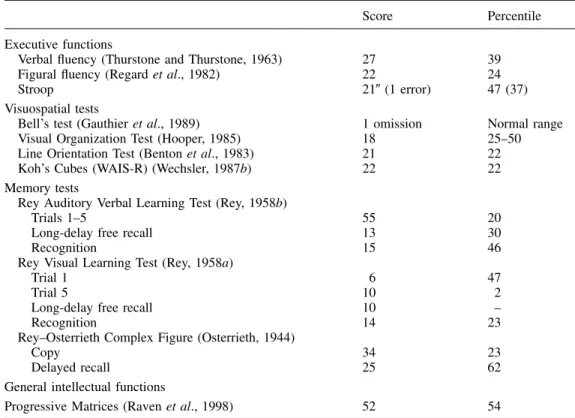
(2) 2288. N. Valenza et al. Table 1 Neuropsychological assessment. Executive functions Verbal fluency (Thurstone and Thurstone, 1963) Figural fluency (Regard et al., 1982) Stroop Visuospatial tests Bell’s test (Gauthier et al., 1989) Visual Organization Test (Hooper, 1985) Line Orientation Test (Benton et al., 1983) Koh’s Cubes (WAIS-R) (Wechsler, 1987b) Memory tests Rey Auditory Verbal Learning Test (Rey, 1958b) Trials 1–5 Long-delay free recall Recognition Rey Visual Learning Test (Rey, 1958a) Trial 1 Trial 5 Long-delay free recall Recognition Rey–Osterrieth Complex Figure (Osterrieth, 1944) Copy Delayed recall General intellectual functions Progressive Matrices (Raven et al., 1998). of an object requires the use of a contour-following strategy, i.e. a dynamic and time-consuming exploratory procedure in which the hand maintains contact with the contour of the object (Lederman and Klatzky, 1987). Despite the evidence from studies with healthy participants, some authors (e.g. Caselli, 1991a, b, 1993) suggest that tactile exploration strategies are not primordial for TOR, basing their assumption on clinical observation of patients with relatively severe motor disturbance but preserved TOR. Patients with tactile agnosia also seem to employ relatively normal exploratory movements (Endo et al., 1992; Platz, 1996; Reed et al., 1996; Nakamura et al., 1998). Tactile agnosia has therefore been interpreted as inability to integrate the tactile features of a shape into a modality-specific tactile representation despite correct exploration (Endo et al., 1992; Platz, 1996; Nakamura et al., 1998). As emphasized in a recent paper (Binkofski et al., 2001), the inverse deficit pattern, i.e. TOR failure based on selectively impaired exploratory procedures with preserved feature extraction, has never been described. Here, we present a patient showing a dissociation between impaired shape and object identification when using spontaneous exploratory procedures (active touch) and intact shape identification when exploration is guided by the experimenter (passive touch).. Score. Percentile. 27 22 21⬙ (1 error). 39 24 47 (37). 1 omission 18 21 22. Normal range 25–50 22 22. 55 13 15. 20 30 46. 6 10 10 14. 47 2 – 23. 34 25. 23 62. 52. 54. artery. The aneurysm of the right middle cerebral artery and a second aneurysm of the posterior communicating artery were clipped. The postoperative course was complicated by severe arterial spasms and an infarction of the right middle cerebral artery. On admission, the neurological examination revealed slight paresis and slight sensory loss of the left side of the face and the left arm. Neuropsychological examination revealed discrete left-sided neglect, disturbed visuospatial capacities and a moderate deficit of non-verbal memory and executive functions. The patient was distractible and showed perseverative tendencies and a lack of insight into her cognitive deficits. Her most apparent deficit was an inability to name objects held in her left hand. The neuropsychological deficits disappeared rapidly, except for the left-sided disturbance of TOR. Two months after admission, motor functions had completely recovered and only slight hypoaesthesia of the left arm persisted. The only complaint mentioned by the patient at that time was the persistence of her inability to recognize objects by touch with the left hand, although she could feel their temperature and substance and crudely recognized their size and smoothness. For instance, she complained of her inability to discriminate small wooden objects used in occupational therapy (e.g. a cone, a mushroom, a ring) when they were put in a bag.. Case report A 28-year-old right-handed woman suffering from renal polycystosis was admitted to rehabilitation 5 weeks after a bleeding from an aneurysm of the right middle cerebral. Neuropsychological evaluation A detailed neuropsychological examination was made 4 months after hospital admission. General intellectual.
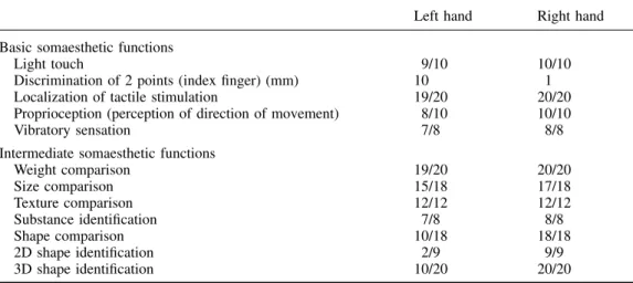
(3) Tactile apraxia without tactile agnosia. 2289. Table 2 Somatosensory evaluation of the left and right hand according to the criteria of Caselli (1997) Left hand. Right hand. Basic somaesthetic functions Light touch Discrimination of 2 points (index finger) (mm) Localization of tactile stimulation Proprioception (perception of direction of movement) Vibratory sensation. 9/10 10 19/20 8/10 7/8. 10/10 1 20/20 10/10 8/8. Intermediate somaesthetic functions Weight comparison Size comparison Texture comparison Substance identification Shape comparison 2D shape identification 3D shape identification. 19/20 15/18 12/12 7/8 10/18 2/9 10/20. 20/20 17/18 12/12 8/8 18/18 9/9 20/20. functions, tested with a short version of the Progressive Matrices of Raven (Raven et al., 1998) were within the normal range. Language, arithmetic skills, praxis and perceptual functions were normal. Visual fields were intact. Assessment of anterograde memory revealed normal verbal (Rey, 1958b) but slightly impaired non-verbal memory (Osterrieth, 1944; Rey, 1958a). Verbal and non-verbal fluency (Thurstone and Thurstone, 1963; Regard et al., 1982) were normal. Assessment of visuospatial and constructive functions revealed normal performance in the Benton Line Orientation Test (Benton et al., 1983), in the Hooper Visual Organization Test (Hooper, 1985) and in the Koh’s cubes of the Wechsler Intelligence Scale—Revised (Wechsler, 1987). There were no signs of neglect in a cancellation test (Gauthier et al., 1989) or in line bisection, and no visual extinction. In summary, the patient showed only a minor non-verbal learning memory deficit (Table 1). The following experiments were conducted after the patient had given written informed consent. The study was approved by the Ethics Committee of the University of Geneva.. patient was asked to compare two objects given successively in either the left or the right hand. Weight perception (discrimination of cylinders of the same size weighing 50, 90, 150, 200 or 270 g), texture perception (discrimination of four grades of sandpaper) and dimension perception (discrimination of sticks of length varying between 1 and 10 cm) were equivalent for both hands. Substance perception (naming of eight substances, such as metal, wood and plastic) did not differ between hands. In contrast, 3D form perception (discrimination of simple forms, such as a cube, a cone and a cylinder) was severely deficient for the left hand. Similarly, perception of 2D and 3D forms (tactile–visual matching of forms palpated with one hand) revealed a severe deficit for the left hand (Table 2). In the latter tasks the patient made striking errors; e.g. she confounded a hemisphere with a cone.. Tactile object recognition. Primary somaesthetic functions were tested according to the criteria of Caselli (Caselli, 1997). In all examinations, the right hand served as a control for the left hand. The following somaesthetic functions were tested: light touch, position sense, vibratory sensation and two-point discrimination. There were no differences between the right and left hands except for moderately deficient two-point discrimination at the fingertips of the left hand (Table 2).. The patient was asked to name 14 objects (e.g. toothbrush, pen, penny, fork, lock) given successively to either hand. She was able to name 13 objects palpated with the right hand but only three objects palpated with the left hand. During this task, palpation with the left hand appeared to be qualitatively different from palpation with the right hand. When using her right hand, the patient first wrapped the object with her hand, searched for its salient features (e.g. the hairs of a toothbrush) and named it very quickly. In contrast, exploration by the left hand was characterized by slow and iterative rubbing movements with the index and the thumb, often limited to non-discriminative features of the object. The patient hardly ever explored the whole object and never appeared to search for its salient features.. Intermediate somaesthetic functions. Motor functions: writing with the left hand. There was no tactile extinction on double simultaneous stimulation. To test intermediate somaesthetic functions, the. To assess fine motor functions, we asked the patient to write down digits from 0 to 9 out of vision, with the hand covered. Evaluation of somaesthetic functions Primary somaesthetic functions.
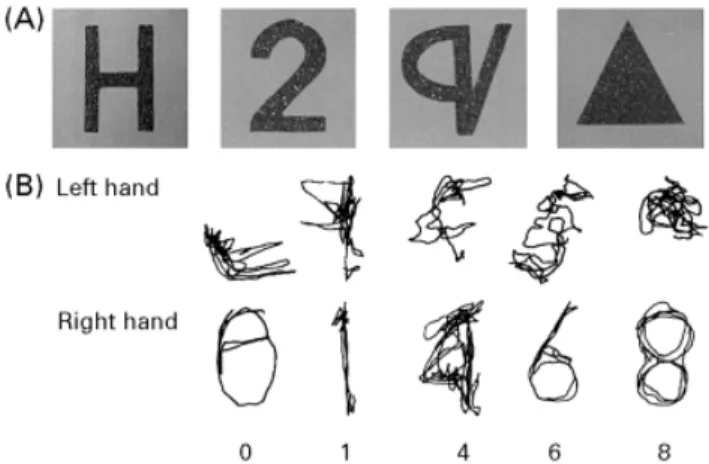
(4) 2290. N. Valenza et al.. Fig. 1 Writing without visual control: examples of digits drawn by the left and right hands.. by a wooden box. The different digits were dictated in random order. The patient was asked to write as quickly as possible, with her left hand first. Examples of her writing in this test are shown in Fig. 1. The patient was able to draw the digits with both her right and her left hand. This residual capacity for writing out of visual control is evidence that fine motor control was preserved in both hands. In summary, detailed investigation of somatosensory functions and of TOR showed that, despite essentially normal sensory functions in both hands, the patient was severely impaired in identifying simple geometric shapes and objects when using her left hand. The deficit could not be attributed to a motor deficit, as motor functions of the left hand had recovered almost completely. As visuospatial functions were normal, the impaired TOR was not attributable to a more general supramodal spatial impairment. The following experiments were motivated by the observation that the patient appeared to use abnormal exploratory strategies with the left hand.. Fig. 2 (A) Examples of 2D sandpaper shapes used in different recognition tasks (Experiments 1, 4, 5 and 6). Items belonged to different categories: letters, digits, elementary geometrical forms and pseudoletters. (B) Exploratory trajectories performed with the left and right hands during tactile recognition of these stimuli.. Table 3 Experiment 1: number of correct answers for the left and right hands in the 2D shape recognition task Hand. Letters. Digits. Forms. Pseudoletters. Left Right. 0/10 9/10. 2/10 9/10. 3/10 10/10. 0/10 9/10. Experiment 1: recognition and exploration of 2D shapes. and 10 pseudoletters (Fig. 2A). Every category was tested separately. Within a category, items were presented randomly and in a different order to the left and the right hand. Each geometrical form was presented twice. When tested with letters and digits, the patient was asked to respond verbally. When tested with forms and pseudoletters, the patient was given a list with all items and asked to point with the other hand to the item corresponding to the felt one. During the exploration of digits, the patient’s hands were videotaped using a video camera placed perpendicularly to the table. A small coloured point at the centre of the nail of her index finger served as a reference point. The patient was asked to explore the 2D shapes using her index finger as often as possible. The tracking movements of her index finger were subsequently traced on sheets of transparent paper using picture-by-picture analysis of still frames.. We assessed the patient’s ability to recognize 2D shapes with her left and right hands. In all conditions, the left hand was tested first to prevent performance bias due to cueing with knowledge obtained by the right, intact hand.. Results Response accuracy. Table 3 summarizes the results. In. Experimental study of tactile exploration The experiments described below were conducted to determine the nature of our patient’s tactile recognition deficit. In order to evaluate distinct stages of tactile shape processing, the patient was asked to recognize shapes by touch in different experimental conditions. In all experiments, the patient’s hands were hidden under a wooden box.. Methods Forms of different significance were cut out of sandpaper and glued on to sheets of paper. The forms had an approximate size of 8⫻4 cm. We used four categories of forms: 10 letters (A, B, H, L, N, O, S, T, V and Z), the 10 digits, five common 2D forms (triangle, rectangle, cross, square and circle). total, the patient gave only five correct answers in 40 trials with the left hand compared with 37 correct responses with the right hand (Fisher’s exact test, P ⫽ 0.003). There was no obvious dissociation between the different categories; the left hand performed badly in all conditions.. Qualitative analysis of explorative movements. Figure 2B shows the qualitative differences between the.
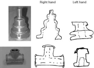
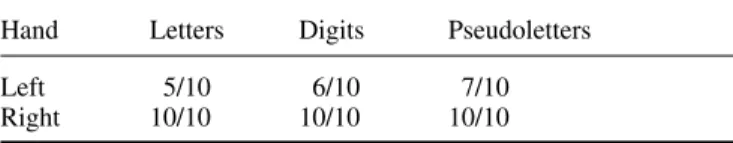
(5) Tactile apraxia without tactile agnosia. 2291. Table 4 Experiment 3: number of correct answers for the left and right hands in the passive recognition task (graphaesthesia) Hand. Letters. Digits. Forms. Pseudoletters. Left Right. 7/10 8/10. 8/10 10/10. 8/10 8/10. 7/10 10/10. Results. Fig. 3 Examples of metallic unfamiliar objects used in Experiment 2 and of the patient’s drawings after palpation of the object with the left and the right hands.. exploratory movements of the left hand and the right hand in the digit-recognition task. The trajectories of the right hand were precise and followed the entire contour of the digit. In contrast, the movements of the left hand seemed chaotic and the trajectories were short and disorganized. Exploration was often limited to one part of a digit and jumped suddenly to another part. Moreover, exploration by the left hand was much slower than that by the right hand.. Experiment 2: tactile representation of unfamiliar objects The previous experiment showed severely impaired tactile recognition of 2D shapes in the left hand, associated with an important alteration of exploratory procedures. According to several studies, agnosic patients are able to draw objects palpated by their agnosic hand correctly but nevertheless fail to recognize them (Endo et al., 1992; Caselli, 1997; Nakamura et al., 1998). This observation suggests that manual exploratory movements made by these patients allow them to elaborate an adequate representation of an object’s shape. We assumed that our patient’s defective manual exploration of objects prevented her from building an adequate representation of the shape of palpated items. Therefore, we asked her to palpate objects and to draw them immediately afterwards.. Methods We used a set of five metallic objects consisting of different types of drainpipe; examples are shown in Fig. 3. Each object could be enclosed entirely by one hand. The objects were placed singly into the patient’s left and right hands. The patient was allowed to palpate the object without constraint, and was then asked to draw it with her right (dominant) hand.. A selection of drawings is presented in Fig. 3. After palpation with the left hand, the patient generally failed to reproduce the global shape of the palpated objects and, although she was able to extract some details, she failed to place them accurately. In contrast, after palpation with her right hand she reproduced adequately the global configuration of the explored object and some of its internal details. Thus, Experiments 1 and 2 show that our patient presents a unimanual disorder of object exploration that might prevent her from extracting an integrated representation of the shapes of objects palpated by her left hand. The following experiments were designed to determine whether our patient had a specific deficit of active tactile exploration or of higher cognitive stages of TOR, in particular whether she failed to access stored tactile shape representations or lexical representations of tactile knowledge.. Experiment 3: identification of passively explored 2D shapes This experiment was designed to determine whether tactile shape recognition with the left hand was preserved when active exploration was not required. If our patient’s TOR impairment is caused by deficient exploration strategies during palpation, elimination of the active exploration process should improve identification of shapes by touch. In order to test this hypothesis, we applied the procedure usually used when testing graphaesthesia.. Methods The same items (letters, digits, shapes, pseudoletters) as those used in Experiment 1 were used. The forms were traced head-down by the experimenter on the patient’s skin using a wooden stick. As in Experiment 1, the different categories of stimuli were tested separately and presented randomly within each category. The patient either named the items (letters, digits) or pointed to the corresponding picture (pseudoletters, geometrical shapes).. Results Table 4 summarizes the results. When the left hand was stimulated, the patient gave 30 correct responses out of 40 trials; with the right hand, she correctly identified 36 stimuli.
(6) 2292. N. Valenza et al.. Table 5 Experiment 4A: number of correct answers for the left and right hands in the 2D shape recognition task under manual guidance of exploration Hand. Letters. Digits. Pseudoletters. Left Right. 9/10 10/10. 10/10 10/10. 9/10 8/10. (Fisher’s exact test, P ⫽ 0.139). Comparison of the left hand’s performance in the present experiment with the results of Experiment 1 showed that passive shape recognition was markedly better than active shape recognition (Fisher’s exact test, P ⬍ 0.001). Thus, passive identification of 2D forms did not differ between the left hand and the right hand. Considering the results of Experiment 2, it may be concluded that the patient presents a dissociation in tactile shape recognition between active and passive touch. The preservation of passive recognition suggests that tactile representations of shapes are intact and appeared to be easily accessible by the left hand. Moreover, it confirms that sensory functions of the left hand are sufficiently preserved to allow correct identification of tactile stimuli, providing further evidence that sensory impairment cannot explain our patient’s TOR deficit. However, graphaesthesia may not be an adequate comparison for active shape exploration. The following experiments were designed to test passive tactile recognition of 2D shapes in conditions closer to those of active tactile recognition.. Experiment 4A: recognition of 2D shapes under manual guidance of exploration Is deficient active TOR secondary to inability to integrate the direction and the succession of exploratory movements? To answer this question, we asked the patient to identify 2D shapes under manually guided exploration, i.e. with her left hand guided by the experimenter.. Methods Thirty sandpaper shapes (letters, pseudoletters and digits) that had been used in Experiment 1 were presented separately, first to the left and then to the right hand. As in the previous experiments, the patient had to name the letters and digits and to point to the corresponding pictorial representation of the palpated pseudoletters. Stimuli were presented in random order to each hand. The experimenter guided the patient’s index finger over the shapes in a trajectory that followed the general pattern of up–down and left–right.. Results Table 5 summarizes the results. The patient correctly identified 28 out of 30 stimuli with both her left hand and her right hand.. It is evident that manually guided exploration significantly reduces the left hand deficit and shows that the left hand is able to access tactile shape representations. This result also implies that our patient is able to derive shape information from hand movements. Importantly, the good performance of the left hand in this experimental condition confirms the existence of a clear-cut dissociation in tactile shape recognition between active and passive touch.. Experiment 4B: recognition of 2D shapes under verbal guidance of exploration This experiment was designed to determine whether our patient was able to execute the exploratory movements necessary to recognize 2D tactile shapes on demand. To answer this question, we asked her to identify 2D shapes under verbally guided exploration.. Methods The stimuli used in this experiment consisted of the five letters and five pseudoletters presented in Experiment 1. The following procedure was applied. A trajectory that followed the pattern of up–down and left–right was defined for each item. After placing the patient’s index finger at the top left corner of the shape, the examiner indicated movement direction to the patient. For example, when presenting the letter L, the examiner placed the patient’s finger at the top of the letter and ordered her to move her finger down. As the patient reached the inferior border of the letter, the examiner told her to stop and to move her finger to the right. Immediately after the end of the movement, she was asked to name letters or to point to the picture representing the pseudoletter. Stimuli were presented in random order within each category, first to the left hand and then to the right hand.. Results The patient correctly recognized all 10 stimuli with both hands. The patient often answered before the end of stimulus exploration. Her answers were rapid even when the exploration was carried out by the left hand. This experiment shows that when free exploration is not required, the left hand demonstrates intact recognition of 2D shapes. The normalization of the left hand’s performance under verbally guided exploration may be explained in two ways. The patient might have translated the verbal orders into a visual representation of the explored form. Thus, her decision did not require tactile information. Alternatively, her tactile exploration might have been improved sufficiently by verbal guidance to allow an informative tactile representation of shape.. Experiment 5: active recognition of 2D shapes with a preliminary hypothesis The three previous experiments showed that when active exploration was not required (Experiment 3) and when.
(7) Tactile apraxia without tactile agnosia Table 6 Experiment 5: number of correct responses for the left and right hands in the 2D shape recognition task with a preliminary hypothesis Hand. Letters. Digits. Pseudoletters. Left Right. 5/10 10/10. 6/10 10/10. 7/10 10/10. exploratory movements were guided by the experimenter (Experiments 4 and 5), 2D shapes were recognized by the left hand as well as by the right hand. Studies about object exploration in healthy subjects show that exploratory movements are guided by internal knowledge about the objects (top-down influences) as well as by the ongoing elaboration of hypotheses during exploration (Klatzky and Lederman, 1993). In this experiment, we investigated whether giving the patient a valid or an invalid cue about stimulus identity would subsequently affect her tactile exploration, i.e. whether top-down information would modulate the patient’s exploratory patterns.. 2293. frontal operculum and the inferoposterior part of the parietal lobe. Lesion reconstruction (Damasio and Damasio, 1989) suggested preservation of the superior right pre- and postcentral gyri [superior portion of Brodmann areas (BA) 2, 3, 4 and 6], areas that correspond to the primary motor and the primary sensory (SI) representation of the left hand (Fig. 4). Since data derived from lesion reconstruction seemed to corroborate our clinical observation of intact sensorimotor functions of the left hand, we used functional imaging to assess whether the preserved cortical area in our patient corresponded to the sensorimotor representation of her left hand. We used functional MRI (fMRI) to measure activation elicited by sensory stimulation of the right and left hands. Our main objective was to examine whether the intact portion of the somatosensory cortex in the right hemisphere was activated by sensory stimulation of the left hand. Secondly, we were interested in the differences between the activated regions in the patient and those in control subjects exposed to the same stimulation.. Material and methods Methods The sandpaper digits, letters and pseudoletters used in Experiment 1 were presented to the patient. Items were presented randomly within each category, first to the left hand and then to the right hand. At the beginning of each trial, before the patient started the exploration of the stimulus, the examiner proposed a possible answer, verbally for the digits and the letters and by pointing to the corresponding pictorial representation for the pseudoletters. The proposed answer was correct (valid cue) in 50% of trials and incorrect (invalid cue) in 50% of trials. The patient was asked to accept or reject verbally our proposition as soon as she had completed stimulus exploration.. The patient and two healthy, age-matched, right-handed women participated in the fMRI study. We used a block paradigm consisting of four cycles of 24 s of activation followed by 24 s of rest. In the activation condition, the palm and the fingers of each hand were stimulated with a small wooden stick which the experimenter moved randomly on the subjects’ skin. The subjects closed their eyes during image acquisition. In the control condition, no sensory stimulation was delivered. Total fMRI acquisition time was 3 min 12 s. The right and left hands were stimulated in separate runs.. MRI Results Table 6 presents the number of correct answers for each hand and every stimulus type. The patient answered correctly in 18 out of 30 trials when exploring stimuli with her left hand. This performance was not different from chance. In contrast, when using her right hand, her answers were correct in 100% of the trials (right–left difference, P ⬍ 0.001). The results of this experiment reveal that the left hand’s performance was not improved by a preliminary hypothesis concerning the identity of the stimulus. Top-down processes did not affect tactile recognition of the left hand, suggesting that the patient was unable to carry out knowledge-driven stimulus exploration.. Neuroimaging study MRI performed 77 days after the stroke showed a large righthemisphere lesion affecting the lateral temporal lobe, the. A 1.5 T Eclipse system (Marconi Medical Systems, Cleveland, OH, USA), equipped with fast gradients (27 mT/m with a slew rate of 72 mT/m/ms) with a standard head coil was used. The fMRI data were obtained from 19 contiguous axial slices of 5 mm parallel to the AC–PC (anterior commissure–posterior commissure) line using single-shot echoplanar imaging [TR (repetition time)/TE (echo time)/ flip angle ⫽ 2 s/40 ms/80°) with in-plane resolution of 1.95 ⫻ 1.95 mm2. Anatomical MRI was obtained in the same session, using gradient echo 2D and high-resolution 3D acquisitions.. fMRI analysis The data were analysed off-line using MedX3.3 software (Sensor Systems, Sterling, Va., USA) on a Unix workstation. All studies were first corrected for head motion (Woods et al., 1998). Activation maps report Z-score values computed from cross-correlation analysis of each voxel time course.
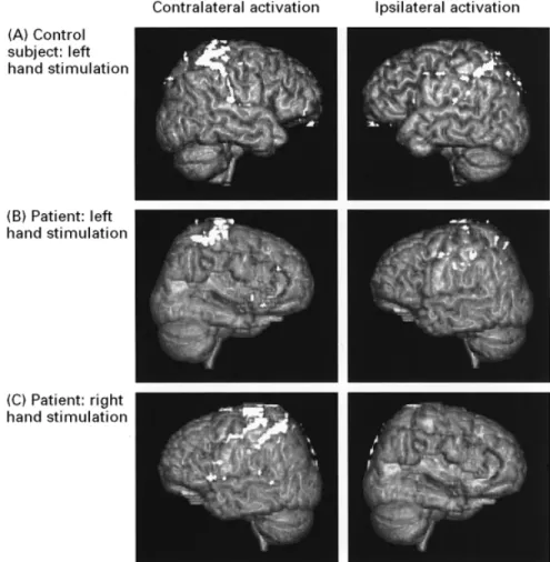
(8) 2294. N. Valenza et al.. Fig. 4 Brain MRI 77 days after stroke, demonstrating a large right-hemisphere lesion affecting the lateral temporal lobe, the frontal operculum and the inferoposterior part of the parietal lobe. The corresponding anatomical templates [using the method of Damasio and Damasio (1989)] suggest that the lesion spares the primary sensory and motor areas of the left hand (superior part of BA 2, 3, 4 and 6).. with a shifted (4 s) boxcar reference function. Voxels with a Z value ⬎1.9, corresponding to a statistical significance of P ⬍ 0.005 after correction for temporal autocorrelation (Worsley and Friston, 1995), were considered active. Functional maps were registered with the 3D MRI and were normalized to Talairach space. Clusters of activated pixels were analysed and compared with the patient’s data.. Results Figure 5 shows the activated areas during stimulation of the left hand in a healthy volunteer (Fig. 5A) and in the patient (Fig. 5B) and during stimulation of the right hand in the patient (Fig. 5C).. Patient Left hand stimulation elicited contralateral activation of the superior part of the postcentral gyrus (BA 1–3) and the superior parietal lobe (BA 5/7) and a small activation of the precentral gyrus (BA 6). In contrast to the control subjects, the patient showed very weak ipsilateral activation and there was no contralateral activation of the inferior postcentral gyrus (BA 43) and of the superior temporal gyrus (BA 22/ 42) (Fig. 5B). Stimulation of the right hand activated the same contralateral areas as in the control subjects, i.e. the postcentral (BA 2/3) and the precentral (BA 6) gyri, the inferior parietal lobule (BA 40) and the superior temporal gyrus (BA 22/41/42). In contrast to the control subjects, the patient showed no ipsilateral activation in this condition (Fig. 5C).. Control subjects Left hand stimulation elicited contralateral activation of the precentral gyrus (BA 4/6), the superior and inferior parts (BA 1–3/BA 43) of the postcentral gyrus, the superior parietal lobe (BA 5/7) and the superior temporal gyrus (BA 22/42). Ipsilateral activation was evoked in the postcentral gyrus (BA 2/3) and in the parietal lobule (BA 7/40) (Fig. 5A). During right hand stimulation, contralateral activation was in the postcentral (BA 2/3) and precentral (BA 6) gyri, the inferior parietal lobule (BA 40) and the superior temporal regions (BA 22/41/42). Ipsilateral activation was located in the parietal lobule (BA 7/40) and more anterior areas (BA 6/44).. Discussion Our patient presented a disorder of object recognition through active touch that was confined to her left contralesional hand after a large right-hemispheric lesion. The deficit could not be explained by impaired elementary sensation, as evidenced by essentially normal basic somaesthetic functions in both hands. The perception of weight, size, substance and texture was also intact in both hands. However, the left hand showed a marked deficit in the tactile identification of 2D and 3D shapes and a severe TOR deficit. The patient was able to write down digits with both hands out of sight, indicating.
(9) Tactile apraxia without tactile agnosia. 2295. Fig. 5 Ipsilateral and contralateral activations elicited by sensory stimulation of the left hand in a control subject (A) and the patient (B), and ipsilateral and contralateral activations elicited by sensory stimulation of the right hand in the patient (C). Areas of activation are shown in white.. that motor functions were sufficiently preserved to allow tactile exploration. It is unlikely that our patient’s TOR deficit was caused by initial cognitive impairments, such as left spatial neglect or difficulties in visual-spatial tasks, as none of these deficits was present at the time of testing. Thus, her left unimanual TOR deficit was attributable neither to an elementary sensorimotor dysfunction nor to a more general supramodal spatial impairment, which has been proposed as an explanation of TOR deficits (Semmes, 1965). The experiments reported here showed a left-hand dissociation in 2D shape recognition between active and passive touch. In recognition tasks based on active touch, i.e. when the patient explored shapes actively, she consistently failed to identify the stimuli with her left hand, whereas her right hand performed perfectly. Exploratory movements of the left hand were disorganized and incomplete. Drawings of objects palpated with the left hand were unrecognizable, suggesting that altered exploration precluded adequate representation of the objects’ shape. In contrast, in situations of passive touch, e.g. in recognition tasks not necessitating active exploration of the stimuli, the performance of the left hand was normal. For example, shapes traced on the skin of. the left hand were recognized accurately. In addition, under manually and verbally guided exploration, the patient was able to recognize correctly the same shapes she failed to recognize with active exploration. The dissociation between active and passive shape recognition suggests that our patient’s difficulty in identifying shapes with her left hand is due to deficient active exploratory movements. The presence of this dissociation excludes an explanation of the TOR deficit in terms of primary sensory impairment, such as twopoint discrimination and proprioception. Our patient’s deficit differs from the known tactile recognition deficits reported in the literature. Reed and colleagues recently reported a patient with TOR impairment restricted to the right hand (Reed et al., 1996). A detailed experimental investigation of her difficulties revealed a specific deficit of shape recognition which could not be attributed to deficient active exploration. The authors interpreted the deficit as apperceptive tactile agnosia resulting from an impairment of modality-specific shape representations. In contrast to our patient, their patient did not improve her performance in passive shape recognition. Another patient could not name, describe or demonstrate the use of objects.
(10) 2296. N. Valenza et al.. placed in his left hand (Platz, 1996). A qualitative analysis of his spontaneous finger movements during TOR revealed a slight alteration in the exploratory movements of his agnosic hand. However, he identified shapes accurately by touch, in contrast to our patient. Platz interpreted the deficit as a pure associative agnosia due to defective integration of distinct tactile features into a coherent tactile percept (Platz, 1996). He stressed that the slight deficit in exploration might be the consequence, rather than the cause, of the recognition deficit. In other cases of associative tactile agnosia (Endo et al., 1992; Nakamura et al., 1998), there was apparently normal exploration of objects and the capacity to analyse tactile features of objects (including their shape) and to draw palpated objects was intact. Despite preserved high-level tactile perception, these patients failed to attribute meaning to correctly explored objects, as evidenced by their inability to classify objects according to their function out of vision. Our patient also differs from patients with tactile anomia who are able to recognize objects tactually, as illustrated by their ability to match them according to their category or function (Beauvois et al., 1978; Endo et al., 1992). However, they are unable to name the recognized object. Recently, two studies reported a deficit of tactile exploration in patients with deficient TOR and severely impaired tactile discrimination consecutive to posterior parietal lesions (Pause et al., 1989; Binkofski et al., 2001). The patients described in these studies showed complete destruction of the temporal characteristics of the finger movements during active touch despite normal frequencies of repetitive finger movements and almost normal force. The authors concluded that the posterior parietal cortex plays a crucial role in the conception and generation of the motor programmes required for the collection of somatosensory information. In contrast to our patient, their patients did not demonstrate intact recognition in either of the conditions tested. However, their passive recognition (e.g. through graphaesthesia) was not tested. A deficit of tactile shape representation may explain their impairment of exploration and TOR. Thus, none of the patients described previously showed the dissociation between active and passive recognition found in our patient. The different clinical expressions of TOR disorders suggest that the tactile identification of objects depends on several processes, the organization of which is at least partly hierarchical. In our patient, the main known causes of TOR deficit can be rejected. As already stressed, an explanation in terms of a primary sensory impairment can be eliminated. Neither is inability to execute the exploratory movements the cause of the alteration of shape exploration, since the patient is perfectly able to perform all movements under verbal guidance. Impaired access to tactile shape representations may not explain her deficient tactile shape identification, since she recognizes forms in experimental situations that do not require the spontaneous generation of exploratory movements. The latter observation also permits us to reject an explanation in terms of a tactile–verbal. disconnection, since our patient can correctly name tactile shapes presented passively to her left hand. As both basic somatosensory functions and tactile shape representations, as well as their links to the lexical memories, are spared in our patient, the deficit in tactile recognition must be located at an intermediate level between basic somatosensory functions and the higher cognitive stages of TOR. The dissociation between active recognition and passive recognition suggests that our patient’s deficit depends on processes linked to the elaboration of exploratory strategies during TOR. Manual exploratory movements during TOR have been studied extensively in healthy subjects (Lederman and Klatzky, 1987). These authors assume that the perception of the precise shape of an object depends on the use of a particular exploratory procedures (EP) (the ‘contourfollowing EP’), during which the hand maintains contact with the contour of the object. According to their observations, the contour-following strategy is the only necessary EP that has to be evoked by subjects to allow correct identification of the exact shape of an object; the remaining EPs are defined as interchangeable (Lederman and Klatzky, 1987). The contour-following EP can thus be conceptualized as a highly specialized EP that occupies a special position among the EP repertoire. Our patient has a selective deficit of shape recognition through active touch but recognizes other tactile properties of objects (substance, length, weight, size) without difficulty. This pattern of deficit is compatible with a selective loss of the contour-following EP and corroborates the suggestion that contour-following is distinct among EPs and may be selectively impaired after brain damage. Why is our patient unable to elaborate a correct tactile exploration of shapes? One possible explanation arises from the notion that exploration is built on continuous interactions between sensory input and stored tactile knowledge about shapes (Klatzky and Lederman, 1993; Platz, 1996). Successful recognition takes place when the perceived information can be matched with stored tactile shape representations. From a purely cognitive point of view, it would be predicted that disconnection between the haptic system and the tactile shape representation system should lead to specific impairment of tactile shape exploration, although the haptic system and tactile shape representation are intact. The hypothesis of such a disconnection is corroborated by the observation that our patient does not profit from a preliminary hypothesis about the identity of shapes. However, this explanation is difficult to match with recent neurophysiological and neuroimaging data about active touch in animals and humans. For instance, Binkofski and colleagues suggest that the parietal lobe is responsible both for ‘the pragmatic and the cognitive aspects of somatosensation’ (Binkofski et al., 2001). A disconnection between two systems would thus be problematic, since both systems would be located within the same brain area. A compatible interpretation of our patient’s dissociation would posit a deficit of the process of sensorimotor transformation (Pause and Freund, 1989), a deficit which would preclude.
(11) Tactile apraxia without tactile agnosia the necessary interactions between sensorimotor input and tactile shape representations that lead to successful TOR. Our patient’s relatively large brain lesion precludes precise analysis of the brain systems involved in active TOR. However, fMRI showed that sensory stimulation of the patient’s left hand activated areas of preserved lateral cortex in the right hemisphere. Sensory stimulation of the left hand elicited bilateral activation of SI, stronger on the contralateral than on the ipsilateral side, and contralateral activation of the superior parietal lobe. These activated regions have been reported to play a role in the discrimination of basic somatosensory attributes (Srinivas and Ogas, 1999), although SI may be the first and most important cortical relay for elementary sensation. The activation in the preserved cortex of the right hemisphere supports the idea that this brain area conveys the preserved basic somatosensory functions of the left hand. The major difference between the patient and the healthy subjects was the absence of activation in regions at the parietotemporal junction (BA 22/42/43), which have been regarded as the human analogues of the secondary sensory area (SII). Its functions and its relationship to SI remain poorly understood and human data about the effects of selective lesion of SII are lacking. Some studies have shown that complete destruction of SII in monkeys evokes impairments in texture and shape discrimination and alters discrimination of size and roughness (Paulesu et al., 1997). Our data suggest that SII may also play an important role in the integration of the tactile information acquired during exploration. Our case demonstrates a pure tactile apraxia without concomitant gnostic deficit, i.e. a sensorimotor integration disorder, originally described clinically by Delay (Delay, 1935) and Klein (Klein, 1931). This disturbance of hand function has been defined as ‘a disorder of the exploration and manipulation of objects, whereas intransitive, expressive and symbolic movements are preserved’ (Pause and Freund, 1989; Binkofski et al., 2001). We propose that tactile apraxia results from impaired integration of sensorimotor feedback with stored tactile shape information—integration that is necessary to generate the exploratory procedures required for successful TOR. Our case study emphasizes that the elaboration of the specific hand movements required for the extraction of distinctive somatosensory information is crucial for successful TOR, and provides new evidence that these exploratory procedures may be selectively impaired after brain damage.. Acknowledgements This work was supported by grants 32-50882.97 and 3238062769.00 from the Swiss National Science Foundation to A.S. References Beauvois M-F, Saillant B, Meininger V, Lhermitte F. Bilateral tactile aphasia: a tacto-verbal dysfunction. Brain 1978; 101: 381–401.. 2297. Benton AL, Hamsher KDS, Varney NR, Spreen O. Contributions to neuropsychological assessment. New York: Oxford University Press; 1983. Binkofski F, Kunesch E, Classen J, Seitz RJ, Freund H-J. Tactile apraxia. Unimodal apractic disorder of tactile object exploration associated with parietal lobe lesions. Brain 2001; 124: 132–44. Caselli RJ. Bilateral impairment of somesthetically mediated object recognition in humans. Mayo Clin Proc 1991a; 66: 357–64. Caselli RJ. Rediscovering tactile agnosia. [Review]. Mayo Clin Proc 1991b; 66: 129–42. Caselli RJ. Ventrolateral and dorsomedial somatosensory association cortex damage produces distinct somesthetic syndromes in humans. Neurology 1993; 43: 762–71. Caselli RJ. Tactile agnosia and disorders of tactile perception. In: Feinberg TE, Farah MJ, editors. Behavorial neurology and neuropsychology. New York: McGraw-Hill; 1997. p. 277–88. Damasio H, Damasio AR. Lesion analysis in neuropsychology. New York: Oxford University Press; 1989. Delay J. Les aste´ re´ ognosies: pathologie du toucher. Clinique, physiologie, topographie. Paris: Masson & Cie; 1935. Endo K, Miyasaka M, Makishita H, Yanagisawa N, Sugishita M. Tactile agnosia and tactile aphasia: symptomatological and anatomical differences. Cortex 1992; 28: 445–69. Gauthier L, Dehaut F, Joanette Y. The Bells Test: a quantitative and qualitative test for visual neglect. Int J Clin Neuropsychol 1989; 11: 49–54. Hooper EH. Hooper Visual Organization Test. Los Angeles: Western Psychological Services; 1985. Klatzky RL, Lederman SJ. Toward a computational model of constraint-driven exploration and haptic object identification. Perception 1993; 22: 597–621. Klein R. Zur Symptomatologie des Parietallapens. Z Ges Neurol Psychiat 1931; 135: 589–608. Lederman SJ, Klatzky RL. Hand movements: a window into haptic object recognition. Cognit Psychol 1987; 19: 342–68. Nakamura J, Endo K, Sumida T, Hasegawa T. Bilateral tactile agnosia: a case report. Cortex 1998; 34: 375–88. Osterrieth PA. Le test de copie d’une figure complexe: contribution a` l’e´ tude de la perception et de la me´ moire. Arch Psychol 1944; 30: 286–356. Paulesu E, Frackowiak RSJ, Bottini G. Maps of somatosensory systems. In: Frackowiak RSJ, Friston KJ, Frith CD, Dolan RJ, Mazziotta JC, editors. Human brain function. San Diego: Academic Press; 1997. p. 183–242. Pause M, Freund HJ. Role of the parietal cortex for sensorimotor transformation. Brain Behav Evol 1989; 33: 136–40. Pause M, Kunesch E, Binkofski F, Freund H-J. Sensorimotor disturbances in patients with lesions of the parietal cortex. Brain 1989; 112: 1599–625..
(12) 2298. N. Valenza et al.. Platz T. Tactile agnosia. Casuistic evidence and theoretical remarks on modality-specific meaning representations and sensorimotor integration. Brain 1996; 119: 1565–74. Raven JC, Court JH, Raven J, editors. Coloured progressive matrices. Paris: Editions & Applications Psychologiques; 1998. Reed CL, Caselli RJ. The nature of tactile agnosia: a case study. Neuropsychologia 1994; 32: 527–39. Reed CL, Caselli RJ, Farah MJ. Tactile agnosia. Underlying impairment and implications for normal tactile object recognition. Brain 1996; 119: 875–88. Regard M, Strauss E, Knapp P. Children’s production on verbal and non-verbal fluency tasks. Percept Mot Skills 1982; 55: 839–44. Rey A. Test de copie et de reproduction de me´ moire de figures ge´ ometriques complexes. Paris: Centre de Psychologie; 1958a. Rey A. L’examen clinique en psychologie. Paris: Presse Universitaire de France; 1958b.. Semmes J. A non-tactual factor in astereognosis. Neuropsychologia 1965; 3: 295–314. Srinivas K, Ogas J. Disorders of somesthetic recognition: a theoretical review. Neurocase 1999; 5: 83–93. Thurstone LL, Thurstone TG. Chicago Test of Primary Mental Abilities. Chicago: Research Associates; 1963. Wechsler D. Wechsler Memory Scale—Revised manual. San Antonio: Psychological Corporation; 1987. Woods RP, Grafton ST, Holmes CJ, Cherry SR, Mazziotta JC. Automated image registration: I. General methods and intrasubject, intramodality validation. J Comput Assist Tomogr 1998; 22: 139–52. Worsley KJ, Friston KJ. Analysis of fMRI time-series revisited— again. Neuroimage 1995; 2: 173–81.. Received March 5, 2001. Revised June 10, 2001. Accepted June 25, 2001.
(13)
Figure
+5


Documents relatifs