. . . .
. . . .
The effects of continuous positive airway pressure
therapy withdrawal on cardiac repolarization: data
from a randomized controlled trial
†
Valentina A. Rossi
1, Anne-Christin Stoewhas
1, Giovanni Camen
1, Jan Steffel
2,
Konrad E. Bloch
1,3, John R. Stradling
4, and Malcolm Kohler
1,3*
1
Sleep Disorders Centre and Pulmonary Division, University Hospital of Zurich, Raemistrasse 100, 8091 Zurich, Switzerland;2
Cardiac Arrhythmia Unit, Cardiovascular Centre, University Hospital of Zurich, Zurich, Switzerland;3
Zurich Centre for Integrative Human Physiology, University of Zurich, Zurich, Switzerland; and4
Oxford Centre for Respiratory Medicine, Churchill Hospital, Oxford, UK
Received 27 October 2011; revised 13 February 2012; accepted 1 March 2012; online publish-ahead-of-print 27 March 2012
This paper was guest edited by Prof. Gerhard Hindricks, University Leipzig, Heart Center, Department of Electrophysiology, Strumpellstrasse 39, Leipzig D-04289, Germany.
Aims The preliminary evidence supports an association between obstructive sleep apnoea (OSA), disturbed cardiac
repo-larization, and consequent cardiac dysrhythmias. The aim of the current trial was to assess the effects of continuous positive airway pressure (CPAP) therapy withdrawal on the measures of cardiac repolarization in patients with OSA.
Methods and results
Forty-one OSA patients established on CPAP treatment were randomized to either CPAP withdrawal (subtherapeu-tic CPAP) or continue therapeu(subtherapeu-tic CPAP for 2 weeks. Polysomnography was performed, and indices of cardiac
repo-larization (QTc, TpTec intervals) and dispersion of repolarization (TpTe/QT ratio) were derived from 12-lead
electrocardiography (ECG) at baseline and 2 weeks. Continuous positive airway pressure withdrawal led to a recur-rence of OSA. Compared with therapeutic CPAP, subtherapeutic CPAP for 2 weeks was associated with a significant
increase in the length of the QTcand TpTecintervals (mean difference between groups 21.4 ms, 95% CI 11.3 – 1.6 ms,
P , 0.001 and 14.4 ms, 95% CI 7.2 – 21.5 ms, P , 0.001, respectively) and in the TpTe/QT ratio (mean difference between groups 0.02, 95% CI 0.00 – 0.03, P ¼ 0.020). There was a statistically significant correlation between
the change in apnoea/hypopnoea index (AHI) from baseline, and both the change in the QTc interval and the
TpTec interval (r ¼ 0.60, 95% CI 0.36 – 0.77, P , 0.001 and r ¼ 0.45, 95% CI 0.17 – 0.67, P ¼ 0.003, n ¼ 41,
respectively).
Conclusion Continuous positive airway pressure withdrawal is associated with the prolongation of the QTcand TpTecintervals
and TpTe/QT ratio, which may provide a possible mechanistic link between OSA, cardiac dysrhythmias, and thus sudden cardiac death.
-Keywords Obstructive sleep apnoea † Continuous positive airway pressure † Cardiac dysrhythmias † Cardiac repolarization
† Sudden cardiac death
Introduction
Obstructive sleep apnoea (OSA) is a highly prevalent sleep-related
breathing disorder affecting up to 30% of middle-aged adults.1,2
Obstructive sleep apnoea is characterized by the repetitive inter-ruption of ventilation during sleep caused by the collapse of the
upper airway, resulting in apnoeas and hypopnoeas associated with oxygen desaturations and arousals from sleep. As a conse-quence of the fragmented sleep, patients with OSA may suffer from increased daytime sleepiness leading to impaired quality of
life and the increased risk of traffic accidents.3,4
†The work was conducted at Sleep Disorders Centre and Pulmonary Division, University Hospital of Zurich, Switzerland.
*Corresponding author. Tel:+41 44 255 97 51, Fax: +41 44 255 44 51, Email:[email protected],[email protected]
Symptomatic OSA has been implicated as an important factor in
the pathogenesis of hypertension and cardiovascular disease.4–6
Furthermore, the findings of observational studies suggest that OSA may be causally related with cardiac dysrhythmias, such as
premature ventricular contractions, atrial fibrillation (AF),
ventricular arrhythmias, atrioventricular block, and sinus arrest.7–
12
The observation that patients with OSA have an increased risk of sudden cardiac death (SCD) at night suggests an association
between OSA and SCD.12The mechanistic link between OSA and
SCD, however, has not yet been thoroughly established. An increase in the dispersion of cardiac repolarization has been sug-gested as a potential mechanism of ventricular arrhythmias and
SCD.13–15The various markers derived from the surface
electro-cardiography (ECG) as well as from the electrophysiological studies provide information about the transmural dispersion of repolarization and the electrical inhomogeneity in the ventricles
during repolarization.16–18
There is preliminary evidence from uncontrolled studies sug-gesting that cardiac repolarization is prolonged and the transmural dispersion of repolarization is increased, in patients with OSA. These ECG abnormalities seem to reverse with continuous
posi-tive airway pressure (CPAP) treatment.19 However, there is no
data from randomized controlled interventional trials proving this association, and thus a controversy remains as to whether OSA is an independent aetiologic risk factor for disturbed cardiac
repolarization.11
To address this uncertainty, we have analysed the ECG data from a randomized controlled trial exploring the effects of a 2-week CPAP withdrawal vs. continuing CPAP in patients with
previously treated OSA,20 in order to investigate the effects of
CPAP withdrawal and a return of OSA on ECG-derived measures of cardiac repolarization.
Methods
Patients, trial design, and sample calculation
Patients previously diagnosed with OSA, and treated with CPAP, who were registered in a database of the Sleep Disorders Centre and Pulmonary Division of the University Hospital of Zurich, Switzerland, were eligible for the trial if they were aged between 20 and 75 years, had an oxygen desaturation index (ODI, ≥4% dips) of .10/h during their initial sleep study, an ODI of .10/h during an ambulatory nocturnal pulse-oximetry performed on the last night of a four-night period without CPAP prior to the main study, and if they had been treated with CPAP for .12 months with a average compliance of≥4 h per night.Patients with previous ventilatory failure, Cheyne – Stokes breathing, unstable and untreated coronary or peripheral artery disease, severe and inadequately controlled arterial hypertension, a history of any sleep-related accident, or with a heavy goods vehicle driver’s licence, were excluded from the study. The trial was performed according to the Declaration of Helsinki, was approved by the University Hospital of Zurich research ethics committee (EK-1600) and registered (ISRCTN 93153804). Written informed consent was obtained from all participants. The primary outcomes of this trial [change in the ODI and apnoea/hypopnoea index (AHI)] are reported elsewhere in more detail.20
After confirming the persistence of OSA by home overnight pulse-oximetry at the end of a four-night period without CPAP, eligible patients returned to therapy with CPAP for≥7 days. After baseline assessments, the patients were randomized to either continue with CPAP therapy or switch to subtherapeutic CPAP for 2 weeks by a series of presealed and numbered envelopes. The follow-up assess-ments of ECG were performed at 1 and 2 weeks. Patients remained blinded as to whether they were receiving therapeutic or subtherapeu-tic CPAP, as did the investigators. The member of the research team who randomized the patients did not take part in the outcome assessments.
A sample size estimation was performed based on the assumption that a clinically relevant difference in the ODI (≥4% drop of oxygen saturation/h) between active treatment and placebo is 10/h (SD 10) and a clinically relevant difference in the QTc is 20 ms (SD 15).21
Based on these assumptions, the power calculation indicated that 41 patients were required in total in order to not miss clinically relevant differences in the ODI (with a power of 90%) and in the QTc(with a
power of 99%).
Sleep studies and continuous positive airway
pressure
At baseline and 2 weeks, attended polysomnographic sleep studies were performed and analysed according to the standard methods. Apnoeas were defined as a reduction in the amplitude of chest wall motion by .90% from baseline over the previous 2 min for .10 s, hypopnoeas were defined as a reduction in the amplitude of chest wall motion by .50% from baseline over the previous 2 min for .10 s, associated with a ≥4% drop in oxygen saturation.22 The severity of sleep-disordered breathing was quantified as the number of apnoeas/hypopnoeas (AHI) and oxygen desaturations ≥4% per hour of study (ODI). In patients randomized to subtherapeutic CPAP, the subtherapeutic pressure was achieved by setting the CPAP machine to the lowest pressure, inserting a flow-restricting connector at the machine outlet, and inserting six extra holes in the collar of the main tubing at the end of the mask to allow air escape, thus preventing rebreathing of carbon dioxide, as previously described.23Sleepiness was assessed using the Epworth sleepiness score (ESS).24,25
Electrocardiography
The participants were asked to abstain from alcohol, tobacco, or caffeine on the day the measurements were done. The room tempera-ture and lighting were set at the same level for all measurements. The participants rested for 5 min in the supine position before measure-ments were done. For all electrocardiographic recordings a commer-cially available 12-lead ECG (AT 104 PC, Schiller-Reomed AG, Switzerland) was used and set at 25-mm/s paper speed and 10-mm/ mV amplitude. The measurements of the ECG intervals were per-formed in duplicate with a dedicated ECG analysis software (DatInfw Measure 2.1d, DatInf GmbH, Tu¨bingen, Germany) by two investigators who were blinded to the patient’s group allocation. The mean of the measurements by the two investigators was used for stat-istical analysis. Lead V5 was used and when unsuitable, lead V4 or V6. The indices of cardiac repolarization were determined as follows: the QT interval was defined as the time from the earliest onset of the QRS complex to the point at which the downward slope of the T-wave returned to baseline and the Tpeak-to-Tend interval (TpTe) as the time from the peak of the T-wave to the end of the T-wave.26The end of the T-wave was defined as the cutting point of the tangent to the downward slope of the T-wave and the isoelectric line.18Both the QT and TpTe intervals were corrected for the heart rate as
described previously.27As a measure of dispersion of repolarization, TpTe/QT ratio was calculated.14
Data analysis
All values are presented as mean (SD) unless otherwise stated. Statis-tical analysis was performed with Statistica V6.0 (StatSoft, Tulsa, OK, USA). Differences in baseline characteristics between the groups were assessed by independent t-tests and x2 tests as appropriate. Comparisons of changes between groups were assessed by inde-pendent t-tests and Mann – Whitney U tests as appropriate. The analyses were performed on an intention-to-treat basis with no change assumed for the missing follow-up data. Correlations between changes in the indices of OSA severity and the ECG measures of cardiac repolarization were assessed by Pearson correlation
coefficients. A two-sided P value of ,0.05 was considered to be stat-istically significant.
Results
Trial profile and patient characteristics
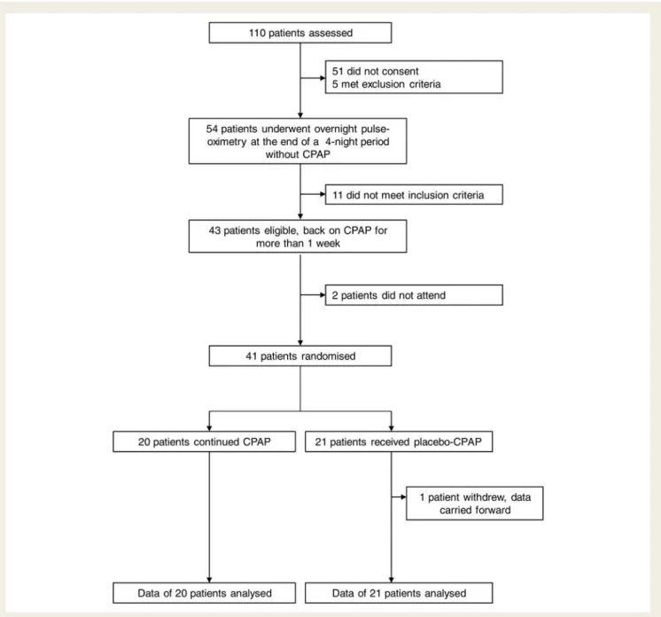
Figure 1 shows the trial profile. The two groups were similar
regarding all patients’ characteristics at baseline (Table 1). Data
on AHI downloaded from the patients’ CPAP machines, as well as ESS values within the normal range, proved successful treatment
of patients with CPAP prior to the study (Table1). One patient in
the subtherapeutic CPAP group withdrew from the study 4 days after randomization because of intolerable daytime symptoms.
Sleep-disordered breathing and sleepiness
Compared with therapeutic CPAP, AHI increased significantly with the subtherapeutic CPAP at 2 weeks (mean difference in the AHI change 30.2, 95% CI 18.8 – 41.7) and ODI (mean difference in the ODI change 24.0, 95% CI 13.5 – 34.4, P , 0.001 for both compar-isons). ESS increased significantly at 2 weeks in the subtherapeutic group compared with the therapeutic group (mean difference in
ESS change 3.1, 95% CI 1.4 – 4.7, P , 0.001, Table2).
Effect of continuous positive airway
pressure withdrawal on cardiac
repolarization indices
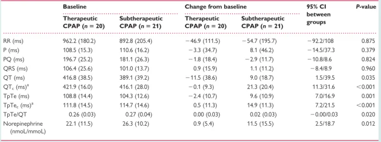
ECG interval times at baseline, and changes from baseline, are
shown in Table 3. There were no relevant differences between
the two groups at baseline.
Compared with therapeutic CPAP, subtherapeutic CPAP for 2 weeks was associated with a significant increase in the length of
the QTc and TpTec intervals (mean difference 21.4 ms, 95%
CI 11.3 – 31.6 ms, P , 0.001 and 14.4 ms, 95% CI 7.2 – 21.5 ms,
P , 0.001, respectively) (Figures 2 and 3) and in the TpTe/QT
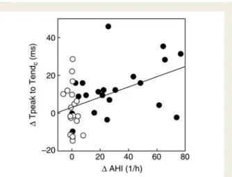
ratio (mean difference 0.02, 95% CI 20.00 – 0.03, P ¼ 0.020). A statistically significant correlation was observed between the
change in the AHI, and both the changes in the QTc and the
TpTec intervals (r ¼ 0.60, 95% CI 0.36 – 0.77, P , 0.001 and r ¼
0.45, 95% CI 0.17 – 0.67, P ¼ 0.003, respectively, n ¼ 41, Figures4
and5).
A statistically significant correlation was also found between the
change in the ODI, and both the changes in the QTcand the TpTec
intervals (r ¼ 0.44, 95% CI 0.16 – 0.66, P ¼ 0.004, and r ¼ 0.51, 95% CI 0.24 – 0.71, P , 0.001, respectively, n ¼ 41).
Discussion
This is the first randomized controlled trial providing data on the effects of OSA on ECG measures of cardiac repolarization. We found that CPAP withdrawal was associated with a considerable prolongation and an increase in the dispersion of cardiac repolar-ization. These findings provide further evidence suggesting a possible link between OSA, cardiac dysrhythmias, and SCD.
The QT interval represents the electrocardiographic correlate of ventricular de- and repolarization, including the vulnerable period for reentry tachycardia. As such, numerous studies have
. . . .
. . . . Table 2 Effects of continuous positive airway pressure withdrawal on sleep-disordered breathing and sleepiness
Baseline Change from baseline 95% CI between groups P-value Therapeutic CPAP (n 5 20) Subtherapeutic CPAP (n 5 21) Therapeutic CPAP (n 5 20) Subtherapeutic CPAP (n 5 21) AHI (1/h) 1.7 (1.8) 2.2 (2.5) 0.4 (2.8) 30.6 (25.2) 18.8/41.7 ,0.001 ODI (1/h) 0.5 (0.8) 0.9 (2.0) 20.2 (1.0) 23.8 (23.1) 13.5/34.4 ,0.001 ESS 7.4 (3.1) 6.6 (2.7) 20.9 (2.2) 2.2 (2.9) 1.4/4.7 ,0.001
Values are mean (SD). 95% CI ¼ 95% confidence interval of the difference between groups in change from baseline. AHI, apnoea/hypopnoea index; ODI, oxygen desaturation index; ESS, Epworth sleepiness score.
. . . . Table 1 Baseline characteristics
Therapeutic CPAP (n 5 20) Subtherapeutic CPAP (n 5 21) Age (years) 63.6 (5.1) 61.8 (7.5) Females (n) 1/20 0/21 Body mass index (kg/m2) 32.9 (6.5) 33.1 (4.4) Waist/hip circumference ratio 1.0 (0.1) 1.0 (0.0) Neck circumference (cm) 46.4 (3.7) 46.1 (4.2) Current smokers (n) 1/20 4/21 Ex-smokers (n) 5/20 8/21 Hypertension (n) 14/20 17/21 Diabetes (n) 4/20 5/21 CAD (n) 2/20 1/21
Sokolow– Lyon index (mV) 21.1 (5.3) 21.1 (5.7) Cardiac dysrhythmiaa(n) 0/20 1/21 Antihypertensive medication (n) 13/20 16/21 Beta-blockers (n) 7/20 6/21 Diuretics (n) 5/20 8/21 Cholesterol-lowering medication (n) 7/20 7/21 Glucose-lowering medication (n) 3/20 2/21 Potassium (mmol/L) 3.9 (0.4) 3.9 (0.3) AHI original sleep study 36.0 (17.3) 45.3 (22.3) ODI original sleep study 26.6 (13.5) 37.3 (22.7) ODI 4-day withdrawal 25.4 (8.6) 28.9 (16.2) AHI on CPAPb 5.1 (2.7) 4.3 (2.3) CPAP compliance (min) 373.1 (67.9) 362.8 (72.3) CPAP use relative to time in
bed (%)c
80.8 (14.5) 79.2 (20.0)
Duration of CPAP treatment (years)
5.6 (3.4) 7.4 (3.2)
ESS before therapy 7.4 (3.1) 6.6 (2.7) Time from screening to
randomization (days)
75.9 (29.0) 68.3 (29.9)
Values are mean (SD) where applicable.
CAD, coronary heart disease; AHI, apnoea/hypopnoea index; ODI, oxygen desaturation index; AHI on CPAP, AHI downloaded from CPAP machine averaged from previous 6 months; ESS, Epworth sleepiness score.
a
Atrial fibrillation.
b
Derived from CPAP machine.
c
identified the inherited as well as acquired QT prolongation as a risk factor for the occurrence of malignant cardiac dysrhythmias
and SCD.28,29
The Tpeak-to-Tend (TpTe) interval is a measure of cardiac transmural dispersion of repolarization, which is explained by a gradient of action potential duration from endo- (longest) to
epicardial cells (shortest).30,31A prolongation of the TpTe interval
leads to increased vulnerability for the occurrence of early after-depolarizations and has been associated with ventricular
tachy-cardia and an increased risk for SCD.32–34 Specifically, recent
findings from Panikkath et al.33suggest that patients with an
uncor-rected TpTe interval .100 ms in the resting ECG are at increased risk of SCD.
In the current study, QTcand TpTecintervals at baseline were
already considerably longer in the CPAP-treated study population than in a group of healthy young adults in one of our previous
studies35 (416.1 vs. 403.8 and 114.7 vs. 87.3 ms, respectively)
possibly due to a high rate of comorbidities seen in our OSA popu-lation. However, we found that CPAP withdrawal for 2 weeks was
. . . .
. . . . Table 3 Effect of continuous positive airway pressure withdrawal on the electrocardiographic intervals and urinary norepinephrine
Baseline Change from baseline 95% CI between groups P-value Therapeutic CPAP (n 5 20) Subtherapeutic CPAP (n 5 21) Therapeutic CPAP (n 5 20) Subtherapeutic CPAP (n 5 21) RR (ms) 962.2 (180.2) 892.8 (205.4) 246.9 (111.5) 254.7 (195.7) 292.2/108 0.875 P (ms) 108.5 (15.3) 110.6 (16.2) 23.3 (34.7) 8.1 (46.2) 214.5/37.3 0.379 PQ (ms) 196.7 (25.2) 181.1 (26.3) 21.8 (18.4) 22.9 (11.7) 210.8/8.6 0.824 QRS (ms) 106.4 (25.6) 101.0 (13.7) 0.9 (15.9) 1.1 (11.2) 28.4/8.9 0.960 QT (ms) 416.8 (38.5) 389.1 (39.2) 211.5 (38.6) 9.0 (18.7) 1.5/39.5 0.035 QTc(ms)a 421.9 (16.0) 416.1 (28.0) 20.1 (9.3) 21.3 (20.4) 11.3/31.6 ,0.001 TpTe (ms) 108.8 (14.4) 104.3 (12.6) 22.4 (10.7) 9.6 (10.9) 7.0/16.9 0.001 TpTec(ms)a 111.8 (14.5) 114.7 (14.6) 0.5 (11.3) 14.9 (11.3) 7.2/21.5 ,0.001 TpTe/QT 0.26 (0.03) 0.27 (0.04) 0.00 (0.03) 0.02 (0.03) 20.00/0.03 0.020 Norepinephrine (nmoL/mmoL) 22.1 (11.5) 26.3 (10.2) 0.9 (5.4) 11.5 (15.5) 2.5/18.7 0.012
Values are mean (SD).
95% CI ¼ 95% confidence interval of the difference between groups in change from baseline. N ¼ 17 and 18 for urinary norepinephrine (nmoL/mmoL of creatinine) in the therapeutic and subtherapeutic CPAP group, respectively.
a
Heart-rate corrected.
Figure 2 Data are presented as mean and standard error. Black bars represent the subtherapeutic continuous positive airway pressure group and white bars represent the therapeutic continu-ous positive airway pressure group. There was a significant increase in the length of the QTcat 2 weeks in the subtherapeutic
continuous positive airway pressure group compared with the therapeutic continuous positive airway pressure group. *P , 0.001 for comparisons of changes from baseline between groups.
Figure 3 Data are presented as mean and standard error. Black bars represent the subtherapeutic continuous positive airway pressure group and white bars represent the therapeutic continu-ous positive airway pressure group. There was a significant in-crease in the length of the TpTec at 2 weeks in the
subtherapeutic continuous positive airway pressure group com-pared with the therapeutic continuous positive airway pressure group. *P , 0.001 for comparisons of changes from baseline between groups.
associated with further, statistically significant prolongation of the
QTcand TpTecintervals. The withdrawal of CPAP was also
asso-ciated with an increase in the uncorrected mean TpTe interval from 104 to 114 ms; as a result, 71% of patients in the subthera-peutic CPAP group were shifted to TpTe values .100 ms and
55% were shifted to QTc values .430 ms. Since these cut-offs
were previously associated with an elevated risk for SCD,33 our
data imply that many OSA patients without CPAP treatment may fall into this category. Moreover, our data demonstrate an increase in the effect of CPAP withdrawal on cardiac repolarization
parameters across 2 weeks (Figures2and3), indicating that further
withdrawal may result in an even more pronounced increase in these values.
The increase in the length of the QTcand TpTecintervals was
positively correlated with the change in the severity of
sleep-disordered breathing (Figures4and5) suggesting that the risk for
SCD increases with the severity of OSA. There is also preliminary data from uncontrolled studies in patients with OSA showing that QT intervals are longer during the night-time compared with the
daytime,21and longer at the onset of an apnoea episode compared
with the period after an apnoea.36In an observational study, OSA
has been shown to be associated with the increased QT interval variability during sleep, possibly reflecting alterations in the
sympa-thetic outflow to the ventricular myocardium.37
In addition to the prolonged QTc and TpTec intervals, an
increased TpTe/QT ratio, a measure of disproportional prolonga-tion of global dispersion relative to the QT interval, may have an
important role in arrhythmogenesis.16This ratio has the advantage
of more reliably eliminating the confounding effects of the heart rate variability in the ECG and the inter-individual variation in
the length of the QT interval.14 We were able to demonstrate
an increase in the TpTe/QT ratio after CPAP withdrawal, further substantiating a potential risk for arrhythmias in this situation.
These findings are in line with the preliminary evidence from observational studies, suggesting that the severity of OSA is also associated with the worsening of QT dynamicity parameters (QT/RR slope), an indicator of abnormal rate adaption of
ventricular repolarization.19,37This may be due to a relative lack
of parasympathetic tone in patients with OSA and, thus, be a predictor of myocardial vulnerability and tendency to cardiac
dysrhythmias.19,38
A prolongation of QRS duration, i.e. a prolongation of ventricular depolarization, has been suggested to be associated
with an increased propensity for SCD.39In our study, we found
a small increase in QRS duration in the CPAP withdrawal group, which was, however, not statistically significant (1.1 ms, P ¼ 0.96). The risk of SCD associated with QRS prolongation is mainly interpreted as a result of overt coronary artery disease
(ischaemia or scar).40,41 However, in our study population, only
three patients (two in the CPAP group and one in the CPAP with-drawal group) had (stable) coronary artery disease. Thus the chances of finding a significant prolongation of the QRS complex during the CPAP withdrawal were likely to be much smaller than it would have been in patients with overt coronary heart disease. This is in contrast to the more subtle markers of
repolar-ization such as TpTec. It hence remains unclear at present whether
OSA directly affects ventricular depolarization in the longer term, and further studies are required to conclusively answer this question.
There may be several mechanisms explaining the disturbance of repolarization and the high incidence of atrial and ventricular dys-rhythmias and SCD in OSA patients. Obstructive apnoea and hypopnoea are associated with repeated inspiratory efforts against the collapsed upper airways associated with large swings in the intrathoracic pressure, which may be as high as 60 – 80 mmHg. During each obstructive apnoea and hypopnoea, the
Figure 5 Correlation between the change in the TpTecinterval
(change from baseline to 2 weeks) and change in the apnoea/ hypopnoea index (r ¼ 0.45, 95% CI 0.17 – 0.67, P ¼ 0.003, n ¼ 41). Black circles represent patients in the subtherapeutic continuous positive airway pressure group and white circles rep-resent patients in the therapeutic continuous positive airway pressure group.
Figure 4 Correlation between the change in the QTcinterval
(change from baseline to 2 weeks) and change in the apnoea/ hypopnoea index (r ¼ 0.60, 95% CI 0.36 – .77, P , 0.001, n ¼ 41). Black circles represent patients in the subtherapeutic continuous positive airway pressure group and white circles rep-resent patients in the therapeutic continuous positive airway pressure group.
negative intrathoracic pressure exerts a direct distending force on
the heart and aorta.42Cardiac wall stress is additionally augmented
by considerable acute changes in the atrial and ventricular volume
during obstructive apnoea and hypopnoea.43–45 In vitro
experi-ments on myocardial tissue have shown that acute atrial stretch (similar to the atrial stretch occurring during apnoea/hypopnoea) induces both early and delayed after-depolarizations which may
trigger premature myocardial contractions.44,45 In an animal
model, Linz et al.46reported shortening of the right atrial
refrac-tory period and the increased susceptibility to premature beats and AF induced by negative thoracic pressure during obstructive respiratory events. This mechanism, repeated during each apnoeic phase, may lead in the long-term to the electrical and
mechanical remodelling of both the atria and left ventricle,47,48
thereby possibly explaining the high prevalence of AF in patients
with OSA.49,50
A further possible mechanism for the alteration of cardiac repo-larization may be autonomic nervous system imbalance which has
been shown to be present in OSA patients.6 Baumert et al.51
showed that experimentally induced increased cardiac sympathetic activity is positively correlated with QT variability in hypertensive patients. We previously reported an increased sympathetic activity as assessed by the urinary noradrenaline (mean increase 43.7%) and heart rate (mean increase 9.7%) after 2 weeks of CPAP
with-drawal in the current study population.20In addition, there was a
correlation between the change in the QTc and change in the
heart rate (r ¼ 0.45, P ¼ 0.003, data not shown) as well as
between the change in the QTc and change in the urinary
noradrenaline levels (r ¼ 0.37, P ¼ 0.020, data not shown). These findings suggest that increased sympathetic nervous system activity may be one of the underlying mechanisms between OSA and the increased dispersion of cardiac repolarization, but further studies are warranted.
In summary, the increased susceptibility to dysrhythmias may therefore be explained by structural remodelling (resulting in the
formation of an arrhythmogenic substrate),48electrical remodelling
(manifesting as disturbed repolarization), increased sympathetic activation, and triggers (including acute episodes of apnoea and hypopnoea), which may eventually lead to the onset of ventricular
dysrhythmias (Coumel’s triangle).47
The current study has some inherent limitations. We per-formed standard resting 12-lead ECG and not continuous 24-h ECG, thus it is not possible to determine from the current study, the effects of CPAP withdrawal on ECG for longer periods, or during sleep. Therefore, further data from rando-mized controlled studies are needed to evaluate the effect of CPAP on measures of repolarization during sleep and longer daytime periods. Future interventional studies will also need to address the question as to which mechanisms underpin the asso-ciation between OSA and both the prolongation and the increased dispersion of cardiac repolarization. The question as to whether OSA is associated with chronic and irreversible alterations of the ECG will need to be addressed in future trials with conventional design, however, due to ethical issues related to a long-term control group, it will be very difficult to perform a randomized controlled trial to answer this question. In addition, the findings of a study that selects patients with
high CPAP compliance cannot be generalized without caution to the untreated OSA population usually seen in a sleep clinic.
In conclusion, we have found that CPAP withdrawal for 2 weeks was associated with prolongation as well as increased dispersion of cardiac repolarization. These findings may provide a possible mechanistic link between OSA, cardiac arrhythmias, and SCD.
Funding
This work was supported by the Swiss National Science Foundation (32003B_124915), the Swiss Society of Pneumology, and the Univer-sity of Zurich (Matching Funds).
Conflict of interest: None declared.
References
1. Stradling JR, Crosby JH. Predictors and prevalence of obstructive sleep apnoea and snoring in 1001 middle aged men. Thorax 1991;46:85 – 90.
2. Young T, Palta M, Dempsey J, Skatrud J, Weber S, Badr S. The occurrence of sleep-disordered breathing among middle-aged adults. N Engl J Med 1993;328: 1230 – 1235.
3. George CF. Reduction in motor vehicle collisions following treatment of sleep apnoea with nasal CPAP. Thorax 2001;56:508 – 512.
4. Kohler M, Stradling JR. Mechanisms of vascular damage in obstructive sleep apnea. Nat Rev Cardiol 2010;7:677 – 685.
5. Marin JM, Carrizo SJ, Vicente E, Agusti AG. Long-term cardiovascular outcomes in men with obstructive sleep apnoea – hypopnoea with or without treatment with continuous positive airway pressure: an observational study. Lancet 2005;365: 1046 – 1053.
6. Kohler M, Pepperell JC, Casadei B, Craig S, Crosthwaite N, Stradling JR, Davies RJ. CPAP and measures of cardiovascular risk in males with OSAS. Eur Respir J 2008; 32:1488 – 1496.
7. Guilleminault C, Connolly SJ, Winkle RA. Cardiac arrhythmia and conduction dis-turbances during sleep in 400 patients with sleep apnea syndrome. Am J Cardiol 1983;52:490 – 494.
8. Namtvedt SK, Randby A, Einvik G, Hrubos-Strøm H, Somers VK, Røsjø H, Omland T. Cardiac arrhythmias in obstructive sleep apnea (from the Akershus Sleep Apnea Project). Am J Cardiol 2011;108:1141 – 1146.
9. Hoffstein V, Mateika S. Cardiac arrhythmias, snoring, and sleep apnea. 1994. Chest 2009;136:e30.
10. Gami AS, Pressman G, Caples SM, Kanagala R, Gard JJ, Davison DE, Malouf JF, Ammash NM, Friedman PA, Somers VK. Association of atrial fibrillation and ob-structive sleep apnea. Circulation 2004;110:364 – 367.
11. Somers VK, White DP, Amin R, Abraham WT, Costa F, Culebras A, Daniels S, Floras JS, Hunt CE, Olson LJ, Pickering TG, Russell R, Woo M, Young T. Sleep apnea and cardiovascular disease. Circulation 2008;118:1080 – 1111.
12. Gami AS, Howard DE, Olson EJ, Somers VK. Day – night pattern of sudden death in obstructive sleep apnea. N Engl J Med 2005;352:1206 – 1214.
13. Kors JA, Ritsema van Eck HJ, van HG. The meaning of the Tp – Te interval and its diagnostic value. J Electrocardiol 2008;41:575 – 580.
14. Gupta P, Patel C, Patel H, Narayanaswamy S, Malhotra B, Green JT, Yan GX. T(p – e)/QT ratio as an index of arrhythmogenesis. J Electrocardiol 2008;41: 567 – 574.
15. Castro Hevia J, Antzelevitch C, Torne´s Ba´rzaga F, Dorantes Sa´nchez M, Dortico´s Balea F, Zayas Molina R, Quinhones Pe´rez MA, Fayad Rodrı´guez Y. Tpeak – Tend and Tpeak – Tend dispersion as risk factors for ventricular tachycardia/ventricular fibrillation in patients with the Brugada syndrome. J Am Coll Cardiol 2006;47: 1828 – 1834.
16. Yan GX, Antzelevitch C. Cellular basis for the normal T wave and the electrocar-diographic manifestations of the long-QT syndrome. Circulation 1998;98: 1928 – 1936.
17. Antzelevitch C, Fish J. Electrical heterogeneity within the ventricular wall. Basic Res Cardiol 2001;96:517 – 527.
18. Antzelevitch C. T peak-Tend interval as an index of transmural dispersion of repolarization. Eur J Clin Invest 2001;31:555 – 557.
19. Roche F, Barthelemy JC, Garet M, Duverney D, Pichot V, Sforza E. Continuous positive airway pressure treatment improves the QT rate dependence adaptation of obstructive sleep apnea patients. Pacing Clin Electrophysiol 2005;28:819 – 825. 20. Kohler M, Stoewhas AC, Ayers L, Senn O, Bloch KE, Russi EW, Stradling JR. The
effects of CPAP therapy withdrawal in patients with obstructive sleep apnea: a randomised controlled trial. Am J Respir Crit Care Med 2011;184:1192 – 1199.
21. Barta K, Szabo´ Z, Kun C, Munka´csy C, Bene O, Magyar M, Csiba L, Lo¨rincz I. The effect of sleep apnea on QT interval, QT dispersion and arrhythmias. Clin Cardiol 2009;33:E35 – E39.
22. Peppard PE, Young T, Palta M, Skatrud J. Prospective study of the association between sleep-disordered breathing and hypertension. N Engl J Med 2000;342: 1378 – 1384.
23. Pepperell JC, Ramdassingh-Dow S, Crosthwaite N, Mullins R, Jenkinson C, Stradling JR, Davies RJ. Ambulatory blood pressure after therapeutic and subther-apeutic nasal continuous positive airway pressure for obstructive sleep apnoea: a randomised parallel trial. Lancet 2002;359:204 – 210.
24. Johns MW. A new method for measuring daytime sleepiness: the Epworth sleepi-ness scale. Sleep 1991;14:540 – 545.
25. Bloch KE, Schoch OD, Zhang JN, Russi EW. German version of the Epworth Sleepiness Scale. Respiration 1999;66:40 – 447.
26. Lepeschkin E, Surawicz B. The measurement of the Q – T interval of the electro-cardiogram. Circulation 1952;6:378 – 388.
27. Bazett HC. The time relations of the blood-pressure changes after excision of the adrenal glands, with some observations on blood volume changes. J Physiol 1920; 53:320 – 339.
28. Elming H, Brendorp B, Kober L, Sahebzadah N, Torp-Petersen C. QTc interval in the assessment of cardiac risk. Card Electrophysiol Rev 2002;6:289 – 294. 29. Viskin S. Long QT syndromes and torsade de pointes. Lancet 1999;354:
1625 – 1633.
30. Antzelevitch C. Cellular basis for the repolarization waves of the ECG. Ann N Y Acad Sci 2006;1080:268 – 281.
31. Xia Y, Liang Y, Kongstad O, Holm M, Olsson B, Yuan S. Tpeak – Tend interval as an index of global dispersion of ventricular repolarization: evaluations using monophasic action potential mapping of the epi- and endocardium in swine. J Interv Card Electrophysiol 2005;14:79 – 87.
32. Yamaguchi M, Shimizu M, Ino H, Terai H, Uchiyama K, Oe K, Mabuchi T, Konno T, Kaneda T, Mabuchi H. T wave peak-to-end interval and QT dispersion in acquired long QT syndrome: a new index for arrhythmogenicity. Clin Sci (Lond) 2003;105: 671 – 676.
33. Panikkath R, Reinier K, Uy-Evanado A, Teodorescu C, Hattenhauer J, Mariani R, Gunson K, Jui J, Chugh SS. Prolonged Tpeak-to-tend interval on the resting ECG is associated with increased risk of sudden cardiac death. Circ Arrhythm Elec-trophysiol 2011;4:441 – 447.
34. Topilski I, Rogowski O, Rosso R, Justo D, Copperman Y, Glikson M, Belhassen B, Hochenberg M, Viskin S. The morphology of the QT interval predicts torsade de pointes during acquired bradyarrhythmias. J Am Coll Cardiol 2007;49:320 – 328. 35. Camen G, Clarenbach CF, Sto¨whas AC, Rossi VA, Sievi NA, Stradling JR,
Kohler M. The effect of simulated obstructive apnea and hypopnea on heart rhythm. Swiss Arch Neurol Psych 2011;162(Suppl. 4):19S(abstract).
36. Gillis AM, Stoohs R, Guilleminault C. Changes in the QT interval during obstruct-ive sleep apnea. Sleep 1991;14:346 – 350.
37. Baumert M, Schlaich MP, Nalivaiko E, Lambert E, Sari CI, Kaye DM, Elser MD, Sanders P, Lambert G. Relation between QT interval variability and cardiac sym-pathetic activity in hypertension. Am J Physiol Heart Circ Physiol 2011;300: 1412 – 1417.
38. Roche F, Gaspoz JM, Court-Fortune I, Costes F, Geyssant A, Duverney D, Pichot V, Barthe´le´my JC. Alteration of QT rate dependence reflects cardiac auto-nomic imbalance in patients with obstructive sleep apnea syndrome. Pacing Clin Electrophysiol 2003;26:1446 – 1453.
39. Goldberger JJ, Cain ME, Hohnloser SH, Kadish AH, Knight BP, Lauer MS, Maron BJ, Page RL, Passman RS, Siscovick D, Stevenson WG, Zipes DP. American Heart Association/American College of Cardiovasculary Foundation/Heart Rhythm Society Scientific statement on noninvasive risk stratification techniques for identifying patients at risk for sudden cardiac death. Circulation 2008;118: 1497 – 1518.
40. Brenyo A, Zareba W. Prognostic significance of QRS duration and morphology. Cardiol J 2011;18:8 – 17.
41. Englbom H, Wagner GS, Setser RM, Selvester RH, Billgren T, Kasper JM, Maynard C, Pahlm O, Arheden H, White RD. Quantitative clinical assessment of chronic anterior myocardial infarction with delayed enhancement magnetic resonance imaging and QRS scoring. Am Heart J 2003;146:359 – 366. 42. Stoewhas AC, Namdar M, Biaggi P, Russi EW, Bloch KE, Stradling JR, Kohler M.
The effect of simulated obstructive apnea and hypopnea on aortic diameter and BP. Chest 2011;140:675 – 680.
43. Orban M, Bruce CJ, Pressman GS, Leinveber P, Romero-Corral A, Korinek J, Konecny T, Villarraga HR, Kara T, Caples SM, Somers VK. Dynamic changes of left ventricular performance and left atrial volume induced by the mueller man-euver in healthy young adults and implications for obstructive sleep apnea, atrial fibrillation, and heart failure. Am J Cardiol 2008;102:1557 – 1561. 44. Virolainen J, Ventila M, Turto H, Kupari M. Effect of negative intrathoracic
pres-sure on left ventricular prespres-sure dynamics and relaxation. J Appl Physiol 1995; 79:455 – 460.
45. Koshino Y, Villarraga HR, Orban M, Bruce CJ, Pressman GS, Leinveber P, Saleh HK, Konecny T, Kara T, Somers VK, Lopez-Jimenez F. Changes in left and right ventricular mechanics during the Mueller maneuver in healthy adults: a pos-sible mechanism for abnormal cardiac function in patients with obstructive sleep apnea. Circ Cardiovasc Imaging 2010;3:282 – 289.
46. Linz D, Schotten U, Neuberger HR, Bohm M, Wirth K. Negative tracheal pressure during obstructive respiratory events promotes atrial fibrillation by vagal activa-tion. Heart Rhythm 2011;8:1436 – 1443.
47. Chan KH, Wilcox I. Obstructive sleep apnea: novel trigger and potential thera-peutic target for cardiac arrhythmias. Expert Rev Cardiovasc Ther 2010;8:981 – 994. 48. Dimitri H, Ng M, Books AG, Kuklik P, Stiles MK, Lau DH, Antic N, Thornton A, Saint DA, Mc Evoy D, Antic R, Kalman JM, Sanders P. Atrial remodelling in obstructive sleep apnea: implications for atrial fibrillation. Heart Rhythm 2012;9: 321 – 327.
49. Hoyer FF, Lickfett LM, Mittmann-Braun E, Ruland C, Kreuz J, Pabst S, Schrickel J, Juergens U, Nickenig G, Skowasch D. High prevalence of obstructive sleep apnea in patients with resistant paroxysmal atrial fibrillation after pulmonary vein isola-tion. J Interv Card Electrophysiol 2010;1:37 – 41.
50. Andreas S, von Breska B, Schaumann A, Gonska BD, Kreuzer H. Obstructive sleep apnoea and signal averaged electrocardiogram. Eur Respir J 1995;8:546 – 550. 51. Baumert M, Smith J, Catcheside P, McEvoy DR, Abbott D, Sander P, Nalivaiko E. Variability of QT interval duration in obstructive sleep apnoea: an indicator of disease severity. Sleep 2008;31:959 – 966.