Human Molecular Genetics, Vol. 1, No. 8 579-585
A YAC contig in Xp21 containing the adrenal hypoplasia
congenita and glycerol kinase deficiency genes
Ann P.Walker*, Jamel Chelly, Donald R.Love
1+, Yumiko Ishikawa Brush, Dominique Recan
2,
Jean-Louis Chaussain
3, Christine A.OIey
4§, J.Michael Connor
5John Yates
6, David A.Price
7,
Maurice Super
7, Armand Bottani
8^, Beat Steinman
8, Jean-Claude Kaplan
2, Kay E.Davies
1and
Anthony P.Monaco
ICRF Laboratories and
1Molecular Genetics Group, Institute of Molecular Medicine, John Radcliffe Hospital, Oxford
0X3 9DU, UK,
2Laboratoire de Biochimie Genetique, Hopital Cochin-Port Royal, 75014 Paris,
3H6pital St Vincent de
Paul, 75014 Paris, France,
4Mater Misericordiae Mother's Hospital, South Brisbane, Queensland 4101, Australia,
department of Medical Genetics, Duncan Guthrie Institute, Glasgow G3 8SJ, department of Pathology, University of
Cambridge, Cambridge CB2 1QP,
7Royal Manchester Children's Hospital, Pendlebury, Manchester M27 1HA, UK and
8Division of Metabolism, Department of Paediatrics, University Children's Hospital, Zurich, CH-8032 Zurich, Switzerland
Received August 10, 1992; Revised and Accepted September 4, 1992
ABSTRACT
The gene loci for adrenal hypoplasia congenita (AHC) and
glycerol kinase deficiency (GK) map in Xp21 distal to
Duchenne muscular dystrophy (DMD), and proximal to
DXS28 (C7), by analysis of patient deletions. We have
constructed a yeast artificial chromosome (YAC) contig
encompassing a 1.2 Mb region extending distally from DMD,
and containing DXS708 (JC-1), the distal junction clone of
a patient with GK and DMD. A pulsed-field gel
electrophoresis map of the YAC contig identified 3 potential
CpG islands. Whole YAC hybridization identified cosmids
both for construction of cosmid contigs, and isolation of single
copy probes. Thirteen new single copy probes and DXS28 and
DXS708 were hybridized on a panel of patients; the deletion
mapping indicates that the YAC contig contains both GK and
at least part of AHC, and together with the physical map
defines a GK critical region of 50-250 kb. In one AHC
patient with a cytogenetically detectable deletion we used the
new probes to characterize a complex double deletion.
Non-overlapping deletions observed in other unrelated AHC
patients indicate that the AHC gene is large, extending over
at least 200-500 kb. This mapping provides the basis for the
identification of the AHC and GK genes.
INTRODUCTION
The Xp21 contiguous gene deletion syndrome of glycerol kinase
deficiency (GK), adrenal hypoplasia congenita (AHC) and/or
Duchenne muscular dystrophy (DMD) results from deletion of
physically linked loci in chromosome band Xp21. Patients with
Xp21 deletions will exhibit the combination of independent
phenotypes corresponding to loss of individual critical genes in
the deleted DNA segment. By cytogenetic and molecular analysis
of patient deletions, the genes underlying AHC and GK have been
positioned in Xp21.3-Xp21.2, between the 3' end of the DMD
locus and the distal group of probes LI - 4 (DXS68) - B24
(DXS67) - C7 (DXS28), (1-13). This group of DNA probes
had been described in conflicting orders when based on patient
deletions (1, 12, 14), and on genetic linkage data (15). However,
the order Xpter-DXS68-DXS67-DXS28-Xcen is defined by three
independent reports, involving: i) reanalysis of deletion data and
analysis of a new informative deletion (16); ii) fluorescent in situ
hybridization of interphase nuclei (17); and iii) YAC contig data
(18), all in agreement with published pulsed-field gel
electrophoresis (PFGE) studies on genomic DNA (19, 20). Thus
the AHC and GK gene loci map in the region between DXS28
(C7) and the 3' end of the DMD gene. Until recently, just one
probe had been accurately placed in this interval with respect
to the published order: probe JC-1 (DXS708), the distal junction
fragment of a patient with GK and DMD but not AHC (21).
DXS708 therefore maps proximal to AHC, and either within or
distal to GK. Long range PFGE studies revealed that JC-1 detects
fragments of approximately 3.5 Mb and 4.2 Mb in partial BssH
II digests from a 48, XXXX cell line; these large fragments are
not detected by DXS28 (C7), or by DXS268 (J66) in the 3' end
of the DMD gene (21). Thus the interval containing the AHC
and GK genes is indicated to be at least 4 Mb in size. More
recently, two new probes YHX39 (DXS727) and QST59
(DXS319) have been placed in this large interval, and a third
probe FT1 (DXS726) has been mapped within 10 kb of the 3'
end of the DMD gene. (22). Both DXS727 and DXS319 were
deleted in a patient with AHC only, indicating that they are distal
to GK and close to the AHC locus. An additional patient with
AHC, GK and DMD was found to have DXS28 and DXS727
present, but DXS319 absent, defining the order of these new
probes as Xpter-DXS28-DXS727-DXS319-DXS726-DMD-Xcen
(22).
* To whom correspondence should be addressed
Present addresses: +CRC Human Cancer Genetics Research Group, Department of Pathology, University of Cambridge, Cambridge CB2 1QP, §Northern Region Genetics Service, Royal Victoria Infirmary, Newcastle upon Tyne NE2 4AA, UK and ^Division de Genetique M&iicale, Centre Medical Universitaire, CH 1211 Geneve 4, Switzerland
Alu-3144 Alu-3143 Table 1. Mapping of cosmid clones identified with YAC DMD 3'-23. 23 9.4 — 2.3 2.0 0.56
Figure 1. Hybridization of Alu PCR pools on the panel of 3'-DMD YACs. Alu
PCR pools were amplified from DMD YAC 3'-23 using either primer 3144 or 3143, and hybridized to Hind UI digests of 3'-DMD YACs (24). The YACs are numbered above each lane as previously designated (24). Lambda/Hind E markers are indicated (sizes in kb). The AJu 3143 pool detected 2 fragments in YAC 3'-21 (24) not seen in the cognate YAC, 3'-23. One of these was the junction fragment of YAC 3'-21 with the left vector arm; the second may result from a previously undetected chimeric insert or rearrangement of YAC 3'-21, or a polymorphism.
We have employed a large insert YAC library (23) to link
physically the 3' end of DMD to DXS708 (JC-1). Cosmid, phage,
plasmid and Alu-PCR probes derived from the YACs have been
used to construct a physical map. Hybridization of 13 new single
copy probes and DXS28 and DXS708 to a panel of 20 patient
DNA samples indicates that the YAC contig contains the whole
of the GK gene, and also at least part of the AHC gene. The
physical map and deletion results together define mapping
intervals for these genes.
RESULTS
Mapping of Alu repeat primed PCR probes
The most distal YAC of the previously described 3.2 Mb DMD
YAC contig (24) was DMD 3'-23 (890 kb), which we
demonstrate contains both the 3 ' end of the DMD gene and
DXS708 (JC-1), and therefore must contain at least part of the
GK gene. Two pools of Alu PCR products were amplified from
3'-23 using two independent primers, 3143 and 3144 (see
Methods). Each pool was mapped by hybridization to Hind UJ
digests of the 10 most distal YACs of the DMD contig (24),
spanning and extending beyond the 3' end of the DMD gene
(Figure 1). The Alu 3144 pool of PCR products identified
strongly hybridizing bands in the cognate YAC DMD 3'-23, plus
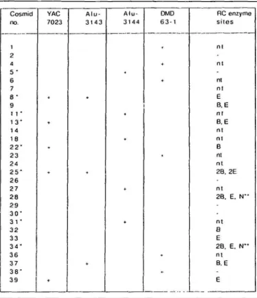
Cosmid no. 1 2 4 5 • 6 7 8 -9 1 1 • 1 3 " 1 4 18 2 2 " 2 3 24 2 5 " 2 6 27 2 8 2 9 3 ( T 31 • 3 2 33 3 4 ' 3 6 3 7 3 8 -3 9 YAC 7023 * + A l u -3 1 4 -3 + A l u -3 1 4 4 + DMD 6 3 - 1 RC enzyme sites n l n l n l n l E B. E n l B, E n l n l B nt nt 2B. 2E n l 2B. E. N " n t B E 2B, E. N " n t B, E E
The 29 viable cosmids which contained genomic inserts are listed. + denotes hybridization of the indicated probe with the cosmid DNA spot on the ICRF flow sorted human chromosome X gridded cosmid library filters. Rare cutting (RC) enzyme sites are: B, BssH II; E, Eag I; N, Not I; - , none of these sites; nt, not tested. *denotes cosmid selected for isolation of unique subclones; **identifies the 2 overlapping cosmids containing a Not I site. Twenty three of these 29 cosmids (79%) have been tested and confirmed to map back to YAC 3'-23.
4 other YACs: 3'-19, 21, 22 and 24, but did not identify the
DMD region control YACs, 3'-20 and 3'-17. From the physical
map of the DMD contig (24), these Alu 3144 products map distal
to DMD within the region approximately 250-450 kb from the
proximal (DMD) end of the YAC. In contrast, the Alu 3143 pool
detected strongly hybridizing bands in the cognate YAC 3'-23
and 3'-21, these being two of the three YACs extending furthest
distally from the DMD gene. The third YAC, 3'-22, did not
hybridize due to a probable distal chimeric insert. The Alu 3143
pool of PCR products was therefore identified as mapping in the
distal 500 kb of YAC 3'-23. The two pools of Alu PCR products
were used in mapping the cosmids, identified by whole YAC
hybridization, so that cosmids spanning the length of 3'-23 were
chosen for subcloning.
Chacterization of cosmids and isolation of probes
Hybridization of YAC DMD 3'-23 to approximately 3 - 4
chromosome equivalents of the ICRF flow sorted human
chromosome X gridded cosmid library (25) identified 39
positives. They were mapped regionally within YAC 3'-23 by
hybridizing the cosmid filters with Alu PCR pools 3143 and 3144,
DMD cDNA 63-1, and the newly isolated JC-1 YAC 7023 (400
kb). Of the 39 original positives, 5 never grew from the frozen
glycerol stock, and 5 were deduced to contain nonrecombinant
polyvector inserts, by hybridization with Bluescript (Stratagene).
Hybridization results of the gridded cosmid library filters are
Probes Xpter 4965-5 Xcen 19RB3 1A-1.1A-5 8-5 Consensus map 2 2 - 9 2 5 - 1 13-7 JC-1 E B E B F F
I II I
5-6 1 1-5 30-1 31-6 34RN2 34RNB2 3 8 - 2 DMD 63-1 a E (E) B B (N)NI I
0.2 0.4 0.6 B F FE (E) B 0.8 E E B B F F N F N N 1.0 YACS 7072 E B F F B BI II I I I , , l I I
1.2 (Mb) 3'-19 7023 E B B FF F E E BF F- I I HI I
R RJ1.L
R " E B B EE(E) F F E E E E E (E) B B ( E ) (N) N (B) E B B 3'-23, I I I I I 1 1 ^ , I l l l l l
Figure 2. A 1.2 Mb YAC contig and physical map localizing probes in the AHC-GK region. The lower part of the figure shows physical maps of individual YACs
7072, 7023, DMD 3'-23 and 3'-19, as determined by partial digestion mapping with Sfi I, F; Not I, N; BssH II, B; and Eag I, E. Sites exhibiting partial digestion in YACs are shown in brackets, ( ). A double slash across the YAC indicates the probable position of a chimeric junction; a dashed lines indicates the portion of chimeric YACs that is not colinear with this Xp21 region. Three potential CpG islands were observed; these and other rare cutting enzyme sites observed consistently are positioned in the centre of the figure (scale in Mb), on the consensus map. Sites observed only in one YAC are marked only on that YAC (see Discussion). At the top of the figure, 15 new subclones plus JC-1 and DMD 63-la are mapped according to their hybridization to pulsed-field gel electrophoresis fragments. The primary data path supporting the mapping of these probes is available on request from the authors. The positions of 19RB3, the subclone isolated from the right end phage from DMD YAC 3'-19, and 4965-5, the subclone isolated from the left end cosmid of YAC 7072, are indicated by arrows, as defined by the position of these YACs in the contig. Probes 1A-1 and 1A-5 could be ordered as shown because they mapped on either side of the chimeric junction of the YAC 7023 insert. D numbers have been assigned to one probe from each cosmid contig distal to the DMD locus : 4965-5 (DXS1074); 1A-1 (DXS1075); 13-7 (DXS1076)-25-1 (DXS1077); 8-5 (DXS1078); 19RB3 (DXS1079); 30-1 (DXS1080); 34R1 (DXS1081).
given in Table 1 for the remaining 29 viable cosmids. YAC JC-1
7023 identified 5 cosmids in common with YAC 3'-23 (cosmids
8, 13, 22, 25 and 39), representing the most distal cosmids within
YAC 3'-23, and 1 new cosmid (7023-1A, not tabulated), distal
to YAC DMD 3'-23. All cosmids detected with the DMD 3'-23
Alu PCR pools had been previously identified as positive with
the DMD 3'-23 whole YAC hybridization. Three cosmids were
detected with the Alu 3143 pool (cosmids 8, 25 and 37), and
of these, two also hybridized with YAC 7023 (cosmids 8 and
25). A total of 5 cosmids (5, 11, 18, 27 and 31) were detected
with the Alu 3144 pool, and 6 independent cosmids (1, 4, 6,
23, 36 and 38) were detected with DMD cDNA 63-1,
representing the most proximal cosmids within YAC 3'-23.
Some cosmids were additionally characterized by digestion with
Hind ID alone and in double digests with the rare-cutting enzymes
Not I, BssH II and Eag I. Two overlapping cosmids contained
a Not I site (cosmids 28 and 34); one of these (cosmid 34), plus
9 cosmids spanning the length of YAC DMD 3'-23 (cosmids 5,
8, 11, 13, 22, 25, 30, 31 and 38; Table 1), and the single cosmid
unique to YAC 7023 (cosmid 7023-1A), were selected for
isolation of single copy probes. The right (distal) end of DMD
YAC 3'-19 was isolated as a phage clone, and a probe (19RB3)
was isolated from it as a unique subclone. A probe was also
isolated from a cosmid (4965) identified by the vectorette PCR
product amplified from the left (distal) end of the 660 kb JC-1
YAC 7072, representing the most distal extent of the YAC contig.
Physical map of the YAC contig
The newly isolated JC-1 YACs 7072 and 7023, and distal YACs
from the 3.2 Mb DMD contig (24), 3'-23 and 3'-19, were
subjected to partial digestion with Sfi I, Not I, BssH II and Eag
I, and mapped by PFGE (Figure 2). Three potential CpG islands,
containing 3 or more rare cutting enzyme sites, were observed:
one immediately distal to JC-1, and two separated by 50 kb,
approximately 30 kb distal to the PFGE fragment hybridizing
with the 3' end of the DMD gene. These CpG islands and other
rare cutting enzyme sites observed consistently in all YACs
spanning a particular site were used to construct a consensus
physical map of the contig. This defined the maximum extent
of the contig from 3'-19 to 7072 as 1.2 Mb, extending the 3.2
Mb DMD contig (24) to 3.5 Mb. Some sites were observed only
in one YAC, and these are shown in Figure 2 only on that YAC
(see Discussion).
Table 2. Deletion results for AHC-GK patients PATIENT AM GJ BEN AS MC C C * JHt 1477 (JC) 448Z «t 44831 1290 JM 3 4 8 AB PHENOTYPE AHC GK | DMD - -+ * + REF 7 2 7 10,21 28 4,10 8 4.10.30 2 9 PROBE C7 0 0 0 4965-51 1A-1 | 1A-5 0 + 0 0 0 0 0 0 0 0 0 0 0 0 0 0 0 0 0 0 0 0 0 0 0 0 0 0 13-7-+ 0 0 0 0 0 0 0 22-9 | 25-1 | JC-1 + 0 0 0 0 0 0 0 0 0 0 0 0 0 0 0 0 0 0 0 0 J 0 0 0 0 0 8-5 jl9RB3| 5-6 | 11-5 0 0 0 0 0 0 0 0 0 0 0 0 0 0 0 0 0 0 0 0 0 0 0 0 0 0 0 0 30-1 0 0 0 0 0 0 34Rl-ll 38-2 0 0 0 0 0 0 0 + 0 0 0 0
Deletion results are shown for C7, JC-1 and 13 new probes on Hind III digested patient DNA. For phenotype, + and - indicate presence and absence of the disease phenotype, respectively. Where available, patient literature references are cited (REF). Six patients were not deleted for any of the probes tested: patients SO, ST, SL, BF and EK (AHC only), and DK (GK only). For probe results, + and o indicate presence and deletion in patient DNA, respectively; J, junction fragment. The order of probes in the table is Xpter-Xcen, except that the orders of 13-7/22-9 and of 5-6/11-5/30-1 could not be determined. *, 13-7 detects a Hind III polymorphism; t , patients CC and JH are cousins; #, patients 4482 and 4483 are brothers.
Fourteen new subclones plus JC-1 and DMD 63-la were
mapped according to their hybridization to PFGE fragments
(Figure 2). D numbers have been assigned to one probe from
each cosmid contig distal to the DMD locus (see the legend to
Figure 2).
Deletion mapping
The patients with deletions are listed in Table 2, with their disease
phenotype (4, 7, 8, 10, 21, 27-30) and hybridization results
for C7, JC-1 and the new subclones. Four new deletions were
detected which would not have been seen with C7 and DMD
probes. A deletion was detected in all (5/5) unrelated patients
with the phenotype AHC-GK-DMD, in all (3/3) unrelated patients
with AHC-GK, and in the single patient with GK-DMD. In
contrast, a deletion was detected in 2/7 (29%) of unrelated patients
with isolated AHC; no deletion has yet been detected in the single
patient with isolated GK.
Patient BEN was observed to have a complex double deletion:
a distal deletion encompassing 4965-5, 1A-1 and 1A-5 and a
second proximal deletion encompassing 25-1, JC-1 and 8-5
(Figure 3). DXS28 was not deleted. The double deletion mapping
was confirmed on 3 separate blots. Further probes will need to
be isolated between C7 and 4965-5 in order to map the distal
deletion breakpoint accurately. The distal and proximal deletions
are separated by 60-230 kb of DNA, and the proximal deletion
spans 50—100 kb. Non-overlapping deletions were observed in
other unrelated AHC patients (AM, GJ versus MC; Figure 3),
indicating that the AHC gene is large, extending at least between
4965-5/1 A-1 and 25-1, a region of 200-500 kb.
A minimum mapping interval is defined by patient deletions
to contain at least part of the GK gene. This GK critical region
is defined distally by patient 1477 (JC), and proximally by BEN,
AS, MC, CC and JH (Figure 3). The GK critical region therefore
extends from JC-1 to 8-5/19RB3, a region of 50-250 kb.
Probe 13-7 (DXS1076) was found to detect a frequent Hind
IH polymorphism (alleles 2.6 kb (frequency 0.47) and 0.9 kb
(0.53), 57 X chromosomes tested). Figure 4 illustrates
hybridization of probes 13-7 and 8-5 together on Hind UII digests
of normal controls and patient DNA, showing the 13-7
polymorphism. Mendelian segregation of the 13-7 alleles through
a 3-generation AHC family was observed (11 individuals tested),
so that 13-7 can be used for concurrent analysis of deletions and
polymorphism in AHC families, and as a distal flanking marker
for DMD.
DISCUSSION
Physical map of the AHC-GK region
The 890 kb YAC DMD 3'-23 was observed to contain DMD
Hind III exon-containing fragments and DXS708 (JC-1),
demonstrating physical linkage of these two loci for the first time.
This physical linkage of loci is consistent with the observation
that some GK patients have deletions of the DMD gene (8, 9,
11). JC-1 mapped on the 50 kb Sfi I fragment between the
potential CpG islands located at 900 kb and 850 kb of the
consensus map (Figure 2), corresponding to Sfi I sites J and J'
of the long range physical map (24, 26). This is in agreement
with studies on genomic DNA, where JC-1 was observed on a
Sfi I fragment of approximately 80 kb (21).
Three potential CpG islands were observed in the YACs. More
rare cutting enzyme sites are observed in hypomethylated YAC
DNA propagated in yeast than in human genomic DNA. The
relationship of sites seen in YACs and in genomic DNA remains
to be fully elucidated; however, the Sfi I sites J, J' and the site
at 500 kb of the YAC consensus map are in agreement with
published data on sperm and leukocyte DNA (21, 26).
Preliminary data indicates that in DNA from a 48, XXXX
lymphoblastoid cell line, at least the CpG island at 900 kb of
the consensus map is partially digested (unpublished
observations). These CpG islands therefore represent candidate
regions to search for potential 5' coding sequences (31), in order
to identify the AHC and GK genes.
A number of discrepancies were observed in the detection of
rare cutting enzyme sites in overlapping YACs. Possible
explanations for these discrepancies include chimeric YAC inserts
or rearrangement events, polymorphisms, and the reproducibility
of partial digestion of rare-cutting enzymes on large YACs. The
regions showing such inconsistencies were the proximal (right
arm) 200 kb of 3'-23; possibly a very small ( < 50 kb) chimeric
or rearranged insert at the proximal end of 3-19, which shows
Xpter Xcen I 1 1 1 I 1 1 0 0.2 0.4 0.6 0.8 1.0 1.2 (Mb) Probes 4965-5 1A-5 2 5 - 1 19RB3 DMD YACs 7072 7023 DMD 3-23 Cosmids Key Patients AM GJ BEN AS MC CC, JH 1477 (JC) (AHC) (AHC) (AHC, (AHC, (AHC, (AHC, GK) GK) GK) GK) (GK, DMD)
to
I I I I DMD 3'-19Figure 3. Map of AHC and GK critical regions. At the top of the figure are shown the positions of probes spanning the AHC-GK region, and the YAC contig, with the 3 potential CpG islands marked on the YACs as vertical bars. The distance between probes C7 and 4965-5 is not to scale. Beneath this are the cosmid contigs constructed around each probe. In the lower part of the figure, key patient deletions define critical regions for AHC and GK. A solid line indicates DNA which is present; brackets flanking an interruption in this line represent the deletion. The GK critical region is defined distally by patient 1477 (JC), and proximally by BEN, AS, MC, CC and JH. The GK critical region extends from JC-1 to 8-5/19RB3, a region of 50-250 kb. Patient BEN (AHC.GK) was observed to have a complex double deletion: a distal deletion encompassing 4965-5, 1A-1 and 1A-5 and a second proximal deletion encompassing 25-1, JC-1 and 8-5. DXS28 was not deleted. Non-overlapping deletions were observed in other unrelated AHC patients (AM, GJ versus MC), indicating that the AHC gene is large, extending at least between 4965-5/A1-1 and 25-1, a region of 200-500 kb. The AHC and GK critical regions as defined by patient deletions are shown as boxes. The portion of the critical region which must contain part of the gene is unshaded; the portion which may contain part of the gene, as defined by deletions, is shaded.
2 different Hind III fragments hybridizing with the left vector
arm probe; and the distal 170 kb of 7023. These possible chimeric
YAC inserts do not detract from the consistent region bounded
by probes 1A-5 and 38-2, and verified in this region by both
probe hybridizations confirmed across at least 2 YACs, and by
consistent features of the consensus physical map across at least
2 YACs. The distal (right) end of 3'-19, and the distal (left) end
of 7072, have both been confirmed as non-chimeric, by mapping
the end clones back onto the YAC and cosmid contigs, and
mapping on deletions in DNA from patients. Thus YACs from
the contig are shown to span the entire region from 4965-5 to
38-2; earher, wojk ^hpws,the cpntig,extends further proximally
to DMD Hind til exon containing fragment 53 (24).
Complex deletion of patient BEN
Patient BEN (AHC, GK) and his mother have an interstitial
deletion involving Xp21^2 and possibly Xp21.3, detected on
'
Tpr6metapliasi'chr6m*os6mes (27)-The chromosomal deletion was
found only upon high resolution chromosome banding. No
molecular deletion was detected after hybridization of 11 probes
from Xpil.2, including the DMD locus (27). Here we detected
the proband's deletion at the molecular level using the new
a double deletions Such a complex double
deletion has been described previously in a DMD family, where
2 brothers, 1 affected and 1 unaffected, and their mother,
displayed deletion of DMD intron 7 probes XJ10.1 and XJ1.1
(DXS208). Only the affected brother displayed a second deletion
detected with DMD cDNA 44-1 (32). It was proposed that the
DXS208 deletion in one of the mother's X chromosomes may
have contributed to the second unique deletion in the affected
DMD male. A double deletion and translocation has also been
reported on the Y chromosome (33), and 2 deletions separated
by a > 110 bp inversion have been characterized in the Xq28
red/green colour pigment gene in an adrenoleukodystrophy patient
(34). It will be interesting to characterize the deletion in the
mother of AHC-GK proband BEN at the molecular level.
Certainly the origin of such double deletion events may be
complex.
The GK critical region
The DNA of patient 1477 (JC) was used as a reference, by which
probes could be mapped relative to the distal deletion breakpoint,
isolated as probe JC-1 (DXS708). The proximal breakpoint of
this deletion has been mapped between exons 42 and 43 (Hind
III fragments 30 and 31) (21), within the DMD gene. Therefore,
probes derived from YAC 3'-23 which are deleted in the DNA
8 - 5
13-7 upper —
in unrelated DMD patients was an early indication that the DMD
gene is large (35). The complete DMD gene has now been
mapped by PFGE to span approximately 2400 kb (14, 19, 20,
26, 30, 36-38). The AHC gene is located between DXS28 (C7)
and 25-1, a region estimated at approximately 3000 kb. Isolation
and mapping of new probes in the interval between C7 and 4965-5
will be important to test AHC patients for distal deletions.
The map presented here provides the basis to identify the genes
for AHC and GK, by characterization of CpG island regions,
and by searching for coding sequence within the defined critical
regions. In this hunt for exons, sequence analysis, cross-species
hybridization using both single copy and whole cosmid probes,
and exon amplification procedures (39) should lead ultimately
to the identification and cloning of the GK and AHC genes.
13-7
lower-Figure 4. Hybridization of probes 8-5 and 13-7 to Hind III digests of control
and patient DNA. Hind III digested DNA from a normal male (M) and normal female (F; 10 /tg), and from a subset of the patients listed in Table 2 (5 /*g), were hybridized with pooled probes 8-5 and 13-7. Probe 8-5 detects a constant band at 4.5 kb. The polymorphic probe 13-7 detects alleles of 2.6 and 0.9 kb. Deletions are demonstrated in patients JM, AB, BEN, MC and JC (1477); the latter deletion maps 8-5 proximal and 13-7 distal to the distal deletion breakpoint of patient JC (1477) (DXS7O8, JC-1).
of patient 1477 are proximal to JC-1; if present, the probe must
be distal to JC-1 (Figure 3). This allowed 25-1 to be localized
distal to JC-1 (Table 2); these probes could not be resolved by
PFGE mapping (Figure 2).
The AHC and GK genes were previously known to flank
DXS708, and were localized within a genomic region of 4 Mb.
The patient deletions and physical map described here permit
more accurate localization of a critical region for each gene,
defined as the minimum region which must contain at least part
of the gene. This will allow targeting of subsequent searches for
coding sequence to those regions known to contain exons. The
GK critical region is flanked distally by JC-1 and proximally by
8-5/19RB3, a region of 50-250 kb.
Extent of the AHC gene
Deletions were detected in all 9 independent patients with a
complex phenotype of at least 2 diseases, reflecting the presence
of relatively large deletions (Table 2). In contrast, a deletion was
detected in only 2/7 independent patients with a single disease,
either GK or AHC. This low frequency of deletion detection in
patients with a single disease may result from small gene
deletions, possibly removing only part of the gene, or from point
mutations. In the case of AHC, it is also possible that the gene
may extend further distally beyond our most telomeric probe;
isolation of further distal probes will be important to investigate
this possibility.
Non-overlapping deletions were observed in unrelated AHC
patients, indicating that the AHC gene is large, extending over
at least 200-500 kb. The finding of non-overlapping deletions
MATERIALS AND METHODS
YAC library screening and YAC DNA preparation
The ICRF Human YAC Reference Library was screened with probe JC-1 (DXS7O8) (21) by colony hybridization (23). Positive yeast clones were streaked onto selective agar plates (-uracil, -tryptophan). Isolated colonies were inoculated into selective medium (-uracil, -tryptophan) for preparation of chromosomes in agarose blocks (23, 40).
Alu repeat primed PCR probes
YAC DMD 3'-23 (24) was used as template for Alu repeat primed PCR, using either 5' Alu primer 3143 (H.Lehrach, unpublished) or 3 Alu primer 3144 (41). Approximately 100-200 ng total DNA was amplified using reaction conditions as described (41). PCR products were analysed on ethidium bromide stained 1 % agarose, 1 % NuSieve (F.M.C.) gels and shown to contain 3 to 4 products, ranging from 0.3 to 2 kb. No amplification was observed from yeast DNA template or from no DNA controls. The pools of Alu PCR products were separated from Alu primers over Qiagen (Hybaid) columns, and approximately 50 ng DNA was radioactively labelled (42). The labelled Alu PCR products were denatured and preannealed by addition of sheared total human DNA (Sigma) to 2.5 mg/ml, and SSC to 5xdenaturation at 100°C for 10 min, ice for 1 min, and incubation at 65°C for 120 min (Cot = 300 mg.min/ml). The preannealed probes were hybridized to Hind III digests of YACs and to the ICRF flow sorted human chromosome X cosmid reference library (25). The filters were incubated for 18 h in prehybridization solution (23) plus 100 /ig/ml sheared total human DNA, and hybridized with labelled Alu PCR fragments in the same solution without human DNA. Blots were washed twice for 30 min in 4 x SSC at room temperature, and twice for 30 min in 0.1 xSSC, 1 % SDS at 65°C, and exposed to X-ray film for 1 - 4 days at -70°C.
Whole YAC screening of cosmid library
DMD 3'-23 and JC-1 7023 YAC DNAs were purified from yeast chromosomes on 1.1 % SeaPlaque (F.M.C.) low melt agarose, 0.5 XTBE gels, under appropriate PFGE conditions. The gels were stained with ethidium bromide, and the YAC DNA band was excised under long wave U.V. light. Agarose was removed by digestion with agarase (Sigma) according to the supplier's protocol, followed by phenol-chloroform extraction and ethanol precipitation. Whole YAC probes were labelled and hybridized to cosmid library filters (43).
Subcloning of cosmids
Cosmids were digested with Hind III, subcloned into Hind IN digested and phosphatased Bluescript, and transformed into competent XL 1-Blue cells (Stratagene). Fragments not hybridizing with total human genomic DNA were selected as probes. When no unique Hind HJ fragment was obtained, the subclones were subjected to double digests of Hind III with EcoR I, BamH I, Bgl II, Pst
I and Xba I.
Construction of a phage library from YAC DMD 3'-19
A phage library was prepared from a partial Mbo I digest of DMD YAC 3'19 and BamH I and EcoR I digested EMBL 3 vector DNA (Stratagene), according to the Stratagene protocol. Packaging was performed using freeze thaw lysate and sonic extract (Gigapack Gold II, Stratagene). The phage library was plated on P2392 host strain, and filter lifts were prepared on Hybond N+ membrane (Amersham) by the manufacturer's protocol. The phage library filters were hybridized with vector left and right probes, prepared directly by PCR amplification of pBR322 sequences in pYAC4 template DNA (44) to identify the phage clones
corresponding to the left and right ends of the YAC. The right arm phage was plaque purified and DNA was extracted (45), for subcloning and identification of non-repetitive probe fragments.
Vectorette PCR
The left end of YAC 7072 was isolated by Hind III vectorette PCR (46). The 200 bp product was cloned into Bluescript (Stratagene) and transformed into competent XL-1 Blue cells (Stratagene), and the purified insert was used to screen the ICRF flow sorted human chromosome X cosmid reference library (25), identifying 2 cosmids. One of these (cosmid 4965) was subcloned as described to yield unique probe 4965-5.
Partial digestion mapping of YACs
Approximately 170-340 ng total DNA was digested with 30, 5, 1 and 0.1 units ofSfil, Not I and Eag I, and 12, 1, 0.1 and 0.001 units of BssH n (New England Biolabs), in a final volume of 200 ji\ of T4 DNA polymerase buffer (45). The reaction mixtures were preincubated at 4°C for 18 h, and DNA was digested for 30 min at 50°C (Sfi I) or 37°C (all other enzymes). The reaction was stopped by the addition of 8 yA 0.5 M EDTA, pH 8.0, on ice. The digested DNA was size-fractionated in 1% agarose gels, 0.5XTBE, on a CHEF DR n (Bio-Rad) apparatus at 170 V with switch times of 25 s for 18 h, 60 s for 15 h and 100 s for 5 h. Gels were alkali-blotted to Hybond N+ membranes (Amersham), neutralized and air dried before hybridization sequentially with pBR322 left and right arm probes (44, 47) and the new single copy probes. Blots were washed as described above.
Patients
Twenty male patients from 18 families were studied (Table 2). Lymphocyte or lymphoblastoid cell line DNA (5 /tg) was digested with Hind III in T4 DNA polymerase buffer (45) for 18 h at 37°C, size fractionated on 1 % agarose gels, blotted to Hybond N + membranes, and hybridized and washed as described above.
ACKNOWLEDGEMENTS
We thank Hans Lehrach, Mark Ross, Sebastian Meier-Ewert, Dean Nizetic, Giinther Zehetner and Christal Douglas for robot spotted YAC and cosmid filters and clone retrieval; David Barton, Marco Machler and Georgia Chenevix-Trench for patient DNA samples; Douglas Higgs for genomic panels used to determine allele frequencies and Franchise Muscatelli for critical reading of the manuscript. Work was supported by the Imperial Cancer Research Fund, Human Frontiers Science Program, Medical Research Council and Muscular Dystrophy Group.
REFERENCES
1. Patil.S.R., Bartley,J.A., Murray,J.C, Ionasescu,V.V. and Pearson,P.L. (1985) Cytogenet. Cell Genet. 40, 720-721.
2. Weiringa,B., Hustinx,T., Scheres.J., Renner.W. and ter Haar,B. (1985) Clin.
Genet. 27, 522-523.
3. BartleyJ.A., Shivanand.P., Davenport,S., Goldstein.D. and Pickens,J. (1986)
J. Pediatrics 108, 189-192.
4. Dunger.D.B., Davies.K.E., Pembrey,M., Lake,B., Pearson.P., Williams,D., Whitfield,A. and Dillon,M.J.D. (1986) Lancet 1, 585-587.
5. Wilcox.D.E., Cooke,A., Colgan.J., Boyd,E., Aitken,D.A., Sinclair,L., Glascow,L., Stephenson.J.B.P. and Ferguson-Smith,M.A. (1986) Hum.
Genet. 73, 175-180.
6. Francke.U., Harper,J.F., Darras.B.T., Cowan.J.M., McCabe.E.R.B., Kohlschutter.A., Seltzer.W.K., Saito.F., Goto,J., HarpeyJ.P. and WiseJ.E. (1987) Am. J. Hum. Genet. 40, 212-227.
7. Yates,J.R.W., Gillard.E.F., Cooke.A., Colgan.J.M., Evans.T.J., and Ferguson-Smith,M.A. (1987) Cytogenet. Cell Genet. 46, 723-724. 8. Chelly.J., Marlhens.F., Dutrillaux,B., Van Ommen.G.J., Lambert,M.,
Haioun.B., Boissinot.G. and Fardeau.M. (1988) Hum. Genet. 78, 222-227. 9. Darras.B.T. and Francke.U. (1988) Am. J. Hum. Genet. 43, 126-130. 10. Davies,K.E., Patterson,M.N., Kenwrick.S.J., Bell,M.V., Sloan,H.R., Westman,J.A., Elsas.L.J. and Mahan.J. (1988) Am. J. Med. Genet. 29, 557-564.
11. McCabe,E.R.B., Towbin.J., Chamberlain,J., Baumbach.L., Witkowski.J., van Ommen.G.J.B., Koenig,M., Kunkel.L.M. and Seltzer.W.K. (1989)/
Clin. Invest. 83, 9 5 - 9 9 .
12. Towbin,J.A., Chamberlain,.!.S., Wu,D., Pillers,D.-A.M., Seltzer,W.K. and McCabe.E.R.B. (1990) Genomics 7, 442-444.
13. Davies.K.E., MandelJ.-L., Monaco,A.P., Nussbaum.R.L. and Willard,H.F. (1991) HGM11. Cytogenet Cell Genet. 58, 853-966.
14. van Ommen.G.J.B., Verkerk,J.M.H., Hofker.M.H., Monaco.A.P., Kunkel.L.M., Ray,P., Worton.R., Wieringa,B., Bakker.E. and Pearson.P.L. (1986) Cell 47, 499-504.
15. Nerby.S. and Schwartz.M. (1987) Clin. Genet. 31, 192-197. 16. Bartley,J. and Gies.C. (1989) Cytogenet. Cell. Genet. 51, 958. 17. Trask.B.J., Massa.H.F. and Burmeister,M. (1992) Genomics 13,455-457. 18. Walker.A.P., Larin.Z., Lehrach,H. and Monaco.A.P. (1991) Cytogenet.
Cell Genet. 58, 2087.
19. Burmeister,M. and Lehrach.H. (1986) Nature 324, 582-585.
20. Burmeister,M., Monaco.A.P., Gillard.E.F., van Ommen,G.-J.B., Affara.N.A., Ferguson-Smith,M.A., Kunkel.L.M. and Lehrach.H. (1988)
Genomics 2, 189-202.
21. Love.D.R., Bloomfield,J.F., Kenwrick.S.J., Yates.J.R.W. and Davies.K.E. (1990) Genomics 8, 106-112.
22. Worley,K.C, Towbin.J.A., Zhu,X.M., Barker.D.F., Ballabio.A., Chamberlain.J., Biesecker.L.G., Blethen.S.L., Brosnan,P., Fox.J.E., Rizzo.W.B., Romeo.G., Sakuragawa.N., Seltzer.W.K., Yamaguchi,S. and McCabe.E.R.B. (1992) Genomics 13, 957-961.
23. Larin.Z., Monaco.A.P. and Lehrach.H. (1991) Proc. Natl. Acad. Sci. USA
88, 4123-4127.
24. Monaco.A.P., Walker.A.P., MUlwood.I., Larin.Z. and Lehrach.H. (1992)
Genomics 12, 465-473.
25. Nizetic,D., Zehetner.G., Monaco,A.P., Gellen.L., Young,B.D. and Lehrach.H. (1991) Proc. Natl. Acad. Sci. USA 88, 3233-3237. 26. Den Dunnen.J.T., Grootscholten,P.M., Bakker.E., Blonden.L.A.J.,
Ginjaar.H.B., Wapenaar.M.C, van Passen.H.M.B., van Broeckhoven.C, Pearson.P.L. and van Ommen.G.J.B. (1989) Am. J. Hum. Genet. 45, 835-847.
27. Marlhens.F., Chelly.J., Kaplan.J.C, Lefrancois.D., HarpeyJ.P. and Dutrillaux.B. (1987) Hum. Genet. 77, 379-383.
28. Love.D.R., Flint,T.J., Marsden.R.F., Bloomfield.J.F., Daniels,R.J., Forrest,S.M., Gabrielli.O., Giorgi,P., Novelli.G. and Davies.K.E. (1990)
Am. J. Med. Genet. 37, 136-142.
29. Recan.D., Chafey,P., Leturcq.F., Hugnot,J.-P., Vincent.N., Tome\F., Collin.H., Simon,D., Czernichow.P., Nicholson,L.V.B., Fardeau,M. and Kaplan,J.-C. (1992) J. Clin. Invest. 89, 712-716.
30. Kenwrick,S., Patterson,M., Speer.A., Fischbeck,K. and Davies.K. (1987) CW/48, 351-357.
31. Bird.A.P. (1986) Nature 321, 209-213.
32. Bartlett.R.J., Walker.A.P., Laing,N.G., Koh,J., Secore.S.L., Speer,M.C, Pericak-Vance.M.A., Hung,W.-Y., Yamaoka.L.H., Siddique.T., Kandt.R. and Roses,A.D. (1989) Lancet 1, 496-497.
33. Fisher,E.M.C, Beer-Romero.P., Brown,L.G., Ridley,A., McNeil,J.A., Bentley Lawrence,J., Willard.H.F., Bieber.F.R. and Page,D.C. (1990) Cell 63, 1205-1218.
34. Feil,R., Aubourg.P., Mosser.J., Douar,A.-M., Le Paslier.D., Philippe.C. and Mandel,J.-L. (1991) Am. J. Hum. Genet. 49, 1361-1371. 35. Kunkel.L.M. et al. (1986) Nature 322, 7 3 - 7 7 .
36. van Ommen,G.J.B., Bertelson,C.E., Ginjaar.H.B., Den Dunnen.J.T., Bakker,E., Chelly.J., Matton.M, Van Essen.A.J., Bartley.J., Kunkel.L.M. and Pearson.P.L. (1987) Genomics 1, 329-336.
37. Feener.C.A., Koenig.M. and Kunkel.L.M. (1989) Nature 338, 509-511. 38. Boyce.F.M., Beggs,A.H., Feener,C. and Kunkel.L.M. (1991) Proc. Natl.
Acad. Sci. USA 88, 1276-1280.
39. Buckler,A.J., Chang.D.D., Graw.S.L., Brook.J.D., Haber.D.A., Sharp.P.A. and Housman.D.E. (1991) Proc. Natl. Acad. Sci. USA 88, 4005-4009. 40. Southem.E.N., Anand,R., Brown.W.R.A. and Fletcher,D.S. (1987) Nucleic
Acids Res. 15, 5925-5943.
41. Monaco.A.P., Lam,V.M.S., Zehetner.G., Lennon.G.G., Douglas.C, Nizetic.D., Goodfellow.P.N. and Lehrach.H. (1991) Nucleic Acids Res. 19, 3315-3318.
42. Feinberg,A.P. and Vogelstein.B. (1983) Anal. Biochem. 132, 6 - 1 3 . 43. Baxendale.S., Bates.G.P., MacDonald.M.E., Gusella.J.F. and Lehrach.H.
(1991) Nucleic Acids Res. 19, 6651.
44. Hirst,M.C, Rack.K., Nakahori.Y., Roche.A., Bell.M.V., Flynn.G., Christadoulou.Z., MacKinnon,R.N., Francis,M., Littler.A.J., Anand.R., Poustka,A.-M., Lehrach.H., Schlessinger,D., D'Urso.M., Buckle.V.J. and Davies.K.E. (1991) Nucleic Acids Res. 19, 3283-3288.
45. Sambrook,J., Fritsch.E.F. and Maniatis.T. (1989) Molecular Cloning: A
Laboratory Manual (2nd Edition). Cold Spring Harbor Laboratory Press,
Cold Spring Harbor, NY.
46. Riley,J., Butler,R., Ogilvie,D., Finniear,R., Jenner.D., PoweU.S., Anand.R., Smith,J.C. and Markham,A.F. (1990) Nucleic Acids Res. 18, 2887-2890. 47. Burke.D.T., Carle,G.F. and 01son,M.V. (1987) Science 236, 806-812.