Restenosis after coronary angioplasty:
review of the literature
B . M E I E R
Centre de Cardiologie, Hopital Cantonal Universitaire, CH-1211 Geneve 4, Switzerland
KEY WORDS: Coronary angioplasty, restenosis after angioplasty.
Histopathology of restenosis
Knowledge on the histopathology of restenosis is scarce. This indicates that, despite its frequent occurrence, restenosis after coronary angioplasty is rarely a lethal disease. Animal models provide some information on what might occur after angioplasty. However, the results of these studies should only be applied to the situation in the atherosclerotic human artery with great caution.
Steele et al. described the stages of healing and restenosis in carotid arteries of pigs receiving no medication'11. One hour after balloon angioplasty, endothelial denudation, medial tears and platelet deposition were found. One day later, there was necrosis of smooth muscle cells. A week later, the endothelium had completely regrown. About a month later, there was mild proliferation of smooth-muscle cells. Significant restenoses, however, were due to organized intraluminal thrombi.
Waller et al. reported on necropsies in three patients who died 80 to 150 days after coronary angioplasty*21. Only one of these patients showed clinical evidence of recurrence, but all showed sig-nificant stenoses at the presumed site of antioplasty. The lesions were not discernible from genuine atherosclerotic plaques and there was no evidence of healed dissection.
More in keeping with the current understanding of the mechanism of balloon angioplasty is a case report by Essed et al. on a patient who died at the beginning of a redilatation attempt 5 months after a successful angioplasty with recurrence of symptoms at 3 months'3'. These authors found medial dissec-tion with a narrowing of the new channel by fibrocellular tissue.
Coronary restenosis: a new disease entity
Restenosis was rapidly recognized as one of the major problems with coronary angioplasty. It is
now a well known disease entity accounting for numerous hospitalizations and interventions.
The restenosis rate of the first 169 consecutive patients treated by Gruentzig in Zurich was 25% after a mean of 18 months'41.
In 1982, Balcon et al. published a restenosis rate of 67% 3 months after the six successful ahgioplasties in their first 11 patients'51. With the benefit of angio-plasty 3 months after the intervention in only two of 11 patients, their lack of enthusiasm for this treatment was understandable.
At the same time, the PTCA Registry of the National Heart, Lung, and Blood Institute (NHLB1) of the United States reported a clinical recurrence rate, after a mean of 12 months, of 17% in 232 patients treated at 34 different centres'61.
In 1983, Meyer et al. reported a recurrence rate of 21 % in 100 consecutive patients after an average of 6 months'71. Their patients were fully anticoagulated with coumadin. The recurrence rate of patients with unstable angina was higher than that of patients with stable angina (24% vs. 17%).
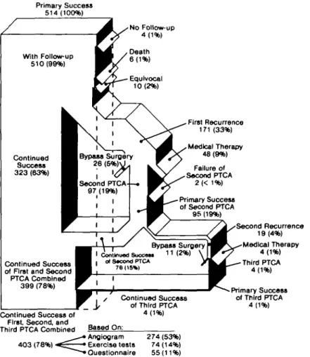
In 1984, Meier et al. reported, from Atlanta, a recurrence rate of 33% in 514 consecutive patients"1. The recurrences were documented at a mean of 5 months (range 1-9 months) after angio-plasty. Most of the patients were treated with acetylsalicylic acid. Male sex and stenoses in the left anterior descending coronary artery (LAD) were identified as risk factors for restenosis. Figure 1 provides the details of the long-term outcome and management of these patients.
Another report from the PTCA Registry of NHLBI on 665 patients followed for 6 months indi-cated an angiographic recurrence rate of 34% that correlated well with clinical symptoms'91.
In 1985, Bertrand et al. reported a recurrence rate of 27% in 269 patients followed for an average of 6 months"01. In the same year the PTCA Registry of the NHLBI confirmed male sex as a risk factor for 0I95-668X/88/09CO01 + 0 6 $02.00/0 © 1988 The European Society of Cardiology
Primary Success 514 (100%) No Follow-up 4(1%) Continued Success 323 (63%) First Recurrence 171 (33%) Medical Therapy 48(9%) Primary Success of Second PTCA 96 (19%) Bypass Surgery I Contlnuad Succ«s> 1 1 . o» 8*condPTCA 76(15*1 Failure of Second PTCA 2 (< 1 %) Continued Success of First and Second PTCA Combined
399 (78%)
r J,
Continued Success ofFirst Second, and Third PTCA Combined
403 (78%) • Continued Success of Third PTCA 4(1%) Second Recurrence 19(4%) Medical Therapy 4(1%) Third PTCA 4(1%) -Primary Success of Third PTCA 4(1%) Based On: > Angtogram 274 (53%) • Exercise tests 74(14%) •Questionnaire 55(11%)
Figure 1 Follow-up for about I year of 514 consecutive patients with successful coronary angioplasty at Emory University in Atlanta, Georgia, U.S.A.
recurrence in the 3079 patients included up to that time1"1. The global recurrence rate was again calcu-lated at 33%. In men it was 36% and in women it was22%(/><001).
Kaltenbach et alP] and Kober et a/."3' reported the recurrence rates of the first 356 and 1000 patients in Frankfurt, respectively. The recurrence rate was about 15% for both initial patients fol-lowed for 6 months and for later patients folfol-lowed for roughly 12 months. Virtually all the recurrences occurred within the first 3 months after angioplasty. They attributed their remarkably low recurrence rate to stringent adherence to a powerful follow-up regimen including high daily doses of acetylsalicylic acid (1-5 g), isosorbide dinitrate (120 mg), and verapamil (240-480 mg) or gallopamil (100 mg).
A third report from Atlanta on 1880 patients followed over an average of 7 months revealed a
recurrence rate of 28%[M1. The presence of a visible local dissection after angioplasty in conjunction with a residual pressure gradient of < 15 mmHg identified a favourable subgroup with a recurrence rate of only 19%.
Mabin et al. reported a recurrence rate of 33% from the Mayo Clinic in 229 patients followed for a mean of 13 months'151. In patients with symptoms at the time of the follow-up examination, restenosis was present in 71%. In those without symptoms, restenosis was found in only 14%
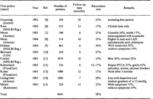
Table 1 summarizes the restenosis rates of some of the cited reports.
Recurrence in mulfHesion angioplasty or moltivessel angioplasty
Table 1 Reported recurrence rates of coronary angioplasty (in chronological order) First author (place) Gruentzig (Zurich) Kent (NHLBI Reg.) Meyer (Aachen) Meier (Atlanta) Holmes (NHLBI Reg.) Bertrand (Lille) Cowley (NHLBI Reg.) Kaltenbach (Frankfurt) Kober (Frankfurt) Leimgruber (Atlanta) Mabin (Rochester) Total Year 1982 1982 1983 1984 1984 1985 1985 1985 1985 1985 1985 Ref. [4] [6] [7] [8] [9] [10] [11] [12] [13] [14] [15] Number of patients 169 232 100 514 665 269 3079 356 1000 1880 229 8493 Follow-up (months) 18 12 6 10 6 6 18 6 12 7 13 Recurrence rate 25% 17% 2 1 % 33% 34% 27% 33% 12-17% 15% 28% 33% 28% Remarks
Including first patient Clinical data only
Unstable 24%, stable 17%; anticoagulated with coumadin Higher in men and LAD acetylsalicylic acid, nifedipine
With symptoms 56%; without symptoms 14%
Men 36%, women 22% Repeat PTCA 33%, grafts 45% salicylate, nitrate, calcium blockers None after 3 months
Less with dissection and residual gradient of ^ 15 mmHg With symptoms 71%;
without symptoms 14%
Table 2 Theoretical recurrence rates in multilesion angio-plasty (assumed recurrence rate per lesion: 33%)
Number
oflesions Recurrence rate per patient
33% 56% 70% 80% 87% 1/3 5/9 19/27 65/81 211/243 (3 x 3 " " ' - 2 x 2""')/3" x 100%
33% per lesion, the theoretical patient recurrence rate (recurrence in > 1 stenosis) in double lesion angioplasty (in 1 or 2 vessels) would be 56%. The formula used for this calculation and the respective recurrence rates for multiple lesion angjoplasties are indicated in Table 2.
Hollman et al. reported a recurrence rate in single-vessel angioplasty of 38% and in multivessel angioplasty of 68%(l6]. His findings concur with the expected recurrence rates and demonstrate that
recurrence is a lesion-related phenomenon rather than a patient-related phenomenon. These figures should not be confused with follow-up data on patients with multivessel disease in whom angio-plasty is done in one artery only1'71.
Quantitative studies on restenosis
Fleck et al. reported on computer-assisted analysis of angiograms in 95 patients with follow-up angiography1181. These authors manually traced two orthogonal views of the stenosis before and after angioplasty and again at follow-up. The catheter was used as calibration reference and the cross-sectional area of the stenosis was indicated in mm2. Their definition for restenosis was a loss of ^ 1 mm2 of the cross-sectional area resulting in a > 70% narrowing of the cross-section. The restenosis rate was 33%. It correlated well with subjective symptoms and stress-test data.
Johnson et al. compared a similar computer-assisted method for evaluating coronary stenoses with videodensitometry in 23 patients re-examined after coronary angioplasty1"1. The recurrence rate
was 35%. The two methods correlated fairly well (r = 0-77).
Non-invasive detection of resteuosis
Several non-invasive tests have been advocated for follow-up after coronary angioplasty. Bicycle exercise tests proved valid in documenting restenosis in the original patients of Gruentzig1201. Treadmill exercise tests combined with thallium-201 scintigra-phy detected six out of nine recurrences in a study from the Montreal Heart Institute1211.
Wijns et al. analyzed the predictive values of stress test and thallium-201 scintigraphy performed 1 month after angioplasty in relation to clinical and angiographic findings at 6 months'22'. For angina, the predicitive values of stress test and thallium-201 scintigraphy were 38% and 66% and for restenosis 50% and 74%, respectively (/><0O05 in favour of thallium-201 scintigraphy).
DePuey et al. found a similar predicitive value for restenosis (73%) with exercise radionuclide ventri-culography performed shortly after angioplasty1231. At the time of follow-up angiography, the predictive value of the same test was 75%.
Restenosis and drugs
In rabbits, both a combination of acetylsalicylic acid with dipyridamole and sulphinpyrazone sig-nificantly decreased the extent of restenosis 4 months after iliac ballon angioplasty1241.
A randomized study from Atlanta compared a daily dose of 325 mg acetylsalicylic acid with anti-coagulation with coumadin1251. Restenosis was slightly less frequent in patients treated with acetyl-salicylic acid. However, statistical significance was only attained for patients in whom symptoms occurred > 6 months before angioplasty where retensosis rates were 21% for acetylsalicylic acid and 44% for coumadin. The compliance in the coumadin group was rather poor and therapuetic prothrombin times were only documented in about
one third of these patients.
Two calcium antagonists have been studied in randomized trials. Both failed to prove efficacious in preventing or significantly diminishing restenosis. Diltiazem, as an adjunct to a combination of acetyl-salicylic acid and dipyridamole, did not reduce recurrence rate significantly in a study on 92 patients followed for 10 months at the Montreal Heart Institute1261. The recurrence rate was 15% with diltiazem and 22% without. Nifedipine was
Table 3 Riskfactorsfor restenosis after coronary angioplasty
Male sex Diabetes mellitus Recent angina Unstable angina Variant angina Stenosis in LAD
Stenosis in venous bypass graft High initial degree of stenosis High residual degree of stenosis High residual pressure gradient Absence of dissection after angioplasty
Ref. [9,11] [9,16] [28] [7,9,28] [29] [8,16] [9,12] [16] [28] [14] [14,16]
Coumadin therapy in stenoses older than 6 months
Interruption of therapy with acetylsalicyclic acid
[25]
[18]
examined as an adjunct to acetylsalicylic acid in a double-blind protocol on 241 patients followed for 4 months in Atlanta1271. The restenosis rate was similar in patients receiving nifedipine (29%) and in patients receiving a placebo (33%).
Risk factors for restenosis
The risk factors for restenosis identified so far are listed in Table 3. The following risk factors were documented in at least two independent studies: male sex, diabetes mellitus, stenosis in the left anterior descending coronary artery or in a venous bypass graft, and absence of intimal dissection after angioplasty.
The conclusion from a study done in Munich1'81 that the discontinuation of acetylsalicylic acid is a risk factor for the development of restenosis may be challenged. Acetylsalicylic acid was discontinued for gastric pain in all cases concerned and, there-fore, these symptoms may already have been an expression of recurrent coronary stenosis.
It must be admitted that currently there is no proved and practicable method for reducing the recurrence rate after coronary angioplasty.
Summary
The average restenosis rate reported so far in the literature is just below 30%. Although restenosis correlates well with the recurrence of symptoms,
the two factors are not identical. The incidence of myocardial infarction during the first 2 years after coronary angioplasty is 4% and the incidence of death is 2%1301. These two cardiac events are rarely
the first symptom of restenosis. Restenosis, there-fore, is not primarily a life threatening disease but still deserves prompt evaluation and correction.
Restenosis is stenosis-related rather than patient-related. Thus, restenosis rate per patient increases with the number of lesions or arteries treated.
Restenosis rates vary considerably with centres. Serial analyses of restenosis rates at individual centres revealed that the restenosis rates remained constant at a centre-specific level. Differences in case selection and particularities in data definition and analysis may account for both these obser-vations. There is no sound evidence that procedural factors (ballon size, number, duration, or pressure of inflations, etc.) or drug regimens are capable of reducing the recurrence rate. All risk factors for restenosis identified so far are difficult to influence. Extinguishable factors such as smoking seem of little importance in this particular problem.
Efforts to find ways of reducing restenoses after coronary angioplasty are commendable and necess-ary. Their chance of success, however, is small. 'Old customers' will continue to represent 20-30% of the clientele for coronary angioplasty. Their risk for failure and complications is small, but they do carry a considerable risk of restenosis.
References
[1] Steele PM, Chesebro JH, Stanson AW et al. Balloon angioplasty. Natural history of the pathophysiological response to injury in a pig model. Ore Res 1985; 57:
105-12.
[2] Waller BF, McManus BM, Gorfinkel J et al. Status of major epicardial coronary arteries 80 to 150 days after percutaneous transluminal coronary angioplasty. Analysis of 3 necropsy patients. Am J Cardiol 1983; 51: 81-4.
[3] Essed CD, Van den Brand M, Becker AE. Transluminal coronary angioplasty and early restenosis. Rbrocellular occlusion after wall laceration. Br Heart J 1983; 49: 393-6.
[4] Gruentzig A. Results from coronary angioplasty and implications for the future. Am Heart J 1982; 103: 779-82.
[5] Balcon R, Brooks N, Layton C, Rickards A. Percu-taneous transluminal coronary angioplasty (Abstr). Br Heart J 1982; 47: 189.
[6] Kent KM, Bentivoglio LG, Block PC et al. Percutaneous transluminal coronary angioplasty: report from the Registry of the National Heart, Lung, and Blood Institute. Am J Cardiol 1982; 49: 2011-9.
[7] Meyer J, Schmitz HJ, Kiesslich T ei al. Percutaneous
transluminal coronary angioplasty in patients with stable and unstable angina pectoris: analysis of early and late results. Am Heart J 1983; 106:973-80.
[8] Meier B, King III SB, Gruentzig AR et al. Repeat coronary angioplasty. J Am Coll Cardiol 1984; 4:463-6. [9] Holmes DR, Vhetstra RE, Smith HC et al. Restenosis after percutaneous transluminal coronary angioplasty (PTCA): a report from the PTCA Registry of the National Heart, Lung, and Blood Institute. Am J Cardiol 1984; 53: 77C-81C.
[10] Bertrand ME, Thieuleux FA, Lablanche JM, Fourrier JL, Traisnel G. L'angioplastie transluminale coronaire: resultats immediats et a court terme. A propos de 302 cas dilates. Arch Mai Coeur 1986; 79:40-6.
[11] Cowley MJ, Mullin SM, Kelsey SF, Kent KM, Gruentzig AR, Detre KM, Passamani Er. Sex differences in early and long-term results of coronary angioplasty in the NHLBI PTCA Registry. Circulation 1985; 71: 90-7. [12] Kaltenbach M, Kober G, Scherer D, Vallbracht C.
Recurrence rate after successful coronary angioplasty. Eur Heart J 1985; 6: 276-81.
[13] Kober G, Vallbracht C, Lang H et al. Transluminale koronare Angioplastik 1977-1985. Erfahrungen bei 1000 Eingriffen. Radiologe 1985; 25: 346-53.
[14] Leimgruber PP, Roubin GS, Anderson HV et al. Influence of intimal dissection on restenosis after success-ful coronary angioplasty. Circulation 1985; 72: 530-5. [15] Mabin TA, Holmes DR, Smith HC et al. Follow-up
clinical results in patients undergoing percutaneous transluminal coronary angioplasty. Circulation 1985; 71:754-60.
[16] Hollman J, Galan K, Franco I, Simpfendorfer C, Fatica K, Beck G. Recurrent stenosis after coronary angioplasty (Abstr). J Am Coll Cardiol 1986; 7: 20A.
[ 17] Reeder GS, Vlietstra RE, Mock MB, Holmes DR, Smith HC, Piehler JM. Comparison of angioplasty and bypass surgery in multivessel coronary artery disease. Int J Cardiol 1986; 10: 213-21.
[18] Fleck E, Dirschinger J, Rudolph W. Quantitative KoronarangiogTaphie vor und nach PTCA. Resteno-sierungsrate. Analyse beeinflussender Faktoren. Herz 1985; 10: 313-20.
[19] Johnson MR^ Brayden GP, Ericksen EE el al. Changes in cross-sectional area of the coronary lumen in the six months after angioplasty: a quantitative analysis of the variable response to percutaneous transluminal angioplasty. Circulation 1986; 73:467-75.
[20] Meier B, Gruentzig AR, Siegenthaler WE, Schlumpf M. Long-term exercise performance after percutaneous transluminal coronary angioplasty and coronary artery bypass grafting. Circulation 1983; 68: 796-802. [21] Scholl JM, Chaitman BR, David PR et al. Exercise
electrocardiography and myocardial scintigraphy in the serial evaluation of the results of percutaneous trans-luminal coronary angioplasty. Circulation 1982; 66: 380-9.
[22] Wijns W, Serruys PW, Reiber JHC et al. Early detection of restenosis after successful percutaneous transluminal coronary angioplasty by exercise-redistribution thallium scintigraphy. Am J Cardiol 1985; 55: 357-61.
[23] DePuey EG, Leatherman LL, Leachmann RD et al. Restenosis after transluminal coronary angioplasty detected by exercise-gated radionucUde ventriculogra-phy. J Am Coll Cardiol 1984; 4: 1103-13.
[24] Faxon DP, Sanborn TA, Haudenschild CC, Ryan TJ. Effect of antiplatelet therapy on restenosis after exper-imental angioplasty. Am J Cardiol 1984; 53: 72C-6C. [25] Thornton MA, Gmentzig AR, Hollman J, King III SB,
Douglas JS. Coumadin and aspirin in prevention of recurrence after transluminal coronary angioplasty: a randomized study. Circulation 1984; 69: 721-7. [26] Corcos T, David PR, Val PG et al. Failure of diltiazem
to prevent restenosis after percutaneous transluminal coronary angioplasty. Am Heart J 1985; 109:926-31. [27] Whitworth HB, Roubin GS, Hollman J el al. Effect of
nifedipme on recurrent stenosis after percutaneous transluminal coronary angioplasty. J Am Coll Cardiol 1986; 8: 1271-6.
[28] Cowley MJ, Block PC. A review of the NHLBI PTCA Registry data: In: Jang GD, ed. Angioplasty. McGraw-Hill: New York, 1985; 368-78.
[29] David PR, Waters DD, Scholl JM et al. Percutaneous transluminal coronary angioplasty in patients with variant angina. Circulation 1982; 66: 695-702. [30] Kent KM, Bentivoglio LG, Block PC et al. Long-term
efficacy of percutaneous transluminal coronary angio-plasty (PTCA): report from the National Heart, Lung, and Blood Institue PTCA Registry. Am J Cardiol 1984; 53:27C-31C.