Development of Selective Peptide- and Protein-Based Reporters of Kinase
Activity Utilizing Chelation-Enhanced Fluorescence
by
Elvedin Lukovi5
B.A. Chemistry, Haverford College, 2002
Submitted to the Department of Chemistry in Partial Fulfillment of the Requirements for the
Degree of Doctor of Philosophy
at the
Massachusetts Institute of Technology
July 2009
© 2009 Massachusetts Institute of Technology All rights reserved
MASSACHUSETTS INSTTTUTE OF TECHNOLOGY
SEP 2 2 2009
LIBRARIES
ARCHIVES
Signature of Author: Certified by: Department of Chemistry July 24, 2009 Barbara Imperiali Class of 1922 Professor of Chemistry and Professor of Biology Thesis SupervisorAccepted by:
Robert W. Field Haslam and Dewey Professor of Chemistry Chairman, Departmental Committee on Graduate Students
This doctoral thesis has been examined by a committee of the Department of Chemistry as follows:
Sarah E. O'Connor Chair Latham Family Career Development Associate Professor of Chemistry Massachusetts Institute of Technology
Barbara Imperiali Thesis Supervisor Class of 1922 Professor of Chemistry and Professor of Biology Massachusetts Institute of Technology
e)
c3"
62
Timothy F. JamisonProfessor of Chemistry Massachusetts Institute of Technology
Development of Selective Peptide- and Protein-based Reporters of Kinase Activity Utilizing Chelation-enhanced Fluorescence
by
Elvedin Lukovid
Submitted to the Department of Chemistry on July 24, 2009 in partial fulfillment of the requirements for the Degree of Doctor of Philosophy
in Organic Chemistry
Abstract
Catalyzed by kinases, serine/threonine and tyrosine phosphorylation is a vital mechanism of intracellular regulation and is involved in nearly every aspect of normal, as well as aberrant, cell function. With more than 500 protein kinases present in the human genome, the need for probes that can rapidly and selectively report the activity of a single kinase or a discreet subset of related kinases is crucial, particularly as researchers move to increasingly complex, and more relevant, systems to study the effects of dysregulated kinase behavior.
We previously developed sulfonamido-oxine (Sox)-based fluorescent peptides following a P-turn focused (BTF) design. Upon phosphorylation of the Sox-containing peptide, the chromophore binds Mg + and undergoes chelation-enhanced fluorescence (CHEF). However, due to the BTF design limitation, only residues C- or N-terminal to the phosphorylated residue were used to specify the target kinase. To address this drawback, the recognition-domain focused (RDF) strategy, which also relies on CHEF, has been developed. In this approach, the Sox sensing moiety is introduced on the cysteine side chain (C-Sox), thereby allowing inclusion of extended kinase binding determinants, which are used to construct chemosensors for multiple Ser/Thr and Tyr kinases with greatly enhanced selectivity. Moreover, a high throughput mass spectrometry-based screening method that builds additional selectivity into RDF Sox-based probes for Ser/Thr kinases was also developed. Using this approach, it should be possible to construct short peptide probes with enhanced catalytic efficiency for virtually any kinase.
To expand the scope of CHEF-based sensors, beyond kinases that derive specificity from the short consensus sequence, a highly selective ERK sensor was prepared via semisynthesis by combining a recombinant kinase docking domain, PNT, with a synthetic sensing module that included the Sox chromophore. This probe was used to exclusively monitor ERK1/2 activity in unfractionated cell lysates in the absence of off-target kinase inhibitors. Furthermore, to improve the photophysical properties of the probes for cellular studies, we developed several oxine-based CHEF chromophores utilizing numerous approaches including the versatile click chemistry. The most promising derivative, p-bromophenyltriazoyl-oxine (Clk), displays a significant bathochromic shift in the excitation (15 nm) and emission (40 nm) maxima compared to Sox,
and efficiently reports kinase activity when incorporated into peptides as a C-Clk residue.
Together, the results presented in this thesis indicate the power that the CHEF-based sensors have to selectively, rapidly and with great sensitivity deliver new insight into the role of in vitro and endogenous kinases in various processes and under a variety of circumstances.
Thesis Supervisor: Barbara Imperiali
Dedication/Posveta
Dragi Tata,
Zbog tvoje neizmjerne ljubavi, dofivotnog irtvovanja i neprestane podrike, ovu disertaciju posvecujem tebi.
Acknowledgements
Most importantly, I would like to thank my advisor, Professor Barbara Imperiali, for welcoming me into the lab and for teaching me how to tackle difficult problems. Barbara, I admire your insight, your breath of knowledge and your ability to run a group with many diverse projects spanning biology and chemistry. Through my interactions with you and the incredibly gifted team of scientists that you assembled, I have learned a tremendous amount. I am particularly thankful for your support in my next step and for the compassion and understanding that you show in all matters. I also thank my chair Professor Sarah O'Connor and committee member Professor Tim Jamison, for your advice and help throughout my tenure at MIT.
The work described in this thesis would not be possible without several key people. Melissa Shults was instrumental in starting the kinase sensing project and taught me how to be a rigorous analytical chemist. Dr. Dora Carrico-Moniz was never too busy to share her vast synthetic knowledge, and Dr. Juan Antonio Gonzilez Vera was always able to cheer me up as we worked late together. Beth Vogel has not only been an instrumental collaborator, but also welcomed me to Boston when I did not know anybody here. Lastly, none of my peptides would have been properly characterized without Dr. Matthieu Sainlos' willingness to run to MALDI at
all hours of day or night for me. Thank you all for your help.
For the past 7 years the Imperiali lab has been home not only to me, but also to an incredible group of coworkers and friends. For accompanying me to the Muddy and keeping me sane I have to thank Mary O'Reilly, Eranthie Weerapana, Galen Loving, Matthieu Sainlos and Angelyn Larkin. My favorite Sri Lankan (aka Eranthie), I have always appreciated and tried to imitate your confidence and poise-so much that I nearly followed you to the West Coast. Mary, through the -30 Fahrenheit wind tunnel of Galileo Galilei Way to blistering heat of 28-30 Plymouth St., you've always been there on my walk home with me, even when you moved 3000 miles away. Galen, you are one of the funniest people with the best stories (despite their length). Because of you I feel bad for Rick Santorum and am excitedly trying to play the trombone (rusty or not); you made the past 6 years much more interesting. Matthieu, I admire your clear-headedness and willingness to talk about science at any time. Plus, around you I feel like I am in a French film. Angelyn, thank you for everything, from late nights at your house to HPLCing with me to much (loud) laughter. You wonder what you'll do without me, but the real question is how I will get on in med school without you?! Drs. Kathy Franz and Mark Nitz, thank you for the encouragement to pursue graduate school when I first got to MIT. Dr. Jebrell Glover, you always put life into perspective with a song recitation. Dr. Bianca Sculimbrene, no one could argue with your reasoning and your sound advice kept coming even after you left. Dr. Anne Reynolds, you are the best editor that I have ever met; thanks for all the help, from papers to personal statements. Brenda Goguen, I'm going to miss our late night conversations and ruminations on all matters-scientific and otherwise. Meredith Hartley, I truly admire your positive attitude and outlook on life. Wendy Iskenderian, thanks for all the tasty baked treats. Langdon Martin, thank you for keeping us entertained with your minimeeting slides. Dr. Jay Troutman, I appreciate your willingness to teach and to listen to Angelyn and me yammer incessantly in your pod. Dr. James Morrison, it's been fun to have a running buddy with whom I shared the agony of marathon training. Good luck with future races. Marcie Jaffee, my favorite second year, you have been an awesome pod-mate. Thanks for great, although sometimes heated, conversations. Dr. Cliff Stains, I am glad to have another Sox-mate; I know that you will keep the project going with exciting new experiments. Susana Gordo, thanks for all the fun
trips-keep them coming. Debby Pheasant, I enjoyed our many interesting lunch-time conversations. Mike Morrison and Michelle Chang, good luck in grad school, although I'm sure you won't need much of it. Of course, I have to thank Elizabeth Fong and Susan Brighton for all that you do to keep the lab and department running smoothly. To all the past members: Carlos Bosques, Maria Ufret, Debbie Rothman, Mayssam Ali, Rob Dempsky, Dr. Eugenio Vazquez, Dr. Christina Carrigan, Dr. Christian Hackenberger, Dr. Guofeng Zhang, Ryu Roshida, Dr. Nelson Olivier, Katja Barthelmes, Dr. Andreas Aemissegger Dr. Melanie Bonnekessel, Mark Chen and Seungjib
Choi, it has been an absolute pleasure working with you and learning from you.
An enormous thank-you to all my eagle-eyed thesis proof-readers: Angelyn, Brenda, Matthieu, Meredith, Wendy and, of course, Barbara. You are to blame if there are spelling
errors!
Outside of lab I have also been surrounded with wonderful people to whom I am forever indebted. Without my chemistry professors at Haverford College, particularly Karin Akerfeldt and Fran Blase, who sparked my interest in organic chemistry, I would not be here. The "Asian girls," Satoko Hirai, HuiWon Choi and Sunghee Son have been with me through all the trials of first year. Our Friday lunches were highlights of my week. Thanks for making our start much more fun. Liz Young, we met during our first day of college 11 years ago and have proceeded to live in rooms next to each other for 10 of those years. We have been through the roller-coaster ride of college and grad school and I cherish our friendship. I am not sure how I will adjust to living without you! Kate Cichucki, Elena Guarinello and Karen Ballantyne, you have provided much needed support from various places in the country. Thank you for always listening to me on my long rides from Boston to NYC. To my other Bosnian, Rusmir, you made this journey so much more fun; from LOST to Salon nights all your efforts have had a captive audience. Lastly, to the Moores (Dick, Barbara, Annie, Meredith, Alec), you have always been my home away from home. With doors to your house and family always open to me, I have been able to thrive here. I cannot express enough gratitude for all that you provided, taught and showed me. Thank you from the bottom of my heart.
Michael Kieffer, I admire your easy-going nature, and appreciate your support and frankness. No matter what Beyonc6 says, you are truly irreplaceable. Thank you for putting up with me.
Takoder se moram zahvaliti Jeleni i Faki za sve sto su za mene uradili ovih godina, od voinje na fakultet pa do 'irkogrudnog dostavljanja organske hrane. A naravno dobrodoglicu zelim mom Mukiju i njenom najnovijem dodatku nagoj prorodici. Napokon, svim mojim prijateljima i porodici od Majdana pa do Zavidovida, Zenice, Sarajeva, Plava, Svedske, Norve'ke i Njujorka moram se duboko i srdaino zahvaliti na velikom i pozitivnom doprinosu ka mom razvoju. Na kraju, naravno najve6u zahvalnicu zasluzuju Mama, Tata, Dienana i Sanida. Bez vas ova disertacija ne bi bila privedena kraju, a mozda ni zapo'eta. Mama, ti si uvijek bila moja inspiracija. Tvoj trud, neprekidna volja za rad i 'elja za znanjem su me uvijek podsticali da dosegnem sto visodiji stupanj obrazovanja. Nije ni 'udo Sto du na jesen da krenem u 23. razred! Dieni, tvoj samouvjereni duh i samostalnost su me stalno impresionirali i ponekad ostavljali bez daha. Zelim da budes sretna u zivotu i da pronades ono za Eim traga'. Dej'e, tvoja ogromna odgovornost, painja i odanost familiji i prijateljima de te dobro slu'iti u bilo kojoj bududoj karijeri. Nadam se da dev u'ivati u ostatku fakultetskih godina i da demo se svi nadi na okupu u Njujorku ubrzo. Tata, tvoj intelekt i analiti'ki duh su naravno imali jak i konstantan uticaj na mene i moj razvoj kao nau'nika. Zelim ti rapidno i potpuno ozdravljenje uz svoje najbli'e. Iz dubine du'e sam zahvalan svima vama na neuvjetnoj ljubavi, podrvci i razumijevanju.
Table of Contents
Abstract ... 3 Dedication/Posveta ... 4 Acknowledgem ents ... 5 Table of Contents ... 7 List of Figures ... 10 List of Schemes ... 12 List of Tables ... 13 List of Abbreviations ... 15C hapter 1.
Introduction ...
...
18
Protein Phosphorylation... 19Detection of Kinase Activity ... 23
Kinase Specificity ... 29
Dissertation Objectives ... 33
References ... 36
Chapter 2.
Improving the Kinetics of Peptide-Based Kinase
Substrates through Recognition-Domain Focused Design ... 39
Introduction... 40
R esults and D iscussion ... 42
Synthesis of R D F Sensors... 42
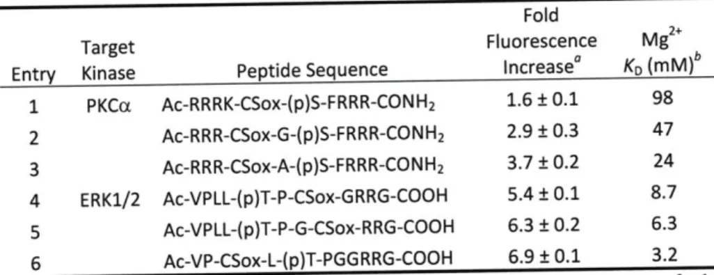
Chromophore Positioning in the RDF Design ... ... 44
Fluorescence Properties of RDF Probes ... 47
Fluorescence Intensity Dependence on [ATP] and [Mg2+] ... ... 49
O rigin of Fluorescence Increase ... 51
Fluorescence Difference under Biochemical Assay Conditions... ... 53
Fluorescence Increase Corresponds to Product Formation... ... 54
Comparison of Kinetic Parameters for BTF and RDF Chemosensors ... 55
Conclusions... 59
Acknowledgements... 60
E xperim ental M ethods ... 61
NMR spectra ... 88
H PL C chrom atogram s ... 9 1 R eferen ces ... 92
Chapter 3.
Development of a Mass Spectrometry-Based Method for
Rapid Discovery of Selective Recognition-Domain
Focused Sensors ...
95
Introduction... 96
Results and Discussion ... 99
Sensors for Aurora A ... ... Development and Validation of the MS-based Screening Method ... Conclusions ... ... Future Directions ... ... A cknow ledgm ents... ... Experim ental M ethods ... ... References ... ...
Chapter 4.
Highly Selective Chimeric Reporters for ERK1/2 in
Cellular Media ...
140
Introduction... 141
R esults and D iscussion ... 145
Semisynthesis of the Sox-PNT Sensor ... 145
Evaluation of the Sox-PNT Probe ... 148
Studies with Crude Cell Lysates ... 151
Direct Titrations of Inhibitor PEA-15 into EGF-stimulated Cell Lysates ... 154
Immunodepletion of ERK1/2 from EGF-stimulated Lysates ... 154
Mircoinjection of Sox-PNT into PtK-1 Cells ... 156
Membrane-Permeable Sox-PNT-TAT ERK1/2 Sensor... 158
Membrane-Targeted Sox-PNT-CAAX ERK1/2 Sensor... 160
Caging the Thr Side Chain to Provide Temporal Control of Phosphorylation... 162
Conclusions... 166
Acknowledgements... 168
E xperim ental M ethods ... 169
References ... 197
Chapter 5. Toward 8-Hydroxyquinoline Derivatives with Improved Photophysical Properties as Reporter Moieties for Phosphorylation ... 202
Introduction... 203
R esults and D iscussion ... 207
Fused Tricyclic Derivatives of Oxn ... 209
Bicyclic Derivatives of Oxn... 212
Synthesis and Screening of Triazole-substituted Oxn Derivatives... 214
Synthesis and Biophysical Evaluation of Clk-based Peptidyl Kinase Substrates... 217
Evaluation of Clk-based Substrates in Enzymatic Assays... 220
Conclusions... 221
Future Directions and Perspectives... 222
Acknowledgements... 224
Experim ental M ethods ... ... 225
G eneral Inform ation ... 225
Instrumentation and Materials ... 225
Synthesis and Characterization of Oxine derivatives ... 227
General Synthesis of the Triazolyl Derivatives 77a-v ... 228
8 103 116 122 123 123 124 137
Peptide Synthesis ... 232
Stock solutions ... 234
Fluorescence experim ents ... 235
NM R Spectra ... 239
References ... 252
Chapter 1 Figure 1-1. Figure 1-2. Figure 1-3. List of Figures Figure Figure Figure 1-4. 1-5. 1-6.
General scheme of kinase-catalyzed phosphoryl transfer...20
A dendrogram of the human kinome. ... ... 21
A scheme summarizing known interactions between constituents of cell-m atrix adhesions. ... 23
Sensing strategies employed to detect kinase activity ... 26
Strategies that protein kinases employ to gain specificity in vivo...30
CH EF-based chem osensors. ... 35
Schematic representations of the BTF and RDF designs from optimized non-fluorescent substrates...41
Structures of amino acids Sox (1) and C-Sox (2) that can be used to install the Sox chromophore ... 42
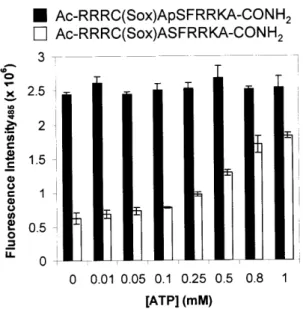
The effect of [ATP] on RDF probe fluorescence...50
M g2+ titration curves. ... ... 52
HPLC chromatogram of the PKA reaction with its RDF sensor after 616 sec of reaction tim e. ... ... 55
Assays of PKC3I and PKC6 enzymes with RDF and BTF chemosensors ... 58
Reaction rates (slopes) of PKCa RDF substrate, PKC3I RDF substrate, PKC6 RDF substrate, and PKCca BTF substrate with PKC isozymes. ... 101
Fold fluorescence difference at 0, 0.1 and 1.1 mM ATP for AurA-S15-S 2 1...10 8 Activation of AurA by TPX2 ... 109
The activity of AurA measured with AurA-S6 in the presence (black bars) or absence (white bars) of TPX(2-43). ... 110
Qualitative comparison of reaction rates of chemosenors with AurA ... 111
AurA docking models with test peptide (H2N-RFSLC(Npl)A-CO2H)...1...113
MS-based screening method to design selective substrates for Ser/Thr k in ases ... 1 18 MALDI-TOF spectrum of the peptide library at the -3 position after its incubation with PKA for 1 h and chemical derivatization with Ba(OH)2 /4-M E P . ... ... 120
The mechanisms of ERK-mediated cell proliferation or apoptosis ... 142
ERK utilizes the PNT domain of its substrate Ets- 1 to drive specificity ... 144
The semisynthesis of the Sox-PNT sensor via NCL... 147
Fluorescence spectra of phosphorylated and unphosphorylated versions of Sox-Peptide and Sox-PNT sensors ... ... 149
In vitro characterization of Sox-PNT ... 151
The EGF signaling pathway results in stimulation of ERK1/2 activity... 152
Chapter 2 Figure 2-1. Figure 2-2. Figure 2-3. Figure 2-4. Figure 2-5. Figure 2-6. Chapter 3 Figure 3-1. Figure 3-2. Figure 3-3. Figure 3-4. Figure Figure Figure 3-5. 3-6. 3-7. Figure 3-8. Chapter 4 Figure 4-1. Figure 4-2. Figure 4-3. Figure 4-4. Figure 4-5. Figure 4-6.
Figure 4-7. Figure 4-8. Figure 4-9. Figure 4-10. Figure 4-11. Figure Figure Figure Figure Figure Figure Figure Figure Figure Figure Figure 4-12. 4-13. 4-14. 4-15. 4-16. 4-17. 4-18. 4-19. 4-20. 4-21. 4-22. Chapter 5 Figure 5-1. Figure 5-2. Figure 5-3. Figure 5-4. Figure 5-5.
Specificity of the Sox-PNT sensor toward ERK1/2 in unfractionated cell
lysates ... 153
Half inhibitory concentration of PEA- 15 with ERK 1/2. ... 154
ERK1/2 activity in immunodepleted and EGF-stimulated lysates ... 155
Overlaid, false-colored images of a panel of cells microinjected with Sox-PNT and Alexa Fluor 568-labeled dextran ... ... 157
Overlaid raw and false-colored images of a cell microinjected with Sox-PNT and Alexa Fluor 568-labeled dextran ... ... 157
The semisynthesis of the Sox-PNT-TAT sensor via NCL ... 159
The semisynthesis of the Sox-PNT-CAAX sensor via NCL ... 161
The rate of uncaging for Sox-Isopeptide. ... 166
The GST-PNT-His6 product. ... 177
Comparison of Sox-PNT and Sox-peptide as substrates for ERK2 ... 182
Detection of ERK1/2 activity by Sox-PNT in lysates from four cell lines...185
Effects of DMSO on ERK1/2 activation. ... 186
Specificity of substrates for ERK. ... 187
Immunodepletion of ERK1/2 ... 189
The GB 1 -PNT-TAT-FGT-His6 product. ... 191
The GST-PNT-FGT-His6-CAAX product ... 193
Derivatives of 8-hydroxyquioline-based amino acids used to report kinase activ ity ... ... 20 5 The novel C-Clk amino acid based on the Oxn core. ... 207
Visual comparison of emission wavelengths and fluorescence intensity for some Oxn derivatives when complexed with Mg2+ ... 213
Qualitative comparison of emission wavelengths of 77a-v ... 2...16
List of Schemes Chapter 2 Scheme 2-1. Chapter 4 Scheme 4-1. Scheme 4-2. Scheme 4-3. Scheme 4-4. Chapter 5 Scheme 5-1. Scheme 5-2. Scheme 5-3. Scheme 5-4. Scheme Scheme Scheme Scheme Scheme Scheme 5-5. 5-6. 5-7. 5-8. 5-9. 5-10.
Synthesis of RDF Chemosensors on Solid Support and Using the Building
B lock A pproach ... ... 44
Synthesis of Caged Thr Amino Acid ... 163
Synthesis of Backbone-Caged Sox-Peptide... 163
Synthesis of Sox-Isopeptide... 164
Uncaging of Sox-Isopeptide and Spontaneous O to N acyl shift to form Sox-Peptide ... 165
A General Representation of CHEF upon Divalent Metal Binding ... 204
Synthesis and Chemical Structures of Oxn Derivatives ... 208
Synthesis of B O xn ... ... 210
Installation of Dimethylsulfonamide and Oxidation to Quinone upon Methoxy Deprotection ... 211
1,3-Dipolar Cycloaddition Reactions of 75 with 76a-v ... 215
Synthesis of the Clk RDF Chemosensors ... 218
Synthesis of an Alternative Tricyclic Core ... .... 222
Synthesis of 5-ArylOxn Derivatives ... ... 223
Preparation of C5 Aryl-substituted BOxn... ... 223
Consensus Phosphorylation Sites of a Representative Sampling of Protein
K inases ... 31
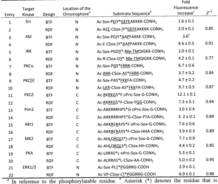
Table 2-1. Peptide Sequences Used to Determine Optimal Positioning of the Sox Chromophore in the RDF Design ... 45
Table 2-2. Fluorescence Increase and Z' Factor Values for Ser/Thr and Tyr Kinase Substrates Described in Table 2-1 ... ... 46
Table 2-3. Comparison of Substrate Sequences and Fluorescence Increases for BTF and RDF Chemosensors... ... ... 48
Table 2-4. Effects of ATP Concentration on Fold Fluorescence Increase of RDF C hem osensors ... ... 50
Table 2-5. Effects of [Mg2+] and [ATP] on Fold Fluorescence Increasea of RDF Tyr Kinase Chem osensors ... ... 51
Table 2-6. Peptide Sequences, Fluorescence Differences and Dissociation Constants for M g2+ . ... ... . . . . 52
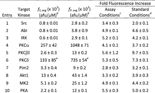
Table 2-7. Fold Fluorescence Increases of RDF Chemosensors Obtained from Substrate (fs) and Phosphopeptide (fp) Intensities under Appropriate Assay Conditions or Standard Conditions ... ... 54
Table 2-8. HPLC and ESI-MS Verification and Quantification of Product Formation Observed by Fluorescence ... 55
Table 2-9. Comparison of Kinetic Parameters Obtained with BTF and RDF Substrate Sequences Presented in Table 2-3... ... 57
Chapter 3 Table 3-1. Table 3-2. Table 3-3. Table Table Table Table Table Table Table Table 3-4. 3-5. 3-6. 3-7. 3-8. 3-9. 3-10. 3-11. Substrate Preferences of 9 PKC Isozymes ... ... 97
Kinetic Parameters of PKC RDF Chemosensors and the Parent Peptides ... 100
The Kinetic Parameters for PKCa RDF Substrate with PKCa, PKC3I and PK CO Isozym es... ... 102
Kinetic Parameters of PKCa Substrate with PKCa, PKC3I and PKC6 ... 102
Sequences of RDF Chemosensors for AurA ... 105
Fold Fluorescence Increase for Selected AurA Chemosensors ... 107
Select Chemosensors were Tested for Reactivity with AurA and AurB... 115
Kinetic Parameters of Selected Chemosensors with AurA... 116
Peptide Libraries for PKA ... 19
Peptide Libraries for AurA ... 121
Sequences and Kinetics of Sox-substrates for AurA ... 122
Chapter 4 Table 4-1. Fold Fluorescence Increase of BTF and RDF Chemosensors in the Presence and Absence of ATP ... 148
Table 4-2. Sequences Surrounding the TP Recognition Region (Red Residues) of the Wild Type Ets-1, MBP and Sox-PNT... 148
List of Tables
Chapter 1 Table 1-1.
Chapter 5
Table 5-1. Relevant Spectroscopic Data for Selected Oxn Derivatives that Form
Fluorescent Com plexes with M g2+ ... 214
Table 5-2. Substrate Sequences of the RDF Chemosensors and Their Fluorescence
List of Abbreviations
Standard 3-letter and 1-letter codes are used for the 20 natural amino acids. Where D precedes the amino acid code, it indicates D-chirality.
Standard 1-letter codes are used for the nucleotides.
afu arbitrary fluorescence unit
Abl Abelson kinase
ADP adenosine-5'-diphosphate
AFP Aequoria victoria fluorescent protein
AIBN azobisisobutyronitrile
ATP adenosine-5'-triphosphate
AurA Aurora kinase A
AurB Aurora kinase B
BME P-mercaptoethanol
Bn benzyl
tBu t-butyl
Boc t-butoxycarbonyl
BOxn 10-hydroxy-2-methylquinoline
BSA bovine serum albumin
BTF P-turn focused
CamK calmodulin-dependent protein kinase
CDK cyclin-dependent kinase
CENP-A centromere protein A
CHEF chelation-enhanced fluorescence
CK casein kinase
Clk 5-(4-(4-bromophenyl)-1H-1 ,2,3-triazol- 1-yl)-2-methylquinolin-8-ol CPEB cytoplasmic polyadenylation element-binding proteins
CPP cell penetrating peptides
DAG diacylglycerol
DIC N,N-diisopropylcarbodiimide
DIEA N,N-diisopropylethylamine
DMAP 4-dimethylaminopyridine
DMEM dulbecco's modified eagle medium
DMF N,N-dimethylformamide
DMNB 4,5-dimethoxy-2-nitrobenzyl
DMSO dimethyl sulfoxide
DTT dithiothreitol
F extinction coefficient or molar absorbtivity
EDT 1,2-ethanedithiol
EDTA ethylenediaminetetraacetic acid
EGFR epidermal growth factor receptor
EGTA glycol-bis(2-aminoethylether)-N,N,N',N'-tetraacetic acid ERKi/2 extracellular signal-regulated kinase 1/2
ESI-MS electrospray ionization mass spectrometry
Fmoc FPLC FPR FTase GB1 GGTase GSK3 GST HeLa HEPES HIV-1 HOAt HOBt HPLC HRMS HT29 IC50 IRK JNK kcat KD Ki KM kem Xex MALDI-TOF MS MeCN 4-MEP MESNa MK2 Mmt NBS NIH-3T3 Nle NMR NMP NVOC Oxn Pbf PBS Pdb PET Pim2 PKA PKB/Akt 9-fluorenylmethoxycarbonyl fast protein liquid chormatography fluorescence plate reader
farnesyltransferase
immunoglobulin binding domain B 1 of streptococcal protein G geranylgeranyltransferase
glycogen synthase kinase-3 glutathione-S-transferase
cervical cancer cell line from Henrietta Lacks
4-(2-hydroxyethyl)-l-piperazine ethane sulfonic acid human immunodeficiency virus-1
7-aza-1 -hydroxybenzotriazole 1 -hydroxybenzotriazole
high performance liquid chromatography high resolution mass spectrometry
human colon adenocarcinoma grade II cell line half inhibitory concentration
insulin receptor kinase c-Jun N-terminal kinase catalytic constant dissociation constant inhibition constant Michaelis constant emission wavelength excitation wavelength
matrix-assisted laser desorption ionization time-of-flight mass spectrometry
acetonitrile
4-mercaptoethylpyridine
2-mercaptoethane sulfonate sodium
mitogen-activated protein kinase-activated protein kinase-2 4-methoxytrityl
N-bromosuccinimide mouse fibroblast cells Norleucine
nuclear magentic resonance N-methylpyrrolidinone
nitroveratryloxycarbonyl oxnie or 8-hydroxyquinoline
2,2,4,6,7-pentamethyldihydrobenzofuran-5-sulfonyl phosphate-buffered saline
protein data bank
photoninduced electron transfer proviral integration site kinase-2
protein kinase A or cAMP-dependent protein kinase protein kinase B (aka Akt)
PKC PKD PS pSer PTD pThr pTyr PtK PyAOP PyBOP Ras Rcel RDF RP-HPLC RT SDS-PAGE s.e.m. Sox-Br SPPS Src std. dev. TAT TBDMS TBDPS TEV TFA TIS TLC TMS TNBS tR Tris Trt UV-Vis Xaa Vmax vol% protein kinase C protein kinase D phosphatidylserine phosphoserine
protein transduction domain phosphothreonine
phosphotyrosine
Potorus tridactylus kidney cells
(7-azabenzotriazol- 1 -yloxy)tripyrrolidinophosphonium hexafluorophosphate
benzotriazol- 1 -yl-oxytripyrrolidinophosphonium hexafluorophosphate rat sarcoma
ras-converting enzyme recognition-domain focused
reverse-phase high performance liquid chromatography room temperature
sodium dodecyl sulfate polyacrylamide gel electrophoresis standard error of measurement
2-bromomethyl-8-t-butyldiphenylsilyloxy-5-(N,N-dimethyl)sulfonamidoquinoline
solid-phase peptide synthesis sarcoma kinase
standard deviation
transactivator of transcription t-butyldimethylsilyl t-butyldiphenylsilyl
tobacco etch virus trifluoroacetic acid triisopropylsilane
thin-layer chromatography tetramethylsilane
2,4,6-trinitrobenzene sulfonic acid retention time
2-amino-2-hydroxymethyl-propane-1,3-diol trityl
ultraviolet-visible
used to denote any amino acid maximum velocity
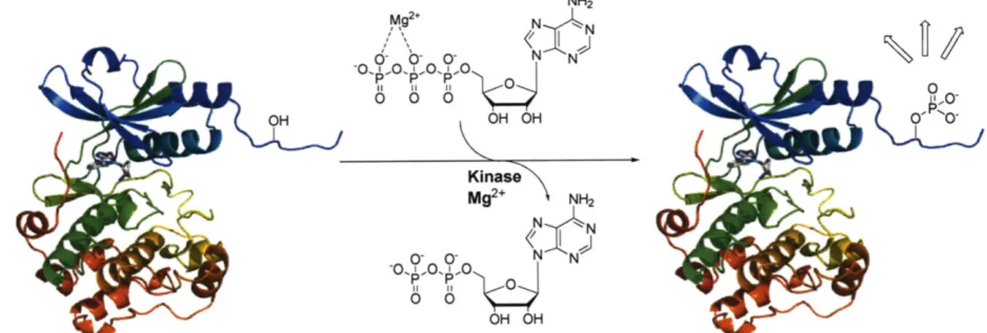
Protein Phosphorylation
Normal cell function heavily depends on protein phosphorylation to accurately relay
multifarious cues from the extracellular environment to appropriate cellular compartments and
proteins.' By catalyzing the transfer of the y-phosphoryl of adenosine-5'-triphosphate (ATP)
onto Ser, Thr, and/or Tyr side chains of protein substrates, kinases have emerged as crucial
regulators of this ubiquitous posttranslational modification.2' 3 Kinases are broadly classified into two major groups: Ser/Thr and Tyr kinases, which are further divided into either nonreceptor or
receptor kinases. Regardless of the classification, divalent metal ions, most commonly Mg2+, are
necessary for catalysis as they prime the nucleotide-triphosphate for nucleophilic attack by the
alcohol4 (Figure 1-1). Although the phosphate group is relatively small compared to the
remainder of the protein, the addition of a densely negatively charged phosphate moiety can
drastically alter protein conformation, biochemical function and/or binding to partner proteins.
These events, in turn, lead to further propagation of the signal until the target is reached. To
efficiently signal the ever-changing extracellular environment, phosphorylation needs to be
highly dynamic and temporally controlled; thus, cells also employ phosphatases to rapidly
remove the phosphate modification. Together with kinases, phosphatases tightly regulate this
OH OH
Figure 1-1. General scheme of kinase-catalyzed phosphoryl transfer. Hydrolysis occurs in the
presence of Mg2+, which is coordinated to the 3 and y phosphates of ATP. ADP and the phosphoproduct are then released.
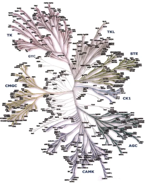
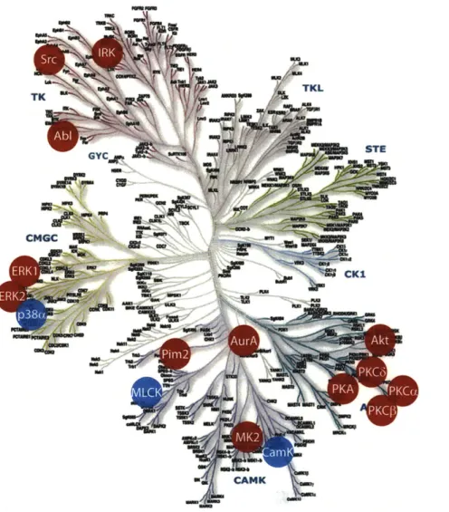
A recent study identified more than 500 different protein kinases encoded in the human
genome (Figure 1-2), constituting nearly 2% of all genes.6 Ser/Thr kinases are more abundant
(-400) than Tyr kinases, but not necessarily more important in cellular homeostasis. Both classes
of kinases modulate aspects of protein function in many ways. They are involved in the control
of subcellular localization, the degradation or stabilization of proteins, the assembly of
multimeric protein complexes, and the allosteric regulation of biochemical activity (e.g.,
activation or repression of an enzyme or transcription factor).7 Because of the ubiquitous
involvement of phosphoproteins in processes such as cell migration, proliferation, cell division,
cell death, immunity, and learning and memory, protein kinases are also instrumental to the
proper function of these listed proceses as enzymes that control the generation of
phosphoproteins.6-8 In addition, since kinases often phosphorylate proteins involved in several
different biochemical pathways, their down- or up-regulation often has effects on the whole cell,
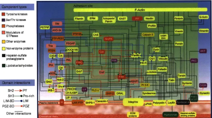
rather than just a single pathway. For example, illustrated in Figure 1-3 are some of the dynamic
interactions involved in cell migration that are mediated by kinases.9Thus, due to the critical role
that kinases play in many aspects of cell function, it is not surprising that the misregulation of
kinase activity is an underlying cause of many human diseases, from neurological disorders to
cancer.'0 U-TK -M Um TKL
I
* STE GYC rr'Ui CMGC --e ow M CK1 AGC CANK u'Figure 1-2. To date, the human kinome encompases 518 Ser/Thr and Tyr kinases divided in 9 major classes. Ser/Thr kinases are the most abundant (-400), followed by Tyr kinases (-90), while only -40 have the dual function. Dendrogram obtained from ref. 6 and www.cellsignal.com.
...
=0r
Mqt
WnM
Recently, much effort has been put forth in the area of drug discovery to identify
molecules that can modulate kinase activity for the treatment of disease. This work is partly
fueled by the remarkable initial success of imitanib mesylate (also known as Gleevec and
STI571) used to treat chronic mylogenous leukemia (CML), gastrointestinal stromal cell tumors
(GIST) and metastatic dermatofibrosarcoma protuberans by direct inhibition of mutated Abl,
c-Kit and PDGFR Tyr kinases, respectively."' 12 Gleevec was the first small molecule to
successfully block the constitutively active Abl kinase and also cause a marked improvement in
individuals suffering from CML. Interestingly, the precursor of Gleevec was discovered by labor
intensive screening of a large library of compounds for in vitro inhibition of protein kinase C
(PKC), a Ser/Thr kinase.13 Although poor inhibitors for PKC, the Gleevec precursor showed
good properties for Abl. Further elaboration lead to Gleevec and its use as an inhibitor of Tyr
kinases. Unfortunately, patients have started to show resistance to Gleevec, which has been
attributed to a single point-mutation in the BCR-ABL gene,14' 15 and further drug screening and
the development of follow-up compounds has been necessary.16
In light of the potential of kinases as therapeutic targets, selective, sensitive and high
throughput methods to detect kinase activity are not only critical in studying the mechanisms of
cellular processes, but are also necessary for the efficient search of potent inhibitors for disease
treatment. As illustrated with Gleevec, kinases are attractive medical targets, but as cancer is
highly adaptive, our search for a complete understanding of roles that kinases play and for more
F-Ain E Tyrosine kinases * Sertrhr kinoase * Phosphutaspes * Modulators of GTPases ] Other onzymes [] Non-enzyme proteins * Hoparan-sulftbe protooglyans * Lipid scarbohyd rauts
SH2 -- IPY
SH3--- Pro-rich
LIM-BO----e LIM PDZ-BD-- PDZ
Other interactions
Figure 1-3. Scheme summarizing known interactions between constituents of cell-matrix
adhesions, illustrating the intricacy of kinase involvement with mutliple targets. Figure obtained from ref. 9 and www.cellmigration.com.
Detection of Kinase Activity
Traditional assays for kinase activity use either phosphopeptide-specific antibodies or,
more commonly, [y-32P]ATP, where transfer of the radioactive y-phosphoryl group to peptide or
protein substrate is quantified by liquid scintillation counting. Although general and sensitive,
[32P]-based assays are not compatible with physiological concentrations of ATP, real-time analysis or high-throughput kinetic determination. These assays require special handling,
generate radioactive waste, and lose much of the kinetic information due to their intrinsic
end-point nature. Furthermore, because [y-32P]ATP is generally required by all kinases, this method
cannot be applied in environments where multiple kinases are present.
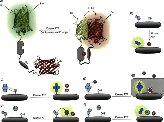
Conversely, continuous assays that rely on fluorescence changes upon sensor
phosphorylation are ideal for high throughput screening (HTS) and thus make discovery of new
inhibitors and substrates with enhanced specificity much more practical. Fluorescence-based
assays may also be compatible with cell lysates, living cells and physiological ATP
concentrations. Existing fluorescent protein- and peptide-based sensors exploit a variety of
detection methods, such as fluorescence polarization, fluorescence resonance energy transfer
(FRET), fluorescence quenching, metal ion-mediated fluorescence and solvatochromism, and
have been recently reviewed.7' 17-19 Herein we will review some of the more recent and practical
approaches for sensing protein phosphorylation (Figure 1-4).
The probes that employ FRET between two Aequorea victoria fluorescent proteins
(AFPs) are perhaps the most widely used for examining kinase activity because they can be
genetically encoded and expressed within cells.2 0 Generally, AFP-based sensors contain a kinase
phosphorylation sequence, a linker region, a phosphopeptide-binding domain, and two AFP
analogs with appropriate photophysical properties, such as CFP and YFP, situated on the termini
of the probe (Figure 1-4a). In these sensors, phosphorylation leads to a conformational change in
the protein, a result of phosphopeptide binding to its cognate recognition region, thereby altering
the distance between the AFPs. Because the distance is related to the efficiency of FRET,
phosphorylation produces a fluorescence response (Figure 1-4a). Accordingly, the fluorescent
signal can either increase or decrease depending on the changes in distance between the AFPs
upon phosphorylation, producing turn-on or turn-off sensors, respectively. Turn-off probes are
less desirable since loss of signal can be mistaken for photobleaching and can affect the accuracy
of readings. Genetically encoded FRET-based reporters have been developed for many kinases
ERK.22 In addition, recent impressive optimization of the AFP-based PKA sensor now allows for
small molecule inhibitor screening in cells, thus expanding the utility of these types of probes.23
Although several AFP analogs containing complementary photophysical properties for FRET
have been developed, and in theory can be applied for dual-kinase sensing, the maximal change
in signal can be fairly low (- 50%), leading to reduced sensitivity and more challenging
fluorescent readouts. Additionally, the large size of the AFP-based sensors (at least 50 kDa for
the two AFPs in each sensor) can be quite disruptive when studying endogenous kinases that
may depend on interactions with other proteins for activity.
On the other hand, peptide-based probes offer distinct advantages over the protein
counterparts, such as relatively small size, ease of large-scale synthesis, and ability to introduce
unnatural elements to aid selectivity and/or sensitivity.17, 24 Importantly, because they generally
do not depend on FRET, the maximal observed change in signal is often well above 100%. This
is a significant benefit, as larger fluorescence changes allow for improved sensitivity, which is
essential when studying endogenously expressed enzymes that are present in reduced amounts or
/ E FRET Ex Kinase, ATP Conformational Change Em a) Ex C) Kinase, ATP OH Kinase, ATP
Figure 1-4. Sensing strategies employed to detect kinase activity. a) AFP-based sensors depend
the change in FRET upon phosphorylation due phosphopeptide-domain binding. b) Solvatochromic chromophores experience a change in solvent polarity after phosphorylation leading to an increase in quantum yield that results in fluorescence enhancement. c) Quenching of pyrene fluorescence by a nearby Tyr residue is disrupted by phosphorylation that enables an enhancement of the fluorescent signal. d) In the "Deep Quench" method, a non-covalent quencher alters the fluorescence of pyrene. After phosphorylation, phosphopeptide-specific protein domain binds to the product and disrupts quenching, giving rise to fluorescence. e) These sensors have low affinity for Ln3+, which significantly increases after the phosphate is present. A nearby antenna (i.e., Trp) can sensitize the peptide-bound Ln3+, which produces a detectable, long-lived luminescence. f) Similarly, sensors bearing CHEF chromophores are silent until phosphorylation enables divalent metal binding, which causes an increase in fluorescence.
The Lawrence group has reported several types of kinase reporters, many of them
utilizing solvatochromic fluorophores that exhibit changes in their fluorescence properties, such
Kinase ATP Kinase, ATP Kinase, ATP I .4)
Y ~
as emission wavelength and quantum yield, due to the polarity of the immediate environment.
Introduction of a phosphoryl group near the chromophore alters the local environment polarity,
resulting in fluorescence change (Figure 1-4b). Notably, this strategy has been used to visualize
PKC activity within live HeLa cells.2 5 In addition, sensors employing quenching strategies have
also been explored.26 For example, when a pyrene moiety is positioned in close proximity to a
Tyr reside, the overall fluorescence is quenched due to xc-7 stacking interactions, reducing
fluorescence quantum yields. The size and charge of the phosphate group disrupts the
pyrene-tyrosine interaction and results in a fluorescent phosphopeptide (Figure 1-4c).27 However, the
method is not applicable to Ser/Thr kinases, as x-r- interactions do not directly contribute to
diminishing pyrene fluorescence. Instead, a modified quenching strategy, called "Deep Quench"
was developed by the Lawrence group for Ser/Thr kinases (Figure 1-4d). In the new system, the
fluorescence was modulated using an in-solution quencher molecule, such as Rose Bengal, that
bound to substrate-tethered pyrene. After phosphate installation, the peptide was sequestered by
a phospho-peptide binding domain (such as 14-3-3-) away from the non-covalent quencher,
thereby restoring fluorescence signal. Such strategy has furnished 21- to 64-fold
phosphorylation-induced fluorescence enhancements, the most dramatic change reported to
date.28 Despite these promising results, the method has not been extended beyond PKA
substrates. One reason for this may be that the probe indirectly senses phosphorylation and
requires several separate components, such as the quencher and the phospho-product binding
domain to be present. This may be problematic if the sensor is to be used in more complex
media, such as cells and cell lysates.
Several strategies that rely on metal ion-mediated luminescence or fluorescence to detect
low metal ion affinity and are predominantly non-fluorescent. However, modification with the
phosphate provides two essential ligands, resulting in a significant increase in the avidity for
binding to hard metal ions such as magnesium or the lanthanide ions, and a subsequent positive
signal. Peptides based on this general strategy have recently been designed to bind lanthanide
ions (Ln3+) such as Tb3+ or Eu3+.29,
30 In these cases, the obligatory nearby antenna (Trp or
carbostyril) absorbs light and sensitizes Ln3+, which produces long-lived luminescence to signal
kinase activity (Figure 1-4e). The peptides produce significant changes (up to 10-fold) in signal
upon phosphorylation; however, the system requires require the presence of free Tb3+ or Eu3+,
which might be problematic if this approach is attempted in cells due to the need for delivering
the lanthanide ion into cells that may be toxic.
On the other hand, chelation-enhanced fluorescence (CHEF) sensing methods utilize
biologically available metal ions, such as Ca2+ and Mg2+, to report phosphorylation (Figure 1-4f).
For example, a CHEF sensor for PKC has exploited elevated Ca2+ levels required for proper
PKC activation and previously reported Ca2+ indicators.31 However, this approach has not been
adapted for use with other ions or to monitor additional kinases.'7 Alternatively, probes
developed in the Imperiali laboratory contain the sulfonamido-oxine (Sox) amino acid32 that
displays good fluorescence increases (4- to 12-fold) upon phosphorylation and binding of
Mg2+.33 Aside from robust fluorescence changes, the use of physiological levels of Mg2+ (low millimolar)34 makes this approach generally useful not only for multiple kinases, but also in a
variety of conditions that are necessary for optimal kinase fuction. The method, termed the
P-turn focused (BTF) design, has been successfully applied in the development of chemosensors
that monitor kinases both in vitro35 and in unfractionated cell extracts.36 However, although
due to the limitations imposed by the requirement for a conformationally-constrained P-turn
motif.
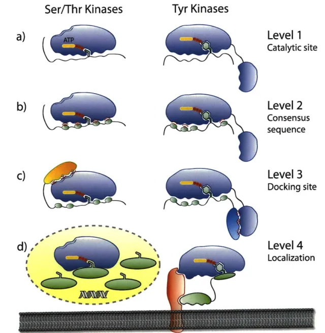
Kinase Specificity
One of the major challenges in the field of kinase analysis is sensor specificity in the
presence of multiple kinases, particularly since most fluorescent sensors employ short peptide
sequences to specify their target. Similar challenges are also encountered inside cells among
kinases and their endogenous substrates. For instance, it has been estimated that 30% of all
cellular proteins are phosphorylated on at least one residue, which translates into nearly 700,000
potential phosphorylation sites for any given kinase.37-39 Although many eukaryotic protein
kinases are structurally homologous,4 0 they often exibit clear preferences for certain substrates
over others. Therefore, our hope is to exploit some methods that endogenous kinases employ to
select their substrates in the development of a new generation of highly targeted sensors.
In vivo substrate specificity is complex and is governed by a number of factors. The first
level of specificity is dictated by the kinase active site and the consensus sequence on the
substrate. Generally the catalytic clefts can either accommodate the Ser/Thr or Tyr side chain
(Figure 1-5a); kinases with dual functionality are less common (-40 out of 518).39 Additionally,
a substantial contribution to substrate recognition comes from the residues that surround the
phsophorylation site (Figure 1-5b). It has been found that typically between 4 to 8 amino acids
are responsible for this substrate specificity, and several laboratories have explored the optimal
Ser/Thr Kinases
Level
1
Catalytic sitea)
b)
c)
Figure 1-5. Strategies that protein kinases employ to gain specificity in vivo. a) The first level of
specificity is found at the catalytic site, which can generally accomodate either Ser/Thr or Tyr side chains. Only -40 of 518 kinases have dual, Ser/Thr and Tyr, capabilities. b) The second tier involves recognition of 4-8 residues surrounding the phosphorylatable amino acid. c) Docking domain interactions are utilized by both types of kinases when the consensus sequence is not specific enough. d) Localization, either to an organelle or to a scaffolding protein, increases the effective local concentration of enzymes and substrates, resulting in an enhanced rate of reaction.
Level 2
Consensus
sequence
Level 3
Docking site
rriTyr Kinases
~-C~Sc~
Table 1-1. Consensus Phosphorylation Sites of a Representative Sampling of Protein Kinasesa
Consensus sequence
Kinase Full name -5 -4 -3 -2 -1 0 +1 +2 +3 +4
PKA Protein kinase A R R X S/T cI
CDK Cyclin-dependent kinase S/T P X K/R
ERK2 Extracellular signal-regulated kinase-2 P X S/T P
CK1 Casein kinase-1 pS X X S/T
CK2 Casein kinase-2 S/T D/E X E/D
GSK3 Glycogen synthase kinase-3 S X X X pS
CaMK2 Calmodulin-dependent protein kinase-2 R X X S/T
AbI Abelson murine leukemia virus tyrosine
kinase I/V/L Y X X P/F
EGFR Epidermal growth factor receptor E E E Y F
Src Rous sarcoma virus tyrosine kinase E E I Y E/G X F
IRK Insulin receptor tyrosine kinase Y M M M
PKB/Akt Protein kinase B R X R X X S/T
PKD Protein kinase D L/I X R X X S/T
Piml-3 Proviral integration site kinases 1-3 R X R X X S/T
a Adapted from ref. 39. X = any amino acid, <D = hydrophobic amino acid, pS =
phospho-serine.
If the consensus sequence alone cannot provide a stringent level of selectivity, often the next specificity determinant involves interactions between docking motifs on the substrate and interaction domains on the kinase (Figure 1-5c). These interaction domains are commonly short
sequences of amino acid residues.44 Such a mechanism has been identified for both Ser/Thr (JNKs, PHK, ERK, MEK, CDK2) and Tyr (TGFBR) kinases. Interestingly, Ser/Thr kinases display docking sites on the catalytic domain, while in Tyr kinases the analogous sites are generally found in modular domains that are separate from the kinase domains, such as Src-homology-2 (SH2), SH3, integrin binding, focal adhesion binding, DNA binding, or Janus tyrosine kinase (JAK) homology domains.4 5 Because of the separation of docking and catalytic domains in the case of Tyr, it has been proposed that Ser/Thr phosphorylation evolved before
interaction partners.46 Conversely, because Tyr kinases evolved later, they progressed toward
using multiple modular interaction domains to drive specificity (Figure 1-5c). Regardless, in both
instances the function of docking motifs is thought to primarily be a mechanical method to
increase the local concentration of the substrate around the kinase. For instance, within a 10 nm
radius from the kinase, substrate effective concentration is proposed to reach 3 mM,4 7 which
would greatly accelerate the rate of phosphorylation of a scarce substrate.
Similarly, localization of the kinase to distinct cellular compartments or structures can
enhance the rate of phosphorylation or promote specificity by limiting the access to the number
of available substrates.39' 46 Additionally, some substrates and kinases also depend on adaptor or
scaffold proteins, which act as dynamic organizing platforms for many molecules to bring them
together (Figure 1-5d).4 8 This is yet another mechanism of localization that serves to increase the
substrate concentration and results in an enhanced overall rate of reaction.
In conclusion, it is evident that in vivo kinases-substrate recognition is a multi-tiered
process that often utilizes multiple strategies in order to achieve a high level of specificity.
However, current fluorescent kinase sensors generally employ short consensus sequences as
selection guides. As the field moves toward cellular work, such probes may not be able to
provide the requisite selectivity, and additional recognition determinants used by native kinases
should be explored. For example, taking advantage of a docking domain on a kinase may turn a
mediocre sensor into a species that is highly specific because it may behave more like an
endogenous substrate. Moreover, a sensor for a specific kinase may be modified to localize it to a
particular cellular compartment or a scaffolding protein, and could, thus, be used to define and
enzymes in cellular environments, designs of kinase sensors will also need to take advantage of
the different strategies that endogenous kinases employ to recognize their substrates.
Dissertation Objectives
Highly modular sensors that can quickly be adapted to take advantage of various
recognition strategies that native kinases employ are of great importance in mapping kinase
networks. This is particularly the case as the use of kinase probes moves from relatively simple
assays with recombinant enzymes toward more complex environments. In order to introduce
fluorescent kinase sensors into cells and obtain relevant data, much work needs to be done to
resolve issues surrounding probe cross-reactivity and sensitivity.
The goal of the work presented in this dissertation is to create sensors with high
selectivity toward kinases and to utilize the CHEF reporting platform previously developed in
the laboratory.33' 35, 36 One way to develop sensors that are better able to discriminate between
multiple kinases, whether in vitro, in vivo or in cell lysates, is by improving the kinetic
parameters, for example, to lower the KM while keeping the Vmax high. This was accomplished by
extending the consensus sequence to include additional amino acids that were absent in the
original BTF probes, due to design requirements. The resulting recognition-domain focused
(RDF) sensors displayed marked improvements in catalytic efficiency (kcat/KM) for multiple
Ser/Thr and Tyr kinases (Chapter 2). Furthermore, to rapidly identify additional specificity
determinants in short peptides, a high-throughput method was also developed that allowed us to
screen individual positions in the consensus sequence with natural and unnatural elements. This
approach yielded an improved peptide sensor for Aurora A kinase (Chapter 3) and should be
In addition, for some kinases, such as ERK1/2, short peptides, based on consensus
sequences do not impart much selectivity. In the development of a sensor for ERK1/2, we took
advantage of important docking interactions that MAPK kinases, such as ERKI/2, often utilize
to differentiate their substrates. A key native chemical ligation (NCL) was employed to build a
chimeric sensor that included the CHEF-sensing moiety and a docking domain specific for
ERK1/2 (Chapter 4). Moreover, the same strategy was applied in the semisynthesis of a
membrane-targeted sensor that will be used to specifically examine the role of ERK1/2 in
migration at the cell edge.
Lastly, in order to be able to monitor kinases in cellulo, we sought to develop new CHEF
derivatives based on the oxine chromophore with improved photophysical properties. Chapter 5
presents the synthesis and characterization of several derivatives that display the required
bathochromic shifts in excitation and emission wavelengths as well as useful quantum yields.
The variety of methods presented in this thesis were used to synthesize sensors for 13
kinases, from Ser/Thr as well as receptor and nonreceptor Tyr families (Figure 1-6). The
high-throughput approaches used here to extend sensor selectivity and sensitivity toward the cognate
kinase should prove to be useful and complementary tools in the quest for better understanding
CMdGC ' ~
-
K1
Am no
n o am;;
S CAMK m
Figure 1-6. CHEF-based chemosensors. Thirteen kinases have been targeted with RDF sensors
(red), while work is ongoing toward the synthesis and validation of several more reporters (blue). Dendrogram obtained from ref. 6 and www.cellsignal.com.
References
1. Hunter, T. Signaling-2000 and beyond. Cell 2000, 100, 113-127.
2. Johnson, L. N.; Lewis, R. J. Structural basis for control by phosphorylation. Chem. Rev.
2001, 101, 2209-2242.
3. Cohen, P. The origins of protein phosphorylation. Nat. Cell Biol. 2002, 4, E127-130. 4. Adams, J. A. Kinetic and Catalytic Mechanisms of Protein Kinases. Chem. Rev. 2001,
101, 2271-2290.
5. Hunter, T. Protein kinases and phosphatases: the yin and yang of protein phosphorylation and signaling. Cell 1995, 80, 225-236.
6. Manning, G.; Whyte, D. B.; Martinez, R.; Hunter, T.; Sudarsanam, S. The Protein Kinase Complement of the Human Genome. Science 2002, 298, 1912-1934.
7. Turk, B. E. Understanding and exploiting substrate recognition by protein kinases. Curr. Opin. Chem. Biol. 2008, 12, 4-10.
8. Manning, G.; Plowman, G. D.; Hunter, T.; Sudarsanam, S. Evolution of protein kinase signaling from yeast to man. Trends Biochem. Sci. 2002, 27, 514-520.
9. Zamir, E.; Geiger, B. Molecular complexity and dynamics of cell-matrix adhesions. J. Cell Sci. 2001, 114, 3583-3590.
10. Cohen, P. Protein kinases--the major drug targets of the twenty-first century? Nat. Rev. Drug Discov. 2002, 1, 309-315.
11. Dancey, J.; Sausville, E. A. Issues and progress with protein kinase inhibitors for cancer treatment. Nat. Rev. Drug Discov. 2003, 2, 296-313.
12. Sawyers, C. Targeted cancer therapy. Nature 2004, 432, 294-297.
13. Roskoski, R., Jr. STI-571: an anticancer protein-tyrosine kinase inhibitor. Biochem. Biophys. Res. Commun. 2003, 309, 709-717.
14. Shah, N. P.; Nicoll, J. M.; Nagar, B.; Gorre, M. E.; Paquette, R. L.; Kuriyan, J.; Sawyers, C. L. Multiple BCR-ABL kinase domain mutations confer polyclonal resistance to the tyrosine kinase inhibitor imatinib (STI571) in chronic phase and blast crisis chronic myeloid leukemia. Cancer Cell 2002, 2, 117-125.
15. Gorre, M. E.; Mohammed, M.; Ellwood, K.; Hsu, N.; Paquette, R.; Rao, P. N.; Sawyers, C. L. Clinical Resistance to STI-571 Cancer Therapy Caused by BCR-ABL Gene Mutation or Amplification. Science 2001, 293, 876-880.
16. Shah, N. P.; Tran, C.; Lee, F. Y.; Chen, P.; Norris, D.; Sawyers, C. L. Overriding Imatinib Resistance with a Novel ABL Kinase Inhibitor. Science 2004, 305, 399-401. 17. Rothman, D. M.; Shults, M. D.; Imperiali, B. Chemical approaches for investigating
phosphorylation in signal transduction networks. Trends Cell Biol. 2005, 15, 502-510. 18. Shanna, V.; Wang, Q.; Lawrence, D. S. Peptide-based fluorescent sensors of protein
kinase activity: Design and applications. Biochim. Biophys. Acta, Proteins Proteomics
2008, 1784, 94-99.
19. Li, Y.; Xie, W.; Fang, G. Fluorescence detection techniques for protein kinase assay. Anal. Bioanal. Chem. 2008, 390, 2049-2057.
20. Zhang, J.; Allen, M. D. FRET-based biosensors for protein kinases: illuminating the kinome. Mol. Biosyst. 2007, 3, 759-765.
21. Wang, Y.; Botvinick, E. L.; Zhao, Y.; Berns, M. W.; Usami, S.; Tsien, R. Y.; Chien, S. Visualizing the mechanical activation of Src. Nature 2005, 434, 1040-1045.
22. Harvey, C. D.; Ehrhardt, A. G.; Cellurale, C.; Zhong, H.; Yasuda, R.; Davis, R. J.; Svoboda, K. A genetically encoded fluorescent sensor of ERK activity. Proc. Natl. Acad. Sci. U.S.A. 2008, 105, 19264-19269.
23. Zhang, J.; Ma, Y.; Taylor, S. S.; Tsien, R. Y. Genetically encoded reporters of protein kinase A activity reveal impact of substrate tethering. Proc. Natl. Acad. Sci. U.S.A. 2001, 98, 14997-5002.
24. Lawrence, D. S.; Wang, Q. Seeing is believing: peptide-based fluorescent sensors of protein tyrosine kinase activity. ChemBioChem 2007, 8, 373-378.
25. Yeh, R. H.; Yan, X. W.; Cammer, M.; Bresnick, A. R.; Lawrence, D. S. Real time visualization of protein kinase activity in living cells. J. Biol. Chem. 2002, 277,
11527-11532.
26. Sharma, V.; Wang, Q.; Lawrence, D. S. Peptide-based fluorescent sensors of protein kinase activity: design and applications. Biochim. Biophys. Acta 2008, 1784, 94-99. 27. Wang, Q.; Cahill, S. M.; Blumenstein, M.; Lawrence, D. S. Self-reporting fluorescent
substrates of protein tyrosine kinases. J. Am. Chem. Soc. 2006, 128, 1808-1809.
28. Sharma, V.; Agnes, R. S.; Lawrence, D. S. Deep quench: An expanded dynamic range for protein kinase sensors. J. Am. Chem. Soc. 2007, 129, 2742-2743.
29. Balakrishnan, S.; Zondlo, N. J. Design of a protein kinase-inducible domain. J Am. Chem. Soc. 2006, 128, 5590-5591.
30. Tremblay, M. S.; Lee, M.; Sames, D. A luminescent sensor for tyrosine phosphorylation. Org. Lett. 2008, 10, 5-8.
31. Chen, C. A.; Yeh, R. H.; Lawrence, D. S. Design and synthesis of a fluorescent reporter of protein kinase activity. J. Am. Chem. Soc. 2002, 124, 3840-3841.
32. Shults, M. D.; Pearce, D. A.; Imperiali, B. Modular and Tunable Chemosensor Scaffold for Divalent Zinc. J. Am. Chem. Soc. 2003, 125, 10591-10597.
33. Shults, M. D.; Carrico-Moniz, D.; Imperiali, B. Optimal Sox-based fluorescent chemosensor design for serine/threonine protein kinases. Anal. Biochem. 2006, 352, 198-207.
34. Haugland, R. P.; Spence, M. T. Z.; Johnson, I. D.; Basey, A. The handbook. a guide to fluorescent probes and labeling technologies. 10th ed.; Molecular Probes: [Eugene, OR],
2005; p iv, 1126 p.
35. Shults, M. D.; Imperiali, B. Versatile Fluorescence Probes of Protein Kinase Activity. J. Am. Chem. Soc. 2003, 125, 14248-14249.
36. Shults, M. D.; Janes, K. A.; Lauffenburger, D. A.; Imperiali, B. A multiplexed homogeneous fluorescence-based assay for protein kinase activity in cell lysates. Nat. Methods 2005, 2, 277-283.
37. Pinna, L. A.; Ruzzene, M. How do protein kinases recognize their substrates? Biochim. Biophys. Acta. 1996, 1314, 191-225.
38. Cohen, P. The regulation of protein function by multisite phosphorylation--a 25 year update. Trends Biochem. Sci. 2000, 25, 596-601.
39. Ubersax, J. A.; Ferrell, J. E. Mechanisms of specificity in protein phosphorylation. Nat. Rev. Mol. Cell Biol. 2007, 8, 530-541.
40. Hanks, S. K.; Hunter, T. Protein kinases 6. The eukaryotic protein kinase superfamily: kinase (catalytic) domain structure and classification. FASEB J. 1995, 9, 576-596.
41. Songyang, Z.; Blechner, S.; Hoagland, N.; Hoekstra, M. F.; Piwnica-Worms, H.; Cantley, L. C. Use of an oriented peptide library to determine the optimal substrates of protein kinases. Curr. Biol. 1994, 4, 973-982.
42. Hutti, J. E.; Jarrell, E. T.; Chang, J. D.; Abbott, D. W.; Storz, P.; Toker, A.; Cantley, L. C.; Turk, B. E. A rapid method for determining protein kinase phosphorylation specificity. Nat. Methods 2004, 1, 27-29.
43. Olsen, J. V.; Blagoev, B.; Gnad, F.; Macek, B.; Kumar, C.; Mortensen, P.; Mann, M. Global, in vivo, and site-specific phosphorylation dynamics in signaling networks. Cell
2006, 127, 635-648.
44. Biondi, R. M.; Nebreda, A. R. Signalling specificity of Ser/Thr protein kinases through docking-site-mediated interactions. Biochem. J. 2003, 372, 1-13.
45. Hubbard, S. R.; Till, J. H. Protein tyrosine kinase structure and function. Annu. Rev. Biochem. 2000, 69, 373-398.
46. Remenyi, A.; Good, M. C.; Lim, W. A. Docking interactions in protein kinase and phosphatase networks. Curr. Op. Struct. Biol. 2006, 16, 676-685.
47. Deshaies, R. J.; Ferrell, J. E., Jr. Multisite phosphorylation and the countdown to S phase.
Cell 2001, 107, 819-822.
48. Pawson, T.; Scott, J. D. Signaling through scaffold, anchoring, and adaptor proteins. Science 1997, 278, 2075-2080.
Chapter 2. Improving the Kinetics of Peptide-Based Kinase Substrates through Recognition-Domain Focused Design
A significant portion of the work described in this chapter has been published in:
Lukovid, E.; Gonzalez-Vera, J. A.; Imperiali, B. Recognition-Domain Focused Chemosensors: Versatile and Efficient Reporters of Protein Kinase Activity. J. Am. Chem. Soc. 2008, 130,