HAL Id: tel-01676711
https://tel.archives-ouvertes.fr/tel-01676711
Submitted on 6 Jan 2018
HAL is a multi-disciplinary open access
archive for the deposit and dissemination of sci-entific research documents, whether they are pub-lished or not. The documents may come from teaching and research institutions in France or abroad, or from public or private research centers.
L’archive ouverte pluridisciplinaire HAL, est destinée au dépôt et à la diffusion de documents scientifiques de niveau recherche, publiés ou non, émanant des établissements d’enseignement et de recherche français ou étrangers, des laboratoires publics ou privés.
Epidemiology of leptospirosis in New Caledonia and
Futuna : symptomatic infections in children,
symptomatic reinfections and incidence of
Jarisch-Herxheimer reactions
Gilles Guerrier
To cite this version:
Gilles Guerrier. Epidemiology of leptospirosis in New Caledonia and Futuna : symptomatic infec-tions in children, symptomatic reinfecinfec-tions and incidence of Jarisch-Herxheimer reacinfec-tions. Infectious diseases. Université Pierre et Marie Curie - Paris VI, 2017. English. �NNT : 2017PA066012�. �tel-01676711�
Dossier de
Validation des Acquis de l’Expérience
En vue de l’obtention d’un doctorat
Spécialité : Épidémiologie
École Doctorale Pierre Louis de Santé
Publique : Épidémiologie et Sciences de
l'Information Biomédicale (ED 393)
de l’UPMC
2 Abbreviations
AP-HP : Assistance Publique Hôpitaux de Paris CHT: Centre hospitalier territorial
CT: Cranial traumatism
CRISPR: clustered regulatory interspaced palindromic repeats DDH: DNA hybridization
GDP: Gross domestic product
HuScFv: Humanized-murine single-chain monoclonal antibody Ig: Immunoglobulin
IL: Interleukine
JHR: Jarisch-Herxheimer reaction Lip: Lipoprotein
LPS: Lipopolysaccharide MAT: Microagglutination test MSF: Médecins Sans Frontières MLST: Multilocus sequencing typing OMP: Outer membrane protein PCR: Polymerase chain reaction
SNP: Single nucleotide polymorphisms TNF: Tumor Necrosis Factor
VAE: Validation des acquis de l’experience WHO: World Health Organization
List of tables
Table 1. The burden of leptospirosis per 100,000 compared to global burden diseases estimates for the burden of various tropical and neglected diseases.
Table 2. Estimated annual leptospirosis morbidity and mortality according to GBD region. Table 3. Estimated leptospirosis morbidity and mortality by country, grouped according to WHO sub-region.
Table 4. Variables indepedantly associated with leptospirosis morbidity in 34 countries. Table 5. Estimated age group and gender-specific leptospirosis morbidity and mortality in the Western Pacific WHO sub-region.
Table 6. Typical reservoir hosts of common leptospiral serovars. Table 7. Serogroups and some serovars of L. interrogans sensu lato. Table 8. Leptospiral serovars seen in multiple species.
Table 9. MAT Panel used at Institut Pasteur in New Caledonia and Futuna.
Table 10. Demographic characteristics and medical histories in 60 paediatric leptospirosis in New Caledonia, 2006-2012.
Table 11. Clinical presentation in 60 paediatric leptospirosis in New Caledonia, 2006-2012. Table 12. Initial laboratory findings in 60 children admitted for leptospirosis in New Caledonia, 2006-2012.
Table 13. Characteristics of infection and effects of antibiotics among 60 leptospirosis children in New Caledonia, 2006-2012.
Table 14. Case reports and case series of Jarsich-Herxheimer reaction after administration of antibiotics for the treatment of leptospirosis.
Table 15. Clinical trials for leptospirosis treatment in which adverse events were searched. Table 16. Clinical characteristics of patients admitted with confirmed leptospirosis, 2007-2009. Table 17. Univariate analysis of factors associated with Jarisch-Herxheimer reaction among patients with confirmed leptospirosis, New Caledonia and Futuna, 2007-2009.
Table 18. Multivariable analysis of independent factors associated with Jarisch-Herxheimer reaction among patients with confirmed leptospirosis, New Caledonia and Futuna, 2007-2009. Table 19. Demographic, clinical and biological characteristics of patients.
Table 20. Serogroups characteristics identified in patients diagnosed with repeated episodes of leptospirosis in New Caledonia and Futuna, 2007-2009.
4 List of figures
Figure 1. Global burden of leptospirosis: estimated in terms of disability adjusted life years. Figure 2. Mean relative risk for membership in age and gender groups among leptospirosis cases and deaths.
Figure 3. Cycle of infection.
Figure 4. Pan-genomic comparisons of 20 Leptospira species.
Figure 5. Scanning electron micrograph of L. interrogans serovar icterohaemorrhagiae Figure 6. Schematic diagram of the cell wall of leptospires.
Figure 7. Disease kinetics of leptospirosis. Figure 8. World map.
Figure 9. Map of New Caledonia.
Figure 10. Distribution of primary healthcare centres. Figure 11. Map of Futuna.
Epidemiology of leptospirosis in New Caledonia and Futuna: symptomatic infections in children, symptomatic reinfections and incidence of Jarisch-Herxheimer reactions
This thesis is based on the following papers :
1. Guerrier G, Hie P, Gourinat AC, Huguon E, Polfrit Y, Goarant C, D'Ortenzio E, Missotte I. Association between age and severity to leptospirosis in children. PLoS Negl Trop Dis. 2013 Sep 26;7(9):e2436.
2. Guerrier G, D'Ortenzio E. The Jarisch-Herxheimer reaction in leptospirosis: a systematic review. PLoS One. 2013;8(3):e59266.
3. Guerrier G, Lefèvre P, Chouvin C, Eric D’Ortenzio E.Jarisch-Herxheimer reaction among patients with leptospirosis: incidence and risk factors. Am J Trop Med Hyg 2016 (submitted 5th July 2016).
4. Guerrier G, Chouvin C, Eric D’Ortenzio E.Reinfection in leptospirosis: a 3-year retrospective study in two Pacific islands. Trans R Soc Trop Med 2016 (submitted 24th Oct 2016).
6 II.1. Introduction
Leptospirosis is a zoonosis of ubiquitous distribution, caused by infection with pathogenic Leptospira species. The spectrum of human disease caused by leptospires is extremely wide, ranging from subclinical infection to a severe syndrome of multiorgan infection with high
mortality. This syndrome, icteric leptospirosis with renal failure, was first reported over 100 years ago by Adolf Weil in Heidelberg (Weil, 1886). However, an apparently identical syndrome
occurring in sewer workers was described several years earlier (Landouzy, 1883). Leptospirosis
was certainly recognized as an occupational hazard of rice harvesting in ancient China (Faine,
1994) and the Japanese name akiyami, or autumn fever, persists in modern medicine. With
hindsight, clear descriptions of leptospiral jaundice can be recognized as having appeared earlier in the 19th century, some years before the description by Weil. The etiology of leptospirosis was
demonstrated independently in 1915 in Japan and Germany (Everard, 1996). Ten years before,
Stimson demonstrated by silver staining the presence of clumps of spirochetes in the kidney tubules of a patient who reportedly died of yellow fever (Stimpson, 1907). The spirochetes had hooked ends, and Stimson named them Spirochaeta interrogans because of their resemblance to a question mark.
II.2. Epidemiology
II.2.1. Global morbidity and mortality of leptospirosis
It is estimated that leptospirosis causes 1.03 (95% CI 0.43–1.75) million cases worldwide each year. These estimates place the disease among the leading zoonotic causes of morbidity and mortality. Table 1 illustrates the burden per 100,000 of leptospirosis compared to the estimates of other neglected and tropical diseases estimated by GBD 2010. Thus the burden of leptospirosis appears to be of a similar magnitude to that of schistosomiasis, leishmaniasis and lymphatic filariasis and about 73% of that of cholera. Furthermore, 2.90 million DALYs represent the equivalent of all the inhabitants of a city the size of Rome or Nairobi losing one year of healthy life. Furthermore, the number of estimated deaths (58,900; 95% CI 23.800–95,900) attributable to leptospirosis approaches or exceeds those for causes of haemorrhagic fever which were
investigated in the Global Burden of Disease Study 2010 (Lozano, 2010) and other studies (Bhatt, 2013).
Table 1. The burden of leptospirosis per 100,000 compared to global burden diseases
estimates for the burden of various tropical and neglected diseases (adapted from Torgerson, 2015).
The large majority of the estimated disease burden occurred in tropical regions and the world’s poorest countries (Table 2). Morbidity estimates reflect mainly the incidence of severe
leptospirosis, rather than rates for clinical or symptomatic illness, since selected studies used case definitions that relied on detection of severe manifestations (Agampodi, 2011). Severe
leptospirosis is generally believed to account for a small fraction (5–15%) of all clinical infections (Ko, 2009). Considering the annual number of deaths worldwide, the impact of leptospirosis equals that of canine rabies (59,000 annual deaths) (Hampson, 2015). The burden of leptospirosis, with respect to morbidity, is higher than some other important neglected tropical diseases,
including visceral leishmaniasis and severe dengue, and is similar to others, including echinococcosis and cysticercosis (Hotez, 2014).
Although, the relatively poor data quality and quantity that inform the estimates in Costa et al. (2015) and Togerson et al. (2015), as manifested by the wide confidence intervals, there is a growing recognition that leptospirosis is an important cause of an acute febrile illness:
leptospirosis has been shown to be the cause of 5–69% of acute undifferentiated or non-malarial fever cases in different parts of the world (Mayxay, 2013; Crump, 2013; Reller, 2014).
Leptospirosis, as in the case of dengue (Bhatt, 2013), may therefore account for a much greater burden than indicated by morbidity estimates of severe disease. Moreover, the health outcomes of leptospirosis have been traditionally associated with its acute disease. The disease causes sub-acute and chronic complications, such uveitis (Rathinam, 2002), and persistent complaints (Goris, 2013). However, the frequency and magnitude of long-term sequelae have not been rigorously quantified.
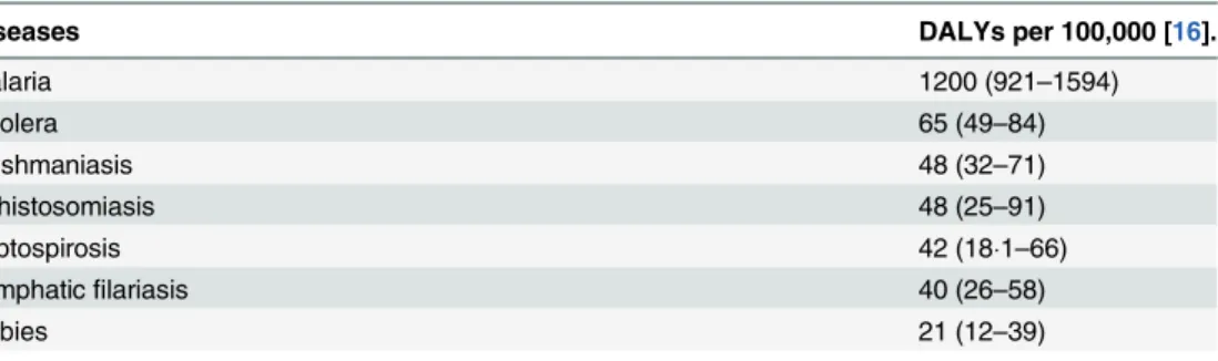
status of leptospirosis by presenting an informed prioritization of this disease among other public health research, prevention, and control priorities. We show that leptospirosis is an important infectious disease worldwide, with a global burden of approximate 2!90 million DALYs per year, most of which occurs among low and middle-income tropical countries. Although GBD 2010 used prevalence based YLDs which will underestimate the burden of chronic diseases where there is increasing populations, comparisons of the burden of leptospi-rosis with diseases that have a major impact on low income countries can be made.Table 5
illustrates the burden per 100,000 of leptospirosis compared to the estimates of other neglected and tropical diseases estimated by GBD 2010 [16]. Thus the burden of leptospirosis appears to be of a similar magnitude to that of schistosomiasis, leishmaniasis and lymphatic filariasis and about 73% of that of cholera. Furthermore, 2.90 million DALYs represent the equivalent of all the inhabitants of a city the size of Rome or Nairobi losing one year of healthy life.
Leptospirosis also predominantly affects males (80% of the total burden), and young adults (52% of the total burden affects adults aged 20–49). Leptospirosis is therefore a disease that
Fig 4. Burden of leptospirosis in terms of DALYs/100,000 per year. doi:10.1371/journal.pntd.0004122.g004
Table 5. The burden of leptopirosis per 100,000 compared to GBD estimates for the burden of various tropical and neglected diseases.
Diseases DALYs per 100,000 [16].
Malaria 1200 (921–1594) Cholera 65 (49–84) Leishmaniasis 48 (32–71) Schistosomiasis 48 (25–91) Leptospirosis 42 (18!1–66) Lymphatic filariasis 40 (26–58) Rabies 21 (12–39) 52 (22–145)* *A higher burden of rabies was estimated by Hampson et al. [30], due to variations in methodology and estimates of mortality rates.
doi:10.1371/journal.pntd.0004122.t005
8 Table 2. Estimated annual leptospirosis morbidity and mortality according to GBD region (adapted from Costa, 2015).
Regions within the developing world where the burden of leptospirosis may be significantly under-recognized include Southeast Asia and Oceania (Table 3). Although transmission is endemic and large outbreaks have been reported in these regions (Punjabi, 2012), active surveillance for leptospirosis has not been routinely performed.
principal risk group for leptospirosis. Based on model predictions, morbidity and mortality was estimated to be high in regions, such as South and Southeast Asia, where leptospirosis is an under-recognized public health problem.
Our approach had to address key challenges in the estimation of leptospirosis burden. First, the available data was sparsely distributed and not representative of all world regions. We therefore developed a model to estimate morbidity and mortality in regions with limited or no information and identified a final model that captured a significant amount of the variability (R2, 0.600) in the data from quality-assured studies. Although 95% confidence intervals for
estimates were calculated to account for the variability in our assumptions, we may not have accounted for all potential uncertainties. Leptospirosis is an environmentally-transmitted dis-ease [1–3,6]; disease risk may therefore vary significantly within a region, which in turn would contribute to spatial uncertainty. We applied criteria, accepted by an independent panel of experts (LERG), to select studies that employed appropriate methodologies with respect to case definitions, case ascertainment and case confirmation. Yet regional differences in access to health care facilities and laboratory testing, which are not explained by country-specific indica-tors of health and socioeconomic wealth, may have contributed to unaccounted variation. The true uncertainty may thus be greater than indicated by the confidence intervals for our esti-mates. Lastly, because specific countries had atypical characteristics, their model-predicted morbidity and mortality had high uncertainty which resulted in inflated estimates due to expo-nentiation from the log scale, which incorporates the standard error into the estimate.
Table 3. Estimated annual leptospirosis morbidity and mortality according to GBD region.
GBD region Morbiditya Cases Mortalitya Deaths
Estimate (95% confidence interval of the prediction)
All GBD regions 14.77 (4.38–25.03) 1,030,000 (434,000–1 750,000) 0.84 (0.34–1.37) 58,900 (23,800–95,900) High Income Asia Pacific 6.95 (2.51–11.87) 14,800 (5,300–25,100) 0.31 (0.13–0.52) 700 (300–1,100) Central Asia 5.53 (2.07–9.51) 4,400 (1,600–7,300) 0.29 (0.12–0.49) 200 (100–400) East Asia 10.28 (3.58–18.28) 142,000 (49,400–252,000) 0.50 (0.19–0.88) 6,900 (2,600–12,200) South Asia 17.97 (6.20–32.25) 289,000 (99,800–519,000) 1.02 (0.41–1.71) 16,500 (6,500–27,600) South–East Asia 55.54 (20.32–99.53) 266,000 (97,500–477,000) 2.96 (1.65–4.93) 14,200 (5,600–24,000) Australasia 9.13 (2.79–16.36) 2,400 (700–4,200) 0.40 (0.16–0.69) 100 (0–200) Caribbean 50.68 (14.93–87.58) 22,300 (6,700–34,700) 2.90 (1.14–4.72) 1,300 (500–1,900) Central Europe 4.02 (1.21–6.85) 4,800 (1,500–8,200) 0.21 (0.08–0.33) 200 (100–400) Eastern Europe 1.43 (0.51–2.69) 2,900 (1,100–5,500) 0.09 (0.04–0.16) 200 (100–300) Western Europe 3.90 (1.45–6.49) 16,300 (6,100–27,100) 0.18 (0.07–0.29) 800 (300–1,200) Andean Latin America 21.90 (7.92–39.82) 11,700 (4,200–21,200) 0.96 (0.37–1.68) 500 (200–900) Central Latin America 15.77 (5.83–27.37) 36,000 (12,300–62,400) 0.68 (0.27–1.15) 1,600 (600–2,600) Southern Latin America 3.87 (1.43–6.74) 2,400 (900–4,100) 0.18 (0.07–0.31) 100 (0–200) Tropical Latin America 13.53 (4.47–26.56) 27,300 (9,000–53,200) 0.66 (0.23–1.28) 1,300 (500–2,600) North Africa / Middle East 7.30 (2.58–11.79) 33,300 (11,800–53,800) 0.34 (0.14–0.56) 1,600 (600–2,500) High Income North America 3.64 (1.02–6.50) 12,800 (3,600–22,900) 0.18 (0.07–0.31) 600 (200–1,100) Oceania 150.68 (40.32–272.29) 16,700 (4,500–30,200) 9.61 (3.56–17.11) 1,100 (400–1,900) Central Sub–Saharan Africa 13.49 (4.48–23.56) 13,100 (4,400–22,900) 1.33 (0.52–2.23) 1,300 (500–2,200) East Sub–Saharan Africa 25.65 (9.29–43.31) 91,100 (33,000–154,000) 1.87 (0.79–3.12) 6,700 (2,800–11,100) Southern Sub–Saharan Africa 3.44 (1.34–5.78) 2,400 (900–4,100) 0.33 (0.13–0.53) 200 (100–400) West Sub–Saharan Africa 9.67 (3.62–16.16) 32,000 (12,000–53,500) 0.85 (0.35–1.36) 2,800 (1,200–4,500)
aAnnual leptospirosis morbidity and mortality rates are shown as cases or deaths, respectively, per 100,000 population.
doi:10.1371/journal.pntd.0003898.t003
Global Morbidity and Mortality of Leptospirosis
Country Morbiditya (95% CI) Mortalitya (95% CI)
Western Pacific region, stratum B
Cambodia 33·65 (10·68 – 63·81) 1·83 (0·65 – 3·35)
China 10·54 (3·52 – 18·39) 0·50 (0·19 – 0·89)
Fiji 54·38 (18·35 – 101·12) 3·08 (1·25 – 5·50)
Kiribati 106·25 (36·04 – 194·67) 6·23 (2·28 – 10·96)
Lao People’s Democratic Republic 19·11 (6·61 – 34·07) 1·09 (0·44 – 1·91)
Malaysia 36·98 (11·36 – 73·20) 1·68 (0·64 – 3·42)
Marshall Islands 25·71 (9·23 – 47·42) 2·23 (0·90 – 4·15) Micronesia, Federated States of 245·09 (77·30 – 490·19) 12·42 (4·59 – 22·37)
Mongolia 2·65 (0·87 – 5·07) 0·14 (0·05 – 0·25)
Nauru 19·90 (5·48 – 44·02) 1·93 (0·65 – 3·98)
Palau 64·06 (20·93 – 120·03) 3·80 (1·38 – 7·30)
Papua New Guinea 195·22 (61·38 – 370·88) 12·48 (4·68 – 24·09)
Philippines 14·98 (4·94 – 26·91) 0·74 (0·26 – 1·31) Republic of Korea 5·02 (1·69 – 8·77) 0·22 (0·08 – 0·39) Samoa 136·69 (47·42 – 274·47) 7·65 (3·08 – 14·22) Solomon Islands 262·93 (84·11 – 518·76) 12·11 (4·37 – 22·80) Tonga 116·07 (38·15 – 225·64) 5·92 (2·17 – 11·28) Tuvalu 74·23 (24·60 – 131·78) 5·03 (1·86 – 8·72) Vanuatu 121·15 (41·99 – 233·46) 6·41 (2·45 – 11·79) Viet Nam 49·69 (16·47 – 98·92) 2·09 (0·79 – 4·15)
Non-independent colonies, states, and territories French Polynesia 97·49 (32·20 – 179·82) 4·55 (1·67 – 8·01) Hong Kong 10·37 (3·28 – 20·19) 0·42 (0·15 – 0·83) Macao 9·72 (3·02 – 19·03) 0·37 (0·13 – 0·71) New Caledonia 68·53 (22·89 – 124·93) 2·95 (1·13 – 5·36) Taiwan 13·14 (4·60 – 24·44) 0·54 (0·20 – 0·97)
Wallis and Funtura 745·65 (209·01 – 1754·34) 27·20 (8·41 – 61·99)
Table 3. Estimated leptospirosis morbidity and mortality by country, grouped according to WHO sub-region (adapted from Costa, 2015).
The reported incidence of leptospirosis reflects the availability of laboratory diagnosis and the clinical index of suspicion as much as the incidence of the disease. A knowledge of the prevalent serovars and their maintenance hosts is essential to understanding the epidemiology of the disease
in any region. Hospital-based surveillance studies, which capture more severe forms leading to
hospitalization, do not measure mild febrile forms that form the base of the iceberg of leptospirosis cases. The global burden of leptospirosis is estimated at 2.90 million DALYs per annum (1.25– 4.54 million). This consisted of 2.80 million YLLs (1.16 million– 4.46 million) and 103,200 (38,800–188,100) YLDs. This represents an incidence of 41.8 DALYs per 100,000 population per
10
year (18.1–65.5). The frequency distribution of the global DALY estimate is illustrated in Figure 1. Males are predominantly affected with an estimated 2.33 million DALYs (0.95–3.66 million) or approximately 80% of the total burden. Young adults aged 20–49 had an estimated burden of 1.5 million DALYs (0.65–2.32 million) or approximately 52% of the total. Of these young men age 20–49 have a burden of 1.30 million (0.56–2.03 million) or 45% of the total burden.
Figure 1. Global burden of leptospirosis: estimated in terms of disability adjusted life years
(adapted from Torgerson, 2015).
II.2.2. Environmental risk factors of infection
Geography, climate, and poverty are the main determinants in the worldwide distribution of leptospirosis. Countries situated in the tropics had the highest estimated disease incidence and accounted for 73% of the world’s estimated cases. This pattern is attributable to environmental and social conditions which promote the abundance of reservoir animals, survival of the bacterium in soil and surface water, and risk of human exposures with these sources of infection (Reis, 2008). Tropical climate favours transmission of leptospirosis, which is often seasonal and increases during periods of heavy rainfall (Lau, 2010). The disease is seasonal, with peak incidence occurring in summer or fall in temperate regions, where temperature is the limiting factor in survival of leptospires, and during rainy seasons in warm-climate regions, where rapid dessication
would otherwise prevent survival. The disease is well-recognized as a health problem of
impoverished rural-subsistence farmers (Sethi, 2010), pastoralists (Biggs, 2013), and urban slum dwellers (Maciel, 2008). Life expectancy, which serves in part as a proxy for poverty, is an independent predictor of country-specific disease incidence (Table 4). Although urban slum environments are an emerging and increasingly important setting for leptospirosis transmission
11
(Maciel, 2008), country percent urbanization is inversely associated with leptospirosis incidence, reflecting in part the high burden of leptospirosis in rural settings, but also the well-recognized association between lower aggregated country-level percent urbanization and poverty.
Table 4. Variables indepedantly associated with leptospirosis morbidity in 34 countries (adapted from Costa, 2015).
Although leptospirosis is often considered to be a rural disease, people living in cities may also be exposed, notably to rats. The risk of such exposure will depend on the living conditions and the level of hygiene both in the house and its immediate environment, and on the general hygienic and sanitary conditions in different areas of a city. By 2037, the majority of the world’s population will be inhabitants of urban centres in developing countries. A large proportion of this population will reside in slum settlements, where poor sanitation has created the conditions for annual rainfall-associated epidemics (Riley, 2007). Extreme weather events and flood-related disasters (Lau, 2010) are predicted to escalate with global climate change (Patz, 2005). As deforestation and agricultural expansion intensify in tropical regions (De Fries, 2010), rural-based farming populations may be increasingly exposed to leptospirosis.
II.2.3. Individual risk factors of transmission
According to Costa et al., the risk of acquiring leptospirosis is higher in adults than children and higher in males than females, and highest among adult males of 20 to 29 years of age (Figure 2A). The age and gender-specific risk for death has a different pattern: the risk for death increases with increasing age (Figure 2B) (Costa, 2015). Unfortunately, raw data for calculating age and gender specific risk for disease and death were exctracted from grey literature by Costa et al (see Annexes and Costa, 2015) limiting our ability to formulate a critical appraisal of those figures. However, male gender preference is a well-recognized phenomenon in leptospirosis and due to the gender-specific occupational and peri-domicilary risk activities (Bovet, 1999). Morbidity and mortality in the Western Pacific region (WHO sub-region) stratified by age and gender is shown in Table 5.
leptospirosis morbidity based on four country-specific variables related to geography and cli-mate (distance from the equator, location on a tropical island), indicators of the population’s overall socioeconomic and health status (life expectancy at birth), and urbanization. The adjusted R2of the prediction model was 0.600. This model was used to estimate age and gen-der-specific morbidity and mortality for 222 countries.
A Monte Carlo model incorporated age and gender-specific incidence and mortality at the country level to obtain country-specific, regional, and global estimates for incidence and mor-tality that were adjusted for incomplete diagnostic testing (Table 3,S11andS12Tables). The annual morbidity and mortality due to leptospirosis worldwide was estimated to be 14.77 cases per 100,000 population (95% CI 4.38–25.03) and 0.84 deaths per 100,000 population (95% CI 0.34–1.37), respectively. Highest disease incidences were estimated in GBD regions of Oceania (150.68 cases per 100,000, 95% CI 40.32–272.29), South-East Asia (55.54, 95% CI 20.32– 99.53), Caribbean (50.68, 95% CI 14.93–87.58), and East Sub-Saharan Africa (25.65, 95% CI 9.29–43.31) (Fig 2andTable 3). Small tropical islands had high estimated incidence of lepto-spirosis; however, in several cases there was also significant uncertainty associated with those predictions. Morbidity and mortality by WHO sub-region (S11 Table) by country, (S12 Table) and stratified by age and gender (S13 Table) are detailed in theS1 Protocol.
The model estimated that worldwide there are 1,030,000 cases (95% CI, 434,000–1,750,000) and 58,900 deaths (95% CI, 23,800–95,900) due to leptospirosis annually (Table 3). The major-ity of leptospirosis cases and deaths occur in tropical regions; 73% of the world’s leptospirosis cases and deaths occur in countries situated between the Tropics of Cancer and Capricorn. Highest morbidity occurred among males with 20–29 years of age (35.27 cases per 100,000, 95% CI 13.79–63.89), while highest estimated mortality occurred in older males with 50–59 years of age (2.89 deaths per 100,000, 95% CI 1.22–4.95). A significant proportion of global burden of cases and deaths due to leptospirosis occurred in the demographic group of males with 20–49 years of age (48% [95% CI 40–61%] and 42% [95% CI 34–53%], respectively).
Discussion
We estimated that leptospirosis causes 1.03 (95% CI 0.43–1.75) million cases worldwide each year. These estimates place the disease among the leading zoonotic causes of morbidity and mortality. Furthermore, the number of estimated deaths (58,900; 95% CI 23.800–95,900) attributable to leptospirosis approaches or exceeds those for causes of haemorrhagic fever which were investigated in the Global Burden of Disease Study 2010 [48] and other studies [58]. The large majority of the estimated disease burden occurred in tropical regions and the world’s poorest countries. The systematic literature review also found that adult males were the Table 2. Variables incorporated in the final multivariable linear regression prediction model for lepto-spirosis morbidity in 34 countries.
Variablea R Squared
Distance from the equator 0.105
Percent urbanization of population 0.243
Life expectancy at birth 0.405
Tropical island 0.478
Complete model 0.600b
aDefinitions and sources of variables are described inS1 Protocolpp. 6–7.
bAdjusted R2is the adjusted proportion of the variance explained by the complete model, with adjustment
for the number of variables in the model. doi:10.1371/journal.pntd.0003898.t002
12 Figure 2. Mean relative risk for membership in age and gender groups among 6011
leptospirosis cases (A) and 164 deaths (B) from compiled data in 10 countries. Mean and standard deviation of the relative risks are presented for males (blues bars) and females (red bars) (adapted from Costa, 2015).
regression models. A linear regression model yielded the best fit multivariable prediction model (Tables2,S9andS10, equation 4). This model predicted the natural logarithm of
Fig 4. Mean relative risk for membership in age and gender groups among leptospirosis cases (A) and deaths (B). Mean and standard deviation of the relative risks are presented for males (blue bars) and females (red bars).
doi:10.1371/journal.pntd.0003898.g004
Global Morbidity and Mortality of Leptospirosis
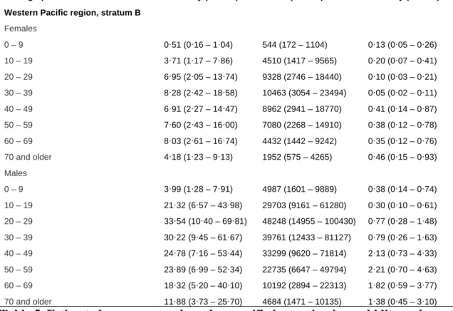
Demographic Morbidity (95% CI) Cases (95% CI) Mortality (95% CI) Western Pacific region, stratum B
Females 0 – 9 0·51 (0·16 – 1·04) 544 (172 – 1104) 0·13 (0·05 – 0·26) 10 – 19 3·71 (1·17 – 7·86) 4510 (1417 – 9565) 0·20 (0·07 – 0·41) 20 – 29 6·95 (2·05 – 13·74) 9328 (2746 – 18440) 0·10 (0·03 – 0·21) 30 – 39 8·28 (2·42 – 18·58) 10463 (3054 – 23494) 0·05 (0·02 – 0·11) 40 – 49 6·91 (2·27 – 14·47) 8962 (2941 – 18770) 0·41 (0·14 – 0·87) 50 – 59 7·60 (2·43 – 16·00) 7080 (2268 – 14910) 0·38 (0·12 – 0·78) 60 – 69 8·03 (2·61 – 16·74) 4432 (1442 – 9242) 0·35 (0·12 – 0·76) 70 and older 4·18 (1·23 – 9·13) 1952 (575 – 4265) 0·46 (0·15 – 0·93) Males 0 – 9 3·99 (1·28 – 7·91) 4987 (1601 – 9889) 0·38 (0·14 – 0·74) 10 – 19 21·32 (6·57 – 43·98) 29703 (9161 – 61280) 0·30 (0·10 – 0·61) 20 – 29 33·54 (10·40 – 69·81) 48248 (14955 – 100430) 0·77 (0·28 – 1·48) 30 – 39 30·22 (9·45 – 61·67) 39761 (12433 – 81127) 0·79 (0·26 – 1·63) 40 – 49 24·78 (7·16 – 53·44) 33299 (9620 – 71814) 2·13 (0·73 – 4·33) 50 – 59 23·89 (6·99 – 52·34) 22735 (6647 – 49794) 2·21 (0·70 – 4·63) 60 – 69 18·32 (5·20 – 40·10) 10192 (2894 – 22313) 1·82 (0·59 – 3·77) 70 and older 11·88 (3·73 – 25·70) 4684 (1471 – 10135) 1·38 (0·45 – 3·10)
Table 5. Estimated age group and gender-specific leptospirosis morbidity and mortality in the Western Pacific WHO sub-region (adapted from Costa, 2015).
Exposure depends on chance contacts between humans and infected animals or a contaminated environment. The names for some forms of leptospirosis (e.g. rice field fever, cane cutter's disease, swineherd's disease, dairy farm fever, mud fever) reflect transmission conditions. The degree and nature of exposure often depend on occupational and/or recreational and social activities (Bovet, 1999). Cattle farmers may be exposed when handling cattle, notably during milking, or when touching dead, aborted fetuses and other procreational products, e.g. amniotic fluid or placenta, or if they come into contact with infectious droplets when cows urinate. Pig farmers may be exposed when tending pigs.
Vegetable farmers and gardeners may be directly or indirectly exposed to infected rodents or their urine. Farmers may be exposed to water contaminated by the urine of rodents or other animals when irrigating fields. Rice farmers, particularly when barefooted, may be exposed to water contaminated by rodents or buffaloes, e.g. when ploughing. Veterinarians and pet keepers may be exposed to infected animals that are ill or have died from leptospirosis or which may be
symptomless carriers/shedders (this also applies to cattle and pig farmers). Abattoir workers and butchers may be exposed when slaughtering infected animals and handling infected carcasses and organs, e.g. kidneys. Those involved in food preparation may be exposed to rat-contaminated surroundings when hygienic measures are unsatisfactory. Sewer workers may be exposed to
14
sewage contaminated by rat urine. Sugar cane workers may be exposed when cutting sugar cane that is contaminated by urine of rodents living in the cane fields. Miners may be exposed to water contaminated with rat urine in mines. People may be exposed when herding cattle. People living in close contact with domestic animals may be exposed if these animals are infected. Inland
fishermen and fish/prawn farmers may be exposed, particularly if the environment and surface waters are contaminated. Soldiers, hunters and hikers may be exposed when wading through contaminated surface waters or swamps, walking on or through contaminated soil, mud or wet vegetation, or by contact with animals. Children may be exposed when playing in yards (in rain puddles or mud) contaminated by animals, such as dogs, pigs, and rats. Those participating in leisure and recreational activities (swimming, sailing, canoeing, rafting, caving, canyoneering, fishing, etc.) or involved in accidents (car accidents, boat accidents) may come into contact with contaminated surface waters, especially with prolonged (head under water) submersion. This also applies to travellers participating in jungle adventure trips or outdoor sports activities. Entire villages may be exposed when the drinking-water source is inadequately treated. Laboratory staff involved in the diagnosis of and research on leptospirosis and other zoonotic research, and
especially field workers are at risk of exposure.
II.2.4. Mechanisms of transmission
The source of infection in humans is usually either direct or indirect contact with the urine of an
infected animal. Animals may be maintenance hosts of some serovars but incidental hosts of
others, infection with which may cause severe or fatal disease. It is believed that the most important maintenance hosts are small mammals (Babudieri, 1958), which may transfer infection
to domestic farm animals, dogs, and humans. A particular Leptospira strain will generally have a
preference for a certain animal host with which there is a largely commensal relationship: the host does not suffer or suffers only comparatively mildly from the infection. Different rodent species may be reservoirs of distinct serovars, but rats are generally maintenance hosts for serovars of the serogroups lcterohaemorrhagiae and Ballum, and mice are the maintenance hosts for serogroup Ballum. Domestic animals are also maintenance hosts; dairy cattle may harbor serovars hardjo, pomona, and grippotyphosa; pigs may harbor pomona, tarassovi, or bratislava; sheep may harbor hardjo and pomona; and dogs may harbor canicola (Table 6). Distinct variations in maintenance hosts and the serovars they carry occur throughout the world.
Table 6. Typical reservoir hosts of common leptospiral serovars (adapted from Bharti, 2003).
Animals, including humans, can be divided into maintenance hosts and accidental (incidental) hosts. The disease is maintained in nature by chronic infection of the renal tubules of maintenance hosts (Badudieri, 1958). A maintenance host is defined as a species in which infection is endemic and is usually transferred from animal to animal by direct contact via their urine. Infection is usually acquired at an early age, and the prevalence of chronic excretion in the urine increases with the age of the animal. Other animals (such as humans) may become infected by indirect contact
with the maintenance host. Leptospires live and multiply in the kidneys of a carrier (generally
mammalian) or maintenance animal host. They are excreted in the urine, and animals that excrete leptospires are called shedders. Infected animals transfer leptospires to their offspring either in utero or during the neonatal period. These offspring then transfer the infection to their own offspring, and so on. In this way, a chain of infection is maintained by the maintenance host. By maintaining the infection, such maintenance hosts form the reservoir of infection. The distinction between natural or maintenance and incidental/accidental hosts is not always clear cut, particularly in domestic animals that are kept in crowded conditions that favour transmission. Exceptions and transitional states may also occur. Leptospires may adapt to a new host that may eventually become a natural maintenance host when the infection establishes itself in a population of animals of the same species, thus forming an infection reservoir. An animal may be temporarily carrying and shedding leptospires without being a natural maintenance host if the infection does not establish itself in a population of animals of the same species. The infection may run a chronic course in the maintenance host with or without serious sequelae.
The usual portal of entry is through abrasions or cuts in the skin or via the conjunctiva; infection may take place via intact skin after prolonged immersion in water, but this usually occurs when abrasions are likely to occur and is thus difficult to substantiate. Human infections may be
For personal use. Only reproduce with permission from The Lancet.
THE LANCET Infectious Diseases Vol 3 December 2003 http://infection.thelancet.com
759
for the presence of contaminating bacteria after 3–4 days and
subcultured after 7–21 days, although leptospires can survive
in undisturbed liquid culture for months, sometimes years.
28Media are made selective by the addition of several antibiotics,
the most common being 5-fluorouracil and neomycin
sulphate, although polymyxin B, rifampicin, and vancomycin
have been used.
27A commonly used medium is
Ellinghausen-McCullough-Johnson-Harris (EMJH) medium,
29–31which
contains 1% bovine serum albumin and Tween 80 (source of
long-chain fatty acids); commercial formulations are
available. Serum-containing liquid or semisolid media include
Korthof’s (peptone, NaCl, NaHCO
3, KCl, CaCl
2, KH
2PO
4,
Na
2HPO
4) and Fletcher’s (peptone, beef extract, NaCl, and
agar).
28Taxonomy
The species classification of the genus Leptospira is based on
DNA relatedness (table 1).
13–16The genus is divided into
17 species, defined as being at least 70% DNA-related and
whose related DNA sequences contain at most 5% unpaired
bases (divergence).
15This classification coexists with the
older serological classification in which antisera are used
to establish antigenic relatedness between isolates.
11Leptospiral strains are still commonly referred to by serovar
(tables 1 and 2). Many serovars studied are represented by
only a single reference strain, and as more strains are studied
the number of species is likely to increase.
32Some leptospiral serovars are commonly associated with
particular animal reservoirs (table 3). Typically, leptospires
were divided into two serological species, with most known
or suspected pathogenic leptospires grouped within the
“interrogans” complex (later, Leptospira interrogans sensu
lato). All others were placed in the “biflexa” complex (later,
Leptospira biflexa sensu lato), which contained primarily the
saprophytic strains. Both complexes (L interrogans and
L biflexa) have been divided into several serovars using the
cross-agglutinin adsorption test (CAAT).
11,12Antigenically
related serovars are arranged for convenience into
serogroups. More than 60 serovars of L biflexa sensu lato
have been described and more than 200 serovars, arranged
into 24 serogroups, are recognised within L interrogans sensu
lato.
32Both the antigenic and the more recently developed
genetic classification systems of Leptospira are in use because
genetic characterisation is possible in only a few research
laboratories and reference serological reagents (polyclonal
and monoclonal antibodies) capable of defining serovars
are not readily available. Further, neither serovars nor
serogroups are indicative of the taxonomic relation among
strains, because one serovar (defined by antibodies directed
against its lipopolysaccharide antigen) may belong to
more than one species (table 4) and members of the same
genetic group do not necessarily belong to the same
serogroup.
33Consequently, new Leptospira isolates should be
characterised by both molecular and serological approaches.
Epidemiology
Leptospirosis has a worldwide distribution. The incidence of
human infection is higher in the tropics than in temperate
regions but transmission occurs in both industrialised and
developing countries. Incidence rates are underestimated
due to lack of awareness of the disease and relatively
inaccessible and insufficiently rapid diagnostics.
Symptom-Table 2. Serogroups of Leptospira interrogans sensu lato of clinical importance with some associated serovars
Serogroup Serovar(s)
Australis australis, bratislava Autumnalis autumnalis, fortbragg, bim Ballum ballum, arborea
Bataviae bataviae
Canicola canicola, portlandvere Celledoni celledoni Cynopteri cynopteri Djasiman djasiman Grippotyphosa grippotyphosa Hurstbridge hurstbridge Hebdomadis jules
Icterohaemorrhagiae icterohaemorrhagiae, copenhageni, lai
Javanica javanica Louisiana lanka Lyme lyme Manhao manhao Mini georgia Panama panama Pomona pomona Pyrogenes pyrogenes
Sejroe sejroe, hardjo Tarassovi tarassovi
Adapted from Levett PN. Leptospira and leptonema. In: Murray PR, Baron EJ, Pfaller MA, et al, eds. Manual of clinical microbiology, 8th edn. Washington DC: ASM Press, 2003: 929–36.
Table 3. Typical reservoir hosts of common leptospiral serovars
Reservoir host Serovar(s)
Pigs pomona, tarassovi Cattle hardjo, pomona Horses bratislava
Dogs canicola
Sheep hardjo
Racoon grippotyphosa
Rats icterohaemorrhagiae, copenhageni Mice ballum, arborea, bim
Marsupials grippotyphosa Bats cynopteri, wolffi
Table 4. Leptospiral serovars seen in multiple species
Serovar Species
bataviae L interrogans, L santarosai bulgarica L interrogans, L kischneri grippotyphosa L interrogans, L kischneri
hardjo L borgpetersenii, L interrogans, L meyeri icterohaemorrhagiae L interrogans, L inadai
kremastos L interrogans, L santarosai mwogolo L interrogans, L kischneri paidjan L interrogans, L kischneri pomona L interrogans, L noguchii pyrogenes L interrogans, L santarosai szwajizak L interrogans, L santarosai valbuzzi L interrogans, L kischneri
Adapted from Levett PN. Leptospira and leptonema. In: Murray PR, Baron EJ, Pfaller MA, et al, eds. Manual of clinical microbiology, 8th edn. Washington DC: ASM Press, 2003: 929–36.
16 acquired through occupational, recreational, or avocational exposures. Occupation is a significant
risk factor for humans (Waitkins, 1986). Direct contact with infected animals accounts for most
infections in farmers, veterinarians, abattoir workers, meat inspectors, rodent control workers, and
other occupations which require contact with animals. In tropical wet areas, there are many
serovars infecting humans and animals and larger numbers of reservoir species, including rodents, farm animals, and dogs. Human exposure is not limited by occupation but results more often from the widespread environmental contamination, particularly during the rainy season. Control of rodent populations, drainage of wet areas, and occupational hygiene are all necessary for
prevention of human leptospirosis. Pathogenic serovars have been isolated from water in tropical regions (Alexander, 1975). Survival of pathogenic leptospires in the environment is dependent on several factors, including pH, temperature, and the presence of inhibitory compounds. Many infections result from barefooted walking in damp conditions or gardening with bare hands
(Douglin, 1997).
Mammalian species excrete the pathogen in their urine and serve as reservoirs for transmission. The pathogen is maintained in sylvatic and domestic environments by transmission among rodent species. In these reservoirs, infection produces chronic and persistent asymptomatic carriage in the renal tubules where L. interrogans forms aggregates (Figure 3). Leptospires infect livestock and domestic animals and causes a range of disease manifestations and carrier states. Maintenance of leptospirosis in these populations is due to continued exposure to rodent reservoirs or transmission within animal herds.
Leptospirosis is transmitted to humans by direct contact with reservoir animals or exposure to environmental surface water or soil contaminated with their urine. Leptospires penetrate abraded skin or mucous membranes, infect the bloodstream and disseminate throughout all the body tissue. Infection causes an acute febrile illness during the early “leptospiraemic” phase, which progresses during late “immune” phase to cause severe multi-system manifestations such as hepatic
dysfunction and jaundice, acute renal failure, pulmonary haemorrhage syndrome, myocardidtis and meningoencephilitis. Although the immune response eventually eliminates the pathogen,
leptospires may persist for prolonged periods in immunoprivileged sites, such as the anterior chamber and vitreous of the eye and the renal tubules, where they can produce respectively, uveitis months after exposure and urinary shedding weeks after resolution of the illness. Humans are an accidental host and it is assumed they do not efficiently shed sufficient numbers of leptospires to serve as reservoirs for transmission (Figure 3).
Figure 3. Cycle of infection (adapted from Ko, 2009).
II.3. Bacteriology
II.3.1. Serological classification
Prior to 1989, the genus Leptospira was divided into two species, L. interrogans, comprising all pathogenic strains, and L. biflexa, containing the saprophytic strains isolated from the
environment. L. biflexa was differentiated from L. interrogans by the growth of the former at 13°C and growth in the presence of 8-azaguanine (225 µg/ml) and by the failure of L. biflexa to form spherical cells in 1 mL NaCl.
Both L. interrogans and L. biflexa are divided into numerous serovars defined by agglutination after cross-absorption with homologous antigen. If more than 10% of the homologous titer remains in at least one of the two antisera on repeated testing, two strains are said to belong to different
serovars. Over 60 serovars of L. biflexa have been recorded (Kmety, 1993). Within the species L.
interrogans over 200 serovars are recognized; additional serovars have been isolated but have yet
to be validly published. Serovars that are antigenically related have traditionally been grouped into serogroups (Yasuda, 1987). While serogroups have no taxonomic standing, they have proved useful for epidemiological understanding. The serogroups of L. interrogans and some common
serovars are shown in Table 7.
Copyright/License ► Request permission to reuse FIGURE 2
Cycle of infection
Mammalian species excrete the pathogen in their urine and serve as reservoirs for transmission. The pathogen is maintained in sylvatic and domestic environments by transmission among rodent species (-A-). In these reservoirs, infection produces chronic and persistent asymptomatic carriage in the renal tubules where L. interrogans forms aggregates (Figure 1D). Leptospires infect livestock and domestic animals and causes a range of disease manifestations and carrier states (Box 3). Maintenance of leptospirosis in these populations is due to continued exposure to rodent reservoirs or transmission within animal herds (-B-).
Leptospirosis is transmitted to humans by direct contact with reservoir animals (-C-) or exposure to environmental surface water or soil contaminated with their urine (-D-). Leptospires penetrate abraded skin or mucous membranes (-1-), infect the bloodstream and disseminate throughout all the body tissue. Infection causes an acute febrile illness during the early “leptospiraemic” phase, which progresses during late “immune” phase to cause severe multi-system manifestations such as hepatic dysfunction and jaundice (-2-), acute renal failure (-3-), pulmonary haemorrhage syndrome (-4-), myocardidtis and
meningoencephilitis (-5-). Although the immune response eventually eliminates the pathogen, leptospires may persist for prolonged periods in immunoprivileged sites, such as the anterior chamber and vitreous of the eye and the renal tubules, where they can produce respectively, uveitis (-7-) months after exposure and urinary shedding weeks after resolution of the illness (-8-). Humans are an accidental host and do not efficiently shed sufficient numbers of leptospires to serve as reservoirs for transmission.
18
Table 7. Serogroups and some serovars of L. interrogans sensu lato (adapted from Bharti, 2003).
II.3.2. Genotypic classification
The phenotypic classification of leptospires has been replaced by a genotypic one, in which a number of genomospecies include all serovars of both L. interrogans and L. biflexa. After an extensive study of several hundred strains, workers at the Centers for Disease Control defined 16
genomospecies of Leptospira that included those described previously (Ramadass, 1992) and
adding five new genomospecies (Brenner, 1999), one of which was named L. alexanderi. DNA
hybridization studies have also confirmed the taxonomic status of the monospecific genus
Leptonema (Levett, 2014).
The genomospecies of Leptospira do not correspond to the previous L. interrogans and L. biflexa), and indeed, pathogenic and nonpathogenic serovars occur within the same species. Thus, neither serogroup nor serovar reliably predicts the species of Leptospira. Moreover, some studies have
included multiple strains of some serovars and demonstrated genetic heterogeneity within serovars
(Table 8). In addition, the phenotypic characteristics formerly used to differentiate L. interrogans sensu lato from L. biflexa sensu lato do not differentiate the genomospecies.
nosis of leptospirosis by serological and molecular methods are analyzed.
HISTORICAL ASPECTS
Leptospirosis is a zoonosis of ubiquitous distribution, caused by infection with pathogenic Leptospira species. The spectrum of human disease caused by leptospires is extremely wide, ranging from subclinical infection to a severe syndrome of multiorgan infection with high mortality. This syndrome, ic-teric leptospirosis with renal failure, was first reported over 100 years ago by Adolf Weil in Heidelberg (624). However, an apparently identical syndrome occurring in sewer workers was described several years earlier (337, 338). Earlier descriptions of diseases that were probably leptospirosis were reviewed recently (207, 211). Leptospirosis was certainly recognized as an occupational hazard of rice harvesting in ancient China (211), and the Japanese name akiyami, or autumn fever, per-sists in modern medicine. With hindsight, clear descriptions of leptospiral jaundice can be recognized as having appeared earlier in the 19th century, some years before the description by Weil (211). It has been suggested that Leptospira interrogans serovar icterohaemorrhagiae was introduced to western Eu-rope in the 18th century by westward extension of the range of of Rattus norvegicus from Eurasia (24).
The etiology of leptospirosis was demonstrated indepen-dently in 1915 in Japan and Germany (207). In Japan, Inada and Ido detected both spirochetes and specific antibodies in the blood of Japanese miners with infectious jaundice, and two groups of German physicians studied German soldiers afflicted by “French disease” in the trenches of northeast France. Uhlenhuth and Fromme (588) and Hubener and Reiter (289) detected spirochetes in the blood of guinea pigs inoculated with the blood of infected soldiers. Unfortunately, these two groups became so embroiled in arguments over priority that they overlooked the first publications in English (296) and German of papers by Inada’s group, whose initial publications predated their own by 8 months (207). Confirmation of the occurrence of leptospirosis on both sides of the Western Front was obtained rapidly after the publication in Europe of Inada’s work (131, 145, 543, 630).
Given the initial controversy over nomenclature, it is ironic that the organism had first been described almost 10 years before (542). Stimson demonstrated by silver staining the pres-ence of clumps of spirochetes in the kidney tubules of a patient who reportedly died of yellow fever. The spirochetes had hooked ends, and Stimson named them Spirochaeta interrogans because of their resemblance to a question mark. Unfortu-nately, this sentinel observation was overlooked for many years (211).
The importance of occupation as a risk factor was recog-nized early. The role of the rat as a source of human infection was discovered in 1917 (293), while the potential for leptospiral disease in dogs was recognized, but clear distinction between canine infection with L. interrogans serovars icterohaemorrha-giae and canicola took several years (329). Leptospirosis in livestock was recognized some years later (24). Several mono-graphs provide extensive information on the early develop-ment of knowledge on leptospirosis (24, 211, 213, 596, 634).
BACTERIOLOGY Taxonomy and Classification
Serological classification. Prior to 1989, the genus Lepto-spira was divided into two species, L. interrogans, comprising all pathogenic strains, and L. biflexa, containing the saprophytic strains isolated from the environment (217, 309). L. biflexa was differentiated from L. interrogans by the growth of the former at 13°C and growth in the presence of 8-azaguanine (225 !g/ ml) and by the failure of L. biflexa to form spherical cells in 1 M NaCl.
Both L. interrogans and L. biflexa are divided into numerous serovars defined by agglutination after cross-absorption with homologous antigen (162, 309, 330). If more than 10% of the homologous titer remains in at least one of the two antisera on repeated testing, two strains are said to belong to different serovars (297). Over 60 serovars of L. biflexa have been re-corded (309). Within the species L. interrogans over 200 sero-vars are recognized; additional serosero-vars have been isolated but have yet to be validly published. Serovars that are antigenically related have traditionally been grouped into serogroups (330). While serogroups have no taxonomic standing, they have proved useful for epidemiological understanding. The sero-groups of L. interrogans and some common serovars are shown in Table 1.
Genotypic classification. The phenotypic classification of leptospires has been replaced by a genotypic one, in which a number of genomospecies include all serovars of both L. in-terrogans and L. biflexa. Genetic heterogeneity was demon-strated some time ago (80, 260), and DNA hybridization
stud-TABLE 1. Serogroups and some serovars of
L. interrogans sensu lato
Serogroup Serovar(s)
icterohaemorrhagiae, ...Icterohaemorrhagiae, copenhageni, lai, zimbabwe Hebdomadis ...hebdomadis, jules,
kremastos
Autumnalis ...autumnalis, fortbragg, bim, weerasinghe Pyrogenes...pyrogenes Bataviae ...bataviae Grippotyphosa...grippotyphosa, canalzonae, ratnapura Canicola ...canicola
Australis...australis, bratislava, lora Pomona ...pomona
Javanica...javanica
Sejroe ...sejroe, saxkoebing, hardjo Panama ...panama, mangus Cynopteri ...cynopteri Djasiman...djasiman Sarmin ...sarmin Mini ...mini, georgia Tarassovi ...tarassovi Ballum...ballum, aroborea Celledoni...celledoni Louisiana ...louisiana, lanka Ranarum ...ranarum Manhao ...manhao Shermani...shermani Hurstbridge...hurstbridge VOL. 14, 2001 LEPTOSPIROSIS 297 on July 18, 2016 by guest http://cmr.asm.org/ Downloaded from
19
Table 8. Leptospiral serovars seen in multiple species (adapted from Bharti, 2003)
The reclassification of leptospires on genotypic grounds is taxonomically correct and provides a strong foundation for future classifications. However, the molecular classification is problematic for the clinical microbiologist, because it is clearly incompatible with the system of serogroups which has served clinicians and epidemiologists well for many years. Until simpler DNA-based identification methods are developed and validated, it will be necessary for clinical laboratories to retain the serological classification of pathogenic leptospires for the foreseeable future. In addition, the retention of L. interrogans and L. biflexa as specific names in the genomic classification also allows nomenclatural confusion. In the following pages, specific names refer to the
genomospecies, including L. interrogans sensu stricto and L. biflexa sensu stricto.
In the 1990s, DNA hybridization (DDH) identified 17 ‘genomospecies’, which also distinguished DDH from serovar. DDH complemented by molecular methods and experimental studies have since confirmed the existence of at least 22 species, and grouping of species as infectious (sometimes referred to as group I and group II pathogens, corresponding to “pathogenic” and “intermediately pathogenic”, respectively) and non-infectious (“saprophytic”) (12). The International Committee on Systematics of Prokaryotes, Subcommittee on the Taxonomy of Leptospiraceae recently agreed that genome sequence comparison should replace DDH for species definition (19).
Leptospiral typing is important for carrying out outbreak investigations and in identifying likely mammalian host reservoir sources of infection. Two commonly used molecular methods
performed are pulsed-field gel electrophoresis and multilocus sequencing typing (MLST). MLST
THE LANCET Infectious Diseases Vol 3 December 2003 http://infection.thelancet.com
759
for the presence of contaminating bacteria after 3–4 days and
subcultured after 7–21 days, although leptospires can survive
in undisturbed liquid culture for months, sometimes years.
28Media are made selective by the addition of several antibiotics,
the most common being 5-fluorouracil and neomycin
sulphate, although polymyxin B, rifampicin, and vancomycin
have been used.
27A commonly used medium is
Ellinghausen-McCullough-Johnson-Harris (EMJH) medium,
29–31which
contains 1% bovine serum albumin and Tween 80 (source of
long-chain fatty acids); commercial formulations are
available. Serum-containing liquid or semisolid media include
Korthof’s (peptone, NaCl, NaHCO
3, KCl, CaCl
2, KH
2PO
4,
Na
2HPO
4) and Fletcher’s (peptone, beef extract, NaCl, and
agar).
28Taxonomy
The species classification of the genus Leptospira is based on
DNA relatedness (table 1).
13–16The genus is divided into
17 species, defined as being at least 70% DNA-related and
whose related DNA sequences contain at most 5% unpaired
bases (divergence).
15This classification coexists with the
older serological classification in which antisera are used
to establish antigenic relatedness between isolates.
11Leptospiral strains are still commonly referred to by serovar
(tables 1 and 2). Many serovars studied are represented by
only a single reference strain, and as more strains are studied
the number of species is likely to increase.
32Some leptospiral serovars are commonly associated with
particular animal reservoirs (table 3). Typically, leptospires
were divided into two serological species, with most known
or suspected pathogenic leptospires grouped within the
“interrogans” complex (later, Leptospira interrogans sensu
lato). All others were placed in the “biflexa” complex (later,
Leptospira biflexa sensu lato), which contained primarily the
saprophytic strains. Both complexes (L interrogans and
L biflexa) have been divided into several serovars using the
cross-agglutinin adsorption test (CAAT).
11,12Antigenically
related serovars are arranged for convenience into
serogroups. More than 60 serovars of L biflexa sensu lato
have been described and more than 200 serovars, arranged
into 24 serogroups, are recognised within L interrogans sensu
lato.
32Both the antigenic and the more recently developed
genetic classification systems of Leptospira are in use because
genetic characterisation is possible in only a few research
laboratories and reference serological reagents (polyclonal
and monoclonal antibodies) capable of defining serovars
are not readily available. Further, neither serovars nor
serogroups are indicative of the taxonomic relation among
strains, because one serovar (defined by antibodies directed
against its lipopolysaccharide antigen) may belong to
more than one species (table 4) and members of the same
genetic group do not necessarily belong to the same
serogroup.
33Consequently, new Leptospira isolates should be
characterised by both molecular and serological approaches.
Epidemiology
Leptospirosis has a worldwide distribution. The incidence of
human infection is higher in the tropics than in temperate
regions but transmission occurs in both industrialised and
developing countries. Incidence rates are underestimated
due to lack of awareness of the disease and relatively
inaccessible and insufficiently rapid diagnostics.
Symptom-Table 2. Serogroups of Leptospira interrogans sensu latoof clinical importance with some associated serovars
Serogroup Serovar(s)
Australis australis, bratislava
Autumnalis autumnalis, fortbragg, bim
Ballum ballum, arborea
Bataviae bataviae
Canicola canicola, portlandvere
Celledoni celledoni Cynopteri cynopteri Djasiman djasiman Grippotyphosa grippotyphosa Hurstbridge hurstbridge Hebdomadis jules
Icterohaemorrhagiae icterohaemorrhagiae, copenhageni, lai
Javanica javanica Louisiana lanka Lyme lyme Manhao manhao Mini georgia Panama panama Pomona pomona Pyrogenes pyrogenes
Sejroe sejroe, hardjo
Tarassovi tarassovi
Adapted from Levett PN. Leptospira and leptonema. In: Murray PR, Baron EJ, Pfaller MA, et al, eds. Manual of clinical microbiology, 8th edn. Washington DC: ASM Press, 2003: 929–36.
Table 3. Typical reservoir hosts of common leptospiral serovars
Reservoir host Serovar(s)
Pigs pomona, tarassovi
Cattle hardjo, pomona
Horses bratislava
Dogs canicola
Sheep hardjo
Racoon grippotyphosa
Rats icterohaemorrhagiae, copenhageni
Mice ballum, arborea, bim
Marsupials grippotyphosa
Bats cynopteri, wolffi
Table 4. Leptospiral serovars seen in multiple species
Serovar Species
bataviae L interrogans, L santarosai bulgarica L interrogans, L kischneri grippotyphosa L interrogans, L kischneri
hardjo L borgpetersenii, L interrogans, L meyeri icterohaemorrhagiae L interrogans, L inadai
kremastos L interrogans, L santarosai mwogolo L interrogans, L kischneri paidjan L interrogans, L kischneri pomona L interrogans, L noguchii pyrogenes L interrogans, L santarosai szwajizak L interrogans, L santarosai valbuzzi L interrogans, L kischneri
Adapted from Levett PN. Leptospira and leptonema. In: Murray PR, Baron EJ, Pfaller MA, et al, eds. Manual of clinical microbiology, 8th edn. Washington DC: ASM Press, 2003: 929–36.
20
has the advantage that it reflects the underlying population genetic structure, is reproducible, is robustly supported by experimental data, and even can be used directly to identify infecting Leptospira in clinical samples (Agampodi, 2013). Genome sequencing, which has become widely available, together with automated tools that assign MLST sequence types directly from sequence data, has demonstrated an important potential for typing, with the expectation that automated analysis tools will become sufficiently user-friendly for rapid and efficient whole genome analysis and comparison, including phylogenetic analysis based on the identification of single nucleotide polymorphisms (SNPs) in the core genome.
The Leptospira Genome Project, initiated in 2011, has been to obtain and compare whole genome information for all known Leptospira species. Among the goals of this analysis were the following: i) identifying Leptospira pathogenesis mechanisms that might explain heterogeneity in clinical manifestations of leptospirosis; ii) understanding the relationship of genomic content and context to pathogenesis; iii) determining the definitive evolutionary rela- tionship of Leptospira towards understanding how infectious Leptospira diverged from sapro- phytes; and iv) identifying common antigens for improving diagnosis and vaccine development. There are 9 known pathogenic
Leptospira species, 5 intermediate Leptospira species, and 6 saprophytic Leptospira species for which the whole genome sequence analysis has been compared (Fouts, 2016) (Figure 4).
Figure 4. Pan-genomic comparisons of 20 Leptospira species. Orthologous protein clusters were binned, counted and placed into a Venn diagram by whether clusters contained
proteins from genomes in each of the three Leptospira groups: pathogenic (A), intermediate (B), saprophytic (C) and the Leptonema outgroup (D). Clusters were counted if there was a majority (50%), all-but-one, or all protein members from a particular group or groups (separated by colons). Singleton clusters, representing species-specific or strain-specific genes are noted in circles surrounding the Venn diagram. Clusters not matching any of these criteria or containing at least one protein from another group were considered as ambiguous groupings (adapted from Fouts, 2016).
II.3.3. Biology of leptospires
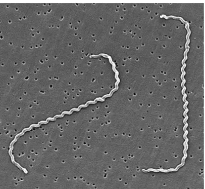
Leptospires are tightly coiled spirochetes, usually 0.1 µm by 6 to 0.1 by 20 µm, but occasional cultures may contain much longer cells. The helical amplitude is approximately 0.1 to 0.15 µm, and the wavelength is approximately 0.5 µm. The cells have pointed ends, either or both of which are usually bent into a distinctive hook (Figure 5). Two axial filaments (periplasmic flagella) with polar insertions are located in the periplasmic space. The structure of the flagellar proteins is complex. Leptospires exhibit two distinct forms of movement, translational and nontranslational. Morphologically all leptospires are indistinguishable, but the morphology of individual isolates varies with subculture in vitro and can be restored by passage in hamsters. Leptospires have a typical double membrane structure in common with other spirochetes, in which the cytoplasmic membrane and peptidoglycan cell wall are closely associated and are overlain by an outer membrane. Leptospiral lipopolysaccharide has a composition similar to that of other gram-negative bacteria, but has lower endotoxic activity. Leptospires may be stained using carbol
Fig 2. Pan-genomic comparisons of 20Leptospira species. Panel A: Orthologous protein clusters were binned, counted and placed into a Venn diagram by whether clusters contained proteins from genomes in each of the threeLeptospira groups: pathogenic (A), intermediate (B), saprophytic (C) and the Leptonema outgroup (D). Clusters were counted if there was a majority (50%), all-but-one, or all protein members from a particular group or groups (separated by colons). Singleton clusters, representing species-specific or strain-specific genes are noted in circles surrounding the Venn diagram. Clusters not matching any of these criteria or containing at least one protein from another group were considered as ambiguous groupings. The Venn diagram is not to scale. Panel B: Protein clusters unique to pathogenic, intermediate, and saprophytic groups or shared only between pathogenic and intermediate groups were counted by main functional role categories. See key for group colors.
doi:10.1371/journal.pntd.0004403.g002
22 fuchsin counterstain. Leptospires are obligate aerobes with an optimum growth temperature of 28 to 30°C. They produce both catalase and oxidase. They grow in simple media enriched with
vitamins (vitamins B2 and B12 are growth factors), long-chain fatty acids, and ammonium salts.
Long-chain fatty acids are utilized as the sole carbon source and are metabolized by β-oxidation.
Figure 5. Scanning electron micrograph of L. interrogans serovar icterohaemorrhagiae strain RGA bound to a 0.2-µm membrane filter (adapted from Levett, 2001).
II.3.4. Immunization
Immunity to leptospirosis is largely humoral and is relatively serovar specific. Thus, immunization protects against disease caused by the homologous serovar or antigenically similar serovars only. Vaccines must therefore contain serovars representative of those present in the population to be immunized. Immunization has been widely used for many years as a means of inducing immunity in animals and humans, with limited success. Early vaccines were composed of suspensions of killed leptospires cultured in serum-containing medium, and side effects were common. Modern vaccines prepared using protein-free medium are generally without such adverse effects. In developed countries, pigs and cattle are widely immunized, as are domestic dogs, but in most developing countries, vaccines which contain the locally relevant serovars are not available. Most vaccines require booster doses at yearly intervals.
Most bovine and porcine vaccines contain serovars hardjo and pomona. Protection against hardjo infection has been suboptimal, but one vaccine has been shown to offer good protection (Zuerner,