Université de Montréal
Impact of a neonatal parenteral nutrition on the hepatic
activity of methionine adenosyltransferase, a limiting step
for glutathione synthesis
ByWesam Elremaly
Département de Nutrition
Faculté du médecine
Mémoire présenté à la Faculté de médecine en vue de l’obtention du grade de maîtrise
en nutrition
Janvier, 2012
Université de Montréal
Faculté des études supérieures et postdoctorales
Ce mémoire intitulé :
Impact of a neonatal parenteral nutrition on the hepatic
activity of methionine adenosyltransferase, a limiting step
for glutathione synthesis
Présenté par : Wesam Elremaly
a été évalué par un jury composé des personnes suivantes :
Stéphanie Fulton, président-rapporteur Jean Claude Lavoie, directeur de recherche
Résumé
Problématique : Le glutathion est une molécule clé de la défense antioxydante. Chez les
enfants sous nutrition parentérale (NP), particulièrement les nouveau-nés, sa concentration tissulaire est anormalement basse. Puisque la capacité de synthèse de glutathion est adéquate, un déficit en cystéine, le substrat limitant, est soupçonnée. À cause de son instabilité en solution, la cystéine est peu présente en NP; la méthionine étant le précurseur endogène de cet acide aminé. L’activité de la méthionine adénosyltransférase (MAT), une enzyme essentielle à la transformation de la méthionine en cystéine, est facilement inhibée par l’oxydation. L’hypothèse : Le faible taux de glutathion chez les enfants sous NP est causé par l’inhibition de la MAT par les peroxydes contaminant ces solutions nutritives.
Objectif: Mesurer l’impact d’une infusion de NP et de H2O2 sur l’activité hépatique de MAT en relation avec le niveau de glutathion. Méthode : Un cathéter est placé dans la jugulaire droite de cobayes de trois jours de vie. Quatre groupes sont comparés:1- Témoin (animaux aucune manipulation, sans cathéter) 2)-(animaux nourris normalement et le cathéter (noué)); 3) NP (animaux nourris exclusivement par voie intraveineuse (acides aminés + dextrose + lipides + vitamines + électrolytes), cette solution génère environ 400 µM de peroxyde. 4) H2O2 (animaux nourris normalement et recevant via le cathéter 400 µM de H2O2). Après quatre jours, le foie et le sang sont prélevés pour la détermination du glutathion, potentiel redox et l’activité de MAT, glutathion peroxydase et glutathion reductase. Résultats : L’activité de MAT est plus faible dans les groupes NP et H2O2. Le potentiel redox du foie et dans le sang est plus oxydé dans le groupe NP. Tandis que la concentration de GSSG du foie est plus élevée dans le groupe NP. Ainsi la concentration de GSH dans le sang et foie est plus faible dans les NP et H2O2 Discussion: La relation entre l’inhibition de MAT et le stress oxydant observée dans le groupe NP pourrait bien expliquer la perturbation du système glutathion observée chez les nouveau-nés prématurés.
Mots-clés : peroxydes, nouveau-né, glutathion peroxydase, glutathion réductase,
Abstract
Introduction: The low glutathione level in premature newborns can partly explain the high
incidence of complications associated with oxidative stress in this population. Since the synthetic activity of glutathione is mature, a lack of substrate, especially cysteine, is suspected. Methionine provided by their parenteral nutrition (PN) is the in vivo precursor of cysteine. Since, methionine adenosyltransferase (MAT), the first enzyme in methionine transformation, has redox sensitive cysteines, we hypothesize that peroxides contaminating PN inhibit the activity of MAT, leading to a lower availability of cysteine for glutathione synthesis. Methods: At 3 days of life, guinea pigs a catheter fixed in jugular vein, the animals were separated in 4 groups: 1) Control: animals without any treatment and no catheter 2) sham: animals fed regular chow, a node closed their catheter; 3) PN: animals fed exclusively with parenteral nutrition (dextrose, amino acids, fat, vitamins) containing about 400 µM peroxides; 4) H2O2: animals fed regular chow and received continuously 400 µM H2O2 through the catheter. Four days later, liver and blood were sampling for determination of reduced glutathione (GSH), oxidized glutathione (GSSG), redox potential and activities of MAT, glutathione peroxidase, and glutathione reductase. Results: MAT activity was lower in groups receiving PN or H2O2. Liver redox was more oxidized in PN group whereas blood redox was more oxidized in PN and H2O2. Liver and blood GSH is lower in PN and H2O2. Liver GSSG was higher in PN. Conclusion: The inhibition of MAT in the PN group could explain the disruption of the glutathione system observed in premature infants. Furthermore the impact of a lower activity of MAT on GSH level is observed in liver and blood. Suggesting that the hepatic synthesis of GSH is insufficient to maintain its own level of glutathione and sustain the rate of export.
Keywords: Peroxides, newborn, glutathione peroxidase, reductase, methionine adenosyltransferase and parenteral nutrition.
Table of contents
1. Introduction ………..1
1.1.1. Oxidative stress……….2
1.1.2. Parenteral nutrition………..…..3
1.1.3. Parenteral nutrition as a source of oxidants………...4
1.1.4. Consequences of oxidative stress in premature infants………...4
1.1.5. Reactive radicals and redox balance………...5
1.2.1 Antioxidant defences……….6
1.2.2 Glutathione ………...7
1.2.3 Glutathione structure……… 7
1.2.4 Glutathione synthesis ………...7
1.2.5 Inter-organ glutathione regulation ………...8
1.3.1. Glutathione function.………….……….………..9
1.3.2. Maintenance of thiol status………..………...9
1.3.3. Antioxidant function………..………...10
1.3.4. Glutathione peroxidase ………..……….10
1.3.5. Glutathione reductase………..…………....11
1.4.1. Methionine as a source of cysteine ………...12
1.4.2. Methionine adenosyl transferase ……...……….14
2. Hypothesis and objectives ………..16
3. Materials and methods………..17
3.1. Materials...…....17
3.2.1. Animals ...…...18
3.2.2. Experimental design ……….………....……….18
3.2.3. Guinea pig as an animal model ………...……….……….20
3.2.4. Determination of peroxides………...21
3.2.5 Protein determination ...21
3.2.6 GSH and GSSG levels……….………... 22
3.2.7. Glutathione reductase and peroxidase activity ………. 22
3.2.8. Methionine adenosyl transferase activity ………..……23
3.2.9. Statistical analysis ……….………24
4. Results ………...25
4.1. Hepatic methionine adenosyl transferase ………...….. 25
4.2. Hepatic glutathione ………26
4.3. Blood glutathione ………...28
4.4. Hepatic and red blood cells glutathione peroxidase activity………...….. 30
4.5. Hepatic and red blood cells glutathione reductase activity………31
4.6. Hepatic GSH level in function of hepatic MAT activity……….32
5. Discussion………...33
6. Limits ………38
7. Future work ……….39
8. Conclusion ………40
List of Abbreviations
PN : Parenteral nutrition GR : Glutathione reductase GPx : Glutathione peroxidase GSSG : Disulfide form of glutathione GSH : Reduced Glutathione
GST : Glutathione-S-transferase ATP : Adenosine triphosphate H2O2 : Hydrogen peroxide
MAT : Methionine adenosyltransferase (EC 2.5.1.6) NADP : Nicotinamide adenine dinucleotide phosphate
NADPH : Reduce Nicotinamide adenine dinucleotide phosphate SAH : S- adenosyl homocysteine
SAM : S- adenosyl methionine SOD : Superoxide dismutase
Lists of figures
Figure 1. Equilibrium redox balance ………3
Figure 2. Redox cycle………...12
Figure 3. Endogenous conversion of methionine to cysteine………..13
Figure 4. Hepatic activity of MAT activity ………...25
Figure 5. Hepatic redox potential in function of treatments………...26
Figure 6. Hepatic GSH and GSSG levels in function of treatment A and B…………...27
Figure 7. Blood redox potential in function of treatments………...28
Figure 8. Blood GSH and GSSG levels in function of treatment A and B………...29
Figure 9. Hepatic and red blood cells glutathione peroxidase activity in function of treatment A and B……….30
Figure10. Hepatic and red blood cells glutathione reductase activity in function of treatment A and B……….31
Figure 11. Hepatic GSH level in function of hepatic MAT activity………..32
« Science built houses does not have a mainstay and ignorance destroys homes of glory and generosity” The Egyptian poet Ahmed Shawqi
Acknowledgements
I would like to thank my supervisor, Dr Jean Claude Lavoie for his constant encouragement and support throughout the period of study, the useful discussions, learning the statistical analysis and his help and support to learning the French language.
I would like to extend my sincere thanks to Thérèse Rouleau: for her support, her help to complete my practical work.
I would like to thank my colleagues in the lab Dr Ibrahim Mohamed and Dr Khalil Miloudi. Finally, I would like to thank my family, especially my husband Mohamed Elbakry, for their help and support.
1.
Introduction
Infants born before 28 weeks of gestation frequently require intravenous nutrition (parenteral nutrition) because of their immature gastrointestinal system. For these infants parenteral nutrition (PN) is the way to provide essential nutrients for growth and development. However, these nutrient solutions are contaminated by oxidizing molecules [1]. In addition to this oxidant load, newborn infants have immature antioxidant defenses including low glutathione levels [2], [3]. Therefore, neonates are at high risk of oxidative stress.
Lee and Frank showed that the pulmonary activity of antioxidant enzymes such as superoxide dismutase, glutathione peroxidase and catalase increases as a function of gestational age in several animal species [4]. Scientists extrapolate this fact to the neonates. Furthermore, the key antioxidant molecule, namely glutathione, is low in premature infants [3]. The intracellular level of this molecule is essential.Glutathione modules, among others, the cellular redox potential which is an important regulator for metabolic functions [5]. The goal of my research is to explore new avenue explaining this abnormally low level of glutathione.
In the next sections of the introduction, I will briefly discuss about the in vitro generation of oxidant molecules in PN solutions, the oxidative stress and its consequences in premature infants, and finally the in vivo generation of glutathione. Then I will formulate my working hypothesis and the objectives of my graduated studies.
1.1.1.
Oxidative stress
Oxidative stress derives from an abnormally high level of oxidative molecules in a cell or organ. Oxidative stress can be induced by an abnormally high production of oxidants, a low capacity of antioxidant defenses or both. Oxidants are produced as a consequence of ATP production in the mitochondria metabolism or by intracellular enzyme system or they can be initiated from external source such as inhaled oxygen and pollutants. An increase in intracellular levels of these oxidants will lead to damage to cellular lipids, proteins, and DNA consequently to impaired macromolecular function, cell function and finally can lead to cell death. Thus oxidative stress plays an important role in development of several pathological complications. For example lung injuries, exposure to polluted air contaminating reactive oxygen species, mineral and dust can lead to airway inflammation, activate the inflammatory and apoptotic process. As show in Figure 1 under the normal condition there is a balance between the exposure to the oxidants and antioxidants leading to human body homeostasis. When the exposure to the oxidants is overload the cell is a situation of oxidative stress will lead to impaired cellular function as result the disease
Figure 1: Equilibrium redox balance
GSH: reduced glutathione; GPx: glutathione peroxidase; GR: glutathione reductase ; SOD : superoxide dismutase
1.1.2. Parenteral nutrition
Parenteral nutrition (PN) is a mixture of amino acids, dextrose, electrolytes, vitamins and lipids. Although PN is essential for the growth and development of the neonates, PN is also associated with undesirable effects caused by oxidant molecules generated in the solution [1]. Hence, PN represents a potential cause of several pathological complications observed in premature newborns that are associated with oxidative phenomena [8]. Since peroxides are an important player in our hypothesis, I will describe how these molecules can contaminate PN.
Antioxidant defense
GSH,GPx,
GR, SOD
,
Oxidants
Health
Oxidative stress
Antioxidant defense
Diseases
1.1.3. PN as a source of oxidants
The generation of oxidant molecules in these solutions is caused by the interaction between reducers such as polyunsaturated fatty acids, several amino acids and vitamin C and a strong oxidant as dissolved oxygen. Several molecules derive from these interactions. Among them, are aldehydes and hydroperoxides [9] derived from lipid peroxidation and hydrogen peroxide (H2O2) from ascorbic acid [10]. Because PN contains a photo-sensitive compound, riboflavin, these reactions are catalyzed by ambient light [10]. Furthermore, these reactions contribute to the loss of fatty acids, amino acids and antioxidant vitamin such as ascorbate. This loss combined with the immaturity of antioxidant defences in premature newborns [11] is dramatic. Even perfect photo-protection of the solution reduces the concentration of peroxides by only half. However, this reduction is sufficient to induce metabolic changes in children [12]. Unfortunately, it is difficult to reach such photo-protection in a clinical setting. Because light exposure during only a few minutes is sufficient to generate significant amounts of oxidant molecules in PN solution [10], the photo-protection must be initiated during the PN preparation in pharmacy without any exposure to the light until the bedside where the bag and delivery tubes must be fully covered [1].
1.1.4. Consequences of oxidative stress in premature infants
The infusion of PN to premature infants without adequate photo-protection is associated with several markers of oxidative stress such as high concentrations of peroxides and isoprostanes [13] in urine and an oxidized redox potential in whole blood [14],[15]. This
stress is associated with a greater incidence of several pathological complications observed in this population such as bronchopulmonary dysplasia [8],[16], [14], [17], [18]. This suggests that the antioxidants systems of premature infants, especially glutathione, are unable to fully scavenger the oxidant load associated with PN [13].
1.1.5. Reactive radicals and redox balance
During normal metabolism, the cell generates oxidizing molecules [19], [20]. Although these oxidant molecules can be derived from detoxification of xenobiotics, the main producer remains the oxidative phosphorylation in mitochondrion [21]. Because the standard redox potential of ½ O2: H2O is among the highest in biology (-0.82 V), oxygen is easily reduced in water through the various protein complexes of mitochondrion. During these processes, about 1 to 3% of O2 will be partially reduced [22], with generation of superoxide anion (.O2-)[23],[24]. Superoxide dismutase (SOD) converts two superoxide anions into hydrogen peroxide (H2O2) and oxygen[25],[26]. H2O2 can be reduced by two enzymes, catalase [27]or glutathione peroxidase (GPx), into O2 and H2O [28]. If not, H2O2 can react with Fe2+ forming the hydroxyl radical (.OH) via the Fenton reaction. This radical initiates lipid peroxidation that generates lipid peroxides, which can be eliminated by the action of GPx and of glutathione-S transferase (GST) [29],[30]. Therefore, peroxides from PN, mainly H2O2, or / and free radicals deriving from its one-electron reduction might be detoxified by the antioxidant defenses of the organism.
1.2.1.
Antioxidant defenses
An antioxidant is defined as a molecule or atom that gives an electron to the oxidant molecule or atom in order to reduce them. In general, the antioxidants are classified as endogenous, exogenous and as secondary. Superoxide dismutase (SOD), glutathione peroxidase (GPx), glutathione and catalase are the main representatives of the endogenous category [31] whereas vitamin C and vitamin E as well as selenium and carotenoids are from exogenous sources (dietary antioxidant) [32]. Riboflavin (vitamin B2), zinc and manganese are considered as secondary antioxidants [33] for their participation in the activity of glutathione reductase and SOD [34]. For example: the action of SOD reduces the free radical anion superoxide in oxygen and hydrogen peroxide that will be reduced in water by catalase or GPx. Catalase is present only in the peroxisome and, to a lesser extent, in mitochondria. In this organelle, GPx is more important than catalase for the reduction of hydrogen peroxide [35]. In addition, the absence of catalase in cytosol, except for the erythrocyte, underlines the importance of GPx.
Vitamin E and vitamin C are obtained from the diet. Vitamin E is a fat soluble vitamin that is incorporated into cell membranes where it protects them from the oxidative damage by scavenging free radicals [36], [37] In this case, vitamin E becomes oxidized and inactive. Vitamin C (water soluble vitamin) is a strong reducing agent [38]. It participates in a variety of oxido-reductive reactions in the body such as the reduction of some metals such as Cu+2 and Fe+3 [39], allowing them to become available for the Fenton reaction, and the recycling of the oxidized form of vitamin E into its reduced and active form [40]. The
oxidized form of vitamin C can be returned to the reducing form by the action of the glutathione system [41].
In addition to the recycling of vitamins C and E, glutathione works as a co-factor of several enzymes, especially glutathione peroxidase and glutathione-S-transferase.
Glutathione is considered a key molecule in the antioxidant defenses of the organism. Hence, the next section is devoted to the glutathione.
1.2.2. Glutathione
1.2.3. Structure of glutathione
Glutathione is the tri-peptide γ-L-glutamyl-L-cysteinyl-glycine (GSH). It is
omnipresent in the body and is especially concentrated in liver. Glutathione exists in two forms: reduced GSH and disulfide (GSSG) [42], frequently named oxidized form. GSH is the main form, existing in millimolar concentrations in most cells (liver 5–10 mM). GSSG normally represents less than 1% of total glutathione concentration [43]. It is noted that the ratio GSSG/GSH may increase during oxidative stress. The concentration of both forms, GSH and GSSG, is important in the maintenance of the intracellular redox potential as discussed below (section 1.3.2).
1.2.4. Glutathione synthesis
Since, GSH cannot freely cross the cellular membranes each cell must synthesize its own GSH [44]. This process requires two ATP per molecule of glutathione. The synthesis is a two-step catalyzed reaction [45]:
This first step of GSH synthesis is catalyzed by glutamate cysteine ligase, in presence of Mg++ and is considered as a rate-limiting step [46]. Moreover, this step is regulated by availability of cysteine [47],[3]. Indeed, the intracellular level of cysteine is close to the km of the enzyme whereas the intracellular concentration of glutamate is far greater the Km. Hence, a little variation in the availability of cysteine will influence greatly the activity of this rate-limiting enzyme [48]. Cysteine is obtained from the diet, from protein or glutathione breakdown, and from endogenous methionine [49]. The availability of cysteine in newborns is particularly problematic [3], especially if they are nourished by a parenteral way (PN). Indeed, this solution contains low level of cysteine because it is oxidized in cystine that precipitates (low solubility) in these nutritive solutions [50]. In fact, the main source of cysteine for glutathione synthesis is derived from the transformation of methionine [51], [52]. The hypothesis (see section 2) of my research project is the transformation of methionine to cysteine is inhibited by oxidants molecules generated in parenteral nutrition. The second step of the glutathione synthesis is catalyzed by GSH synthase. In presence of K+ and Mg++, the enzyme links the glycine to the dipeptide γ-glutamyl-L-cysteine.
γ-glutamyl-L-cysteine + L-glycine + ATP → GSH + ADP + Pi
In the following section, I will discuss to how the tissue concentration of GSH is regulated between organs.
1.2.5. Inter-organ glutathione regulation
For the in vivo regulation of the GSH, some organs work as synthesizers of GSH, whereas others work as exporter [53]. The liver is considered both a synthesizer and
exporters of GSH. It establishes 90% of the glutathione supply in blood via hepatic exportation [54], [55]. Indeed, the liver has a great level of glutathione synthesis for its own needs and for export via sinusoidal protein MRP3 [56] -glutamyltranspeptidase is an enzyme anchored in cell membranes with its catalytic site in the extracellular milieu. This -glutamyl moiety of glutathione on a second amino acid, forming two dipeptides (glutamyl-amino acid and cysteinylglycine). These dipeptides are absorbed by cells and hydrolysed in free amino acids. Free cysteine can be used for a de
novo synthesis of GSH. Thus, we can conclude that the liver plays an important role in the
whole body homeostasis of glutathione in all the other organs.
1.3. Glutathione functions
1.3.1. Maintenance of thiol status
There are many redox couples in the cell that work together to maintains the intra cellular redox. By this abundance (millimolar), one of the most important couple in the cell is GSSG/2GSH [5]. In fact, glutathione is considered the buffer of the intracellular redox potential.
The redox potential can be estimated from the concentrations of both, GSH and GSSG, with the Nernst equation (Ehc = -240 -(59.1/2) log ([GSH] 2/ [GSSG]) mV at 25 oC pH 7. The redox potential Ehc is associated with the biological status of the cell. For example a redox potential of glutathione of -240 mV is associate with proliferating stage whereas a more oxidized potential such as -220 mV occurs during differentiation stage of cell and a more oxidized status, for instance -170 mV, induces apoptotic events [57]. This
fact can be explained by the following reaction in which thiol functions of several proteins can react with GSH or GSSG dependently of the redox potential [49]:
Protein-SSG + GSH protein-SH + GSSG
This reaction regulates a variety of metabolic processes such as enzyme activity, transport activity and gene expression [58].
1.3.2. Antioxidant function
The first antioxidant property of glutathione is related to its participation in reduction of peroxides. For this purpose, it acts as a cofactor for GPx. Since during this reaction, GSH will be oxidized in its disulfure form (GSSG), the redox potential of the cell could be modified and consequently several metabolic pathways. Likely Proteins, in which thiols are important for their function, are subjects to be regulated by the redox potential of the cell. For instance, several phosphatases are regulated this way. Therefore, pathways that use phosphorylation as regulation strategy are susceptible to be influenced by the redox potential. Here, MAT has sensitive thiols that are also influenced by the redox of the cell. Among other pathways that are regulated by the redox potential we find several enzymatic activities (as for the present study), active transports and gene expressions as mentioned in section 1.3.1. Hence, the redox potential could be maintained by the action of glutathione reductase (GR) that recycles GSSG in GSH as described below.
1.3.3. Glutathione peroxidase (GPx)
Glutathione peroxidase (GPx), an enzyme of which the catalytic action is dependent on the micronutrient selenium (Se), catalyzes the reduction of hydrogen peroxide (H2O2) or
organic peroxides to water and corresponding alcohols, respectively [59]. This reaction requires two molecules of glutathione (GSH) as reducing substrate [60]. Glutathione disulfide (GSSG) the oxidized glutathione is formed as the product of this reaction. The activity of this enzyme is tightly controlled since modification of its activity will lead to a alter intracellular concentration of hydrogen peroxide, an important modulator of the activity of several proteins [59] such as methionine adenosyltransferase, the targeted enzyme of my work.
1.3.4. Glutathione reductase (GR)
Because an elevation of GSSG level in cells, following the action of GPx, will affect the cellular redox potential, which interferes with several metabolisms, normally GSSG is reduced back to GSH by the action of glutathione reductase (GR) in the presence of NADPH as electron donor [61], [62]. In case where the action of GPx overpass the activity of GR, the accumulated GSSG will be actively exported to maintain the intracellular redox potential [63] . In this case, lower intracellular level of GSH will affect the activity of GPx [64] because the normal level of GSH in cells is at the level of the Km of GPx. A lower level of GSH leads to a lower activity of GPx, allowing a higher level of H2O2 in cell [65]. To prevent this loss of glutathione, the cell stimulates its synthesis and it recycles the GSSG into GSH by GR. Hence, by maintaining the level of GSH, GR plays a critical role in the oxidant defense of the cell. GR contains two molecules of FAD per enzyme. FAD is derived from vitamin B2 and is why this vitamin is considered as a secondary antioxidant [66].
Figure 2: Redox cycle
1.4. Methionine as source of cysteine
To maintain its pool of glutathione, premature newborns on PN (containing high level of peroxides) need cysteine. However, the instability of cysteine in solution limits its enrichment in PN. In addition, the cellular uptake of cysteine is immature in this population [48]. Therefore, the main source of cysteine for glutathione synthesis remains the
transformation of methionine in cysteine (figure 3). Indeed, it is known that this essential amino acid that is primarily metabolized in the liver, serves as the endogenous source of cysteine and, consequently, for glutathione synthesis [67], [68].
Reduced glutathione 2GSH Glutathione peroxidase Selenium Glutathione reductase Riboflavin (FAD) GSSG Oxidized glutathione Hydrogen peroxide H2 O2 Water 2H2O NADP + NADPH+H+
Methionine
ADP
Methionine adenosyltransferase MAT
Methyl transferase S-adenosyl homocysteine S-adenosyl methionine ATP Cysteine γ-glutamyl-cysteine GSH Glutatione synthase Glutamate cysteine ligase α-Ketobutyrate Homocysteine cystathionine β synthase Serine Cystathionine Trans-Sulfuration
Figure 3: endogenous conversion of methionine to cysteine
As shown in Figure 3, the first enzyme involved in methionine metabolism is the methionine adenosyltransferase (MAT) that catalysis the conversion of methionine and ATP in S-adenosylmethionine (SAM) [69], [70], [71], [72]. SAM is the principal methyl donor and participates in three key metabolic pathways in the liver: polyamine synthesis, transmethylation and transsulfuration [73],[74]. SAM by the action of methyltransferase is converted into S-adenosylhomocysteine (SAH), which is hydrolyzed to homocysteine and
adenosine through a reversible reaction catalyzed by SAH hydrolase. Then homocysteine exits through two pathways. First, it can be remethylated to form methionine by two enzymes: 1) methionine synthase is requires normal levels of folate and vitamin B12 [75], [76] and 2) betaine homocysteine methyltransferase, which requires betaine, a metabolite of choline [77], [78]. The second important metabolism of homocysteine is its conversion into cysteine via the transsulfuration pathway [79]. During this process, homocysteine is transformed into cystathionine by a reaction catalyzed by cystathionine β synthase (CBS). Finally free cysteine is released by vitamin B6-dependent enzyme cystathionase for glutathione synthesis [80], [68].
1.4.1. Methionine adenosyltransferase
An inhibition of this enzyme will lead to the perturbation of metabolism needing methyl group, but also to a lower level of glutathione in subjects with a low exogenous source of cysteine [81], [82], [83], such as newborns on parenteral nutrition. What do we know about this enzyme? Three forms of this enzyme have been identified in mammalian tissues [84], [85]. MAT II, primarily expressed in extrahepatic tissues and fetal liver, is a heterotetramer formed of catalytic α2 and regulatory ß subunits. In adult liver, MAT exists as a tetramer (MAT I) and as a dimer (MAT III) of a single α1 subunit of 43.7 kDa [86], [87], [88]. I have focused my work on the liver. Consequently, the activities measured were from MAT I and III isomers.
We assume that peroxides can inhibit the activity of MAT by two mechanisms. The first mechanism is by a direct effect on MAT cysteines. Knowing that the catalytic site of
these enzymes has cysteinyl residues that are susceptible to oxidation [89], [81]. Avila et al have reported that, in vitro, MAT was inhibited by H2O2 [82].
MAT –SH + H2O2 MAT-SOH (inactive)
The second mechanism is by modification of the redox potential. Therefore, levels of GSH and GSSG were important as show in these equations
MAT-SOH + GSH MAT-SSG (inactive)
MAT-SSG + GSH MAT-SH (active) + GSSG.
Hence the peroxide had oxidized the cysteinyl residues of the enzyme. This fact is the origin of our working hypothesis.
2. Hypothesis
We hypothesize that neonatal exposure to oxidant molecules in parenteral nutrition inhibits the hepatic activity of MAT, leading to a lower availability of cysteine for glutathione synthesis.
2.1.
Objectives
The general objective of this project is to understand why glutathione concentration is low in premature infants, and subsequently find new nutritional strategies to improve the level of this important molecule of antioxidant defenses.
Two specific objectives were:
1- Assess in an animal model of neonatal parenteral nutrition, newborn guinea pig, independently of prematurity, the impact of infusion of a standard parenteral nutrition on the hepatic activity of MAT, level of GSH and GSSG as well as the redox potential of glutathione.
2- Evaluate if the PN impact is associated with its H2O2 content, by comparing data from animals infused with H2O2, at same concentration that measured in PN, to those from animals receiving PN.
3.1. Materials
I mentioned below the list of the products which I had used throughout the period of study. Ferrous chloride, potassium chloride, sodium chloride and xylenol orange were purchased from Agilent Technologies, Mississauga, ON, Canada and magnesium chloride from Anala R BDH Laboratory Suppliers, Poole, Angleterre. Sodium hydroxide was obtained from American Chemicals, Montreal, QC, Canada and potassium hydroxide from Baker, Phillipsburg, NJ, USA. Xylazine was purchased from Bayer, Toronto, ON, Canada and methionine from Baxter, Mississauga, ON, Canada. Blood collection tubes (Vacutainer ® spray coated K2EDTA tubes) were obtained from BD, Oakville, ON, Canada and BioRad protein assay, power supply supply (Model 1000/500 Power Supply), AG 50W-X4 Resin cation exchange resin were from BioRad, Mississauga, ON, Canada. Tetra-acetic acid disodium, tris hydrochloride , bovine serum albumin (BSA), 4 - (2 – hydroxyethyl) -1 - piperazine ethane sulfonic acid (HEPES) were obtained from Boehringer Mannheim GmbH, Laval, QC, Canada. Guinea pigs animal model was purchased from Charles River St-Constant, Montréal, QC, Canada , methanol from Chemical products of ACP, Montreal, QC, Canada, boric acid from JT Baker Chemical Co, Polyurethane catheter from Phillipsburg NJ, USA , Luther Medical Product, Tustin, CA, USA, and Intra lipid 20 %,20% (Pharmacia Upjohn, Baie D’Urfé, QC, Canada), multivitamins and amino acids from Sandoz, Boucherville, Qc, Canada. Adenosine 5`-Triphosphate, Tetrasodium Salt, [2, 8-3H] was obtained from PerkinElmer , reduced glutathione (GSH), oxidized glutathione(GSSG), glutathione reductase (GR), nicotinamide adenine dinucleotide phosphate (NADPH) from Roche Diagnostique, Montréal, QC, Canada, pediatric. Ketamine
from Wyeth whereas xylazine from bayer health care Toronto, Ontario ,centrifuge tubes of 50 ml from Sarstedt, Montréal, QC, Canada, meta phosphoric acid, S-adenosyl-L-methionine from Sigma-Aldrich Co, St-Louis, MO, USA and filter papers from Whatman, Florham Park, NJ, USA. Finally all the solutions were prepared in pure water and all the other reagents were of analytical grade.
3.2.Methods 3.2.1. Animals
At three days of life Hartley guinea pigs (Charles River Laboratories, St.Constant, Quebec,Canada) , were anesthetized by using ketamine (50mg/mL;0, 18mL/100g) and xylazine (20mg/mL; 0,05mL/100g) in order to fix a catheter (Luther
Medical Products, Tustin, CA) in jugular vein. The catheter was placed and externalized in
the scapular region, with a branch connected to the infusion system.
The studied intravenous solutions were infused continuously through the catheter at rate of 22 mL / 100 g body weight / day. The solutions were changed daily.
The research protocol was approved by the institutional committee of animal research and practices of the hospital Sainte-Justine built on the principles of the Canadian Council of animals.
3.2.2. Experimental design
The animals were classified into 4 groups
1- Control group: four animals of same age, without manipulation such as surgery, fed with regular laboratory food for guinea pigs.
2- Sham group: three animals that were not fed through the catheter, the catheter was fixed and closed, but ate the laboratory food for Guinea pigs for the duration of the experiment.
3- PN: five animals fed exclusively with total parenteral nutrition (5% (w.v) dextrose + 0.45% (w,v) NaCl + 4.8 g/kg/day amino acids + 3.8 g/kg/day Intralipid 20 and 1% (v,v) Multi12 pediatrics multivitamins The PN solution used contained a similar composition to that one given to the children in neonatal care units.
4- H2O2: Seven animals receiving 400 µM H2O2 through the catheter. These animals were fed with regular laboratory food for guinea pigs.
Four days later, at seven days of age, animals were sacrificed for collection of liver and blood. The liver samples were removed, processed, aliquot and stored at -80OC until biochemical determinations. An aliquot of the blood was centrifuged to separate plasma from red blood cells. Both fractions and an aliquot of whole blood were and stored at -80OC until biochemical determinations.
3.2.3. The guinea pig as animal model
Our newborn animal model was the guinea pig because, guinea pigs and humans are dependent on vitamin C [90], [91], a strong antioxidant molecule, they are unable to synthesize it and this is essential characteristic when we study mechanisms related to oxidative stress. Also it is an accessible animal model for the study of neonatal PN because at three days of life, the size of its body is sufficient (limit) for insertion of the smallest catheter of the world in its jugular vein , by this age, their glutathione development is not
complete [92], [93] , similarly to humans and weaning occurs early, by three days of life, this animal nourishes itself by itself [94 , 95, 96].
The problematic of PN is observed mainly in premature newborns. Studies on the impact of PN in this population are difficult since no control exist, premature infants of the same gestational age without PN does not exist. By choosing a term newborn animal, we can separate the impact of PN from prematurity. Similarly to premature infants on PN, several markers of oxidative stress have been measured in this animal model on PN [97], [15]As observed with premature infants [17], the absence of photo-protection of PN induced a loss of alveoli [8], a characteristic feature of the bronchopulmonary dysplasia such observed in premature infants [14].
3.2.4. Determination of peroxides
Total peroxides were determined by following at 560 nm the generation of chromophore from the complex Fe3+-xylenol orange [98], [99]. The principle is based on the Fenton reaction in which ferrous ion (Fe2+) is oxidized in presence of H2O2 in its ferric form (Fe3+). Thus, diluted PN was mixed with reactive solution containing 22.5 mM H2SO4, 90 µM xylenol orange, 225 µM FeCl2, and 3.6 mM 2,6-di-tert-butyl-4-methylphenol in methanol. After 30 min of incubation, at the room temperature the solutions were centrifuged 5500 × g for 3 min). The absorbency of the supernatant was read at 560 nm. The results were expressed in µM. H2O2 was used for the external standard curve.
3.2.5. Protein determination
Protein was measured by Bradford method, the principle of this technique is a dye-binding reaction to protein with a differential color change of a dye (Coommassie) occurs in response to the protein concentrations. The absorbance was detected at 595nm.
The pellets from different tissues were solubilized, with 1N NaOH at 37 ° C for 2 hour. Addition dilution (1 / 250) was made; these dilutions were transferred to spectrophotometric cuvettes. Bradford reagent previously diluted (1 / 5) with distal pure water was added. Samples were incubated 10 minutes at room temperature. The absorbance values were compared to the curve of bovine serum albumin (BSA; 0 – 90 pg/μL).
3.2.6. GSH and GSSG levels
Immediately after sampling, 200 μl of blood or 0.5 g of liver were mixed with respectively, 4 and 5 volumes of 5% (w/v) freshly prepared metaphosphoric acid. The blood sample was vortex for 10 seconds whereas the liver sample was homogenated on ice during 20 seconds with Polytron (Biospec Products, Bartlesville, OK, USA). Thereafter, they were centrifugated for 1-3 min at 10000 RPM. Supernatants (for glutathione determinations) and pellets (for protein determination) were separated and frozen at -80°C until the day of the assay.Reduced (GSH) and disulfide (GSSG) forms of glutathione were separated by capillary (75 μm×50 cm silica) electrophoresis (75 mM boric acid +25 mM Bis–Tris buffer, pH 8.4, 28°C, 18 kV) and were detected at 200 nm on a P/ACE MDQ system (Beckman Coulter). GSH and GSSG were used as external standard curves [95]. The redox potential was calculated by using the Nernst equation (25ºC, pH 7): Ehc = -240-(59.1/2) log ([GSH] 2/
[GSSG]) mV.
3.2.7. Determination of glutathione reductase and peroxidase activity
Tissue samples were homogenized in 9 times volume of (50 mM Tris-HCl, 0.1 mM EDTA-Na2, pH 7.6) on ice. The homogenates were centrifuged 1 min at 10000 RPM. The supernatants were taken to determine the enzyme activities and protein levels.
The determination of glutathione reductase (GR) activity is based on the following principle GSSG + NADPH + H+ → 2 GSH + NADP+
The activity of glutathione reductase is proportional to the disappearance of NADPH, which absorbs at 340 nm. The reaction is started by the addition of the biological sample in reactive milieu (50 mM Tris-HCl, 0.1 mM EDTA-Na2, 1 mM GSSG, and 0.1mM NADPH, pH 7.6). Slope of the drop of absorbance recorded during 10 minutes was quantified using the NADPH molar extinction coefficient of 6.22 mM−1cm−1. The activity was expressed as μmol NADPHoxidized/min/mg protein at 30°C.
Glutathione peroxidase activity (GPx) was measured by mixing the supernatants with
reactive solution (250 mM Tris, 0.1mM EDTA-Na2, 1 mM GSH, 1mM tert-butyl hydroperoxide, 4.8 U/mg GR, and 0.1mM NADPH, pH 7.6). Determination of the glutathione peroxidase activity is based on the following principle
2GSH + H2O2 → GSSG + 2H2O
Then GR reduces the GSSG to complete the cycle: GSSG + NADPH + H+ → 2 GSH + NADP+
The decrease in NADPH absorbance measured at 340 nm during the oxidation of NADPH to NADP+ is indicative of GPx activity. Slope of the drop of absorbance recorded during 5 minutes was quantified using the NADPH molar extinction coefficient of 6.22 mM−1cm−1. The activity was expressed as μmol NADPH oxidized/min/mg protein at 30C° [3].
3.2.8. Determination of Methionine adenosyl transferase (MAT)
Liver samples were homogenized on ice during 20 seconds with Polytron (Biospec Products, Bartlesville, OK, USA) in 4 volumes of (10 mM Tris/HCL, 0.3 M sucrose, 1 mM benzamidine, 0.1 mM phenylmethanesulfonylfluoride, and pH 7.5). The homogenate was centrifuged at 40000 g for 2h30 at 4 C°. The supernatants were taken for
determination of MAT activity and protein level. The supernatants were incubated at 37C° for 30 min with (75m M Tris/HCL, 250 mM KCl, 9 mM MgCl2, 60 µM methionine and 5 mM [2-³H]ATP (1Ci/mol), pH 7.8). The reaction was stopped by adding 3 ml of cold water and immediately applied onto the cation exchanger Dowex AG5OW columns (0.5 ml) that was previously prepared and balanced with water, adjusted pH at 7 with NaOH. The chromatography was made at room temperature. Subsequently, the column was washed with 20 ml water to get rid of the unused radioactive substrate. The H3-S-adenosyl methionine was eluted by using 4 ml of 3M NH4OH. The radioactivity of this fraction was measured by liquid scintillation by a beta counter (Beckman Co, Fullerton, CA, USA). The activity was expressed as nmol-S-adenosyl methionine formed/min/mg protein at 37°C [81], [86], [100].
3.2.9. Statistical analysis
Data were presented as mean ± s.e.m. and were orthogonally compared by Two way ANOVA as follows: 1) Control vs. Sham that will inform on the impact of surgery (we expected to find no rejection of null hypothesis); 2) PN vs. H2O2 that will inform on the impact of intravenous nutrition vs. per os alimentation (we expected to find no rejection of null hypothesis); 3) [control – sham] vs. [PN – H2O2] that will inform on the impact of intravenous infusion of peroxides (we expect to find a rejection of null hypothesis). Verification of homoscedasticity was tested by the Bartlett’s Chi squaretestχ2, the level of significance was set at p <0.05.
4. Results
4.1. Hepatic methionine adenosyl transferase (MAT)
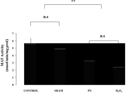
MAT activity was lower (p < 0.01) in groups received PN or H2O2 as shown in Figure 4. There was no difference between PN and H2O2 or between control and sham group
0 1 2 3 4 5 6 7 MA T A ct iv it y (n mo l/ mi n /mg p ro t) ** CONTROL SHAM PN H2O2 n.s n.s
Figure 4: Hepatic activity of MAT as a function of treatments.
Control: animals without any manipulation; Sham: animals with catheter in jugular, without infusion, enterally fed; PN: animals fed exclusively with parenteral nutrition (PN); H2O2: animals received intravenous solution containing 400 µM H2O2, enterally fed. The activity of MAT was lower in groups infused with solution containing peroxides (PN and H2O2). Mean s.e.m.; n.s.: no statistical difference; **: p<0.01.
4.2. Hepatic glutathione
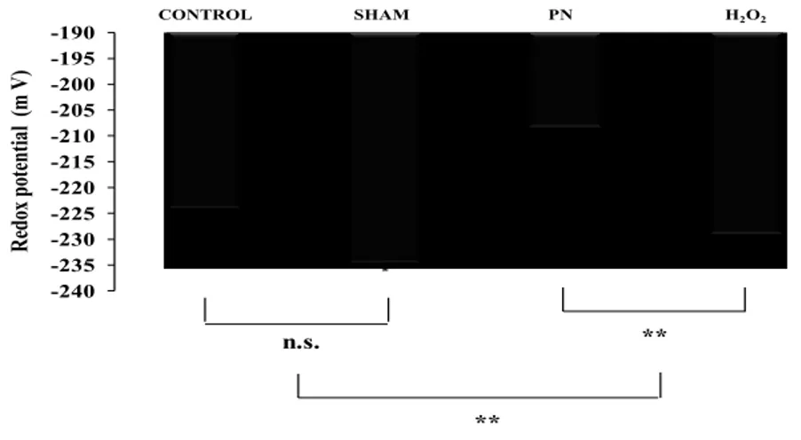
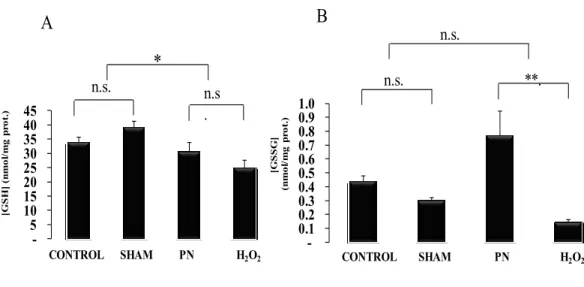
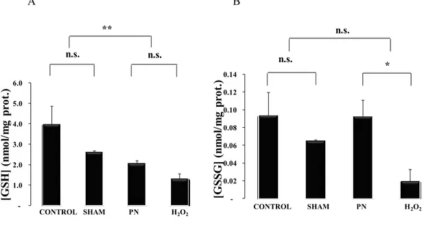
The statistical analysis shown in Figure 5 the redox potential of glutathione was higher (more oxidized) in PN group (p<0.01) than in H2O2 group whereas the impact of surgery (control versus Sham) was without impact. The level of GSH Figure 6, A was low in animals that received PN or H2O2 whereas GSSG levels Figure 6, B were higher (p<0.01) in PN group compared to H2O2 group.
-240 -235 -230 -225 -220 -215 -210 -205 -200 -195 -190 R edo x po te nt ia l (m V ) CONTROL SHAM PN H2O2 ** ** n.s.
Figure 5: Hepatic redox potential as a function of treatments.
Control: animals without any manipulation; Sham: animals with catheter in jugular, without infusion, enterally fed; PN: animals fed exclusively with parenteral nutrition (PN); H2O2: animals received intravenous solution containing 400 µM H2O2, enterally fed. The hepatic redox was more oxidized in groups infused with solution containing peroxides (PN and H2O2). Mean s.e.m.; n.s.: no statistical difference; **: p<0.01.
-5 10 15 20 25 30 35 40 45 [G S H ] (n m o l/ m g p ro t.) CONTROL SHAM PN H2O2 * n.s . n.s. -0.1 0.2 0.3 0.4 0.5 0.6 0.7 0.8 0.9 1.0 [G S S G ] (n m o l/ m g p ro t. ) CONTROL SHAM PN H2O2 **. n.s. n.s. A B
Figure 6: Hepatic GSH and GSSG levels as a function of treatments. Control: animals
without any manipulation; Sham: animals with catheter in jugular, without infusion, enterally fed; PN: animals fed exclusively with parenteral nutrition (PN); H2O2: animals received intravenous solution containing 400 µM H2O2, enterally fed. The hepatic GSH level
(A) was lower in groups infused with solution containing peroxides (PN and H2O2) whereas the hepatic GSSG (B) was higher in PN. Mean s.e.m.; n.s.: no statistical difference; *: p<0.05, **: p<0.01.
4.3 Blood glutathione
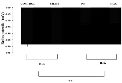
The statistical analysis shown in Figure 7 the redox potential of glutathione in blood was higher (more oxidized) in PN and H2O2 groups. There was no difference between PN and H2O2 groups or between control and sham groups. The level of GSH Figure 8, A was low in animals infused with PN or H2O2 whereas GSSG level Figure 8, A was higher (p<0.01) in PN group compared to H2O2 group.
-195 -190 -185 -180 -175 -170 -165 -160 R edox pot ent ial ( m V) ** CONTROL SHAM PN H2O2 n.s. n.s.
Figure 7: Blood redox as a function of treatments. Control: animals without any
manipulation; Sham: animals with catheter in jugular, without infusion, enterally fed; PN: animals fed exclusively with parenteral nutrition (PN); H2O2: animals received intravenous solution containing 400 µM H2O2, enterally fed. The redox in the blood was lower in groups infused with solution containing peroxides (PN and H2O2). Mean s.e.m; n.s.: no statistical difference; **: p<0.01.
-1.0 2.0 3.0 4.0 5.0 6.0 [G SH ] (n m ol /m g pr ot .) ** n.s. CONTROL SHAM PN H2O2 n.s. -0.02 0.04 0.06 0.08 0.10 0.12 0.14 [G SS G ] (n m ol /m g pr ot .) n.s. * CONTROL SHAM PN H2O2 n.s. A B
Figure 8: Blood GSH and GSSG levels as a function of treatments. Control: animals
without any manipulation; Sham: animals with catheter in jugular, without infusion, enterally fed; PN: animals fed exclusively with parenteral nutrition (PN); H2O2: animals received intravenous solution containing 400 µM H2O2, enterally fed. The blood GSH (A) was lower in groups infused with solution containing peroxides (PN and H2O2) whereas the blood GSSG (B) was lower in animals infused with H2O2. Mean s.e.m.; n.s.: no statistical difference; **: p<0.01 *: p<0.05.
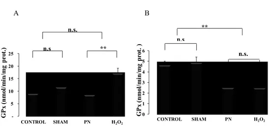
4.4. Hepatic and erythrocyte glutathione peroxidase activity.
Glutathione peroxidase activity was higher (p < 0.01) in liver from animals infused with H2O2 as shown in figure (9, A). In contrast, in blood, the activity of GPx was lower (p< 0.01) in the group received peroxides (PN and H2O2) Figure 9. In both tissues, there was no difference between control and sham groups.
-5 10 15 20 25 G P x (n m ol /m in /m g pr ot .) n.s. ** CONTROL SHAM PN H2O2 n.s 0 1 2 3 4 5 6 G P x (n m ol /m in /m g pr ot . ) ** n.s n.s. CONTROL SHAM PN H2O2 A B
Figure 9: Hepatic and erythrocyte GPx activity as a function of treatments.
Control: animals without any manipulation; Sham: animals with catheter in jugular, without infusion, enterally fed; PN: animals fed exclusively with parenteral nutrition (PN); H2O2: animals received intravenous solution containing 400 µM H2O2, enterally fed. The hepatic GPx activity (A) was higher in group H2O2. Whereas GPx activity in the red blood cells (B) was lower in groups received peroxides PN, H2O2. Mean s.e.m.; n.s.: no statistical difference; **: p<0.01, **: p<0.01.
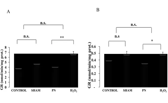
4.5. Hepatic and erythrocyte glutathione reductase activity.
Compared to PN groups, the activity of glutathione reductase were higher (p < 0.05) in liver and erythrocytes from group H2O2 as shown in Figures 10 There was no difference between control and sham groups.
0 1 2 3 4 5 6 7 8 GR (n m ol/ m in /m g p ro t. ) CONTROL SHAM PN H2O2 n.s. ** n.s. -0.1 0.2 0.3 0.4 0.5 0.6 G R (n m ol /m in /m g p rot. ) n.s. n.s * CONTROL SHAM PN H2O2 A B
Figure 10: Hepatic and erythrocyte GR activity as a function of treatments. Control:
animals without any manipulation; Sham: animals with catheter in jugular, without infusion, enterally fed; PN: animals fed exclusively with parenteral nutrition (PN); H2O2: animals received intravenous solution containing 400 µM H2O2, enterally fed. The activity of hepatic GR (A) was lower in groups infused with peroxide H2O2. While the activity of GR in the red blood cells (B) was lower in PN group Mean s.e.m.; n.s.: no statistical difference; **: p<0.01, *: p<0.05.
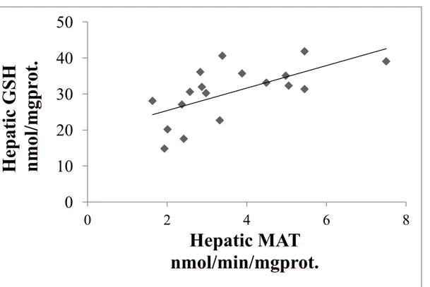
4.6. Association between the hepatic MAT activity and hepatic GSH level
Hepatic MAT activity was correlated significantly and positively (r2 = 0.41, p <0.01) with hepatic GSH level as in Figure 11.
Figure 11: Hepatic GSH level in function of hepatic MAT activity. The hepatic GSH
level was positively correlated (r2 = 0.41, p <0.01) with hepatic MAT activity.
0
10
20
30
40
50
0
2
4
6
8
He
patic
GSH
n
mol
/mgp
rot
.
Hepatic MAT
nmol/min/mgprot.
5. Discussion
This study tested the hypothesis that peroxides contaminating PN inhibit the activity of MAT, leading to a lower availability of cysteine for glutathione synthesis. Our data demonstrated that indeed, the PN during four days inhibits strongly the activity of hepatic MAT. This inhibition is reproduced by an infusion of a solution containing a concentration of H2O2 similar to that measured in PN. In addition, we identified an association between the inhibition of MAT activity and GSH level. This is the first study demonstrating this new avenue (inhibition in transformation of the methionine to cysteine) explaining the abnormally low level of glutathione in premature infants receiving PN.
Because PN contains oxidant molecules such as peroxides the inhibition of MAT can result from a direct effect of peroxides on active thiols of the enzyme or from a lower recycling of the enzyme that is dependent on the redox potential which is oxidized in animals on PN. Indeed, the in vivo total activity of MAT is dependent of the equilibrium between its active (MAT-SH) and inactive (MAT-SOH) form. Peroxides react with thiol to generate the inactive form (MAT-SOH) whereas the redox status favors the reduction of MAT–SOH in MAT–SH. The discrepancy between hepatic redox potential observed in animals infused with PN or H2O2 suggests that the inhibition of MAT did not result from a more oxidized hepatic redox.
With inhibition of MAT following the infusion of peroxides, we expected to observe a more oxidized redox potential in PN and H2O2 groups. Indeed, the lower level of GSH in liver and in blood contributes to a more oxidized redox status. On the other side, a greater level of GSSG also induces a more oxidized redox. However the accumulation of GSSG was
observed only in liver of animals received PN. This last observation is sufficient to explain the more oxidized redox potential calculated only in PN group. Interestingly, the infusion of H2O2 has induced a lower level of both GSH and GSSG. Hence, the redox potential is maintained in this group. The difference between PN and H2O2 on GSSG suggests that PN exerts an oxidizing activity which is not only related to H2O2. This effect of PN may be related to others oxidants molecules such as ascorbyperoxide, lipid peroxides and 4-hydroxynonenal that also contaminate the solution of PN [101], [9] , [102]. Alternatively, the explanation can come from the fact that the infusion of H2O2 induces free radicals [103] which are known to activate transcription factors such as NF-E2-related factor 2 (Nrf2) [104], [105]. This factor plays a physiological role in the regulation of oxidative stress by activation of gene transcription for GPx and GR [106]. This is supported by the higher hepatic activities of both enzymes observed only in the H2O2 group. These two enzymes play an important role on the redox potential, especially GR that recycles GSSG in GSH. This fact can explain the low GSSG in animals infused H2O2. The absence of modification of activity of these two enzymes in PN group can be explained by the presence of the free radical scavenging vitamin C and vitamin E [107]. The activation of this transcription factor is not possible in red blood cell because the absence of nucleus. The activities of both enzymes were not increased in this tissue. On the contrary, we observed a lower activity of GPx in animals infused with solutions containing peroxides. It is known that peroxides in high concentration inhibit GPx activity [108], [109].
The impact of a lower activity of MAT on GSH level is also observed in blood. The level of glutathione in extrahepatic tissues is dependent of the level of GSH exported by the liver. This extracellular level of GSH serves as a source of essential cysteine for GSH synthesis. Hence, results from blood highlight that the hepatic synthesis of GSH is insufficient to maintain its own level of glutathione and sustain the rate of export into the plasma.
A major difference between the PN and H2O2 groups in regards of the glutathione level resides in the fact that animals infused with PN did not receive cysteine from their nutrition whereas animals infused with H2O2 fed regular chow that contains cysteine [95]. Given that the availability of cysteine is a rate-limiting step in the synthesis of GSH, the comparison of both groups informs also on the impact of the mode of nutrition. We can expect that the presence of cysteine in enteral food of H2O2 group enables them to sustain GSH synthesis and consequently its level in liver and blood. Our results show that this source of cysteine did not correct for the impact of the inhibition of MAT on the glutathione level. This observation could explain the absence of impact of cysteine supplementation on glutathione level in premature infants [50]. Therefore, these data suggest that, in neonates, endogenous conversion of methionine to cysteine is more important for glutathione synthesis than exogenous source of cysteine.
The impact of abnormally low MAT activity can also explain, at least partially, two major complications observed in premature infants: cholestasis and frequent blood transfusion. A lower level of SAM, following inhibition of MAT, could induce hepatic cholestasis. Indeed, several studies reported that SAM prevents the development of
cholestasis by maintaining normal liver membrane Na+-K+-ATPase activity through preservation of the membrane lipid environment [110, 111, 112]. In addition, cholestasis associated with PN can also be explained by a low glutathione production following a low level of SAM; glutathione favors bile formation [113, 114, 115]. The second impact of our conclusion is related to the oxidative stress of red blood cells (more oxidized redox potential, lower level of GSH, lower activity of an enzyme detoxifying peroxides (GPx). Oxidative stress is a major cause of aging of these cells, shortening their lifespan [116] , [117]. Hence, inhibition of MAT may also be responsible for the high number of transfusions required by premature infants.
In summary, the results of this study support our hypothesis that the inhibition of MAT in the PN group could contribute to the disruption of the glutathione system observed in premature infants. Moreover this inhibition appears to be caused by H2O2 contaminating PN. The impact of a lower activity of MAT on GSH level is observed in liver and blood. Knowing that the liver exports actively GSH for the benefit of other organs, results suggest that the hepatic synthesis of GSH is insufficient to maintain its own level of glutathione and sustain the rate of export. The differences between PN and H2O2 on GSSG levels suggesting that PN exerts another oxidizing activity
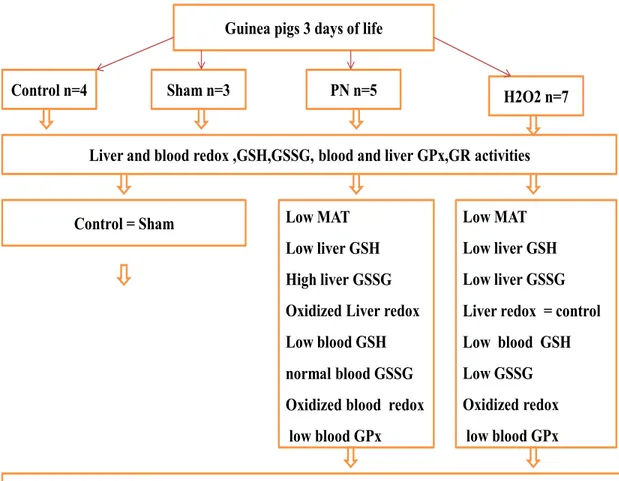
Guinea pigs 3 days of life Control n=4
Liver and blood redox ,GSH,GSSG, blood and liver GPx,GR activities
PN n=5 H2O2 n=7
Sham n=3
Peroxides contaminating perenteral nutrition inhibit the MAT activity leading to low GSH Low MAT
Low liver GSH High liver GSSG Oxidized Liver redox Low blood GSH normal blood GSSG Oxidized blood redox
low blood GPx
Low MAT Low liver GSH Low liver GSSG Liver redox = control Low blood GSH Low GSSG Oxidized redox
low blood GPx Control = Sham
urgency to develop safer intravenous nutritive solutions
6. Limits of this study
PN is a mode of nutrition frequently used in neonates born before 28 weeks of gestation. The investigation of the impact of PN in this population is difficult because control groups of premature newborns without PN cannot exist. The use of newborn animals born at term enabled us to study the impact of PN during a neonatal period independently of the prematurity. Therefore, we need to take caution in the extrapolation of data collected with this animal model to human premature newborn. However, we could expect to find similar impacts in premature infants as observed with the animals. Unfortunately the absence of control groups of premature newborns without PN thus, it is possible that the prematurity, itself, causes the elevation of all the of oxidative stress markers.
Additionally all the animals in our experiment are males alternatively, Lavoie has been shown that in cells extracted from endotracheal aspirates from the baby girl and boy the increased curve of glutathione levels in function of the gestational age is slower in boys. These results suggesting that the baby girl is potentially better protected against an oxidative stress than boy. As I mentioned in section 1.1.4 (consequences of oxidative stress in premature infants) oxidative stress is associated with a greater incidence of several pathological complications observed in this population such as bronchopulmonary dysplasia. Knowing that sex is an important determinant of the incidence of this disease among premature infants, the boys are much higher incidence to this disease. In addition the recent published results demonstrate there is a correlation between this disease and the redox potential of the glutathione. Thus this the main reason for choosing the animals male to achieve our hypothesis with the population that more vulnerable for oxidative stress.
Given that the level of the glutathione is different between baby girls and baby boys as well as their vulnerability to the oxidative stress differs according to the sex. So, in my opinion, it would be interesting to repeat the experiments with female animals in order to separate the effect of sex.
7. Future work
Further studies are needed to investigate the impact of PN or peroxides on the activation of Nrf2 which control the gene transcription of Glutamate-cysteine ligase, the rate-limiting step in the synthesis of glutathione (GSH). In addition, the determination of cysteine level in the liver and blood. Since activity of MAT is sensitive to the redox state of their cysteine residues; its activity is inhibited by peroxides. It will be interestingly to challenge the hypothesis of the protective effect of the reducing agents such as hexapeptides which derived from human milk or Dithiothreitol on the hepatic MAT activity. Supporting this hypothesis our lab team demonstrated that by the addition of hexapeptides which derived from human milk limit the generation of peroxides in PN moreover correct the redox potential and prevent PN-induced hepatic oxidative stress. Moreover it will be important to apply these studies for the neonates in order to decrease the incidence of all the complications which related to oxidative stress for this population by increase the production of GSH.
As consequence, the hepatic redox potential of glutathione switches to a more oxidized status, which could be a first factor leading to development of several pathological complications of neonates on PN. An important possibility in the clinic the blood status could be a representative reflection of liver disturbance.
8. Conclusion
Oxidative stress plays a potential role of several medical complications in premature infants such as bronchopulmonary dysplasia. This stress is associated with their treatment PN and their low antioxidant defense, including glutathione. Our results demonstrated that peroxides contaminating PN inhibit the activity of MAT, leading to a lower availability of cysteine for glutathione synthesis. Thus, with the increasing number of premature births in Canada, our results underline the urgency to develop safer intravenous nutritive solutions, which do not generate unwanted molecules such as peroxides. With such nutritive solution, it would be possible to improve the antioxidant defense of neonates and consequently diminish the incidence of pathological complications related to oxidative stress.
The participants for this study
1- Surgery for the animals at 3 days of life were performed by Thérèse Rouleau 2- All statistical analysis have done by my supervisor Dr Jean Claude Lavoie
3- All the biochemical determinations, enzyme activities, and writing this thesis were carried out by myself