HAL Id: hal-01948082
https://hal.archives-ouvertes.fr/hal-01948082
Submitted on 3 Dec 2019
HAL is a multi-disciplinary open access
archive for the deposit and dissemination of
sci-entific research documents, whether they are
pub-lished or not. The documents may come from
teaching and research institutions in France or
abroad, or from public or private research centers.
L’archive ouverte pluridisciplinaire HAL, est
destinée au dépôt et à la diffusion de documents
scientifiques de niveau recherche, publiés ou non,
émanant des établissements d’enseignement et de
recherche français ou étrangers, des laboratoires
publics ou privés.
Distributed under a Creative Commons Attribution| 4.0 International License
Potential of Wolbachia-Based Interventions in an IVM
Framework
El Hadji Amadou Niang, Hubert Bassene, Florence Fenollar, Oleg
Mediannikov
To cite this version:
El Hadji Amadou Niang, Hubert Bassene, Florence Fenollar, Oleg Mediannikov. Biological Control
of Mosquito-Borne Diseases: The Potential of Wolbachia-Based Interventions in an IVM
Frame-work. Journal of Tropical Medicine, Hindawi Publishing Corporation, 2018, 2018, 1470459 (15 p.).
�10.1155/2018/1470459�. �hal-01948082�
Review Article
Biological Control of Mosquito-Borne Diseases: The Potential of
Wolbachia-Based Interventions in an IVM Framework
El Hadji Amadou Niang
,
1,2,3Hubert Bassene,
1,4Florence Fenollar,
4and Oleg Mediannikov
21VITROME, Campus International UCAD-IRD, Dakar, Senegal
2Aix-Marseille Univ, IRD, AP-HM, MEPHI, IHU-M´editerran´ee Infection, Marseille, France 3Laboratoire d’Ecologie Vectorielle et Parasitaire, Facult´e des Sciences et Techniques,
Universit´e Cheikh Anta Diop (UCAD) de Dakar, Senegal
4Aix Marseille Univ, IRD, AP-HM, SSA, VITROME, IHU-M´editerran´ee Infection, Marseille, France
Correspondence should be addressed to El Hadji Amadou Niang; eaniang1@yahoo.fr
Received 15 September 2018; Revised 17 October 2018; Accepted 23 October 2018; Published 15 November 2018 Academic Editor: Pedro P. Chieffi
Copyright © 2018 El Hadji Amadou Niang et al. This is an open access article distributed under the Creative Commons Attribution License, which permits unrestricted use, distribution, and reproduction in any medium, provided the original work is properly cited.
People living in the tropical and subtropical regions of the world face an enormous health burden due to mosquito-borne diseases such as malaria, dengue fever, and filariasis. Historically and today, targeting mosquito vectors with, primarily, insecticide-based control strategies have been a key control strategy against major mosquito-borne diseases. However, the success to date of such approaches is under threat from multiple insecticide resistance mechanisms while vector control (VC) options are still limited. The situation therefore requires the development of innovative control measures against major mosquito-borne diseases. Transinfecting mosquitos with symbiotic bacteria that can compete with targeted pathogens or manipulate host biology to reduce their vectorial capacity are a promising and innovative biological control approach. In this review, we discuss the current state of knowledge about the association between mosquitoes and Wolbachia, emphasizing the limitations of different mosquito control strategies and the use of mosquitoes’ commensal microbiota as innovative approaches to control mosquito-borne diseases.
1. Introduction
Mosquitoes of the Anopheles, Aedes, and Culex genera include a number of main vector species of protozoan, virus, and nematode pathogens [1]. Therefore, since their first associa-tion with the transmission of such pathogens to humans and other vertebrates in the late nineteenth century [1], targeting mosquito vectors to interrupt the transmission of diseases has been a key control strategy against major mosquito-borne diseases such as malaria, yellow fever, dengue, chikungunya fever, and Zika virus infection. During the first quarter of the twentieth century, mosquito control activities were primarily based on source reduction, through larviciding using petroleum oils and larvivorous fish, together with environmental-based management [2]. With the advent of Dichlorodiphenyltrichloroethane (DDT) and the discovery
of its insecticidal properties in the early 1940s, began the chemical era of vector control (VC) with mainly DDT-based interventions, both as larvicide and adulticide [3]. The publication of “Silent Spring” by Rachel Carson in 1962 raised public concerns about the use of DDT, characterized by high mammalian toxicity, poisoning risks to nontarget organisms, persistence in the biosphere surface, and an accumulation in food-chains [2]. Increasing public concerns about Persistent Organic Pollutants (POPs) led to DDT being banned. Fortunately, in the 1980s, a few years before the prohibition of DDT, synthetic pyrethroids compounds were added to the arsenal of public health insecticides [4]. Between 2000 and 2015, pyrethroid-treated bed-nets (ITNs), indoor residual spraying (IRS) with residual insecticides, and other insecticide-based strategies were widely used as front-line tools against the vectors of malaria and other mosquito-borne Volume 2018, Article ID 1470459, 15 pages
diseases [5] in an Integrated Vector Management (IVM) framework [3]. However, despite controversial, growing mul-tiple insecticide resistance mechanisms threaten to reverse the progresses made so far to eliminate or control main mosquito-borne diseases [1, 6]. In this context, attention has turned toward research onto biological control, transgenic and paratransgenic approaches as potential alternatives, or complements to current chemical strategies [7].
Mosquito transgenesis is based on genetic modifications to introduce novel elements into the mosquito genomes. According to Abraham et al. [8], the two major transgenic approaches are (i) the genetic suppression or limitation of the vectors’ ability to serve as competent hosts for parasite development, thus decreasing or eliminating their ability to transmit pathogens (vector competence), and (ii) the genetic suppression of insect populations by reducing the lifespans of known vectors. These approaches can, potentially, be used to control mosquito populations by reducing their ability to transmit human or animal pathogens [9]. For instance, Anopheles gambiae and Anopheles stephensi, the respective main malarial vectors in Africa and Asia, have been successfully engineered to interfere with malaria parasites, to stop or at least reduce transmission of the disease [10, 11]. Nowadays, there is a huge potential for transgenic vector control strategies. However, genetic manipulation tends to reduce the fitness of the modified mosquitoes thus reducing the chance of successfully spreading of genes of interest among natural populations of the targeted vector species [12]. Moreover, failure of spreading transgenes (Weill M. personal communication), mutation, and recombination rates could seriously undermine the feasibility or durability of such an approach as anticipated for the engineered geminivirus aiming to protect crops in the agricultural sector [5].
The recent discovery of a number of symbiotic bacteria inhabiting the gut and/or reproductive tissues of arthropods has opened the way for innovative control strategies against some of the major vector-borne diseases [13, 14]. Indeed, bacterial symbionts associated with mosquitoes can directly exert a pathogenic effect on their host [15], interfere with its reproduction [16, 17], and reduce vector competence [18]. Furthermore, the use of genetically modified bacteria to deliver antiparasite molecules has several advantages over the use of genetically modified vectors [19]. Strategies to exploit symbiotic microorganisms to control vector-borne diseases are known as paratransgenesis, i.e., the generation of engineered symbionts expressing antiparasite molecules [20]. Moreover, organisms that are able to manipulate their host biology and even shorten their lifespan may be of the highest interest for use as biological control agents.
Over the last decade, the focus has been put upon symbiotic microorganisms to identify potential candidates which could be used in new vector control approaches. Among the most promising candidates, several strains of the genus Wolbachia, a dominant endosymbiotic bacterium of numerous insects including major vectors of zoonotic pathogens, are of highest interest for the scientific commu-nity. Indeed, Wolbachia is a maternally inherited that can infect mosquitoes’ reproductive organs to self-sustain itself in host populations, but also somatic tissues where pathogens
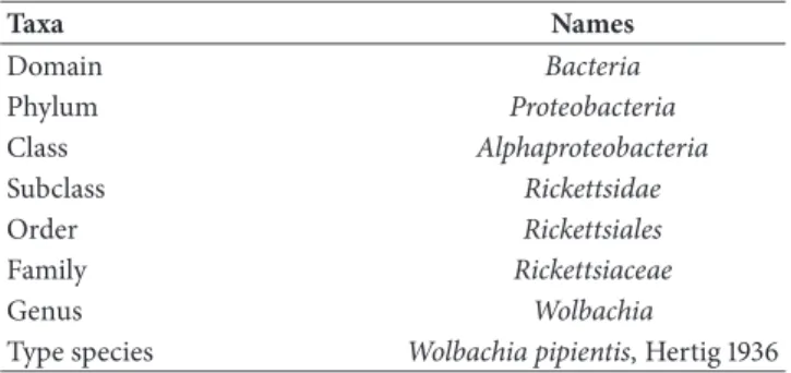
Table 1: Taxonomic classification of Wolbachia.
Taxa Names Domain Bacteria Phylum Proteobacteria Class Alphaproteobacteria Subclass Rickettsidae Order Rickettsiales Family Rickettsiaceae Genus Wolbachia
Type species Wolbachia pipientis, Hertig 1936
development occurs and compete with them. It is, therefore, an interesting biological control agent which can be used to stop or prevent the transmission of several vertebrate pathogens to humans and domestic animals [21].
In this review, we discuss the current state of knowledge about the association between mosquitoes and Wolbachia, emphasizing the limitation of different mosquito control strategies and the use of mosquitoes’ commensal microbiota as innovative approaches to control mosquito-borne diseases.
2. Methodology
2.1. Search Methods. Peer-reviewed literature search was
con-ducted using online databases including PubMed, Bibliovie, INSERM databases, Web of Knowledge, and Google Scholar for articles. Gray literature searches were conducted using World Health Organization (WHO) webpage. The key search terms used was different combinations of “mosquito”, “wol-bachia”, “biological control”, “control”, and “mosquito-borne diseases” using the Boolean operator “OR”, and combinations between concepts used the logical “AND”.
2.2. Data Screening. All documents were quickly checked to
assess their relevance to the project using information in the title and the abstract. A subset of all relevant documents was selected, sorted by section, further reviewed and compiled in the manuscript.
3. Main Text
3.1. The Genus of Wolbachia (Alphaproteobacteria)
3.1.1. Description, Classification and Phylogeny. Bacteria of
the genus Wolbachia are obligate intracellular Gram-negative bacteria belonging to the Alphaproteobacteria class (Table 1) found into the cytoplasmic vacuoles inside the cells of their insects, isopods, mites, arachnids and nematodes hosts [22]. The genus was first discovered in 1924 by Marshall Hertig and S. Burt Wolbach in the reproductive organs of Culex pipiens [23], then subsequently described by Hertig in 1936, who named the genus after his collaborator [24].
Wolbachia pipientis is the unique valid species of the
genus. Noteworthily, the two other species that have been pre-viously described as belonging to the genus Wolbachia [25]:
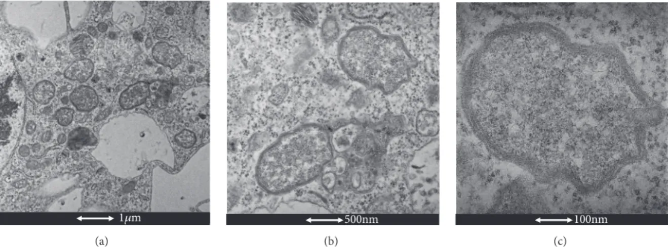
1m (a) 500nm (b) 100nm (c)
Figure 1: Electron microscopy of Wolbachia. (a) Wolbachia cocci (Scale bar: 1𝜇m). (b) Zoom of two Wolbachia cells (Scale bar: 500 nm). (c) Zoom of a single Wolbachia cell (Scale bar: 100 nm). (by El Hadji Amadou Niang).
latter on [26]. W. melophagi is now considered as nomen
nudum, because no strain of this species has been found
to date. While W. persica, which was isolated from the soft tick Argas persicus, was erroneously attributed to the genus as revealed by phylogenetic analysis of the 16S rRNA gene showing its close relatedness to the genus Francisella [27].
Morphologically, Wolbachia is a pleomorphic bacterium (Figure 1) that appears as small rods [0.5–1.3𝜇l in length] and coccoid forms [0.25–1𝜇l]; large forms [1–1.8 𝜇l in diameter] growing inside vacuoles in the cytoplasm of host cells [28]. Despite its Gram-negative cell wall structure, Wolbachia is poorly stained by Gram staining, but can be well visualized using the Diff-Quik and May-Grunwald-Giemsa staining methods. Using the Gimenez stain, they can also be visualized as dark-blue structures within a blue-green cytoplasm [29]. Since they do not form morulae and exclusively infect arthropods and filarial nematodes, Wolbachia are easily differentiated from other closely related genera [28].
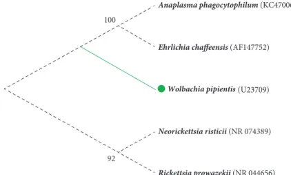
Phylogenetic analysis of the 16S rRNA gene (Figure 2), showed that W. pipientis, the nomen of the genus, forms a monophyletic clade within the Alphaproteobacteria class, closely related to the Anaplasma, Ehrlichia and Neorickettsia genera of the Anaplasmataceae family [28].
Further analysis based on the 16S rRNA and the
Wol-bachia Surface Protein (wsp) genes, were used to cluster
the species into finer taxonomic scales. A system based on the level of similarity in the wsp gene sequences has been proposed for strain grouping. So far, 16 main evolutionary lineages from different host taxa known as “supergroups” have been identified and designated by the letters A to Q, with the exception of G [22]. The usefulness of such an assemblage remains controversial and the suggestion of splitting W.
pipientis into multiple species has its pros and cons [32, 33].
Typically, the supergroups A and B are widely spread across many arthropod taxa [34]. Their common ancestor has probably diverged approximately 58–67 million years ago, at a time when all modern Arthropoda orders already existed [22]. The supergroups C and D are obligate and beneficial endosymbionts in some filarial nematodes [34–37]. While the
F supergroup is peculiar and includes both nematode and arthropod Wolbachia strains [37–40]. More specific to certain host lineages, strains in supergroup E have been reported from Collembola [41, 42], in H from termites [40], and in M and N from aphids [43]. Further distinct supergroups have been identified either in nematodes or arthropods [34].
3.1.2. Obligate Intracellular Lifestyle. A range of microbial
pathogens interact with their host in numerous and complex ways. Many are extracellular, while others invade organs and multiply within specific vector cells [44]. Wolbachia belongs to the latter group and has an original lifestyle as an obligate intracellular symbiont (endosymbiont) in close relationship with infected eukaryotic cells [45]. In arthropods, Wolbachia grow inside vacuoles often within the cytoplasm in the host’s reproductive cells. However, they can also be found in somatic tissues, including nervous tissue and hemocytes [28]. Growing research has provided exciting insights into various aspects of the Wolbachia’s biology [46]. One of the most obvi-ous consequences of their presence inside reproductive cells is the facilitation of their transovarian transmission to their host’s offspring. Analysis of the sequenced genomes of several members of the a-Proteobacteria group, to which belong the
Wolbachia genus, has also provided greater understanding of
their reductive genome evolution and antigenic variation as well as how they manipulate host cells [44]. However, the intracellular lifestyle has led to the loss of several genes as a consequence of the reduced genome size, varying from 1.1 Mb to 1.5 Mb, including less than 1000 protein-coding genes [45]. Furthermore, it has been reported that intracellular symbionts, such as Wolbachia, transfer genes into the host nucleus and vice versa [47, 48]. Leclercq et al. [49] showed high affinity between coding sequences of the f-element of the common pillbug (Armadillidium vulgare) with a large piece of the genome of the feminizing wVulC Wolbachia strain. Symbionts may also acquire genes from other symbionts [47]. The high level of genetic exchange in Wolbachia mentioned above suggests that its core genome is completed by an extensive auxiliary genome. As explained by Ishmael et al.
Anaplasma phagocytophilum (KC470064)
Ehrlichia chaffeensis (AF147752)
Wolbachia pipientis (U23709)
Neorickettsia risticii (NR 074389)
Rickettsia prowazekii (NR 044656)
100
92
Figure 2: Molecular Phylogenetic analysis of Anaplasmataceae by Maximum Likelihood method. The evolutionary history was inferred using the Maximum Likelihood method based on the Tamura-Nei model [30]. The tree with the highest log likelihood (-4338.5700) is shown. The percentage of trees in which the associated taxa clustered together is shown next to the branches. Initial tree(s) for the heuristic search were obtained automatically by applying the Neighbor-Join and BioNJ algorithms to a matrix of pairwise distances estimated using the Maximum Composite Likelihood (MCL) approach, and then selecting the topology with the higher log likelihood value. The analysis involved five nucleotide sequences. All positions containing gaps and missing data were eliminated. There was a total of 1,411 positions in the final dataset. Evolutionary analyses were conducted in MEGA7 [31].
[50], the core genome contains all the housekeeping genes shared by all (or almost all) sequenced strains for a given taxon, while all other genes constitute the auxiliary genome, encompassing the genetic variation within the species.
3.1.3. Host Reproductive Manipulation. Wolbachia are
typ-ically transmitted verttyp-ically through host eggs and alter host biology in diverse ways. They induce reproductive manipulations (Figure 3), such as (i) the feminization of infected males (i.e., turning genetic males into females); (ii) induced parthenogenesis (i.e., reproduction without males); (iii) killing of infected males; and (iv) cytoplasmic incom-patibility (i.e., modification of sperm from infected males resulting in embryonic defects and death) [34, 51].
(1) Feminization. Infected males are “dead ends” for Wol-bachia inheritance, because they do not transmit WolWol-bachia
infection to their offspring. Thus, converting infected male offspring into females increases the potential for Wolbachia to be transmitted to the next generation. The phenomenon was first described in isopods such as Armadillidium vulgare and more recently in insects [52], where it involves different mechanisms, operating at the embryonic stage [51, 52]. In sev-eral species of terrestrial isopod within the order Oniscidae,
Wolbachia invade the androgenic gland. The hypertrophied
gland is then inhibited, causing genetic males to develop as females [53]. Among insects, feminizing strains have been reported in Ostrinia furnacalis (Lepidoptera) and in Eurema
hecabe (Lepidoptera) and Zyginidia pullula (Hemiptera), in
which the involved mechanisms remain unclear [51].
(2) Parthenogenesis. Another beneficial strategy to increase
the maternal inheritance of Wolbachia is to induce the production of female offspring without fertilization by sperm,
a process known as parthenogenesis (thelytoky). Wolbachia-induced female parthenogenesis is less common and has only been documented in haplodiploid species such as thrips (Thysanoptera), mites (Acari) and wasps (Hymenoptera) [51]. In these organisms, males normally develop from unfertil-ized haploid eggs (arrhenotokous parthenogenesis), whereas females develop from fertilized diploid eggs. Wolbachia disrupt the cells’ early embryonic development, doubling the number of chromosomes in the unfertilized haploid eggs and rendering them diploid. This leads to development as an asexually produced female, so that infected females produce twice as many daughters as uninfected ones, allowing their cytoplasm to be transmitted to twice as many granddaughters as possible [54].
(3) Male Killing. In Coleoptera, Lepidoptera, Diptera (Insecta)
and Pseudoscorpiones (Arachnida), Wolbachia induce male killing of infected females’ male progeny. This phenotype, occurring mainly during embryogenesis, provides fitness benefits to the female progeny in terms of the competi-tion for resources. -induced male killing occurs through lethal feminization. Indeed, when Insight into the mech-anism has shown that Wolbachia infected mothers were treated with tetracycline to remove Wolbachia, genetic males survive, whereas in the presence of Wolbachia, genetic males become feminized and die during larval development [51, 54].
(4) Cytoplasmic Incompatibility (CI). Wolbachia-induced
cytoplasmic incompatibility (CI) is the most commonly described reproductive manipulation phenotype. Reproduc-tive incompatibility between populations of the Culex pipiens mosquito was first reported in the 1950s, but Wolbachia was only identified as the causative agent in the 1970s [55]. This
Infected Infected Uninfected Infected Uninfected
FEMINIZATION PARTHENOGENESIS MALE KILLING CYTOPLASMIC INCOMPATIBILITY
Males are turned into females Reproduction without males Wolbachia kills genetic males Infected males are incompatible with uninfected females
Infected Uninfected = MALE = FEMALE
Dead
Figure 3: Different phenotypes of Wolbachia’s host reproductive manipulation.
phenotype comprises two distinct components: Wolbachia-induced modification of sperm during spermatogenesis and rescue of this modification in embryos infected with the same strain [51]. The incompatible cross, due to the asynchrony of the male and female pronuclei phases at the initial stage of mitosis, occurs when Wolbachia-infected males mate with uninfected females (unidirectional CI). Bidirectional CI occurs when both partners are infected by different but incompatible Wolbachia strains, causing cross lethality in both directions. CI has been widely described in numerous arthropod host species infected by Wolbachia strains belong-ing to both the A and B supergroups [56].
3.1.4. Wolbachia spp. and Insects. The “pandemic” nature of Wolbachia [46] resulting in their widespread distribution in
various invertebrate hosts (Figure 4) is explained by their ability to manipulate host reproduction, but also by their ability to move horizontally across species boundaries [51, 54]. It has been estimated that different strains of Wolbachia may infect more than 65% of insect species [34]. Among these, several mosquito species belonging to different genera have been found carrying different strains.
3.2. Mosquitoes (Diptera, Culicidae)
3.2.1. Taxonomy, Classification and Phylogeny. Mosquitoes
are a monophyletic group that belongs to the order of Diptera (Table 2 and Figure 5) [58]. The origin and phylogenetic his-tory of the family of Culicidae dates back to the Mesozoic Era. It is estimated that the main lineages of current mosquitoes date from the early Cretaceous period (145-100 million years) [58, 59].
The family of Culicidae is a large and abundant group which is distributed from tropical latitudes to temperate regions, well beyond the Arctic Circle. It includes approx-imately 4,000 species, classified into two subfamilies and 112 genera. The subfamily Anophelinae has three genera and
Culicinae has 109 genera, segregated into 11 tribes [58].
Mosquitoes are of prime medical and veterinary impor-tance. In nearly all Culicidae species, only females feed on vertebrates, because of their need for blood to produce their offspring. During blood-sucking, a complex salivary
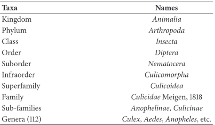
Table 2: Taxonomic classification of mosquitoes (Diptera: Culici-dae). Taxa Names Kingdom Animalia Phylum Arthropoda Class Insecta Order Diptera Suborder Nematocera Infraorder Culicomorpha Superfamily Culicoidea
Family Culicidae Meigen, 1818
Sub-families Anophelinae, Culicinae
Genera (112) Culex, Aedes, Anopheles, etc.
secretion facilitates feeding but also enables several pathogens (viruses, protozoa, and nematode worms) to be directly injected into the capillaries of their vertebrate hosts [60].
3.2.2. Mosquitoes of Medical and Veterinary Importance.
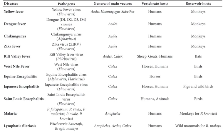
Mosquito-borne diseases such as malaria, filariases, dengue, chikungunya, Zika, and West Nile fevers represent significant medical and veterinary problems around the world and lead to major economic problems [61]. Table 3 summarises some of the most devastating mosquito-borne diseases [59].
3.2.3. Wolbachia and Mosquitoes. Among Culicidae, two types
of Wolbachia infections can be distinguished: natural
Wol-bachia infections and transinfected mosquito lines.
(1) Natural Wolbachia Infections. The interest in the Wolbachia
genus has renewed when the biological connection between cytoplasmic incompatibility and Wolbachia infection was established and documented by Yen & Barr [55] in the early 1970s. Subsequently, Yen [62] reported the presence of
Wol-bachia within the ovaries and eggs of mosquito members of
the Aedes scutellaris group (Aedes cooki, Aedes polynesiensis,
Aedes albopictus, and Aedes riversi). In 2002, while screening
several mosquitoes species, Ricci et al. [63] found arthropod strains of Wolbachia in Culex modestus, Culex pipiens, and
Lepidoptera Diptera Hymenoptera Coleoptera Phthiraptera Hemiptera Thysanoptera Blattaria Isoptera Orthoptera Odonata Oribatida Prostigmata Ixodida Mesostigmata Araneae Scorpiones Amphipoda Isopoda (a) Diptera 0 0.2 0.4 0.6 0.8 1 Bactrocera Anastrepha Lutzomya Drosophila Culex Anopheles ∗ Aedes Culicoides Protocalliphora C u licidae (b)
Figure 4: Wolbachia in arthropods. (Modified from Russell & Steiner 2012, Myrmecological News Journal [57]). (a) Graph illustrating
Wolbachia-infected (Blue shaded portion) and Wolbachia-uninfected (white portion) proportions by host taxon. (b) Histogram highlighting
the frequencies Wolbachia infection of some dipteral families. The asterisk (∗) indicates the recent discovery of native Wolbachia within the
Anopheles genus.
FAMILY SUBFAMILIES GENERA
Anopheles Bironella Chagasia Culicidae Anophelinae Culicinae Aedeomyia Aedes Armigeres Coquillettidia Culex Culiseta Deinocerites Eretmapodites Ficalbia Galindomyia Haemagogus Heizmannia Hodgesia Isostomyia Johnbelkinia Kimia Limatus Lutzia Malaya Mansonia Maorigoeldia Mimomyia Onirion Opifex Orthopodomyia Psorophora Runchomyia Sabethes Shannoniana Topomyia Toxorhynchites Trichoprosopon Tripteroides Udaya Uranotaenia Verrallina Wyeomyia Zeugnomyia
Figure 5: Classification of mosquitoes (DIPTERA: CULICIDAE) (by El Hadji Amadou Niang).
cinereus, Aedes detritus, and Ae. geniculatus) were infected
with filarial strains previously described from Dirofilaria
immitis, and two mosquitoes (Aedes punctor and Culex tor-rentium) were positive for both arthropod and filarial strains.
Later, the development of PCR and sequencing techniques has led to the discovery of many other Wolbachia strains from several other mosquito species.
A recent meta-analysis of the distribution of Wolbachia in mosquitoes indicated that, of 185 mosquito screened, 31.4%
was Wolbachia-infected but also demonstrated the nonran-dom distribution of Wolbachia among different mosquito taxa [64]. Indeed, Wolbachia was found in 39.5% of the 147 mosquito species screened, but never in Ae. aegypti, the primary vector of Dengue, Chikungunya, and Zika viruses [9, 64]. Moreover, prior to 2014 no Wolbachia infection has been documented in 38 anopheline species, including several important malarial vector species (An. gambiae, An.
Table 3: Main diseases transmitted by mosquito.
Diseases Pathogens Genera of main vectors Vertebrate hosts Reservoir hosts
Yellow fever Yellow Fever virus
(Flavivirus) Aedes Haemagogus Sabethes Humans Monkeys
Dengue fever
Dengue (D1, D2, D3, D4) viruses (Flavivirus)
Aedes Humans Monkeys
Chikungunya Chikungunya virus
(Alphavirus) Aedes Humans Monkeys
Zika fever Zika virus (ZIKV)
(Flavivirus) Aedes Humans Monkeys
Rift Valley fever Rift Valley fever virus
(Phlebovirus) Aedes, Culex Sheep, Goats, Humans Bats
West Nile Fever West Nile virus
(Flavivirus) Culex Horses, Humans Birds
Equine Encephalitis Equine Encephalitis virus
(Alphavirus, Flavivirus) Culex Horses Birds
Japanese Encephalitis Japanese Encephalitis virus
(Flavivirus) Culex Horses, Humans Pigs and wild birds
Saint Louis Encephalitis
Saint Louis Encephalitis virus
(Flavivirus)
Culex Humans, Animals Birds
Malaria
P. falciparum, P. vivax, P. malariae, P. ovale, P.
knowlesi
Anopheles Humans Monkeys for P. knowlesi
Lymphatic filariasis Wuchereria bancrofti,
Brugia malaya Anopheles, Aedes, Culex Humans Wild mammals for B. malaya
dirus, An. Albimanus, and An. darlingi), which has led to
the previous believe that Wolbachia were unable to infect
Anopheles species until their very recent discovery in natural
populations of Anopheles gambiae and Anopheles coluzzii in Burkina Faso [21, 65] and in Mali (Gomes et al. 2017). More recently Ayala et al. [66] and Jeffries et al. [67] have revealed that native Wolbachia infections was wider than expected among natural anopheline populations with at least 16 species naturally infected (Table 4). Furthermore, Ayala et al. [66] revealed a large diversity of Wolbachia strains in wild anopheline populations, which offers an unexpected opportunity to discover suitable phenotypes to suppress
Plasmodium transmission and/or to manipulate Anopheles
reproduction and reduces the malaria burden in Africa [66].
(2) Mosquito Transinfection. The absence of natural infection
in some dominant vector species has been a limiting factor for the potential operational use of Wolbachia to control vectors and the diseases they transmit. The ability of the bacterium to adapt to new intracellular environments has been exploited to transinfect vector species of medical and veterinary importance [9]. Transinfection via embryonic microinjection was used to transfer several Wolbachia strains into Ae. albopictus [64] and Ae. aegypti. For instance, the life-shortening strain of Wolbachia (wMelPop-CLA) from
Drosophila melanogaster was successfully and stably
intro-duced into Ae. aegypti to reduce its life-span. Given the proof that wMelPop strain being protective against RNA viruses in Drosophila, its derivate has been used latter to block
Dengue, Chikungunya transmitted by Ae. aegypti, while the wMel Wolbachia strain (wMel Br) has been used successfully against Zika infections in Brazil [69, 70]. Contrary to the complex Wolbachia-Arbovirus vectors, current views about the impact of Wolbachia on Plasmodium infections are almost entirely based on artificially transfected mosquito models [71]. In the Anophelinae sub-family, Wolbachia transinfection has been successful in Anopheles gambiae [72, 73] and in
Anopheles stephensi [74], respectively major vectors of human
malaria in Africa and the Middle East, and South Asia [75, 76].
3.2.4. Vector Control Approaches for the Control of Mosquito-Borne Diseases. In the past century, significant advances
have been made in controlling main vector-borne diseases. Malaria disappeared from the northern hemisphere, diseases such as typhus, Bartonella, and yellow fever prevalence were drastically reduced in many countries with effective vector control methods [9]. Despite these successes, there are currently no effective vaccines against dengue, filariasis, or malaria, and specific treatments are only available for malaria and some filariases. Historically and today, targeting mosquito vectors has been a key control strategy against major mosquito-borne diseases. Vector control is an essential component of mosquito-borne disease prevention and con-trol. Its aim is to interrupt or eliminate local transmission or reduce vulnerability to disease and prevent secondary infec-tions from introduced diseases and prevent outbreaks. Before the Second World War, vector control was predominantly
T a ble 4 :N at iv e W o lbac h ia inf ec tio n s in na tu ral m os q u it o p o p ula tio n s. H o st T ax a Su p er gr o u p s Stra in s GeneB ank # R ef er ences Su b fa m il y G enera Sp ecies w sp Cu li cin ae C u le x C x p. pipi en s B wPi p A F 02 0060 H er ti g an d W o lb ac h 19 24 ,Z h o u et al .,1 9 98 C x p. quin qu ef a sc ia tu s B wPi p A F 02 006 1 Z h o u et al .,1 9 98 Cx. br ev ipalp is A wB re AF3 17 4 77 R u ang -A re er at e et al .,2 0 03 Cx. (Eu me lano m yia ) sp p . A wE u m AF3 17 4 80 Cx. fu sco ce ph al a B wF us AF3 17 4 81 Cx. G el id u s B wGe l AF3 17 4 82 Cx (Lo ph oc er ao m yi a )s p p . A WLo p AF3 17 490 Cx. si tie n s B wS it AF3 17 49 1 Cx. m od es tu s B wPi p -R icci et al .,2 0 02 Cq .r ich iar d ii B wC on -Cx. to rr en ti u m B, C wPi p, wDi -Ae d es A e. al bo pi ct u s A, B wA lb A ,wA lb B AF0 20 0 59 ,A F0 20 059 W ri gh t and W an g 19 80 ;Z ho u et al .,1 9 98 Ae .a lb ot ae n iat u s w Al bo AF3 17 4 75 R u ang -A re er at e et al .,2 0 03 Ae .cr ag gi W C ra g AF3 17 4 78 A e. n ov oniv eu s w N ov AF3 17 4 84 Ae .n iv eu s w N iv AF3 17 4 85 A e. pseuda lb op ict us, w P seu AF3 17 4 87 A e. per pl ex us wP er p AF3 17 4 86 Ae .c ooki -Y en1 97 5; D ea n &D o b so n( 20 0 4 ); T ak ke n &K o en ra ad t2 0 13 Ae .p ol yn esi en si s -Ae .ri ve rs i -A e. ci n er eus C wDi -R icci et al .,2 0 02 Ae .d et ri tu s C wDi -Ae .g en ic u lat u s C wDi -Ae .p u n ct or B, C wPi p, wDi -Ae .fl u vi at il is B wFl u GQ 9813 15 M o re ir a et al., 20 0 9
Ta b le 4 :C o n ti n u ed . H o st T ax a Su p er gr o u p s Stra in s GeneB ank # R ef er ences Ar m ig ere s A rm .s u ba lb at u s A wS u b AF3 17 4 88 R u ang -A re er at e et al .,2 0 03 Ma n so n ia Mn .u n if or m is B wU n if AF3 17 49 3 M n .i ndiana B wI n d AF3 17 49 2 Coq u il le tt id ia Cq .c ra ss ipe s B wC ra AF3 17 4 79 A n op h eli nae A nop h el es A n .g amb iae A, B wA n ga -BF ,w A n ga -M al i KJ 72 87 39 -M F 94 4 22 3 Ba ld in i et al .,2 0 14 ;S h aw et al .,2 0 16 ;G o m es et al .,2 0 17 A n .c ol u zzi i A, B wA n ga -BF ,w A n ga -M al i KJ7 28 755-MF 94 422 3 An .a rabi en si s A, B wA n ga K J72 8739-K J7 28 75 5 Sh aw et al .,2 0 16 An .f u n es tu s A, B wA n fu -N ia n g et al .,2 0 18 [6 8] ,A ya la et al .,2 0 18 An .c ar n ev al ei A, B --Ay al a et al ., 20 18 An .c ou st an i B --A n .h anco ck i B --An .i m pl ex u s B --An .j eb u d en si s B --A n .m ar sha ll ii B --An .m ou ch et i B --A n .n ige rie n sis B --An .n il i B --An .p al u d is B --An .v in ck ei A, B
--based on the environmental control of the proliferation of mosquitoes [3]. The so-called “chemical period” then began with the advent of DDT and other organochlorine pesticides in the late 1940s. During this period, widescale spraying of the indoor surfaces of houses and shelters drastically reduced the number of malarial mosquitoes and other insects and led to the successful eradication of malaria in the United States, European countries, the Soviet Union, South East Asia, India, and South America [4, 77]. But the Malaria Eradication Pro-gramme failed in several malarial pilot areas in the African continent, due to the extremely high malaria heterogeneity and vector behavioral plasticity [78]. However, during the past decade there has been a global renewed focus on vector control with the widespread use of impregnated (LLINs) and sprayed materials (IRS), particularly against malaria vectors. Large community-based distribution and/or IRS campaigns have led to significant ITN and IRS coverage in several African countries, resulting in a substantial drop in the preva-lence of malaria in that region [3]. However, to make vector control more effective, cost effective, ecologically sound, and sustainable the WHO adopted in 2004 the Global Strategic Framework on Integrated Vector Management (IVM) as the first step towards the search and the implementation of new approaches to control vectors and the diseases they transmit [3]. Defined as “a rational decision-making process for the optimal use of resources for vector control,” IVM is not a new concept since its basic principles have been used over the past century in the USA through the vast network of Mosquito Abatement Districts implemented to protect people from nuisance-biting and vector species of mosquitoes [79]. Lately, the WHO called for the strengthening of IVM as one of the strategic areas for action in the global plan framework to combat neglected tropical diseases for 2008–2015.
Although insecticides have been successful in controlling vectors, current ecological and environmental protection standards make insecticide-based strategies unsustainable, due to the adverse effects of many insecticides on nontarget species, their environmental impact, the contamination of soil and water and the development of selective processes, and subsequent mosquito resistance to insecticides [1]. Moreover, a number of malaria prevention and control tools currently available are quite expensive, while arbovirus vector man-agement also has to face significant challenges, due to the peculiar traits of Aedes vectors, which have huge physiological and ecological plasticity making them difficult to control [80]. A broad spectrum of resistance to insecticides has evolved in the Culex genus, involving both “Metabolic” (enhanced esterase, glutathione-S-transferase, or p450 monooxygenase activities) and “Target Site” (modification of the acetyl-cholinesterase; the GABA receptors; or the sodium channels) mechanisms [81]. There is, therefore, an urgent need for effective alternative vector control strategies that can be used on a large scale and which are environmentally friendly. This is critical to sustaining control efforts and to achieving the goal of malaria elimination. Potential alternatives or com-plementary strategies to current core interventions include genetic control approaches, using refractory mosquitoes to replace vector populations or the release of mosquitoes carrying a lethal gene to suppress the targeted populations
[1]. In addition to transgenic mosquitoes, paratransgenic and biological control approaches provide concrete possibilities for innovative vector control strategies [7].
3.2.5. Biological Control of Mosquito-Borne Diseases. Beyond
the paratransgenic VC approaches taking advantage of the naturally/transinfected mosquitoes microbiota and defined as the use of symbiotic organisms naturally inhabit or successfully introduced into mosquitoes to deliver an effector molecule to inhibit, compete or kill the pathogen in insects [1, 9], their utilisation to directly interfere with or modulate vector immunity against pathogens constitutes a Biological approach to control MBD. The feasibility of the paratrans-genic approach was demonstrated by Durvasula et al. [82], when they successfully transformed a commensal symbiont in the hindgut lumen of Rhodnius prolixus, Rhodococcus
rhodnii, to express the cecropin A protein to kill the causative
agent of Chagas disease, and Trypanosoma cruzi inside their host. Similarly, the recent use of the life-shortening Wolbachia wMelPop-CLA strain is a prelude for an innovative Biological approach to control MBD. Indeed, intracellular bacteria such as Wolbachia that can manipulate their host biology, includ-ing their immune system, are unduly regarded as promisinclud-ing innovative biocontrol approach to control insect-transmitted diseases. Therefore, several studies have attempted to show the potential for Wolbachia to be used in such a strategy to control mosquito-transmitted diseases [74]. Wolbachia has several characteristics, including the capacity to perturb insect ecology, behaviour, and physiology, making it one of the best candidates for blocking, or at least significantly reducing, the transmission of pathogens of medical and veterinary importance [21]. However, before the operational implementation of any Wolbachia-based approach, an impor-tant prerequisite is to better characterize all potential the strain of the genus and their host manipulation phenotypes which could make them good biocontrol agents candidates, to develop predictive models, and to perform a compre-hensive risk assessment for their use to control mosquitoes and disease they transmit. As stated before, Wolbachia-transinfection technology has already shown promise in con-trolling the transmission of arboviruses by Ae. aegypti using different Wolbachia strains which can shorten vector lifes-pans, limit susceptibility to infection, and induce cytoplasmic incompatibility to reduce vector density. Furthermore, in An.
gambiae and An. stephensi, the presence of Wolbachia appears
to negatively impact the Plasmodium developmental cycle and egg laying [21, 74, 83]. Although potentially eligible as an inovative weapon, our knowledge of Wolbachia-mediated antiparasite mechanisms is fragmented, if not completely lacking. A significant delay in the virus-induced mortality of the pathogenic Drosophila C, Cricket paralysis and Flock House virus have been related to the presence of Wolbachia in the host. Johnson hypothesized that by reducing the viral load Wolbachia endosymbionts enhance host survival [84]. However, since different Wolbachia strains affect a wide variety of insect viruses this likely suggests that the underlying mechanisms are not pathogen specific/Wolbachia interactions but involve putatively broad processes targeting a
wide-range of viral types, including competition for resources and the upregulation of hosts’ immune responses.
(1) Wolbachia-Based Approach to Control Arboviruses Dis-eases. A new era for controlling arboviruses started with
the successful introduction of the life-shortening wMelPop-CLA Wolbachia strain into Ae. aegypti to reduce its natural populations life span [85, 86]. Primary data gathered from field trials in Australia has made it possible to validate theoretical models for Wolbachia population dynamics and has demonstrated the feasibility and sustainability of such a strategy to control mosquito populations and the diseases they transmit [87]. However, barriers to dispersal respon-sible for a slower than anticipated spread of transinfected
Aedes aegypti mosquito in Cairns (Australia) [88] should
be taken into account in future releases. Moreover, how
Wolbachia strains of interest interfere with pathogens is a
critical aspect that needs to be better understood when dealing with Wolbachia-based approaches. Several authors have attempted to unravel the basis of Wolbachia pathogen blocking. To that aim, Terradas and McGraw discussed the possible mechanistic basis of Wolbachia-mediated pathogen blocking and have evaluated the existence of evidences from field mosquitoes and related insects [89]. They showed that the amount of Wolbachia inside host cells and tissues appears to correlate with the strength of Wolbachia-mediated blocking. They revealed that the highly replicative Wolbachia strain (wMelPop) by exhibiting great cellular loads causes tissue damage thus inducing near perfect blocking in Ae.
aegypti [89]. Another possible mode of action through which Wolbachia interferes with pathogen infection is by priming
the host immune system, with the preactivation of the immune response which could then theoretically protect the insect from a range of pathogens. Gene regulation is another way by which Wolbachia modulates the host immune system as demonstrated by recent studies about the potential role of the Vago protein on the innate immune pathways of Culex
quinquefasciatus and Ae. aegypti to restrict West Nile and
dengue virus replication [90]. For instance, Asad et al. have shown that in Wolbachia-infected cells, knocking-down the Vago1 gene led to significant increases in DENV replication with no effect on Wolbachia density, and concluded that in
Ae. aegypti the induction of the AeVago1 protein, mediated
by Wolbachia in infected cells, may function as a host factor to suppress DENV replication [90].
(2) Wolbachia-Based Approach to Control Malaria. As
re-ported in the last World Malaria Report 2017, despite sig-nificant progress made since 2000, the rate of decline of malaria has stalled and even reversed in some region since 2014 [91]. Reasons for this are the spread of resistance of parasite to antimalarial drugs and vectors to insecticides [4]. Beside the implementation of a strategic insecticide resistance monitoring for malaria endemic countries, the WHO’s Global plan for insecticide resistance management in malaria vectors (GPIRM) highlighted also the need for the development of innovative approaches for sustainable vector control at global scale [92]. As a response to that, attention has been drawn to mosquitoes’ microbiota and their potential
impact on host fitness and parasite evolution [93]. Wolbachia-mediated parasite interference in other insect systems has raised the exciting possibility of using them to control or limit the spread of malaria. However, the development of
Wolbachia-based antimalarial strategies has been impeded by
the lack of stable Wolbachia infections in natural anopheline populations, as well as the failure to establish stable inherited transinfections in anopheline mosquitoes. Both issues have recently been overcome with the successful establishment of a stable Wolbachia strain wAlbB infection in Anopheles
stephensi, an important malarial vector in Asia [74], and the
recent report of stable Wolbachia infections in natural popu-lations of two important malarial vectors, Anopheles gambiae and Anopheles coluzzii, in Burkina Faso [65]. Furthermore, Shaw et al. showed that the wAnga strain stably infects reproductive tissues (ovaries), and certainly somatic tissues where the Plasmodium development occurs, and where it may effectively compete for resources or upregulate the immune response to effectively kill the malaria parasite [21]. Similar results were reported recently in Mali with a new anopheline
Wolbachia strain (wAnga-Mali) [83]. Interestingly,
experi-mental infection showed that wAnga-Mali has strong impact on late sporozoites stages and reduces malaria transmission [83]. Both studies showed the potential for the release of
Wol-bachia-infected mosquitoes as a promising strategy to reduce
malaria transmission, but also raised the great limitation due to the apparent lack of clear Cytoplasm Incompatibility [21] to ensure released population self-sustenance in the nature. The recent discovery of native Wolbachia infections in 16 out of 25 wild African Anopheles species, including both vectors and non-vectors of malaria confirm that natural Wolbachia infection in anopheline mosquitoes is more common than expected [66, 67]. This offers an unprecedented opportunity to further studies the diversity of anopheline Wolbachia strains to identify suitable phenotypes naturally impeding the development of Plasmodium parasites in mosquitoes, especially among Wolbachia strains associated with non-malaria vectors.
4. Conclusions and Future Directions
This review discussed the current state of knowledge about the association between mosquitoes and Wolbachia, empha-sizing the limitation of different mosquito control strategies and the use of mosquitoes’ commensal/introduced micro-biota as innovative VC intervention against mosquito-borne diseases.
In summary,
(1) Several human, animal and zoonotic diseases are transmitted by mosquitoes of the Anopheles, Aedes, and Culex genera. Insecticide-based vector control tools/strategies are keys components in the fight against major mosquito-borne diseases.
(2) The increasing emergence/ resurgence of mosquito-borne diseases such as malaria, yellow fever, dengue, chikungunya, and Zika fevers, and the spread of drug resistant parasites and insecticide resistant mosquito strains threatens the sustainability of current control
methods and stresses the urgent need for the devel-opment of additional control methods for mosquito-borne diseases.
(3) Wolbachia is one of the most promising mosquito symbionts for innovative vector control approaches.
Wolbachia has several characteristics which can be
used in such a strategy to reduce host fitness and competes or kills the pathogens.
(4) Wolbachia was first discovered in 1924 and described in 1936 by Marshall Hertig and S. Burt Wolbach in the reproductive organs of Culex pipiens. The “pandemic” nature of Wolbachia results from their ability to manipulate host reproduction and to move horizontally across species’ boundaries.
(5) About 31.4% of mosquito species naturally harbour one or several Wolbachia strains. Moreover, it is now possible to stably transinfect mosquito vector species of medical and veterinary importance with nonnative
Wolbachia strains which can shorten vector lifespan,
limit susceptibility to infection, or induce cytoplasmic incompatibility to reduce vector density.
(6) Wolbachia-based approach is certainly a promising innovative strategy for mosquito vector control. How-ever, our knowledge of Wolbachia-mediated antipara-site mechanisms is fragmented if not entirely lacking. (7) Additional studies, including laboratory experiments, semifield, and field trial on several mosquito vector species in different geographical population urgently need to be reinforced to better understand Wolbachia-mediated antiparasite mechanisms and interaction between hosts and parasites but also to provide empir-ical data to test theoretempir-ical models for Wolbachia population dynamics, and demonstrate the feasibil-ity and sustainabilfeasibil-ity of Wolbachia-based approach approaches to control mosquito and diseases they transmit.
Conflicts of Interest
The authors declare that there are no conflicts of interest regarding the publication of this paper.
Acknowledgments
The authors thank Dr Florian M. Steiner, Editor in Chief of Myrmecological News Journal, and Dr Jacob A. Russell of Drexel University for allowing them to reproduce their Fig 1 of Myrmecology News 16: 7-23.
References
[1] A. B. B. Wilke and M. T. Marrelli, “Paratransgenesis: A promis-ing new strategy for mosquito vector control,” Parasites &
Vectors, vol. 8, no. 1, 2015.
[2] M. S. Mulla, “Mosquito control then, now, and in the future,”
Journal of the American Mosquito Control Association, vol. 10,
no. 4, pp. 574-575, 1994.
[3] WHO, Handbook for Integrated Vector Management, WHO Press, Geneva, Switzerland, 2013.
[4] S. Sougoufara, S. Doucour´e, P. M. B. Semb´ene, M. Harry, and C. Sokhna, “Challenges for malaria vector control in sub-Saharan Africa: Resistance and behavioral adaptations in Anopheles populations,” Journal of Vector Borne Diseases, vol. 54, no. 1, pp. 4–15, 2017.
[5] K. Karunamoorthi and S. Sabesan, “Insecticide resistance in insect vectors of disease with special reference to mosquitoes: a potential threat to global public health,” Health Scope, vol. 2, no. 1, pp. 4–18, 2013.
[6] H. Alout, P. Labb´e, F. Chandre, and A. Cohuet, “Malaria Vector Control Still Matters despite Insecticide Resistance,” Trends in
Parasitology, vol. 33, no. 8, pp. 610–618, 2017.
[7] M. V. Mancini, R. Spaccapelo, C. Damiani et al., “Paratransgene-sis to control malaria vectors: A semi-field pilot study,” Parasites
& Vectors, vol. 9, no. 1, 2016.
[8] E. G. Abraham, S.-J. Cha, and M. Jacobs-Lorena, “Towards the genetic control of insect vectors: An overview,” Entomological
Research, vol. 37, no. 4, pp. 213–220, 2007.
[9] W. Takken and C. J. Koenraadt, Ecology of Parasite-Vector
Interactions, Wageningen Academic Publishers, Wageningen,
the Netherlands, 2013.
[10] G. L. Grossman, C. S. Rafferty, J. R. Clayton, T. K. Stevens, O. Mukabayire, and M. Q. Benedict, “Germline transformation of the malaria vector, Anopheles gambiae, with the piggyBac transposable element,” Insect Molecular Biology, vol. 10, no. 6, pp. 597–604, 2001.
[11] F. Catteruccia, T. Nolan, T. G. Loukeris et al., “Stable germline transformation of the malaria mosquito Anopheles stephensi,”
Nature, vol. 405, no. 6789, pp. 959–962, 2000.
[12] F. Catteruccia, J. C. H. Godfray, and A. Crisanti, “Impact of genetic manipulation on the fitness of Anopheles stephensi mosquitoes,” Science, vol. 299, no. 5610, pp. 1225–1227, 2003. [13] G. Favia, I. Ricci, C. Damiani et al., “Bacteria of the genus
Asaia stably associate with Anopheles stephensi, an Asian malarial mosquito vector,” Proceedings of the National Acadamy
of Sciences of the United States of America, vol. 104, no. 21, pp.
9047–9051, 2007.
[14] G. Favia, I. Ricci, M. Marzorati et al., “Bacteria of the genus asaia: A potential paratransgenic weapon against malaria,”
Advances in Experimental Medicine and Biology, vol. 627, pp.
49–59, 2008.
[15] E. Schnepf, N. Crickmore, J. Van Rie et al., “Bacillus thuringien-sis and its pesticidal crystal proteins,” Microbiology and
Molec-ular Biology Reviews, vol. 62, no. 3, pp. 775–806, 1998.
[16] E. Zchori-Fein, Y. Gottlieb, S. E. Kelly et al., “A newly discovered bacterium associated with parthenogenesis and a change in host selection behavior in parasitoid wasps,” Proceedings of the
National Acadamy of Sciences of the United States of America,
vol. 98, no. 22, pp. 12555–12560, 2001.
[17] S. Zabalou, M. Riegler, M. Theodorakopoulou, C. Stauffer, C. Savakis, and K. Bourtzis, “Wolbachia-induced cytoplasmic incompatibility as a means for insect pest population control,”
Proceedings of the National Acadamy of Sciences of the United States of America, vol. 101, no. 42, pp. 15042–15045, 2004.
[18] C. B. Beard, E. M. Dotson, P. M. Pennington, S. Eichler, C. Cordon-Rosales, and R. V. Durvasula, “Bacterial symbiosis and paratransgenic control of vector-borne Chagas disease,”
International Journal for Parasitology, vol. 31, no. 5-6, pp. 621–
[19] M. A. Riehle and M. Jacobs-Lorena, “Using bacteria to express and display anti-parasite molecules in mosquitoes: Current and future strategies,” Insect Biochemistry and Molecular Biology, vol. 35, no. 7, pp. 699–707, 2005.
[20] I. V. Coutinho-Abreu, K. Y. Zhu, and M. Ramalho-Ortigao, “Transgenesis and paratransgenesis to control insect-borne diseases: Current status and future challenges,” Parasitology
International, vol. 59, no. 1, pp. 1–8, 2010.
[21] W. R. Shaw, P. Marcenac, L. M. Childs et al., “Wolbachia infec-tions in natural Anopheles populainfec-tions affect egg laying and negatively correlate with Plasmodium development,” Nature
Communications, vol. 7, 2016.
[22] Y. Ilinsky and O. E. Kosterin, “Molecular diversity of Wolbachia in Lepidoptera: Prevalent allelic content and high recombina-tion of MLST genes,” Molecular Phylogenetics and Evolurecombina-tion, vol. 109, pp. 164–179, 2017.
[23] M. Hertig and S. B. Wolbach, “Studies on Rickettsia-Like Micro-Organisms in Insects,” The Journal of Medical Research, vol. 44, no. 3, pp. 329–374, 1924.
[24] M. Hertig, “The Rickettsia, Wolbachia pipientis (Gen. Et Sp. N.) and Associated Inclusions of the Mosquito, Culex pipiens,”
Parasitology, vol. 28, no. 4, pp. 453–486, 1936.
[25] V. B. D. Skerman, V. McGowan, and P. H. A. Sneath, “Approved lists of bacterial names,” International Journal of Systematic
Bacteriology, vol. 30, no. 1, pp. 225–420, 1980.
[26] N. Lo, C. Paraskevopoulos, K. Bourtzis et al., “Taxonomic status of the intracellular bacterium Wolbachia pipientis,”
Interna-tional Journal of Systematic and Evolutionary Microbiology, vol.
57, no. 3, pp. 654–657, 2007.
[27] M. Forsman, G. Sandstrom, and A. Sjostedt, “Analysis of 16S ribosomal DNA sequences of Francisella strains and utilization for determination of the phylogeny of the genus and for iden-tification of strains by PCR,” International Journal of Systematic
Bacteriology, vol. 44, no. 1, pp. 38–46, 1994.
[28] B. La Scola, C. Bandi, and D. Raoult, “Wolbachia,” in Bergeys
Manual of Systematics of Archaea and Bacteria, pp. 1–12, John
Wiley & Sons, Ltd, Chichester, UK, 2015.
[29] F. Fenollar, B. La Scola, H. Inokuma, J. S. Dumler, M. J. Taylor, and D. Raoult, “Culture and Phenotypic Characterization of a Wolbachia pipientis Isolate,” Journal of Clinical Microbiology, vol. 41, no. 12, pp. 5434–5441, 2003.
[30] K. Tamura and M. Nei, “Estimation of the number of nucleotide substitutions in the control region of mitochondrial DNA in humans and chimpanzees,” Molecular Biology and Evolution, 1993.
[31] S. Kumar and C. Barillas-Mury, “Ookinete-induced midgut peroxidases detonate the time bomb in anopheline mosquitoes,”
Insect Biochemistry and Molecular Biology, vol. 35, no. 7, pp. 721–
727, 2005.
[32] S. T. Ram´ırez-Puebla, L. E. Serv´ın-Garcidue˜nas, E. Orme˜no-Orrillo et al., “A response to Lindsey et al. “Wolbachia pipientis should not be split into multiple species: A response to Ram´ırez-Puebla et al.”,” Systematic and Applied Microbiology, vol. 39, no. 3, pp. 223–225, 2016.
[33] A. R. I. Lindsey, S. R. Bordenstein, I. L. G. Newton, and J. L. Rasgon, “Wolbachia pipientis should not be split into multiple species: A response to Ram´ırez-Puebla et al.,” Systematic and
Applied Microbiology, vol. 39, no. 3, pp. 220–222, 2016.
[34] E. Glowska, A. Dragun-Damian, M. Dabert, and M. Gerth, “New Wolbachia supergroups detected in quill mites (Acari: Syringophilidae),” Infection, Genetics and Evolution, vol. 30, pp. 140–146, 2015.
[35] K. Henkle-D¨uhrsen, V. H. Eckelt, G. Wildenburg, M. Blaxter, and R. D. Walter, “Gene structure, activity and localization of a catalase from intracellular bacteria in Onchocerca volvu-lus1Note: Nucleotide sequence data reported in this paper are available in the EMBL, GenBank and DDJB databases under the accession numbers X82176, AF069070 and AF069069.1,”
Molecular and Biochemical Parasitology, vol. 96, no. 1-2, pp. 69–
81, 1998.
[36] M. Sironi, C. Bandi, L. Sacchi, B. D. Sacco, G. Damiani, and C. Genchi, “Molecular evidence for a close relative of the arthropod endosymbiont Wolbachia in a filarial worm,”
Molecular and Biochemical Parasitology, vol. 74, no. 2, pp. 223–
227, 1995.
[37] M. J. Taylor, K. Bilo, H. F. Cross, J. P. Archer, and A. P. Underwood, “168 rDNA phylogeny and ultrastructural char-acterization of Wolbachia intracellular bacteria of the filarial nematodes Brugia malayi, B. pahangi, and Wuchereria ban-crofti,” Experimental Parasitology emphasizes, vol. 91, no. 4, pp. 356–361, 1999.
[38] N. Lo, M. Casiraghi, E. Salati, C. Bazzocchi, and C. Bandi, “How Many Wolbachia Supergroups Exist?” Molecular Biology and
Evolution, vol. 19, no. 3, pp. 341–346, 2002.
[39] J. M. Sakamoto, J. Feinstein, and J. L. Rasgon, “Wolbachia infections in the Cimicidae: Museum specimens as an untapped resource for endosymbiont surveys,” Applied and
Environmen-tal Microbiology, vol. 72, no. 5, pp. 3161–3167, 2006.
[40] B. C. Campbell, T. S. Bragg, and C. E. Turner, “Phylogeny of symbiotic bacteria of four weevil species (coleoptera: cur-culionidae) based on analysis of 16S ribosomal DNA,” Insect
Biochemistry and Molecular Biology, vol. 22, no. 5, pp. 415–421,
1992.
[41] T. T. M. Vandekerckhove, S. Watteyne, A. Willems, J. G. Swings, J. Mertens, and M. Gillis, “Phylogenetic analysis of the 16S rDNA of the cytoplasmic bacterium Wolbachia from the novel host Folsomia candida (Hexapoda, Collembola) and its implications for wolbachial taxonomy,” FEMS Microbiology
Letters, vol. 180, no. 2, pp. 279–286, 1999.
[42] A. B. Czarnetzki and C. C. Tebbe, “Detection and phylogenetic analysis of Wolbachia in Collembola,” Environmental
Microbi-ology, vol. 6, no. 1, pp. 35–44, 2004.
[43] Z. Wang, X.-M. Su, J. Wen, L.-Y. Jiang, and G.-X. Qiao, “Widespread infection and diverse infection patterns of Wol-bachia in Chinese aphids,” Insect Science, vol. 21, no. 3, pp. 313– 325, 2014.
[44] A. C. Darby, N.-H. Cho, H.-H. Fuxelius, J. Westberg, and S. G. E. Andersson, “Intracellular pathogens go extreme: genome evolution in the Rickettsiales,” Trends in Genetics, vol. 23, no. 10, pp. 511–520, 2007.
[45] P. T. Le, P. Pontarotti, and D. Raoult, “Alphaproteobacteria species as a source and target of lateral sequence transfers,”
Trends in Microbiology, vol. 22, no. 3, pp. 147–156, 2014.
[46] A. Saridaki and K. Bourtzis, “Wolbachia: more than just a bug in insects genitals,” Current Opinion in Microbiology, vol. 13, no. 1, pp. 67–72, 2010.
[47] O. Duron and G. D. D. Hurst, “Arthropods and inherited bacteria: From counting the symbionts to understanding how symbionts count,” BMC Biology, vol. 11, article no. 45, 2013. [48] N. A. Moran, “Tracing the evolution of gene loss in obligate
bacterial symbionts,” Current Opinion in Microbiology, vol. 6, no. 5, pp. 512–518, 2003.
[49] S. Leclercq, J. Th´ez´e, M. A. Chebbi et al., “Birth of a W sex chromosome by horizontal transfer of Wolbachia bacterial
symbiont genome,” Proceedings of the National Acadamy of
Sciences of the United States of America, vol. 113, no. 52, pp.
15036–15041, 2016.
[50] N. Ishmael, J. C. D. Hotopp, P. Loanidis et al., “Extensive genomic diversity of closely related wolbachia strains,”
Micro-biology, vol. 155, no. 7, pp. 2211–2222, 2009.
[51] J. H. Werren, L. Baldo, and M. E. Clark, “Wolbachia: master manipulators of invertebrate biology,” Nature Reviews
Microbi-ology, vol. 6, no. 10, pp. 741–751, 2008.
[52] D. Kageyama, G. Nishimura, S. Hoshizaki, and Y. Ishikawa, “Feminizing Wolbachia in an insect, Ostrinia furnacalis (Lep-idoptera: Crambidae),” Heredity, vol. 88, no. 6, pp. 444–449, 2002.
[53] T. T. M. Vandekerckhove, S. Watteyne, W. Bonne et al., “Evolu-tionary trends in feminization and intersexuality in woodlice (Crustacea, Isopoda) infected with Wolbachia pipientis (𝛼-Proteobacteria),” Belgian Journal of Zoology, vol. 133, no. 1, pp. 61–69, 2003.
[54] S. Charlat, G. D. D. Hurst, and H. Merc¸ot, “Evolutionary consequences of Wolbachia infections,” Trends in Genetics, vol. 19, no. 4, pp. 217–223, 2003.
[55] J. H. Yen and A. R. Barr, “New hypothesis of the cause of cytoplasmic incompatibility in Culex pipiens L. [31],” Nature, vol. 232, no. 5313, pp. 657-658, 1971.
[56] G. D. D. Hurst, F. M. Jiggins, and A. Pomiankowski, “Which way to manipulate host reproduction? Wolbachia that cause cytoplasmic incompatibility are easily invaded by sex ratio-distorting mutants,” The American Naturalist, vol. 160, no. 3, pp. 360–373, 2002.
[57] J. A. Russell, “The ants (Hymenoptera: Formicidae) are unique and enigmatic hosts of prevalent Wolbachia (Alphaproteobac-teria) symbionts,” Myrmecological News, vol. 16, pp. 7–23, 2012. [58] R. E. Harbach, “The Culicidae (Diptera): A review of taxonomy, classification and phylogeny,” Zootaxa, no. 1668, pp. 591–638, 2007.
[59] G. D. Duvallet, “D. (1958-. . . . ). Fontenille, and V. (1956-. . . . ). Robert,” Entomologie m´edicale et v´et´erinaire, 2017.
[60] P. Reiter, “Climate change and mosquito-borne disease,”
Envi-ronmental Health Perspectives, vol. 109, no. 1, pp. 141–161, 2001.
[61] I. Iturbe-Ormaetxe, T. Walker, and S. L. O’ Neill, “Wolbachia and the biological control of mosquito-borne disease,” EMBO
Reports, vol. 12, no. 6, pp. 508–518, 2011.
[62] J. H. Yen, “Transovarial transmission of rickettsia-like microor-ganisms in mosquitoes,” Annals of the New York Academy of
Sciences, vol. 266, no. 1, pp. 152–161, 1975.
[63] I. Ricci, G. Cancrini, S. Gabrielli, S. D’Amelio, and G. Favia, “Searching for Wolbachia (Rickettsiales: Rickettsiaceae) in mosquitoes (Diptera: Culicidae): Large polymerase chain reac-tion survey and new identificareac-tions,” Journal of Medical
Ento-mology, vol. 39, no. 4, pp. 562–567, 2002.
[64] K. Bourtzis, S. L. Dobson, Z. Xi et al., “Harnessing mosquito-Wolbachia symbiosis for vector and disease control,” Acta
Tropica, vol. 132, no. 1, pp. S150–S163, 2014.
[65] F. Baldini, N. Segata, J. Pompon et al., “Evidence of natural Wol-bachia infections in field populations of Anopheles gambiae,”
Nature Communications, vol. 5, article no. 3985, 2014.
[66] D. Ayala, O. Akone-Ella, N. Rahola et al., “Natural Wolbachia infections are common in the major malaria vectors in Central Africa,” bioRxiv, Article ID 343715, 2018.
[67] C. L. Jeffries, G. G. Lawrence, G. Golovko et al., “Novel Wolbachia strains in Anopheles malaria vectors from Sub-Saharan Africa,” bioRxiv, Article ID 338434, 2018.
[68] E. H. Niang, H. Bassene, P. Makoundou, F. Fenollar, M. Weill, and O. Mediannikov, “First report of natural wolbachia infection in wild anopheles funestus population in senegal,”
Malaria Journal, vol. 17, no. 1, p. 408, 2018.
[69] L. A. Moreira, I. Iturbe-Ormaetxe, J. A. Jeffery et al., “A Wolbachia symbiont in Aedes aegypti limits infection with dengue, Chikungunya, and Plasmodium,” Cell, vol. 139, no. 7, pp. 1268–1278, 2009.
[70] H. L. C. Dutra, M. N. Rocha, F. B. S. Dias, S. B. Mansur, E. P. Caragata, and L. A. Moreira, “Wolbachia Blocks Currently Circulating Zika Virus Isolates in Brazilian Aedes aegypti Mosquitoes,” Cell Host & Microbe, vol. 19, no. 6, pp. 771–774, 2016.
[71] F. Z´el´e, A. Nicot, O. Duron, and A. Rivero, “Infection with Wolbachia protects mosquitoes against Plasmodium-induced mortality in a natural system,” Journal of Evolutionary Biology, vol. 25, no. 7, pp. 1243–1252, 2012.
[72] Z. Kambris, A. M. Blagborough, S. B. Pinto et al., “Wolbachia stimulates immune gene expression and inhibits plasmodium development in anopheles gambiae,” PLoS Pathogens, vol. 6, no. 10, 2010.
[73] G. L. Hughes, R. Koga, P. Xue, T. Fukatsu, J. L. Rasgon, and D. S. Schneider, “Wolbachia Infections Are Virulent and Inhibit the Human Malaria Parasite Plasmodium Falciparum in Anopheles Gambiae,” PLoS Pathogens, vol. 7, no. 5, p. e1002043, 2011. [74] G. Bian, D. Joshi, Y. Dong et al., “Wolbachia invades Anopheles
stephensi populations and induces refractoriness to Plasmod-ium infection,” Science, vol. 340, no. 6133, pp. 748–751, 2013. [75] M. E. Sinka, M. J. Bangs, S. Manguin et al., “The dominant
Anopheles vectors of human malaria in Africa, Europe and the Middle East: occurrence data, distribution maps and bionomic pr´ecis,” Parasites & Vectors, vol. 3, no. 1, article 117, 2010. [76] M. E. Sinka, M. J. Bangs, S. Manguin et al., “A global map of
dominant malaria vectors,” Parasites & Vectors, vol. 5, no. 1, 2012. [77] L. J. Bruce-Chwatt, “Malaria research and eradication in the USSR. A review of Soviet achievements in the field of malar-iology,” Bulletin of the World Health Organization, vol. 21, pp. 737–772, 1959.
[78] L. Molineaux and G. Gramiccia, he Garki project: Research on
The Epidemiology And Control of Malaria in The Sudan Savanna of West Africa, 1980.
[79] J. C. Beier, J. Keating, J. I. Githure, M. B. Macdonald, D. E. Impoinvil, and R. J. Novak, “Integrated vector management for malaria control,” Malaria Journal, vol. 7, no. Suppl 1, p. S4, 2008. [80] G. Benelli and H. Mehlhorn, “Declining malaria, rising of dengue and Zika virus: insights for mosquito vector control,”
Parasitology Research, vol. 115, no. 5, pp. 1747–1754, 2016.
[81] A. Rivero, J. V´ezilier, M. Weill, A. F. Read, S. Gandon, and M. Manchester, “Insecticide Control of Vector-Borne Diseases: When Is Insecticide Resistance a Problem?” PLoS Pathogens, vol. 6, no. 8, p. e1001000, 2010.
[82] R. V. Durvasula, A. Gumbs, A. Panackal et al., “Prevention of insect-borne disease: an approach using transgenic symbiotic bacteria,” Proceedings of the National Acadamy of Sciences of the
United States of America, vol. 94, no. 7, pp. 3274–3278, 1997.
[83] F. M. Gomes, B. L. Hixson, M. D. W. Tyner et al., “Effect of naturally occurring Wolbachia in Anopheles gambiae s.l. mosquitoes from Mali on Plasmodium falciparum malaria transmission,” Proceedings of the National Acadamy of Sciences
of the United States of America, vol. 114, no. 47, pp. 12566–12571,
![Figure 4: Wolbachia in arthropods. (Modified from Russell & Steiner 2012, Myrmecological News Journal [57])](https://thumb-eu.123doks.com/thumbv2/123doknet/13412462.407203/7.900.180.731.111.496/figure-wolbachia-arthropods-modified-russell-steiner-myrmecological-journal.webp)