HAL Id: hal-01405097
https://hal.sorbonne-universite.fr/hal-01405097
Submitted on 29 Nov 2016HAL is a multi-disciplinary open access archive for the deposit and dissemination of sci-entific research documents, whether they are pub-lished or not. The documents may come from teaching and research institutions in France or abroad, or from public or private research centers.
L’archive ouverte pluridisciplinaire HAL, est destinée au dépôt et à la diffusion de documents scientifiques de niveau recherche, publiés ou non, émanant des établissements d’enseignement et de recherche français ou étrangers, des laboratoires publics ou privés.
neurogenesis
Fabian Rentzsch, Michael Layden, Michaël Manuel
To cite this version:
Fabian Rentzsch, Michael Layden, Michaël Manuel. The cellular and molecular basis of cnidarian neurogenesis. Wiley Interdisciplinary Reviews: Developmental Biology, Wiley, 2016, 6 (1), pp.e257 �10.1002/wdev.257�. �hal-01405097�
The cellular and molecular basis
of cnidarian neurogenesis
Fabian Rentzsch,
1* Michael Layden
2and Michaël Manuel
3Neurogenesis initiates during early development and it continues through later developmental stages and in adult animals to enable expansion, remodeling, and homeostasis of the nervous system. The generation of nerve cells has been ana-lyzed in detail in few bilaterian model organisms, leaving open many questions about the evolution of this process. As the sister group to bilaterians, cnidarians occupy an informative phylogenetic position to address the early evolution of cellular and molecular aspects of neurogenesis and to understand common prin-ciples of neural development. Here we review studies in several cnidarian model systems that have revealed significant similarities and interesting differences compared to neurogenesis in bilaterian species, and between different cnidarian taxa. Cnidarian neurogenesis is currently best understood in the sea anemone Nematostella vectensis, where it includes epithelial neural progenitor cells that express transcription factors of the soxB and atonal families. Notch signaling reg-ulates the number of these neural progenitor cells, achaete-scute and dmrt genes are required for their further development and Wnt and BMP signaling appear to be involved in the patterning of the nervous system. In contrast to many
verte-brates and Drosophila, cnidarians have a high capacity to generate neurons
throughout their lifetime and during regeneration. Utilizing this feature of cni-darian biology will likely allow gaining new insights into the similarities and dif-ferences of embryonic and regenerative neurogenesis. The use of different cnidarian model systems and their expanding experimental toolkits will thus continue to provide a better understanding of evolutionary and developmental aspects of nervous system formation.© 2016 The Authors.WIREs Developmental Biology pub-lished by Wiley Periodicals, Inc.
How to cite this article:
WIREs Dev Biol 2016. doi: 10.1002/wdev.257
INTRODUCTION
C
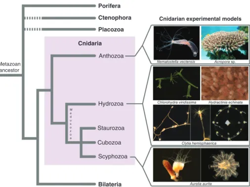
nidarians are an early offshoot in the evolution of animals, having separated from the lineage that led to the emergence of bilaterians more than600 million years ago.1 Their importance for under-standing the evolution of nervous systems has long been recognized, based both on their phylogenetic position as an outgroup to bilaterians (Figure 1) and on the structure of their nervous system, which is predominantly organized as nerve nets. The cnidar-ians comprise two major clades, the anthozoans and
the medusozoans (Figure 1),2,3 which separated
before 550 million years ago and thus not long after the cnidarian lineage separated from all other ani-mals.4 Two distinct body forms can be found in cni-darians: sessile polyps, which are tube-shaped animals with a single terminal body opening (called the mouth) surrounded by prey-catching tentacles; *Correspondence to: [email protected]
1Sars Centre for Marine Molecular Biology, University of Bergen,
Bergen, Norway
2Lehigh University, Bethlehem, PA, USA
3Sorbonne Universités, UMPC Univ Paris 06, CNRS, Evolution
Paris-Seine, Institut de Biologie Paris-Seine (IBPS), Paris, France Conflict of interest: The authors have declared no conflicts of inter-est for this article.
and free-swimming medusae, which swim by rhyth-mic contractions of their bell. Polyps are present in both cnidarian clades, while medusae are only pres-ent in the medusozoans (Figure 1), suggesting that a polyp stage was present in the last common ancestor of cnidarians, whereas parsimony favors the evolu-tion of the medusa stage after the separaevolu-tion of the two clades. The nervous system of polyps can infirst approximation be described as a nerve net with more or less pronounced regionalisation, including in some taxa instances of local condensations of neurites (in the form of nerve cords or nerve rings), for exam-ple in the oral region, or along the internal musculature-bearing mesenteries.5–7 Medusae often possess sensory organs, in particular eyes and gravity sensors (lacking in polyps), associated with a consid-erable degree of centralized signal processing, best understood in hydrozoan medusae where it involves two peripheral and parallel nerve rings.8–11 These sense organs and CNS-like nervous system elements offer an excellent opportunity to study the independ-ent evolution of advanced signal receiving and inte-grating structures and how they control the behavior and the unique mode of locomotion of jellyfish (see also Boxes 1 and 2).
In cnidarians, the nerve cell concept embraces three different but related classes of cells. Sensory
(or sensory-motor) cells generally have an elongated cell body, and always bear an apical cilium that emerges at the body surface. Ganglion cells have their cell body located in a deep, basi-epithelial position; they are often considered equivalent to interneurons, but they can also synapse on muscle cells and nemato-cytes.12 Nematocytes are the cnidarian-specific sting-ing cells, characterized by a complex intracytoplasmic capsule (nematocyst) housing a coiled tubule, and an apical sensory ciliary cone. That nematocytes are modified nerve cells is supported by a vast array of data relating to their neurophysiological properties, ultrastructural features, and expression of neurogenic genes in nematocyte precursors.13,14 Sensory cells, ganglion cells, and some nematocytes bear neurites and establish synaptic contacts with other cells. Both morphological and molecular observations show that each of the three general neural cell types consists of several or many subtypes that can be characterized for example by the number of neurites or the expres-sion of various neuropeptides.15,16 Glial cells have not been identified in cnidarians.
Today, several cnidarian model species can be reared in the laboratory to provide access to embryos and the availability of new molecular tools allows addressing longstanding questions about the develop-ment and architecture of their nervous system
Metazoan ancestor
Porifera Ctenophora Placozoa
Cnidarian experimental models
Nematostella vectensis
Chlorohydra viridissima Hydractinia echinata
Clytia hemisphaerica Aurelia aurita Acropora sp. Cnidaria Anthozoa Hydrozoa Staurozoa M e d u s o z o a Cubozoa Scyphozoa Bilateria
FIGURE 1| Position of cnidarians in the animal tree of life and phylogenetic diversity within the phylum, with a selection of experimental models used for developmental biology studies. Photo credits:Nematostella vectensis: Chiara Sinigaglia;Clytia hemisphaerica(medusa): Michaël Manuel;Acroporasp.: www.aquaportail.com;Chlorohydra viridissimaandAurelia aurita: Thomas Condamine;Hydractinia echinata: Uri Frank;
(Table 1). Transgenic reporter lines can be used to label the nervous system in unprecedented detail and to trace the progeny of cells that express a given gene at a particular time point.6,25,30,35 Inhibition of gene function by RNA interference or morpholino antisense oligonucleotides has been established and can be com-plemented by overexpression of in vitro synthesized mRNAs.23,26,31,36–38,43 Heritable genome manipula-tions by TALENs and the CRISPR/Cas9 system have been reported39 and the availability of genome and transcriptome resources allows analyzing the effects of gene manipulations and identifying new regulators of neural development beyond candidate genes.
In this review, we focus on the generation of neurons and the patterning of the nervous system in cnidarians during embryogenesis and in adult polyps and we briefly discuss aspects of the establishment of neural connectivity. Among cnidarian model organ-isms, neural development is currently best under-stood in the anthozoan Nematostella vectensis, and accordingly this sea anemone is central to our discus-sion on the cellular and molecular basis of neurogen-esis, particularly during embryonic development.
THE GENETIC TOOLKIT FOR
NEUROGENESIS IS CONSERVED IN
CNIDARIANS
The genetic toolkit that controls the generation of nerve cells and the patterning of the nervous system in bilaterian animals is represented in cnidarian genomes by an almost full repertoire of ortholo-gues. A few families of neurogenic genes are of more ancient origin than the metazoan lineage itself as deduced from their presence in unicellular holozoan genomes, e.g., several families of bZIP transcription factors (TFs) including CREB; puta-tive members of the paired-like, POU and LIM classes of homeoboxes, and of the Sox/TCF family of TFs.44,45 However, the vast majority of genes with regulatory functions in neural development of bilaterians belong to gene families that were estab-lished either in a common metazoan ancestor after divergence of choanoflagellates but before diver-gence of sponges, or in an exclusive ancestor of cnidarians and bilaterians after divergence of sponges46,47 (leaving here apart ctenophores and placozoans, whose position in the animal tree BOX 1
SENSE ORGANS IN CNIDARIANS
In the medusae of scyphozoans and cubozoans, the multifunctional neuro-sensory organ known as the rhopalia (which is a highly modified ten-tacle) contains at least seven groups of sensory cells arranged in complex bilateral pattern.150 The rhopalia nervous system is primarily ecto-dermal and its development is a highly ordered process.151Rhopalia are involved in gravity
sen-sing, chemoreception, and in some cases, photoreception. Hydrozoan medusae do not have rhopalia; they either lack sensory organs or possess isolated statocysts (equilibration organs) and in some instances, simple eyes (e.g., in Cladonema).
Apical organs in anthozoan planulae are characterized by a tuft of long cilia, which are thought to have a chemo- and/or mechanosen-sory function that might be required for the transition from planula to polyp stage.36,140The tuft region, however, does not contain
neu-rons6,152 and apical tuft formation in
Nematos-tella is not affected by knockdown of NvSoxB
(2) or NvAth-like.79,89 Developmentally, there is
thus no evidence that the cells of the apical tuft region are part of the nervous system.
BOX 2
NEUROGENESIS IN MEDUSAE
In hydrozoan medusae, nematogenesis appears restricted to the manubrium and the tentacle bulbs.153 Tentacle bulbs are basal swellings of the tentacles, attached to the medusa bell periphery. Stem cell marker genes are expressed in two restricted symmetrical areas at the proximal extremity of the tentacle bulb ectoderm (presumably populations of intersti-tial stem cells) in Clytia hemisphaerica76,153 and in Podocoryne carnea.154 There is some degree of spatial sorting of nematogenesis stages along the tentacle bulb axis, reflected by stag-gered expression of nematogenesis genes asso-ciated with different stages from stem cells to early differentiation and maturation (the latter restricted to the distal third of the bulb).24,77,153
Therefore, the tentacle bulb ectoderm behaves as a cellular conveyor belt,155,156 which can facilitate experimental approaches to under-stand the temporal progression of nematocyte development. Formation of sensory and gan-glion cells has not been described in hydrozoan medusae.
remains uncertain, Figure 1). Conservation of the neurogenic toolkit between cnidarians and bilater-ians extends to all functional gene categories from TFs (see comprehensive lists compiled in Refs 14,47,48) and components of signaling pathways (Notch, Wnt, TGF-β, FGF, Hedgehog, and Jak/Stat) to post-transcriptional regulators acting at the mRNA level (e.g., Elav and Musashi).6,49
As a general rule, multigenic families of
neu-rogenic genes diversified before the last common
ancestor of cnidarians and bilaterians, but many
of them underwent further diversification within
the bilaterian lineage(s). As a result, there are examples of well-known bilaterian neural develop-ment genes with no strict orthologue in cnidarians,
such as Neurogenin and NeuroD (bHLH TFs),50
SoxD (HMG domain-containing TFs),46,51,52 or
Engrailed (Antp-class homeodomain-containing
TFs).53 These deductions rely on gene phylogenies that are often difficult to interpret, however, such that in most cases it is hard to say if absence of a given orthologue in cnidarians reflects a primitive state (with respect to a genetic novelty of bilater-ians) or is the result of gene loss. This difficulty also causes frequent discrepancies with respect to orthology assessments among studies (for example
within the Hox, Sox, and Wnt multigenic
families). Finally, it must be recognized that our view of the cnidarian neurogenic toolkit is strongly biased toward searching for homologues of known bilaterian genes, with the consequence that the
extent of cnidarian-specific genetic innovations
associated with the nervous system remains
entirely unevaluated, except for
nematocyte-specific genes.54,55
THE CELLULAR BASIS OF CNIDARIAN
NEUROGENESIS
Interstitial Stem Cells Function in
Hydrozoan Neurogenesis
Until recently, neurogenesis in cnidarians had been mainly studied in the context of the adult polyp of the freshwater Hydra, in which production of new nerve cells takes place continuously for tissue homeo-stasis and is also needed for budding or regeneration of lost body parts.56–58 In Hydra, the three types of nerve cells derive from a common pool of stem cells known as interstitial stem cells, located in the inter-spaces between ectodermal epithelial cells of the body column. The cell lineage deriving from interstitial stem cells, which also comprises glandular cells and germ cells, is in Hydra independent from the
ectoder-mal and endodermal epithelio-muscular cell
lineages,59–62although in Hydractinia, i-cells can also generate epithelio-muscular cells.63,64 Within the Hydra interstitial cell lineage, there is a subset of i-cells that is restricted to the generation of nemato-cytes and other nerve cell types,65 although it is not known whether individual i-cells in this population can give rise to different neural cell types. In Hydra, stem cells are exclusively found in the body column where they divide, and progenitor cells are then dis-placed (together with epithelial cells) toward one of the body extremities (either the oral or basal pole).66,67 They sequentially differentiate and trans-differentiate into particular subtypes of sensory or ganglion cells depending on their position along the body axis68–71 and are eventually eliminated at the extremities. Production of nematocytes follows a sim-ilar process but nematoblasts, arranged in clusters
TABLE 1| The Experimental Toolkit for Cnidarian Model Systems
Species Genome/ Transcriptome Gain-of-Function Loss-of-Function
Trans-genics Other Refs
Acropora millepora
No/yes No No No 17–19
Aurelia aurita In progress/yes No dsRNA No 20–22
Clytia hemispherica In progress/yes mRNA, plasmid MO No 23,24 Hydra magnipapillata
Yes/yes Transgene RNAi Yes FACS 25–29
Hydractinia echinata
In progress/yes Transgene MO, RNAi Yes FACS 30–34
Nematostella vectensis
Yes/yes mRNA,
plasmid
MO Yes BiFC, histone modifications, CRISPR/
Cas9, TALEN
35–42
Note that references for transcriptome resources are not comprehensive.
BiFC, bimolecular complementationfluorescence; CRISPR, clustered regularly interspaced short palindromic repeats; FACS, fluorescence activated cell sorting; MO, morpholino antisense oligonucleotide; TALEN, transcription activator-like endonucleases.
resulting from several synchronous cell cycle divi-sions in the body column, differentiate (capsule for-mation) before actively migrating toward their final destination, i.e., mainly to the tentacles.61
There are experimental indications of signi fi-cant variation concerning cellular aspects of neuro-genesis in hydrozoans other than Hydra. Sensory cells (but not ganglion cells or nematocytes) can form in the absence of interstitial cells, and thus probably from epithelial cells, in the planula larvae of Pennaria tiarella and Clytia (formerly Phialidium) gre-garia.72,73 Isolated striated muscular cells of the medusa of Podocoryne carnea were observed in Petri dishes to transdifferentiate into smooth muscle cells and then into nerve cells.74
During embryonic development in hydrozoans, interstitial cells first appear in the endoderm shortly after gastrulation.75,76In the early planula, the endo-dermal interstitial cells give rise to nematoblasts and neuroblasts, which then migrate to the ectoderm (Figure 2).75 Therefore in hydrozoans, whereas adult interstitial stem cells are located in the ectoderm, the nervous system is of endodermal embryonic origin.
Dedicated Neural Progenitor Cells
Contribute to Neurogenesis in
Nematostella
Neurogenesis in Nematostella commences at blastula stage with the emergence of neural progenitor cells (NPC); differentiation of neural cells can first be observed in the ectoderm during gastrulation and sub-sequently also in the endoderm6,49,79 (Figure 2). Transplantation experiments have been used to gener-ate chimeric embryos in which the endoderm carries the neuron-specific NvElav1::mOrange transgene and these embryos revealed that the endoderm itself can generate neurons.6 In Nematostella, no morphologi-cal equivalent of i-cells has been identified; instead, neural cells are derived from epithelial progenitors. A transgenic line expressing mOrange under the control of regulatory elements of the NvSoxB(2) gene revealed that the population of NvSoxB(2)-expressing cells gives rise to sensory cells, ganglion cells, and nematocytes but not to non-neural cell types.79
In bilaterian neurogenesis, differences in cell cycle characteristics often reflect functionally asym-metric cell divisions that result in different fates of the daughter cells, generating for example one neu-ron and one intermediate progenitor cell or two dif-ferent types of intermediate progenitor cells.80–82 A similar situation can be observed in Nematostella. Small clusters of NvSoxB(2)::mOrange positive cells (assumed to be clones derived from one NPC) can contain both even and odd numbers of cells,
suggesting that there is no strictly synchronous cell division in the progeny of an NPC. This is further supported by EdU pulse labeling experiments, which showed that even after a 2 h pulse, only one cell in pairs of NvSoxB(2)::mOrange positive cells has incorporated EdU and thus been in S-phase.79 These observations suggest that NPCs in Nematostella can divide more than once and that their developmental program includes asymmetric divisions. The develop-mental potential of individual NvSoxB(2) expressing cells is currently not known, e.g., whether a single cell can give rise to all three principal classes of neu-ral cells or whether different NvSoxB(2)+ NPCs are limited to the generation of either sensory cells, gan-glion cells or nematocytes (Figure 3). It is also not
clear whether NvSoxB(2)+ cells self-renew,
i.e., whether they are stem cells. Alternatively, they might represent progenitor cells that after a series of divisions differentiate into neurons and that are con-tinuously replenished from an as yet unidentified pool of self-renewing stem cells or directly from epidermal cells (Figure 3).
Neurogenesis in
Scyphozoa
As in anthozoans, there is no evidence for the exist-ence of an interstitial cell lineage in scyphozoans.83 During embryonic development, differentiating nerve cells arefirst observed in the ectoderm of the planula and there is no indication that their progenitors would originate in the endoderm.84
THE MOLECULAR REGULATION OF
CNIDARIAN NEUROGENESIS
As discussed above, cnidarian genomes contain the bilaterian complement of‘neurogenic’ genes. A table summarizing expression of known bilaterian neural genes across cnidarian species has been provided in a previous review paper.14 Here, we focus on func-tional evidence that suggests that neurogenesis in cni-darians and bilaterians is likely conserved beyond the superficial observation that they possess a similar complement of genes.
Sox Action Upstream of bHLH Proneural
Gene Transcription Factors Represents a
Conserved Neurogenic Cascade
Most functional data has come from disruption of candidate neurogenic genes (Table 2). bHLH pro-neural genes belonging to the achaete-scute and atonal families have been the focus of study in multi-ple cnidarians because of their highly conserved roles
in bilaterian neurogenesis (reviewed in Ref 85). Cnash, a cnidarian achaete-scute homologue (ash) gene identified in Hydra, is expressed in developing nematocytes and sensory neurons.86,87 The endoge-nous neurogenic function of cnidarian ash genes was unclear until NvAshA was shown to be both neces-sary and sufficient for development of a subset of the Nematostella nervous system.88 Although there is a conserved role for ash genes between multiple cnidar-ian species and bilatercnidar-ians, some key expression
differences exist. Expression of cnidarian ash genes appears restricted to non-proliferative differentiating neurons,86,89 whereas in bilaterian species ash genes are expressed in both proliferative progenitor/stem cells and early differentiating neurons.85,90
Nematostella has multiple atonal-like bHLH genes, but exact homology assignments are not clear.50 Regardless, NvAth-like (also called NvArp3) promotes neural development in Nematostella.89,92 Another atonal family gene NvArp6 is necessary for
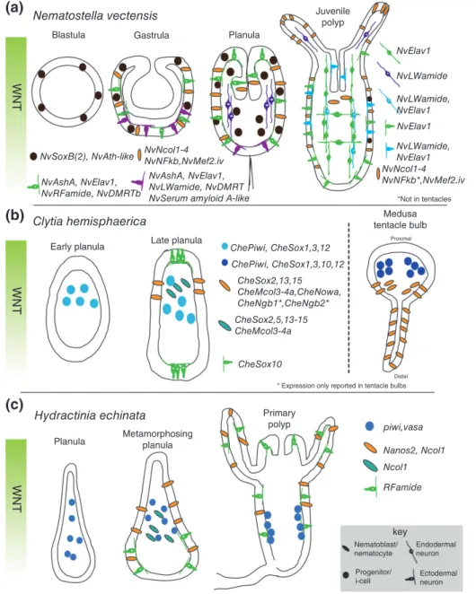
Blastula (a) (b) (c) WNT WNT WNT Nematostella vectensis Clytia hemisphaerica Hydractinia echinata
Planula Metamorphosingplanula
Gastrula Planula Juvenile polyp NvElav1 NvLWamide NvLWamide, NvElav1 NvLWamide, NvElav1 NvNcol1-4 NvNFkb*,NvMef2.iv NvNcol1-4 NvNFkb,NvMef2.iv NvSoxB(2), NvAth-like
Early planula Late planula
Primary polyp piwi,vasa Nanos2, Ncol1 Ncol1 RFamide key Nematoblast/ nematocyte Endodermal neuron Ectodermal neuron Progenitor/ i-cell ChePiwi, CheSox1,3,12 ChePiwi, CheSox1,3,10,12 CheSox2,13,15 CheMcol3-4a,CheNowa, CheNgb1*,CheNgb2* CheSox2,5,13-15 CheMcol3-4a CheSox10
* Expression only reported in tentacle bulbs
Distal
NvAshA, NvElav1, NvLWamide, NvDMRT NvSerum amyloid A-like NvAshA, NvElav1, NvRFamide, NvDMRTb *Not in tentacles Medusa tentacle bulb Proximal NvElav1
FIGURE 2| Summary of neural gene expression during embryogenesis. Some of the known neural gene expression patterns forNematostella vectensis(a),Clytia hemisphaerica(b), andHydractinia echinata(c) are shown. Developmental time progresses from left to right with depicted stage indicated above each image. In all planulae and polyp stages, the images are oriented with oral pole facing up. The orientation of theClytia
tentacle (b, right side) is proximal up and distal down. Note that the expression patterns ofCheSoxgenes are depicted in a simplified manner.77
development of GLWamide+ neurons during larval development of Nematostella. Unlike NvAsh genes, NvAth-like is clearly expressed in proliferating pro-genitor cells,89 and loss of NvAth-like function results in a decrease in the expression of NvAsh and other neural markers such as NvElav1.89,92It is still unclear if the exact function of NvAth-like is to pro-mote neurogenesis by regulating the fate of already existing neural progenitors or whether it functions in their initial specification.
sox family TFs are one of the earliest expressed genes in the neural ectoderm of both Drosophila and
vertebrates98,99and sox function is required for neu-rogenesis in both groups.98,100,101 Expression of sox genes has been characterized in both hydrozoans (Clytia hemisphaerica) and anthozoans (N. vectensis, Acropora millepora) during development51,79,102and in an adult medusa.77 Interestingly, sox genes are expressed in neural progenitor/stem cells in Clytia and Nematostella (Figures 2 and 3). Morpholino mediated knockdown of NvSoxB(2) reduces the number of neurons and the expression of NvAshA and NvAth-like.79,89 Additionally, knockdown of a different soxB gene, NvSoxB2a, reduces expression Nematostella (a) (b) Hydra NvMEK Notch signaling Wnt signaling? NvAth-like NvSoxB2 NvAshA, NvDmrtB, NvBMPs?
NvElav1, NvRFa, NvNCol3
Notch? Notch Cnox-2 Progressive restriction of developmental potential Unipotent progenitors
Expression: CnAsh, Hyzic, prdl-A&B, hyCOUP-TF (and others)
Non-neural cells
Neural progenitor cells Multipotent stem cells?
Embryonic neurogenesis
Adult neurogenesis
Multipotent stem cells Neural progenitor cells?
FIGURE 3 | Neurogenesis inNematostellaembryos and in adultHydra.(a) InNematostella, a pool of dedicated neural progenitor cells (NPCs) gives rise to the three major classes of neural cells (sensory cells, ganglion cells, and nematocytes) during embryogenesis. Individual NPCs may give rise to different classes (upper part) or to only one class of neural cells (lower part). Note that the existence of these two types of NPCs is not mutually exclusive. NPCs might be derived from multipotent stem cells, but experimental evidence for such stem cells is missing. Bars above the figure depict the stages at which the indicated genes act during the progression of neurogenesis, according to functional data described in the text (except forNvNCol3andNvRFa). Notch signaling has a role in regulating the number of NPCs and likely in the differentiation of nematocytes. (b) In adultHydra, multipotent interstitial stem cells (i-cells) give rise to the different classes of neural cells, but also to non-neural cells. As for
NematostellaNPCs, the developmental potential of individual i-cellsin vivois not clear. The generation of neural cells may involve a dedicated NPC. Except forcnox-2, there are no functional data for the indicated genes.
of NvAshA, NvAshB, and NvAth-like.92 The obser-vations that two distinct NvSoxB genes act upstream of proneural gene TFs and that NvSoxB(2) is expressed in progenitor cells suggest that soxB func-tion upstream of proneural genes at early steps in neurogenesis is a conserved feature of cnidarian and bilaterian neural programs.
Notch in Cnidarian Neurogenesis
A highly conserved bilaterian neural regulatory path-way is the Notch signaling pathpath-way. Notch activity in cnidarians has been investigated using both phar-macological and gene specific functional analyses. During Nematostella development, treatment with DAPT, which inhibits Notch activity by disrupting
TABLE 2| Genes Required for Nervous System Development in Cnidarians
Nematostella vectensis
Gene Expression Approach Effect of Manipulation Ref(s)
NvAshA Scattered cells lof—MO Fewer SCs and GCs (ISH, qPCR) 88,91
gof—mRNA More SCs and GCs (ISH, qPCR) 88,91
NvAth-like/ NvArp3
Scattered cells lof—MO Fewer SCs, GCs, and NCs (ISH, qPCR,
NvElav1::mOrange, IHC)
89,92
NvSoxB(2) Scattered cells lof—MO Fewer SCs, GCs, and NCs (ISH, qPCR,
NvElav1::mOrange, IHC)
79
NvNotch Scattered cells (gastrula) lof—inhibitor (DAPT), MO
More NPCs, SCs, and GCs (ISH, qPCR, NvElav1::mOrange), more immature NCs, fewer mature NCs (ISH, IHC)
89,91,93
gof—NICD mRNA FewerNvAshA+neural precursors, fewer SCs
and GCs (ISH, qPCR)
91
NvDelta Scattered cells (gastrula) lof—MO IncreasedNvAshA expression (qPCR) 91
gof—mRNA FewerNvAshA+neural precursors (ISH, qPCR) 91 NvSoxBa/
NvSox1
Broad in oral domain lof—MO Fewer NvRFa+and NvGLWa+neurons (IHC),
no effect onNvElav1::mOrange+neurons 92
NvAshB Broad in oral domain lof—MO Fewer NvRFa+and NvGLWa+neurons (IHC) 92
NvArp6 One sided in endoderm (planula) lof—MO Fewer GLW+neurons (IHC, qPCR) 92
Nvβ-catenin nd lof—inhibitor
(iCRT14)
Fewer NvRFa+, NvGLWa+(IHC), and
NvElav1::mOrange+neurons
92
gof—GSK3 inhibitors
More NvRFa+and NvGLWa+neurons (IHC), no effect onNvElav1::mOrange
92,94
NvElav1 Scattered cells lof—MO Fewer NvRFa+, NvGLWa+(IHC), and
NvElav1::mOrange+neurons
6
NvDmrtB Scattered cells lof—MO Fewer endodermalNvElav1::mOrange+
neurons
95
NvBMPs One sided in oral domain lof—MO Fewer NvRFa+and NvGLWa+neurons (IHC) 92,96
gof—protein No effect at gastrula, fewer NvRFa+, and
NvGLWa+neurons at planula (IHC)
92
NvMEK nd lof—inhibitor
(UO126)
Fewer NPCs, SCs, and GCs (ISH) 97
Hydra
Cnox2 Scattered cells (i-cells, neurons, nematoblasts)
lof—dsRNA Fewer SCs and GCs (ISH, IHC) 65
Hydractinia echinata
nanos 2 Scattered cells (nematoblasts, nematocytes)
lof—MO Fewer NCs (IHC), moreRFa+—neurons (ISH) 32 gof—plasmid/
transgene
More NCs (IHC), fewerRFa+—neurons (ISH) 32
GC, ganglion cell; gof, gain-of-function; ISH, in situ hybridization; IHC, immunohistochemistry; lof, loss-of-function; NC, nematocyte; MO, morpholino anti-sense oligonucleotide; qPCR, quantitative polymerase chain reaction; SC, sensory cell.
γ-secretase mediated cleavage of Notch, results in an increased neural marker expression at embryonic and larval stages.89,91,93Specific knockdown of NvNotch also resulted in increased neurogenesis, and hyper-activation of NvNotch suppresses neural differentia-tion.91 These data are consistent with Notch having conserved neurogenic function between Nematostella and bilaterian animals. More specifically, NvNotch
regulates the number of NvAth-like+ and NvSoxB
(2)+ NPCs,89,91 which is also in line with the
observed role for Notch regulation of neural progeni-tor fates in multiple bilaterian species.103 Interest-ingly, Notch effects on neurogenesis in Nematostella are likely mediated by a hes- and‘suppressor of hair-less’-independent pathway,91 which indicates an ancient role for the still poorly understood non-canonical Notch signaling mechanisms in neurogene-sis. It is not yet known if Notch can act at multiple steps of neurogenesis in Nematostella. For example, in Drosophila Notch activity isfirst required to select neuroblast progenitor cells in the ventral neuroecto-derm and then to regulate neuroblast and ganglion
mother cell fates after each neuroblast
division.104–107 Currently, there is no experimental evidence in Nematostella in support of or refuting the possibility of Notch acting at multiple levels of the neurogenic cascade. On its own the work in Nematostella suggests that Notch is a conserved neu-rogenic signaling pathway.
In contrast to the situation in Nematostella, Hydra polyps treated with DAPT generate normal numbers of neurons and nematocytes, but nemato-cytes fail to fully differentiate.108,109 Nematocyte maturation defects are also observed in Nematostella after DAPT treatment.89 The lack of a clear neural phenotype in DAPT treated Hydra blurs the compar-ison between Notch activity in cnidarians and bilater-ians. However, it is notable that the Hydra studies were done in adult polyps. Without a better compari-son of Notch activity at embryonic stages in other cnidarians, it is difficult to establish that neurogenic phenotypes for Notch in cnidarians and bilaterians represent a deep conservation for this pathway in early neural development.
Does Neurogenesis in
Nematostella Require
an Inductive Cue?
Neurogenesis in many bilaterians requires molecular cues that confer the competence of a tissue to
gener-ate neurons; this process is termed neural
induction.110–113 Whether comparable inductive sig-nals exist in cnidarians is not irrevocably clear, but there is evidence that suggests cells in Nematostella
exhibit differential abilities to become neuronal. For example, ubiquitous misexpression of NvAshA is able to upregulate neural marker expression in Nematos-tella.88However, close examination reveals a number of cells that are unresponsive to the ectopic NvAshA. This is reminiscent of unilateral misexpression of ash genes in Xenopus expanding the nervous system, but not neuralizing an entire half embryo.114 Both the Xenopus and Nematostella proneural misexpres-sion phenotypes indicate that cells require a signal to sensitize them toward competence to respond to proneural gene activity. Additional support that not all cells will generate neurons comes from studies investigating Notch signaling. Notch suppresses neu-rogenesis in Nematostella, but inhibition of Notch by either morpholino or DAPT, does not result in ubiquitous neural marker expression and neither does the simultaneous inhibition of Notch and over-expression of NvAshA.89,91 These data argue that Notch regulates the total number of neurons, but that it does not act on embryonic tissue that has a uniform neurogenic potential. Rather, Notch signal-ing appears to act on a distributed population of cells that has already acquired neurogenic potential. The question then remains whether the differential neurogenic potential is the result of some inductive cue or if it reflects some inherent properties of a subpopulation of embryonic cells.
There are a number of studies of candidate molecules that can provide some insight regarding the potential for neural induction in Nematostella. Inhibition of BMP2/4 signaling is perhaps the best-known neural induction mechanism in bilater-ians.111,112,115,116 In Nematostella embryos, until gastrulation, BMP signaling activity is hardly detecta-ble (as measured by phosphorylation of the signal transducer NvSmad 1/5/8) and the onset of neuro-genesis at blastula stage thus occurs at very low levels of BMP signaling.92,117,118 Treatment with human BMP2 protein until mid-blastula stage does not affect
the number of NvFMRF+ and NvGLW+ neurons at
planula stage; however, it is not clear whether the onset of neurogenesis is delayed until the BMP pro-tein is washed out, which would be expected if inhi-bition of BMP signaling is required for neurogenesis. Prolonged treatment with human BMP2 (until plan-ula stage) reduces expression of neural markers at larval stages of Nematostella development,92 but injection of the NvBMP2/4 morpholino has the same effect.92,96 Thus, it remains unclear whether the absence of BMP activity is a prerequisite for the initi-ation of neural development.
Reports from chicken and zebrafish indicate that FGF signaling promotes neurogenesis by both inducing the expression of the BMP2/4 inhibitors chordin and noggin as well as directly acting to pro-mote expression of neural genes.113,119Broad inhibi-tion of FGF signaling with SU5402 does not impact expression of NvashA and thus does not appear to have a neurogenic role.97However, FGF independent activity of the Mitogen Activated Protein Kinase kinase MEK is necessary for the expression of NvSoxB(2), NvAth-like, and NvAshA.97 Addition-ally, ectopic NvAshA cannot promote neural fates when MEK activity is blocked with the pharmacolog-ical inhibitor U0126,97which taken together suggests that some instruction is necessary to impart neuro-genic potential in embryonic cells.
Wnt signaling has also been linked to neural development in Nematostella. Inhibition of Wnt/ β-catenin signaling impairs neural development and reduces the expression of early markers of neurogen-esis already at blastula stage.92 While the exact step at which Wnt/β-catenin signaling regulates neurogen-esis remains to be determined, it is interesting to note that the activity of this pathway is highest at the oral pole of the blastula and gastrula, whereas expression of neural markers becomes excluded from the high Wnt activity oral domain just prior to gastrula-tion.79,89 Loss of neurogenesis following disruption of Wnt, BMP, and MEK signaling suggests that external cues are able to promote neural fates, and the requirement of MEK activity for NvAshA to pro-mote neural fates suggests that ‘neural’ is not an inherent/default state in some or all of Nematostella cells. Taken together current observations suggest that a process similar to neural induction is required to make cells in Nematostella competent to become neural, but the exact identity of the inductive cue remains elusive.
THE RELATION OF ECTODERMAL
AND NEURAL PATTERNING
Gene expression studies have identified distinct ecto-dermal territories with sharp boundaries along the oral–aboral axis of cnidarian planulae, most promi-nently in Nematostella. In Nematostella, Clytia, Hydractinia, and Hydra Wnt signaling has been shown to be an important regulator of oral–aboral patterning, with high levels of Wnt/β-catenin signal-ing promotsignal-ing the development of oral identity, and
different Wnt genes having nested expression
domains starting from the oral pole.31,94,120–125 In bilaterians, the nervous system is patterned along the
antero-posterior axis in register with the ectodermal expression domains of regulatory genes such as otx or the hox genes. Morphological regionalisation along the oral–aboral axis is not much pronounced in the cnidarian planula larva (except for the mouth at the oral pole and the apical organ at the aboral pole), but when looking at the precise distribution of cell types, it appears that there is oral–aboral region-alization of the planula nervous system. In hydro-zoan planula larvae, RFamide and GLWamide immunoreactive sensory cells are concentrated in the aboral region, and nematocytes at the oral pole, at least in some species (e.g., Clava multicornis126; C. hemisphaerica127 and unpublished observations; Hydractinia echinata16,128–130). There is also regiona-lized distribution of molecularly defined neural cell types in anthozoan and scyphozoan planulae,6,49,131 showing that patterning of the nervous system along the oral–aboral axis is common in cnidarians.
A correspondence of these domains of neural marker expression to those of ectodermal patterning genes remains to be established. Consistent with its function in the patterning of the oral–aboral axis, Wnt/β-catenin signaling is required for the formation of RFamide and GLWamide immunoreactive neu-rons in the oral territory of Nematostella.92Similarly, NvSix3/6, which regulates aboral development, is required for the expression of NvDmrtB in the abo-ral domain.132However, the expression of RFamide, GLWamide and NvDmrtB is not strictly limited to oral and aboral domains, respectively; rather their expression can be detected in different densities along the oral–aboral axis. Thus, while regionalization of the nervous system along the oral–aboral axis is pres-ent in cnidarian planulae, the molecular mechanisms that control this regionalization remain to be explored. As afirst step, a better molecular definition of distinct neural cell types (by the expression of e.g., receptors, neuropeptides, neurotransmitters, or related biosynthetic enzymes and transporters) will be required.
SUBPOPULATIONS OF NEURONS
Subpopulations of neurons can be characterized by their function (e.g., as chemo- or mechanosensory cells), their morphology (e.g., the pattern of neurite projections) and by molecular features (e.g., by neu-rotransmitter or gene expression) or by combinations of these characters. Each of these three categories of features has been used to describe neural cell types in cnidarians, but there is hardly any information on
generation of these cell types. Nematocytes have mechanosensory properties and they contain a
sophisticated extrusive organelle, the
nemato-cyst.133,134 Despite the existence of several different types of nematocysts, the nematocytes might thus be considered a rather well-defined class of neural cells. In Hydra polyps, nests of nematoblasts derived from up to five rounds of synchronous divisions allow the relation of gene expression patterns to nematocyte
development.135 The zinc finger gene HyZic is
expressed in proliferating nematoblast nests but not in sensory or ganglion cells, identifying it as a nematocyte-specific regulator that might be useful to elucidate the molecular basis of nematocyte develop-ment.136Recently, the Hydractinia nanos 2 gene has been shown to promote the formation of nemato-cytes at the expense of neurons, thus acting as a switch between two classes of neural cells.32 In Nematostella, in situ hybridization combined with
counterstaining using neural markers
(e.g., neuropeptides) or the use of transgenic reporter lines can tie expression patterns to specific neural cell types. For the nematocyte lineage, NvNF-κB has been described as a specific regulator,137whereas NvElav1 is expressed in and required for the development of subsets of sensory and ganglion cells, but not nematocytes.6
For a better understanding of the development of classes of neural cells, it will be necessary to find markers for subpopulations of mature neurons. Sub-populations of sensory cells can potentially be identi-fied by genes related to sensory functions in bilaterians, even though it is often not straightfor-ward to assign particular sensory modalities to these genes. Genes related to vertebrate chemoreceptors and to insect gustatory receptors have been identified in the Nematostella genome,138,139 but their expres-sion patterns are either not known, or (in the case of the putative gustatory receptor NvGrl1) do not sug-gest a role in chemoreception. For other candidate sensory cell receptors (e.g., TRP channels, TMCs, and Piezo) expression analyses are so far also limited. A TRPV-like gene is expressed in the apical organ of Nematostella,140 which contains a tuft of long cilia that are thought to have mechano- and/or
chemo-sensory functions; and an antibody against
NvTRPA1 labels potentially mechanosensory hair cells in the tentacles.141The expression of some can-didate regulators of neural development in Nematos-tella (e.g., NvRough and NvEvx142) is confined to a small number of distributed cells, potentially identify-ing neural subpopulations defined by a shared devel-opmental program. Functional analyses, however, have so far focused on early and broadly acting
regulators of neural development in Nematostella (and other cnidarians), and the knowledge derived from these studies can now be used to test whether a particular candidate gene is indeed expressed in the neural lineage. The generation of new transgenic lines will be essential to describe the composition of the Nematostella nervous system and to refine the cellu-lar and molecucellu-lar program that generates neural cell type diversity. This more detailed knowledge will in turn provide the basis for comparisons of cell type specific regulatory programs between cnidarians and bilaterians that can inform the reconstruction of the evolution of neural cell types.
IMPLICATIONS CONCERNING THE
EVOLUTION OF NEURAL
DEVELOPMENT
It is now clear that Nematostella shares several cellu-lar and molecucellu-lar features of neurogenesis with bila-terian model organisms. Epithelial NPCs divide repeatedly to give rise to different neural cell types; their number is regulated by Notch signaling; soxB, atonal, and achaete-scute genes are broadly required for neural development and Wnt signaling is involved in the patterning of the nervous system. These fea-tures are strong candidates for being shared ancestral traits of cnidarians and bilaterians. A more detailed understanding of the transcriptional regulation of conserved neurogenic genes in cnidarians will likely help to understand how their expression became restricted to defined parts of the ectoderm in many groups of bilaterians and how this relates to the evo-lution of centralized nervous systems.
Comparison of neurogenesis in hydrozoans and Nematostella reveals similarities at a general level, like the broad neurogenic potential during develop-ment and regeneration, but also clear differences. The generation of hydrozoan neurons by interstitial stem cells and the lack of an effect of Notch inhibi-tion on the number of neurons108,109 indicate sub-stantial differences in the cellular source and the molecular regulation of neurogenesis (Figure 3). Such differences may provide an opportunity to study the evolutionary plasticity of neural development, but such attempts will require a much improved under-standing of neurogenesis in hydrozoans, Nematos-tella and other cnidarians. Current comparisons are to a large extent based on observations in adult Hydra polyps, whereas data for Nematostella is exclusively derived from embryonic neurogenesis. Other important questions that need to be addressed
Nematostella and the developmental potential of individual i-cells during unperturbed hydrozoan development. Hypothetically, the NvSoxB(2)+ NPCs could be derived from multipotent epithelial stem cells (functionally resembling i-cells) and the i-cells might contain subpopulations that are dedicated to the generation of neural cells (resembling NPCs, Figure 3). New data on these questions will improve the reconstruction of ancestral and derived aspects of cnidarian neurogenesis and in consequence that of
shared features of cnidarian and bilaterian
neurogenesis.
DEVELOPMENT OF CONNECTIVITY
IN CNIDARIAN NERVOUS SYSTEMS
Establishing functional neural circuits requires the outgrowth of neurites and the formation of synaptic connections to other neurons and/or to effector cells, e.g., contractile or secretory cells. In bilaterians, neur-ites are usually distinguished into dendrneur-ites, which receive signals at their postsynaptic sites, and axons, which transmit signals to other cells via presynaptic sites. The mechanisms that control neurite outgrowth differ for dendrites and axons and are better under-stood for the latter.143,144 In cnidarians, it is not known whether a clear separation of neurites into axons and dendrites exists. Neurons, in particular ganglion cells, can have multiple neurites, but they usually do not display obvious morphological fea-tures (e.g., large synaptic terminals or dendritic spines) that would identify them as dendrites or axons. Cnidarian chemical synapses can be unidirec-tional or bidirecunidirec-tional (i.e., with synaptic vesicles on both sides of the synaptic cleft), with varying relative abundance,145–147but neither the distribution of pre-and postsynaptic sites nor the polarity of microtu-bules (which differs between bilaterian dendrites and axons148) have been mapped systematically.
Cnidarians also differ from bilaterians with respect to the development of neural connectivity. Neurons (including ganglion cells) are generated throughout most of the body column in Nematostella and the distribution of i-cells in Clytia and Hydracti-nia planulae suggests that this is also the case in hydrozoans. This widely distributed origin of neu-rons contrasts with the spatially more restricted gen-eration of neurons (in particular interneurons) in the main bilaterian model organisms. In these animals, neurites are guided by a combination of permissive and instructive cues that are provided by the extracel-lular matrix and by intermediate ‘signpost cells’ or the eventual targets for innervation.143,144 The
formation of a nerve net starting from distributed neurons is, however, also conceivable without target-derived guidance cues. Neurites might grow out ran-domly and the formation of stable synaptic contacts could be determined by the ‘availability’ of target cells, i.e., target cells would accept only a limited number of synaptic contacts. In such a scenario, neurites would compete for the available target sites and the consolidation of synapses would potentially depend on activity that reflects integration into neu-ral circuits. Two observations, however, argue that the outgrowth of neurites in Nematostella is not an entirely random process. At the early planula stage, a dense net of basi-ectodermal neurites is present but they are almost entirely excluded from a small region at the aboral pole.79 In contrast, at a slightly later timepoint, the neurites of the NvElav1and GLWa-mide expressing sensory neurons all project in an aboral direction.6 While this is a transient phenome-non (later born NvElav1 and GLWamide neurons project rather in transverse orientation), it suggests that the aboral pole may have a role in regulating the orientation of neurite outgrowth,first negatively and subsequently positively for a subpopulation of neurons.
In anthozoans, bundles of neurites run along the base of the mesenteries, which are endodermal infoldings that structure the gastric cavity and bear the retractor muscles and the gonads.6,49While these neurite bundles are the most prominent morphologi-cal feature of the nervous system, the neurites within these bundles can project in oral or aboral direction, suggesting that there is no uniform mechanism that regulates their formation.6Molecularly, genes encod-ing for several of the major receptor-ligand pairs involved in bilaterian neurite guidance (Semaphorin and Plexin, Ephrin and Eph, Wnt and Ryk, Netrin and Neogenin/DCC, Unc5, RGM) are present in the Nematostella genome.48 Interestingly, Netrin and RGM are expressed in different subdomains in the aboral territory, consistent with a possible role in the attraction and/or repulsion of neurites in this area.117,149Functional characterization of these con-served candidate genes will likely provide interesting first insights into the mechanisms that direct the establishment of the nervous system architecture in cnidarian polyps.
CONCLUSIONS AND FUTURE
PERSPECTIVES
Continued study of cnidarian neurogenesis will impact our understanding of nervous system function,
evolution of nervous systems, and potentially provide critical clues about mechanisms regarding neural regeneration. Now that tools have been developed (namely transgenesis) in multiple cnidarian species it will be possible to begin to unravel the connectivity of cnidarian nerve nets. Use of neural specific promoters to express calcium sensitivefluorescent proteins, light-controlled ion channels, and anterograde and retro-grade labeling reagents, such as the C-terminal frag-ment of the tetanus toxin (TTC) and wheat germ agglutinin (WGA),157will allow us to assemble wiring maps which in turn can help to understand how neu-ral patterning in neurogenesis might contribute to the formation of specific neural circuits.
Functional studies demonstrate that some cni-darian nervous systems are using the same generic neurogenic programs that bilaterian animals deploy. This links cnidarian nerve nets and bilaterian nervous systems to a common origin. Expression profiling of animals in which neurogenesis has been enhanced or decreased experimentally or of fluorescently labeled neural cells now allows looking at cnidarian neural development at broader scale. Such studies will likely also identify roles of non-conserved, taxonomically restricted regulators of neural development. More detailed knowledge of the neurogenic program in cni-darians will likely provide insight as to what
evolu-tionary modifications of neural gene regulatory
networks gave rise to bilaterian nervous systems and in particular to the brain(s) and central nervous system(s).
While neuronal patterning in both cnidarians and bilaterians is tied to axial patterning, the identity of neu-rons within distinct domains cannot be easily homolo-gized and there is no adequate description of cnidarian neuronal cell types that would allow specific compari-sons between cnidarian nerve nets and bilaterian nerv-ous systems. Thus, there is currently insufficient data to make definitive statements about potential homology of neural cell types or particular regions of the nervous sys-tems of cnidarians and bilaterians.
An exciting aspect of cnidarian neurogenesis is the potential to utilize this highly regenerative group of animals to better understand how nervous systems regenerate. During regeneration, new neurons must reintegrate with existing neurons to reform a functional system. This process is still poorly understood in ani-mals. One of the challenges of neural regeneration is neurite pathfinding and re-establishing connectivity in an adult environment. Data about how this occurs nat-urally in animals are relatively limited, but one study in zebrafish suggests that axon pathfinding during regen-eration requires molecular programs distinct from those used during development.158 This observation implies that even if research on regenerative neurogenesis can be guided by development, independent studies speci fi-cally focused on understanding regeneration must be carried out. Multiple cnidarian species are now accessi-ble to experimental manipulation during development and regeneration and they are poised for studies identi-fying and comparing developmental and regenerative neurogenic mechanisms.
ACKNOWLEDGMENTS
We thank Chiara Sinigaglia, Thomas Condemine, Uri Frank, and Muriel Jager for providing photographs for Figure 1. All authors contributed equally to the manuscript and FR co-ordinated the writing.
FURTHER READING
Schmidt-Rhaesa A, Hartzsch S, Purschke G, eds. Structure and Evolution of Invertebrate Nervous Systems. Oxford Univer-sity Press; 2016.
REFERENCES
1. dos Reis M, Thawornwattana Y, Angelis K,
Telford MJ, Donoghue PC, Yang Z. Uncertainty in the timing of origin of animals and the limits of precision in molecular timescales. Curr Biol 2015, 25:2939–2950. 2. Collins AG. Phylogeny of medusozoa and the evolu-tion of cnidarian life cycles. J Evol Biol 2002, 15:418–431.
3. Collins AG, Schuchert P, Marques AC, Jankowski T, Medina M, Schierwater B. Medusozoan phylogeny and character evolution clarified by new large and small sub-unit rDNA data and an assessment of the utility of phy-logenetic mixture models. Syst Biol 2006, 55:97–115. 4. Cartwright P, Collins A. Fossils and phylogenies:
origin of early major metazoan lineages. Integr Comp
Biol 2007, 47:744–751.
5. Koizumi O, Hamada S, Minobe S, Hamaguchi-Hamada K, Kurumata-Shigeto M, Nakamura M, Namikawa H. The nerve ring in cnidarians: its pres-ence and structure in hydrozoan medusae. Zoology
(Jena) 2015, 118:79–88.
6. Nakanishi N, Renfer E, Technau U, Rentzsch F. Nerv-ous systems of the sea anemone Nematostella vectensis are generated by ectoderm and endoderm and shaped
by distinct mechanisms. Development 2012,
139:347–357.
7. Batham EJ, Pantin CFA, Robson EA. The nerve-net of the sea-anemone Metridium senile: the mesenteries and the column. Q J Microsc Sci 1960, 101:487–510. 8. Satterlie RA. Neuronal control of swimming in
jelly-fish: a comparative story. Can J Zool 2002, 80:1654–1669.
9. Satterlie RA. Do jellyfish have central nervous systems?
J Exp Biol 2011, 214:1215–1223.
10. Mackie G, Meech R. Central circuitry in the jellyfish Aglantha. II: the ring giant and carrier systems. J Exp
Biol 1995, 198:2271–2278.
11. Mackie G, Meech R. Central circuitry in the jellyfish Aglantha. I: the relay system. J Exp Biol 1995, 198:2261–2270.
12. Westfall JA, Elliott CF, Carlin RW. Ultrastructural evi-dence for two-cell and three-cell neural pathways in the tentacle epidermis of the sea anemone Aiptasia
pal-lida. J Morphol 2002, 251:83–92.
13. Thurm U, Brinkmann M, Golz R, Holtmann M, Oliver D, Sieger T. Mechanoreception and synaptic transmission of hydrozoan nematocytes.
Hydrobiolo-gia 2004, 530:97–105.
14. Galliot B, Quiquand M, Ghila L, de Rosa R, Miljkovic-Licina M, Chera S. Origins of neurogenesis, a cnidarian view. Dev Biol 2009, 332:2–24.
15. Koizumi O, Sato N, Goto C. Chemical anatomy of hydra nervous system using antibodies against hydra neuropeptides: a review. Hydrobiologia 2004, 530:41–47.
16. Martin VJ. Characterization of a RFamide-positive subset of ganglionic cells in the hydrozoan planular nerve net. Cell Tissue Res 1992, 269:431–438. 17. Moya A, Huisman L, Ball EE, Hayward DC,
Grasso LC, Chua CM, Woo HN, Gattuso JP, Foret S, Miller DJ. Whole transcriptome analysis of the coral
Acropora millepora reveals complex responses to
CO(2)-driven acidification during the initiation of cal-cification. Mol Ecol 2012, 21:2440–2454.
18. Meyer E, Aglyamova GV, Wang S, Buchanan-Carter J, Abrego D, Colbourne JK, Willis BL, Matz MV. Sequencing and de novo analysis of a coral larval tran-scriptome using 454 GSFlx. BMC Genomics 2009, 10:219.
19. Kortschak RD, Samuel G, Saint R, Miller DJ. EST analysis of the cnidarian Acropora millepora reveals extensive gene loss and rapid sequence divergence in
the model invertebrates. Curr Biol 2003,
13:2190–2195.
20. Fuchs B, Wang W, Graspeuntner S, Li Y, Insua S, Herbst EM, Dirksen P, Bohm AM, Hemmrich G, Sommer F, et al. Regulation of polyp-to-jellyfish transi-tion in Aurelia aurita. Curr Biol 2014, 24:263–273. 21. Nakanishi N, Camara AC, Yuan DC, Gold DA,
Jacobs DK. Gene expression data from the moon jelly, Aurelia, provide insights into the evolution of the com-binatorial code controlling animal sense organ devel-opment. PLoS One 2015, 10:e0132544.
22. Rachamim T, Morgenstern D, Aharonovich D, Brekhman V, Lotan T, Sher D. The dynamically evol-ving nematocyst content of an anthozoan, a scypho-zoan, and a hydrozoan. Mol Biol Evol 2015, 32:740–753.
23. Momose T, Houliston E. Two oppositely localised frizzled RNAs as axis determinants in a cnidarian embryo. PLoS Biol 2007, 5:e70.
24. Houliston E, Momose T, Manuel M. Clytia
hemi-sphaerica: a jellyfish cousin joins the laboratory. Trends Genet 2010, 26:159–167.
25. Wittlieb J, Khalturin K, Lohmann JU, Anton-Erxleben F, Bosch TC. Transgenic hydra allow in vivo tracking of individual stem cells during morphogenesis.
Proc Natl Acad Sci U S A 2006, 103:6208–6211.
26. Lohmann JU, Endl I, Bosch TC. Silencing of develop-mental genes in hydra. Dev Biol 1999, 214:211–214. 27. Chapman JA, Kirkness EF, Simakov O, Hampson SE,
Mitros T, Weinmaier T, Rattei T,
Balasubramanian PG, Borman J, Busam D, et al. The
dynamic genome of hydra. Nature 2010,
464:592–596.
28. Chera S, de Rosa R, Miljkovic-Licina M, Dobretz K, Ghila L, Kaloulis K, Galliot B. Silencing of the hydra serine protease inhibitor Kazal1 gene mimics the human SPINK1 pancreatic phenotype. J Cell Sci 2006, 119:846–857.
29. Hemmrich G, Khalturin K, Boehm AM, Puchert M, Anton-Erxleben F, Wittlieb J, Klostermeier UC,
Rosenstiel P, Oberg HH, Domazet-Loso T,
et al. Molecular signatures of the three stem cell lineages in hydra and the emergence of stem cell func-tion at the base of multicellularity. Mol Biol Evol 2012, 29:3267–3280.
30. Kunzel T, Heiermann R, Frank U, Muller W,
Tilmann W, Bause M, Nonn A, Helling M,
Schwarz RS, Plickert G. Migration and differentiation potential of stem cells in the cnidarian Hydractinia analysed in eGFP-transgenic animals and chimeras.
Dev Biol 2010, 348:120–129.
31. Duffy DJ, Plickert G, Kuenzel T, Tilmann W, Frank U. Wnt signaling promotes oral but suppresses aboral
structures in Hydractinia metamorphosis and regenera-tion. Development 2010, 137:3057–3066.
32. Kanska J, Frank U. New roles for Nanos in neural cell fate determination revealed by studies in a cnidarian.
J Cell Sci 2013, 126:3192–3203.
33. Soza-Ried J, Hotz-Wagenblatt A, Glatting KH, del Val C, Fellenberg K, Bode HR, Frank U, Hoheisel JD, Frohme M. The transcriptome of the colonial marine hydroid Hydractinia echinata. FEBS J 2010, 277:197–209.
34. Hensel K. Wnt signalling in the hydrozoan Hydractinia
echinata. PhD Thesis, National University of Ireland Galway, 2013. Available at: https://aran.library. nuigalway.ie/xmlui/handle/10379/4374. (Accessed June 2, 2016).
35. Renfer E, Amon-Hassenzahl A, Steinmetz PR, Technau U. A muscle-specific transgenic reporter line of the sea anemone, Nematostella vectensis. Proc Natl
Acad Sci U S A 2009, 107:104–108.
36. Rentzsch F, Fritzenwanker JH, Scholz CB, Technau U. FGF signalling controls formation of the apical sensory organ in the cnidarian Nematostella vectensis.
Devel-opment 2008, 135:1761–1769.
37. Magie CR, Daly M, Martindale MQ. Gastrulation in the cnidarian Nematostella vectensis occurs via invagi-nation not ingression. Dev Biol 2007, 305:483–497. 38. Wikramanayake AH, Hong M, Lee PN, Pang K,
Byrum CA, Bince JM, Xu R, Martindale MQ. An ancient role for nuclear β-catenin in the evolution of axial polarity and germ layer segregation. Nature 2003, 426:446–450.
39. Ikmi A, McKinney SA, Delventhal KM, Gibson MC. TALEN and CRISPR/Cas9-mediated genome editing in the early-branching metazoan Nematostella vectensis.
Nat Commun 2014, 5:5486.
40. Putnam NH, Srivastava M, Hellsten U, Dirks B, Chapman J, Salamov A, Terry A, Shapiro H, Lindquist E, Kapitonov VV, et al. Sea anemone genome reveals ancestral eumetazoan gene repertoire and genomic organization. Science 2007, 317:86–94.
41. Hudry B, Thomas-Chollier M, Volovik Y,
Duffraisse M, Dard A, Frank D, Technau U, Merabet S. Molecular insights into the origin of the Hox-TALE patterning system. Elife 2014, 3:e01939. 42. Schwaiger M, Schonauer A, Rendeiro AF, Pribitzer C,
Schauer A, Gilles AF, Schinko JB, Renfer E, Fredman D, Technau U. Evolutionary conservation of the eumetazoan gene regulatory landscape. Genome
Res 2014, 24:639–650.
43. Layden MJ, Rentzsch F, Rottinger E. The rise of the starlet sea anemone Nematostella vectensis as a model system to investigate development and regeneration.
WIREs Dev Biol 2016, 5:408–428. doi:10.1002/
wdev.1222.
44. King N, Westbrook MJ, Young SL, Kuo A, Abedin M, Chapman J, Fairclough S, Hellsten U, Isogai Y, Letunic I, et al. The genome of the choanoflagellate
Monosiga brevicollis and the origin of metazoans. Nature 2008, 451:783–788.
45. Sebe-Pedros A, de Mendoza A, Lang BF, Degnan BM, Ruiz-Trillo I. Unexpected repertoire of metazoan tran-scription factors in the unicellular holozoan
Capsas-pora owczarzaki. Mol Biol Evol 2011, 28:1241–1254.
46. Larroux C, Luke GN, Koopman P, Rokhsar DS, Shimeld SM, Degnan BM. Genesis and expansion of metazoan transcription factor gene classes. Mol Biol
Evol 2008, 25:980–996.
47. Galliot B, Quiquand M. A two-step process in the emergence of neurogenesis. Eur J Neurosci 2011, 34:847–862.
48. Watanabe H, Fujisawa T, Holstein TW. Cnidarians and the evolutionary origin of the nervous system. Dev
Growth Differ 2009, 51:167–183.
49. Marlow HQ, Srivastava M, Matus DQ, Rokhsar D, Martindale MQ. Anatomy and development of the nervous system of Nematostella vectensis, an antho-zoan cnidarian. Dev Neurobiol 2009, 69:235–254. 50. Simionato E, Ledent V, Richards G,
Thomas-Chollier M, Kerner P, Coornaert D, Degnan BM, Vervoort M. Origin and diversification of the basic helix-loop-helix gene family in metazoans: insights
from comparative genomics. BMC Evol Biol
2007, 7:33.
51. Shinzato C, Iguchi A, Hayward DC, Technau U, Ball EE, Miller DJ. Sox genes in the coral Acropora
millepora: divergent expression patterns reflect
differ-ences in developmental mechanisms within the Antho-zoa. BMC Evol Biol 2008, 8:311.
52. Jager M, Queinnec E, Houliston E, Manuel M. Expan-sion of the SOX gene family predated the emergence of the Bilateria. Mol Phylogenet Evol 2006, 39:468–477. 53. Ryan JF, Burton PM, Mazza ME, Kwong GK,
Mullikin JC, Finnerty JR. The cnidarian-bilaterian ancestor possessed at least 56 homeoboxes: evidence from the starlet sea anemone, Nematostella vectensis.
Genome Biol 2006, 7:R64.
54. Milde S, Hemmrich G, Anton-Erxleben F,
Khalturin K, Wittlieb J, Bosch TCG. Characterization of taxonomically restricted genes in a phylum-restricted cell type. Genome Biol 2009, 10:R8. doi:10.1186/gb-2009-10-1-r8.
55. Hwang JS, Ohyanagi H, Hayakawa S, Osato N, Nishimiya-Fujisawa C, Ikeo K, David CN, Fujisawa T, Gojobori T. The evolutionary emergence of cell type-specific genes inferred from the gene expression analy-sis of hydra. Proc Natl Acad Sci U S A 2007, 104:14735–14740.
56. Bosch TC, Anton-Erxleben F, Hemmrich G,
stem cell community. Dev Growth Differ 2010, 52:15–25.
57. Watanabe H, Hoang VT, Mattner R, Holstein TW. Immortality and the base of multicellular life: lessons from cnidarian stem cells. Semin Cell Dev Biol 2009, 20:1114–1125.
58. Hobmayer B, Jenewein M, Eder D, Eder MK, Glasauer S, Gufler S, Hartl M, Salvenmoser W. Stem-ness in hydra – a current perspective. Int J Dev Biol 2012, 56:509–517.
59. Bosch TCG, David CN. Stem cells of hydra magnipa-pillata can differentiate into somatic cells and germ line cells. Dev Biol 1987, 121:182–191.
60. David CN, Murphy S. Characterization of interstitial stem cells in hydra by cloning. Dev Biol 1977, 58:372–383.
61. Bode HR. The interstitial cell lineage of hydra: a stem cell system that arose early in evolution. J Cell Sci 1996, 109:1155–1164.
62. Bode HR, David CN. Regulation of a multipotent stem-cell, interstitial cell of hydra. Prog Biophys Mol
Biol 1978, 33:189–206.
63. Muller WA, Teo R, Frank U. Totipotent migratory stem cells in a hydroid. Dev Biol 2004, 275:215–224. 64. Gahan JM, Bradshaw B, Flici H, Frank U. The
intersti-tial stem cells in Hydractinia and their role in regenera-tion. Curr Opin Genet Dev 2016, 40:65–73.
65. Miljkovic-Licina M, Chera S, Ghila L, Galliot B. Head regeneration in wild-type hydra requires de novo neu-rogenesis. Development 2007, 134:1191–1201. 66. David CN, Hager G. Formation of a primitive
nervous-system– nerve-cell differentiation in the polyp hydra. Perspect Dev Neurobiol 1994, 2:135–140. 67. Boehm AM, Bosch TCG. Migration of multipotent
interstitial stem cells in hydra. Zoology 2012, 115:275–282.
68. Koizumi O. Developmental neurobiology of hydra, a model animal of cnidarians. Can J Zool 2002, 80:1678–1689.
69. Koizumi O, Bode HR. Plasticity in the nervous system of adult hydra. I. The position-dependent expression of FMRFamide-like immunoreactivity. Dev Biol 1986, 116:407–421.
70. Koizumi O, Bode HR. Plasticity in the nervous system of adult hydra. III. Conversion of neurons to expres-sion of a vasopressin-like immunoreactivity depends on axial location. J Neurosci 1991, 11:2011–2020. 71. Koizumi O, Heimfeld S, Bode HR. Plasticity in the
nervous system of adult hydra. II. Conversion of gan-glion cells of the body column into epidermal sensory cells of the hypostome. Dev Biol 1988, 129:358–371. 72. Martin VJ, Thomas MB. The origin of the nervous
sys-tem in Pennaria tiarella, as revealed by treatment with colchicine. Biol Bull 1981, 160:303–310.
73. Thomas MB, Freeman G, Martin VJ. The embryonic origin of neurosensory cells and the role of nerve cells in metamorphosis in Phialidium gregarium (Cnidaria, Hydrozoa). Int J Inver Rep Dev 1987, 11:265–285. 74. Alder H, Schmid V. Cell cycles and in vitro
transdiffer-entiation and regeneration of isolated, striated muscle of jellyfish. Dev Biol 1987, 124:358–369.
75. Martin VJ. Cnidarians, the jellyfish and hydras. In: Gilbert SF, Raunio AM, eds. Embryology. Sunderland, MA: Sinauer Associates; 1997, 57–86.
76. Leclére L, Jager M, Barreau C, Chang P, Le Guyader H, Manuel M, Houliston E. Maternally loca-lized germ plasm mRNAs and germ cell/stem cell for-mation in the cnidarian Clytia. Dev Biol 2012, 364:236–248.
77. Jager M, Queinnec E, Le Guyader H, Manuel M. Mul-tiple Sox genes are expressed in stem cells or in differ-entiating neuro-sensory cells in the hydrozoan Clytia
hemisphaerica. Evodevo 2011, 2:12.
78. Lechauve C, Jager M, Laguerre L, Kiger L, Correc G, Leroux C, Vinogradov S, Czjzek M, Marden MC, Bailly X. Neuroglobins, pivotal proteins associated with emerging neural systems and precursors of
meta-zoan globin diversity. J Biol Chem 2013,
288:6957–6967.
79. Richards GS, Rentzsch F. Transgenic analysis of a SoxB gene reveals neural progenitor cells in the cnidar-ian Nematostella vectensis. Development 2014, 141:4681–4689.
80. Cheffer A, Tarnok A, Ulrich H. Cell cycle regulation during neurogenesis in the embryonic and adult brain.
Stem Cell Rev 2013, 9:794–805.
81. Homem CC, Repic M, Knoblich JA. Proliferation con-trol in neural stem and progenitor cells. Nat Rev
Neu-rosci 2015, 16:647–659.
82. Hardwick LJ, Ali FR, Azzarelli R, Philpott A. Cell cycle regulation of proliferation versus differentiation in the central nervous system. Cell Tissue Res 2015, 359:187–200.
83. Gold DA, Jacobs DK. Stem cell dynamics in Cnidaria: are there unifying principles? Dev Genes Evol 2013, 223:53–66.
84. Yuan D, Nakanishi N, Jacobs DK, Hartenstein V. Embryonic development and metamorphosis of the
scyphozoan Aurelia. Dev Genes Evol 2008,
218:525–539.
85. Bertrand N, Castro DS, Guillemot F. Proneural genes and the specification of neural cell types. Nat Rev
Neu-rosci 2002, 3:517–530.
86. Hayakawa E, Fujisawa C, Fujisawa T. Involvement of hydra achaete-scute gene CnASH in the differentiation pathway of sensory neurons in the tentacles. Dev
Genes Evol 2004, 214:486–492.
87. Grens A, Mason E, Marsh JL, Bode HR. Evolutionary conservation of a cell fate specification gene: the hydra