DOI 10.1007/s00221-014-3839-7 REsEaRch aRtIclE
Exploring the world with Bálint syndrome: biased bottom‑up
guidance of gaze by local saliency differences
Radek Ptak · Julia Fellrath
Received: 13 November 2013 / accepted: 9 January 2014 / Published online: 22 January 2014 © springer-Verlag Berlin heidelberg 2014
his gaze is captured by particularly conspicuous features. We propose that oculomotor apraxia in Bálint syndrome reflects a combination of biased representations within a parietal priority map and increased fixational activity due to biased interactions within the oculomotor network. Keywords Bálint syndrome · Eye movements · Parietal lobe · saccade planning · simultanagnosia · spatial attention
Introduction
Bálint syndrome, as originally described by Bálint (1909; traduction in harvey 1995), is characterized by a combina-tion of visual–spatial disturbances including severe con-striction of attention, often around a single object (simul-tanagnosia, which Bálint termed ‘psychic paralysis of gaze’), errors in visually guided pointing or reaching (optic ataxia) and a spatial disorder of attention. Following the initial description, many authors also noted impaired initia-tion and control of saccadic eye movements in patients with Bálint syndrome (oculomotor apraxia; luria et al. 1963; Girotti et al. 1982; Rafal 1997; Rizzo and Vecera 2002). Oculomotor apraxia is evidenced as a failure to disengage gaze from a fixated object (sometimes described as ‘sticky’ fixation), making it often impossible to look at objects shown in the visual periphery, or conversely the failure to maintain fixation on a given object. While numerous exper-imental studies explored the cognitive bases of simultana-gnosia and optic ataxia, oculomotor disturbances of Bálint patients have comparatively rarely been studied. the obvi-ous reason is that the calibration of eye-tracking equipment requires stable fixation of sequentially presented targets, a capacity lacking in most patients with oculomotor apraxia Abstract Bálint syndrome is a combination of severe
deficits affecting spatial attention, visuo-motor control and oculomotor function. While the severe restriction of attention (simultanagnosia) and impairments of visually guided reaching have been extensively studied, oculomo-tor apraxia has received comparatively little attention. the main explanatory hypothesis of oculomotor apraxia is that it is a direct consequence of the severe restriction of attention. here, we examined in a patient with Bálint syndrome to what extent local image features such as luminance and contrast predict whether a region will be fixated or not. During the viewing of natural photographs, the patient made saccades of very small amplitude, but showed strongly increased fixation duration. In addition, the horizontal and vertical range of fixations was severely restrained compared to control subjects. When analysing the local feature content at fixation, we found that central fixations of the patient contained less local luminance and contrast than fixations of controls while he made fixations to peripheral image regions with disproportionately high luminance and contrast. these findings suggest that while our patient gazes at central regions irrespective of their local feature content, he only looks to the periphery when
R. Ptak · J. Fellrath
laboratory of cognitive Neurorehabilitation, Faculty of Medicine, University of Geneva, Geneva, switzerland R. Ptak (*)
Division of Neurorehabilitation, Department of clinical Neurosciences, University hospitals Geneva, av. de Beau-séjour 26, 1211 Geneva 14, switzerland
e-mail: [email protected] R. Ptak
Faculty of Psychology and Educational sciences, University of Geneva, Geneva, switzerland
(Rizzo and hurtig 1987; Ptak and Müri 2013). therefore, only few patients have been examined using eye tracking, and these probably represent a selection of relatively less impaired cases. some of these patients are unable to follow a slowly moving stimulus (Girotti et al. 1982) and show a seemingly chaotic fixation pattern when asked to freely explore natural stimuli (luria et al. 1963). More recent studies found that simultanagnosic patients fail to fixate informative regions (such as the clock hands when reading a clock, or the eye regions when gazing at faces; Nyffeler et al. 2005; Dalrymple et al. 2011), suggesting that irrel-evant perceptual details capture the gaze of these patients and thus lead to an erratic fixation pattern. Based on this observation, some authors have hypothesized that local saliency differences inherent in natural images capture the gaze of simultanagnosic patients and thus determine the observed, chaotic fixation pattern (Nyffeler et al. 2005). On the other hand, top-down control over eye movements may partly be preserved, and at least some patients are able to fixate on specific image regions when instructed to do so, even though they fail to do it spontaneously (Dalrymple et al. 2013b; Jackson et al. 2009). this finding suggests that during spontaneous scene viewing bottom-up factors dom-inate and are the main determinants of whether an image region is fixated or not. however, so far no study examined the local content of image regions fixated by patients with Bálint syndrome.
here, we measured ocular fixations of a Bálint patient asked to freely explore photographs of natural scenes and then examined the local statistics of fixated image
regions. Previous studies with healthy participants using this approach identified local contrast (Reinagel and Zador 1999; Parkhurst and Niebur 2003) and edge density (Man-nan et al. 1996; tatler et al. 2005) as reliable predictors of whether a region was fixated or not. We therefore focused our analysis on these two features and additionally exam-ined whether local luminance and colour differences were particularly high or low at fixated regions. Our findings show that local image features affect ocular fixations in Bálint syndrome qualitatively differently compared to healthy participants and thus suggest biased bottom-up pro-cessing during ocular scanning.
Methods
Patient description
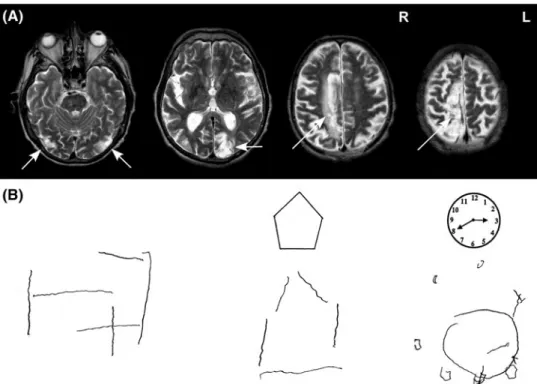
ER, a former cook without special education, suffered from multiple strokes at the age of 66 when undergoing coronary surgery. Upon awakening from anaesthesia, he showed slight left hemiparesis and complete right hemianopia, as well as severe visual–spatial disturbances described in detail below. structural MRI revealed ischemic lesions of the right dorsal frontoparietal cortex, the left medial occipi-tal cortex including primary visual cortex and the lateral occipitotemporal cortex on both sides (Fig. 1a). Medial occipital damage to the left hemisphere extended from the calcarine sulcus dorsally into the precuneus and to the posterior intraparietal sulcus. Right frontoparietal damage
Fig. 1 a t2-weighted MRI scan
performed 9 months post-injury showing ischemic damage to bilateral occipito-temporal cor-tex (left), left medial occipito-parietal cortex (middle) and dor-sal frontoparietal cortex (right).
b ER’s attempts to draw a cube
from memory (left) and to copy a pentagon and a clock face
extended posteriorly into the superior parietal lobule and the anterior intraparietal sulcus.
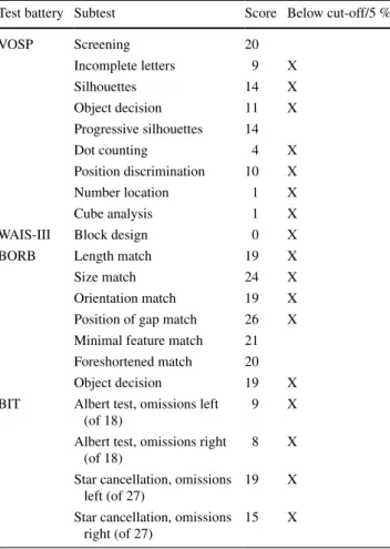
ER initially exhibited all signs of Bálint syndrome including optic ataxia, simultanagnosia, oculomotor dis-turbances and severe visual–spatial confusion, making it difficult for him to identify visually presented objects. the present examination was performed 7 weeks following the stroke when ER was able to fixate an object for several sec-onds. During this period, ER benefited from 1 to 2 daily sessions of therapy targeting compensation of oculomotor deficits (ocular pursuit tasks, visual search and detection of visual targets presented in the left or right visual field) and optic ataxia (pointing to dots presented on a touch screen, reaching and manipulating different objects). he was fully oriented and showed no signs of aphasia or apraxia. Out of 35 common household objects presented as line draw-ings, he named 28 correctly and made visual errors for the remaining objects (e.g. calling a screw a ‘little key’). he scored within average range on verbal fluency, ver-bal abstraction (similarities subtest of WaIs-III) and oral arithmetic, though memory was moderately impaired (six of ten words recalled after 30 min). confrontation testing showed complete right hemianopia (formal perimetry test-ing was impossible because ER was unable to maintain fixation during the examination). Optic ataxia was tested clinically by asking ER to point to the examiner’s finger and to grasp a pencil held out by the examiner in his vis-ual periphery (five trials per hemifield) while maintaining fixation on the examiner’s nose. ER showed deviations of approximately 2–5 cm when pointing in his left hemifield, but much larger errors (10–15 cm) in his right hemifield. Optic ataxia was also manifested in ER’s drawings, where he was often unable to position the pencil correctly (e.g. when asked to cross out visual targets). the patient addi-tionally showed moderate oculomotor apraxia character-ized by ‘sticky’ fixation (often fixating a face or an object for several seconds) and impaired ocular pursuit (loosing contact with the visual target, in particular for movements to the right). simultanagnosia was evidenced when ER was asked to count dots displayed on a sheet of paper or to name several objects arranged on the table. In the lat-ter situation, he would comment that the objects became all intermixed and that it was impossible for him to disentan-gle the individual items. he was able to name individual letters, but failed to read even short words (e.g. he identi-fied the French word ‘lUNE’—moon as ‘lURO’, and the word ‘VIOlONIstE’ as ‘VOlVOlINE’). copying simple shapes or drawing them from memory was extremely diffi-cult for ER as he was unable to position single elements of the drawing correctly and quickly lost track once he lifted the pencil (Fig. 1b). table 1 shows ER’s performance in tests probing visual, visual–spatial and constructional abili-ties. he was impaired in all tests except those that required
single-object identification or matching. at the time of this examination, ER did not show signs of left spatial neglect, but missed many cancellation items in both hemifields. In other tasks or during therapy, he would generally detect tar-get objects that were to his left better than those located on the right. he also did not show clear head or gaze deviation to the right.
Despite his severely impaired exploration of large, cluttered visual displays (such as typically present in can-cellation tasks), ER accurately detected items defined by colour or shape when they were presented in his central visual field. We tested him using a simple visual search task containing four squares that were either red or green and filled or unfilled. the task was to indicate whether the display contained a square defined by the combina-tion of colour/filledness (e.g. a green/unfilled square) while the three distracters were systematically varied. In the dissimilar condition, all distracters differed on both features from the target (e.g. all three were red/filled). In
Table 1 ER’s scores in visual and visual–spatial tests
BIT behavioural inattention test (Wilson et al. 1987), BORB Bir-mingham object recognition battery (Riddoch and humphreys 1993), VOSP visual object and space perception battery (Warrington and James 1991), WAIS-III Wechsler adult Intelligence scale (Wechsler
1997)
test battery subtest score Below cut-off/5 %
VOsP screening 20 Incomplete letters 9 X silhouettes 14 X Object decision 11 X Progressive silhouettes 14 Dot counting 4 X Position discrimination 10 X Number location 1 X cube analysis 1 X
WaIs-III Block design 0 X
BORB length match 19 X
size match 24 X
Orientation match 19 X
Position of gap match 26 X Minimal feature match 21 Foreshortened match 20
Object decision 19 X
BIt albert test, omissions left (of 18)
9 X
albert test, omissions right (of 18)
8 X
star cancellation, omissions left (of 27)
19 X
star cancellation, omissions right (of 27)
the similar condition, all distracters shared one feature with the target (e.g. all three were green/filled). In the
mixed condition, one distracter shared one feature with the target (e.g. green/filled) while the other two distract-ers shared the other feature (e.g. red/unfilled). Figure 2 shows the results of ER compared to a group of ten age-matched controls and 14 patients with left neglect tested in a previous study (Ptak and Valenza 2005). ER made no omission on this task and only one false response when the target was absent. Both control groups made more omissions (controls: 2.7 %, neglect: 5.1 %) and false responses (controls: 4.6 %, neglect: 1.5 %). ER’s reac-tion time data were analysed with an aNOVa with fac-tors visual field (left/right) and condition (mixed, simi-lar, dissimilar). the analysis revealed only a significant main effect of visual field, with slower reactions to tar-gets shown in the right (hemianopic) compared to the left hemifield [F(1,137) = 6.03, p < .05]. We compared ER’s data directly to controls and neglect patients using a Bayesian approach (crawford and Garthwaite 2007). though ER was overall slower than healthy controls, none of the comparisons reached significance. In con-trast, he was significantly faster than neglect patients to detect left hemifield targets in the mixed condition (p < .05). this finding shows that ER processed visual targets presented in his central visual field adequately and with normal speed. any differences between him and control participants in visual exploration of natural images can therefore not be attributed to impaired atten-tion for informaatten-tion presented at fixaatten-tion.
Material and procedure
all participants gave written informed consent and the study was approved by the Ethical committee of the Uni-versity hospital, Geneva. Material and procedure were as described in a previous study (Ptak et al. 2009), which involved 18 healthy controls and 13 right-hemisphere stroke patients (six without and seven with left spatial neglect). twenty colour photographs depicting roughly symmetrical portraits of natural scenes, architecture or regular patterns were shown on a 21″ cRt for 15 s each.
the fixation position of the right eye was measured with an infrared, video-based eye-tracker (highspeed; sMI, Berlin, Germany) at a sampling rate of 240 hz. During calibration, ER was required to fixate sequentially on nine small circles presented at different positions. the calibration procedure was under manual control of the experimenter who indi-cated verbally the position of the current target and veri-fied visually whether eye position was stable. If necessary, calibration was repeated for specific calibration targets, and a verification run was performed to ensure that calibration was adequate. In order to favour bottom-up guidance of gaze, no specific instructions were given to ER other than to freely explore each image.
analysis
saccades and fixation locations were extracted offline using velocity (saccade: ≥50°/sec) and duration (fixation:
≥100 ms) criteria. spatial and temporal parameters of
saccades and fixations were then computed for each par-ticipant. local image features were extracted from patches of 1° × 1° drawn around each fixation and computed as described previously (Ptak et al. 2009). Briefly, local lumi-nance was calculated as the average intensity of all pixels within the patch scaled to the mean luminance of the whole image. Following scaling, values >1 indicated that the patch was brighter than and values <1 that it was relatively darker than the average brightness of the image. chromatic contrast was expressed as the standard deviation of pixel intensities in each RGB colour channel, which were then averaged and finally normalized to the maximal possible contrast. lumi-nance contrast was computed similarly to chromatic trast, but only with one channel (grey). Finally, edge con-tent was extracted by convolving the original image using the ‘canny’ edge detection algorithm (canny 1986) imple-mented in Matlab® Image Processing toolbox.
Results
table 2 shows the results of basic saccade and fixation parameters of ER as compared to 18 healthy participants
Fig. 2 Results of the visual search task of ER compared to healthy
and seven neglect patients tested in our previous study (Ptak et al. 2009). statistical comparisons were performed using a Bayesian approach for small samples. the aim of this approach is to reduce the probability that the patient score is falsely classified as being abnormal (crawford and Garthwaite 2007). It provides a point estimate of the abnormality of the patient’s score relative to a control population by treating population parameters (such as the mean) as random variables with a probabilistic distribution. In comparison with the control (p < .01) and the neglect group (p < .05), ER produced a lower total number of sac-cades and less leftward (controls: p < .05; neglect: ns.) or rightward (both p < .05) saccades. he had a slight direc-tional bias, making significantly more saccades to the left than saccades to the right [t(14) = 2.3, p < .05]. his mean saccade amplitude was significantly smaller than controls (p < .05), but did not differ from neglect patients. Finally, he exhibited significantly longer fixation durations than the control and the neglect group (both p < .01).
Figure 3 shows a scatter plot of all fixations made by ER as compared to controls and neglect patients. the average fixation position was nearly central in healthy controls (hor-izontal: -0.3 degrees; vertical: −1.2°), shifted to the right in neglect patients (horizontal: 4.4°; vertical: −0.7°) and shifted upwards and sligthly to the left in ER (horizontal:
−1.9°; vertical: 4.5°). as can be seen in Fig. 3, the disper-sion of fixations was much smaller in ER compared to both
groups. Most of his fixations were restricted to a sector that covered −7 to +5 degrees horizontally and 0–7° vertically. In contrast, healthy controls had a range in the horizontal and vertical direction that covered the entire image sur-face, and neglect patients also exhibited a larger range than ER—even in the horizontal direction and although their average horizontal position was shifted to the right.
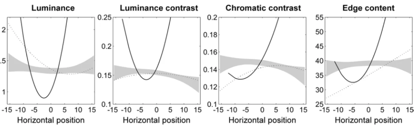
In order to examine whether local image content affected the distribution of ER’s fixations, we extracted statistics for luminance, contrast, chromatic contrast and edge content from image patches drawn around each fixation. Previ-ous work has shown that the distribution of local features in photographs tends to increase or decrease towards the edges (Parkhurst and Niebur 2003). We therefore computed quadratic polynomials for each of the four image features across horizontal fixation positions. Figure 4 shows poly-nomial functions of ER as compared to healthy participants and neglect patients. as reported previously (Ptak et al. 2009), neglect patients tended to look at regions located in the left half of the image only when these regions had par-ticularly high luminance, but low edge content. the poly-nomial functions generated from ER’s data were strongly U-shaped and showed significant deviations (p < .01) from the pattern of control participants for all four local fea-tures. For local luminance and luminance contrast, pre-dicted feature values were below control values for central positions, but significantly above control values for more
Table 2 Means (±sD) of basic saccade parameters
Group saccades, total (N) saccades, leftward (N) saccades, rightward (N) saccade amp (°) Fixation time (ms)
controls 35.6 ± 6.4 15.6 ± 2.8 15.3 ± 3.3 4.8 ± 0.8 240 ± 42
Neglect 32.1 ± 6.8 12.6 ± 3.6 14.3 ± 3.5 3.3 ± 0.9 231 ± 61
ER 16.1 ± 5.2 7.8 ± 3.5 6.3 ± 2.3 2.8 ± 0.8 547 ± 186
Fig. 3 Scatter plot showing all fixations produced by ER as
com-pared to healthy controls and neglect patients. the size of circles rep-resenting each fixation is proportional to fixation duration. the white
cross indicates the mean ± 1 sD of the horizontal and vertical distri-bution of fixations
peripheral positions. the trend was similar for chromatic contrast and edge content, with the difference that only right peripheral fixations were directed to regions with par-ticularly increased local feature values. thus, ER gazed at central regions irrespective of their local feature con-tent, but only looked to the periphery at regions character-ized by particularly conspicuous features. In addition, we examined whether local feature content affected ER’s first fixation similarly to control participants. For this analysis, we compared ER to healthy controls, neglect patients and a random observer by treating ER’s first fixations for all images as if they represented a group of independent obser-vations. analysis of variance revealed a significant differ-ence between groups for local luminance [F(3,54) = 3.49,
p < .05], indicating that healthy controls and neglect patients fixated regions of higher luminance compared to random (lsD-tests, p < .05). In contrast, the luminance of ER’s first fixations did not differ from a random observer. No significant differences between control groups, ER and the random observer were observed for luminance contrast [F(3,54) = 1.39], chromatic contrast [F(3,54) = 1.77] and edge content [F(3,54) = 2.09].
Discussion
We identified several features of oculomotor apraxia in a patient with Bálint syndrome that extend previous observa-tions. ER’s pattern of basic saccade and fixation parameters strongly differed from healthy controls and also differenti-ated him from neglect patients. he made significantly less saccades than both groups and had strongly reduced sac-cade amplitude than healthy controls. however, the latter observation does not appear to be limited to Bálint syn-drome as neglect patients exhibit a similarly reduced sac-cade amplitude during visual exploration. the most sig-nificant difference to controls and neglect patients, and one
that is directly related to the notion of ‘sticky’ fixation in Bálint syndrome, is ER’s significantly prolonged fixation duration. On average, ER produced fixation durations that were more than the double of those of control participants. similar durations were observed in a previous study when a simultanagnosic patient was required to make saccades to peripheral targets while a central fixation stimulus was pre-sent (a so-called overlap task; Nyffeler et al. 2005). how-ever, the sudden appearance of a stimulus at fixation also strongly affects fixation durations in patients with unilat-eral posterior brain damage and spatial neglect (Ptak et al. 2007; Walker and Findlay 1996), while the prolonged dura-tions during visual scanning appear to be specific to Bálint syndrome.
how can ‘sticky’ fixation during ocular exploration be explained? some authors (Rafal 1997; Rizzo and Vecera 2002) proposed that oculomotor apraxia is due to a path-ological constriction of visual attention to a single object. according to this hypothesis, patients make erratic eye movements because of a reduced ‘spatial window’ of atten-tion (Dalrymple et al. 2013a) and consequently the fail-ure to disengage attention from a fixated object. Given the close interdependence of attention and saccade program-ming (Reprogram-mington 1980; hoffman and subramaniam 1995), a reduced ‘spatial window’ of attention would strongly affect the selection of saccade targets. Our analysis of local image content at fixated locations only partly supports this conclusion. ER only made saccades to peripheral locations when these had high luminosity and contrast. Given that ER had a right homonymous hemianopia, it is important to determine to what extent this finding can be explained by his visual field impairment, since for all eye movements directed to the right, the saccade landing position was not visible for him. We would therefore expect that fixations located on the right side be selected randomly, which pre-dicts a strongly asymmetrical distribution of local image features between left and right fixations. contrary to this
Fig. 4 Polynomial functions predicting local feature content across horizontal positions of fixations. the grey area is based on the 99 %
prediction, the pattern of increased local luminosity and contrast was very similar for left and right peripheral loca-tions and is therefore not adequately explained by ER’s right hemianopia. Rather, it suggests that ER’s gaze is only captured by image regions that are particularly conspicu-ous. although factors such as task constraints and expecta-tion significantly influence visual exploraexpecta-tion during active scene viewing (tatler et al. 2011), bottom-up visual sali-ency is a powerful predictor of fixation locations (Itti and Koch 2000; Itti et al. 1998; Parkhurst et al. 2002). Our findings suggest that in Bálint syndrome, this role of local saliency differences is biased: for central positions, ER-fixated image regions that were relatively less conspicuous compared to healthy participants. less conspicuous central regions should make it easier to shift attention (and gaze) away to the periphery, yet ER only looked at peripheral locations when these were disproportionately salient. In our view, this finding reflects two possibly interacting factors. On the one hand, biased attentional priority following dam-age to posterior parietal cortex (PPc) along the intrapari-etal sulcus (IPs). Neurophysiological studies have shown that the IPs encodes stimuli in a feature-independent man-ner and integrates bottom-up saliency signals with top-down task-related factors into a spatiotopic priority map of the environment (Bisley and Goldberg 2010; Gottlieb et al. 1998; Ptak 2012; Vandenberghe et al. 2012). attentional priority may be conceived as emergent property computed from converging inputs from different sensory modali-ties and relevance signals originating in prefrontal cortex (Ptak and Fellrath 2013). If the priority map is crucial for the selection of sensory contents by attention, damage to this representation, in particular if it is bilateral, should have devastating consequences on spatial attention. this is indeed what happens in patients with Bálint syndrome, who in severe cases are described as virtually blind due to their failure to select stimuli for conscious processing (Rizzo and Vecera 2002; holmes and horrax 1919; Kim and Rob-ertson 2001). however, it is unclear why bilateral damage to the parietal priority map should result in a strong ocular bias towards central regions (or ‘sticky’ fixation). this cen-tral bias is more readily explained by functional impairment of structures of the oculomotor network that are involved in fixational activity. the mesencephalic superior colliculus contains neurons that discharge when a stimulus is actively fixated, while other neurons become active when a saccade is prepared and executed (Munoz and Wurtz 1993; Dor-ris et al. 1997). this structure is directly connected to the PPc, and unilateral parietal damage has facilitatory effects on the ipsilateral and inhibitory effects on the contralateral colliculus (sprague 1966; Rafal 2006). Following bilateral damage to the PPc top-down facilitatory influences of the parietal cortex on the superior colliculus is diminished, making it difficult to initiate saccadic eye movements; as
a consequence, fixational activity in both colliculi is dis-inhibited and leads to the strong bias favouring stimuli presented at fixation. such a mechanism could therefore underlie the ‘sticky fixation’ observed in patients with ocu-lomotor apraxia. however, this model, though supported by neurophysiology and some experimental studies on animal models of spatial neglect (Payne et al. 1996; Rushmore and Payne 2003), awaits direct support by human lesion studies.
In sum, based on our findings, we propose that oculomo-tor apraxia, in particular the bias of Bálint patients towards stimuli shown at fixation, reflects a combination of a severe impairment of mechanisms involved in attentional selection and a low-level oculomotor impairment following biased interactions between the PPc and the superior colliculus.
Acknowledgments study supported by the swiss National science
Foundation (Grant 320030-134591) and the De Reuter foundation (Geneva).
References
Bálint R (1909) seelenlähmung des ‘schauens’, optische ataxie, räu-mliche störung der aufmerksamkeit. Msschr Psychiat Neurol 25:51–66
Bisley JW, Goldberg ME (2010) attention, intention, and pri-ority in the parietal lobe. annu Rev Neurosci 33:1–21. doi:10.1146/annurev-neuro-060909-152823
canny J (1986) a computational approach to edge detection. IEEE trans Pattern anal Mach Intell 8(6):679–698
crawford JR, Garthwaite Ph (2007) comparison of a single case to a control or normative sample in neuropsychology: development of a Bayesian approach. cogn Neuropsychol 24(4):343–372. doi:10.1080/02643290701290146
Dalrymple Ka, Birmingham E, Bischof WF, Barton JJ, Kingstone a (2011) Experiencing simultanagnosia through windowed viewing of complex social scenes. Brain Res 1367:265–277. doi:10.1016/j.brainres.2010.10.022
Dalrymple Ka, Barton JJ, Kingstone a (2013a) a world unglued: simultanagnosia as a spatial restriction of attention. Front hum Neurosci 7:145. doi:10.3389/fnhum.2013.00145
Dalrymple Ka, Gray aK, Perler Bl, Birmingham E, Bischof WF, Barton JJ, Kingstone a (2013b) Eyeing the eyes in social scenes: evidence for top-down control of stimulus selection in simultana-gnosia. cogn Neuropsychol 30(1):25–40. doi:10.1080/02643294. 2013.778234
Dorris Mc, Paré M, Munoz DP (1997) Neuronal activity in monkey superior colliculus related to the initiation of saccadic eye move-ments. J Neurosci 17(21):8566–8579
Girotti F, Milanese c, casazza M, allegranza a, corridori F, avanzini G (1982) Oculomotor disturbances in Balint’s syndrome: anato-moclinical findings and electrooculographic analysis in a case. cortex 18(4):603–614
Gottlieb J, Kusunoki M, Goldberg ME (1998) the representation of visual salience in monkey parietal cortex. Nature 391:481–484 harvey M (1995) Psychic paralysis of gaze, optic ataxia, spatial
dis-order of attention’. translated from Balint (1909) ‘seelenläh-mung des “schauens”, optische ataxie, räumliche störung der aufmerksamkeit. cogn Neuropsychol 12:265–282
hoffman JE, subramaniam B (1995) the role of visual attention in saccadic eye movements. Percept Psychophys 57(6):787–795
holmes G, horrax G (1919) Disturbances of spatial orientation and visual attention, with loss of stereoscopic vision. arch Neurol Psychiatry 1(4):385–407
Itti l, Koch c (2000) a saliency-based search mechanism for overt and covert shifts of visual attention. Vis Res 40:1489–1506 Itti l, Koch c, Niebur E (1998) a model of saliency-based visual
attention for rapid scene analysis. IEEE trans Pattern anal Mach Intell 20(11):1254–1259
Jackson GM, swainson R, Mort D, husain M, Jackson sR (2009) attention, competition, and the parietal lobes: insights from Balint’s syndrome. Psychol Res 73(2):263–270. doi:10.1007/ s00426-008-0210-2
Kim M-s, Robertson lc (2001) Implicit representations of space after bilateral parietal lobe damage. J cogn Neurosci 13(8):1080–1087 luria aR, Pravdina-Vinarskaya EN, Yarbus al (1963) Disor-ders of ocular movements in a case of simultanagnosia. Brain 86:219–228
Mannan sK, Ruddock Kh, Wooding Ds (1996) the relationship between the locations of spatial features and those of fixations made during visual examination of briefly presented images. spat Vis 10(3):165–188
Munoz DP, Wurtz Rh (1993) Fixation cells in monkey superior col-liculus. I. characteristics of cell discharge. J Neurophysiol 70(2):559–575
Nyffeler t, Pflugshaupt t, hofer h, Baas U, Gutbrod K, von Wart-burg R, hess cW, Müri RM (2005) Oculomotor behaviour in simultanagnosia: a longitudinal case study. Neuropsychologia 43:1591–1597
Parkhurst DJ, Niebur E (2003) scene content selected by active vision. spat Vis 16(2):125–154
Parkhurst DJ, law K, Niebur E (2002) Modeling the role of salience in the allocation of overt visual attention. Vis Res 42:107–123 Payne BR, lomber sG, Geeraerts s, van der Gucht E, Vandenbussche
E (1996) Reversible visual hemineglect. Proc Natl acad sci Usa 93:290–294
Ptak R (2012) the frontoparietal attention network of the human brain: action, saliency, and a priority map of the environment. Neuroscientist 18(5):502–515. doi:10.1177/1073858411409051
Ptak R, Fellrath J (2013) spatial neglect and the neural coding of attentional priority. Neurosci Biobehav Rev 37(4):705–722. doi:10.1016/j.neubiorev.2013.01.026
Ptak R, Müri RM (2013) the parietal cortex and saccade planning: lessons from human lesion studies. Front hum Neurosci 7:254. doi:10.3389/fnhum.2013.00254
Ptak R, Valenza N (2005) the inferior temporal lobe mediates dis-tracter-resistant visual search of patients with spatial neglect. J cogn Neurosci 17(5):788–799
Ptak R, schnider a, Golay l, Müri R (2007) a non-spatial bias favouring fixated stimuli revealed in patients with spatial neglect. Brain 130(12):3211–3222
Ptak R, Golay l, Müri R, schnider a (2009) looking left with left neglect: the role of spatial attention when active vision selects local image features for fixation. cortex 45(10):1156–1166 Rafal RD (1997) Balint syndrome. In: Feinberg tE, Farah MJ (eds)
Behavioral neurology and neuropsychology. McGraw-hill, New York, pp 337–356
Rafal RD (2006) Oculomotor functions of the parietal lobe: effects of chronic lesions in humans. cortex 42(5):730–739
Reinagel P, Zador aM (1999) Natural scene statistics at the centre of gaze. Netw: comput Neural syst 10:1–10
Remington RW (1980) attention and saccadic eye movements. J Exp Psychol: hum Percept Perform 6(4):726–744
Riddoch MJ, humphreys GW (1993) BORB. Birmingham object rec-ognition battery. lawrence Erlbaum associates, hove
Rizzo M, hurtig R (1987) looking but not seeing: attention, per-ception, and eye movements in simultanagnosia. Neurology 37(10):1642–1648
Rizzo M, Vecera sP (2002) Psychoanatomical substrates of Balint’s syndrome. J Neurol Neurosurg Psychiatry 72(2):162–178 Rushmore RJ, Payne BR (2003) Bilateral impact of unilateral
vis-ual cortex lesions on the superior colliculus. Exp Brain Res 151:524–547
sprague JM (1966) Interaction of cortex and superior colliculus in mediation of visually guided behavior in the cat. science 153:1544–1547
tatler BW, Baddeley RJ, Gilchrist ID (2005) Visual correlates of fixa-tion selecfixa-tion: effects of scale and time. Vis Res 45:643–659 tatler BW, hayhoe MM, land MF, Ballard Dh (2011) Eye
guid-ance in natural vision: reinterpreting salience. J Vis 11(5):5. doi:10.1167/11.5.5
Vandenberghe R, Molenbergs P, Gillebert cR (2012) spatial attention deficits in humans: the critical role of superior compared to infe-rior parietal lesions. Neuropsychologia 50(6):1092–1103 Walker R, Findlay JM (1996) saccadic eye movement programming
in unilateral neglect. Neuropsychologia 34(6):493–508
Warrington EK, James M (1991) the visual object and space percep-tion battery. thames Valley test company, Bury st Edmunds Wechsler D (1997) the Wechsler adult intelligence scale, 3rd edn.
the Psychological corporation, san antonio, tX
Wilson B, cockburn J, halligan P (1987) Behavioural inattention test. thames Valley test company, Bury st Edmunds