DOI 10.1007/s00262-014-1535-x OrIgInal artICle
Influence of natural killer cells and perforin‑mediated cytolysis
on the development of chemically induced lung cancer in A/J mice
Manuela Frese‑Schaper · Andreas Keil ·
Hideo Yagita · Selina Katja Steiner · Werner Falk · Ralph Alexander Schmid · Steffen Frese
received: 24 april 2013 / accepted: 9 March 2014 / Published online: 22 March 2014 © Springer-Verlag Berlin Heidelberg 2014
neither depletion of tnF-α, tnF-related apoptosis-induc-ing ligand, tnF-like weak inducer of apoptosis or Fasl alone nor in combination induced an augmentation of tumour burden. to show whether an alternative cell death pathway is involved, we next generated a/J mice deficient for perforin. after challenging with nnK, mice deficient for perforin showed an increase in tumour number and vol-ume compared to wild-type a/J mice. In summary, our data suggest that nK cells and perforin-mediated cytolysis are critically involved in the protection from lung cancer giving promise for further immunotherapeutic strategies for this disease.
Keywords lung cancer · natural killer cells · Perforin-mediated cytolysis · tumour necrosis factors · apoptosis · Immunogenicity
Abbreviations
nSClC non-small cell lung cancer
nnK nitrosamine 4-(methylnitrosamino)-1-(3-pyridyl)-1-butanone
nK natural killer
tnF tumor necrosis factor
traIl tumor necrosis factor-related apoptosis-induc-ing ligand
tWeaK tnF-like weak inducer of apoptosis PKO Perforin-deficiency
Introduction
lung cancer is the leading cause of cancer death worldwide among men and the second leading cancer site in women. In fact, 1.6 million of people were diagnosed with lung can-cer in 2008 accounting for 13 % of total cases. In the same Abstract One alternative approach for the treatment of
lung cancer might be the activation of the immune system using vaccination strategies. However, most of clinical vac-cination trials for lung cancer did not reach their primary end points, suggesting that lung cancer is of low immuno-genicity. to provide additional experimental information about this important issue, we investigated which type of immune cells contributes to the protection from lung cancer development. therefore, a/J mice induced for lung adeno-mas/adenocarcinomas by the tobacco-specific nitrosamine 4-(methylnitrosamino)-1-(3-pyridyl)-1-butanone (nnK) were depleted of CD4+ or CD8+ t cells, CD11b+
mac-rophages, gr-1+ neutrophils and asialo gM1+ natural killer
(nK) cells. Subsequent analysis of tumour growth showed an increase in tumour number only in mice depleted of nK cells. Further asking by which mechanism nK cells sup-pressed tumour development, we neutralized several death ligands of the tumour necrosis factor (tnF) family known to be involved in nK cell-mediated cytotoxicity. However,
M. Frese-Schaper · a. Keil · S. K. Steiner · S. Frese (*) Department of Clinical research, University of Bern, Murtenstrasse 50, P.O. Box 44, 3010 Bern, Switzerland e-mail: [email protected]
M. Frese-Schaper · a. Keil · S. K. Steiner · r. a. Schmid · S. Frese
Division of general thoracic Surgery, University Hospital Bern, 3010 Bern, Switzerland
H. Yagita
Department of Immunology, Juntendo University School of Medicine, tokyo 113-8421, Japan
W. Falk
Department of Internal Medicine I, University of regensburg, 93042 regensburg, germany
year, 1.4 million patients died from lung cancer according to 18 % of total cancer deaths [1]. Despite improved sur-gery, radio- and chemotherapy, the 5-year survival remains low with approx. 16 % [2], suggesting that alternative treat-ment options are needed for this disease.
Such an alternative treatment strategy might encom-pass immunotherapy which has been successfully applied for other types of cancer such as prostate cancer [3] or melanoma [4]. a prerequisite for the administration of immunotherapy to certain types of cancer is their immu-nogenicity which describes the property of cancer cells to induce a detectable reaction of the immune system. Whether lung cancer is sufficiently accessible to the immune system is an unsolved issue yet; 80 % of lung cancer is associated with smoking [1]. early studies dem-onstrated that carcinogens from tobacco smoke provoke an impaired function of the immune system. In this con-text, a decrease in nK cell function was found in both mice and humans [5, 6]. However, an impaired function of immune effector cells by tobacco smoke would not exclude an immunogenicity of lung tumours. Some pre-liminary answers to the question whether lung cancer is able to induce a sufficient reaction of the immune sys-tem were provided by vaccination studies. these studies made use of tumour antigens such as Mage (melanoma-associated antigen)-a3 or Mucin 1 which are known to be overexpressed in non-small cell lung cancer (nSClC) and associated with poor prognosis [7, 8]. Phase II clini-cal trials were conducted using these proteins for vacci-nation. albeit trends for a positive clinical response to both vaccines were detectable, both studies did not reach their primary endpoint in terms of significantly prolonged survival [9, 10]. Combining the results of these clinical trials together with the demonstrated immunosuppression by tobacco smoke, these data indicate that lung cancer is not or only a weak inducer of an immune response. How-ever, this conclusion might be amended by an encourag-ing clinical trial usencourag-ing the vaccine belagenpumaucel-l (lucanixtM) which is a mixture of allogeneic nSClC cell lines stably transfected to secret an antisense nucleo-tide to tgF-β. this trial demonstrated a survival benefit of patients with nSClC as well as an immune reaction to the vaccine [11].
the aim of the present work was to further shed light into the matter how the immune system is involved in the protection from lung cancer. to this end, we used a mouse model of chemically induced lung cancer. a/J mice treated with the tobacco-specific nitrosamine 4-(methylnitrosamino)-1-(3-pyridyl)-1-butanone (nnK) developed adenomas/adenocarcinomas of the lung [12,
13] and are a well-known mouse model mainly used for chemoprevention studies. In this context, it was shown that certain substances such as isothiocyanates
or methoxsalen which are believed to inhibit the muta-genic activation of nnK were able to decrease tumour growth in a/J mice [14, 15]. also, FtI-276 an inhibitor of the enzyme farnesyltransferase which facilitates the activation of the proto-oncogene ras demonstrated ben-eficial effects in the a/J mouse model [16]. the effect of FtI-276 was attributed to mutations of the K-ras gene shown for a high percentage of lung tumours in a/J mice [17, 18]. However, only a few data exist about how the immune system contributes to the carcinogenesis in a/J mice. Splenic and peripheral nK cell number seems to be lower in a/J mice compared with C57Bl/6 mice [19], and their cytolytic activity was shown to be lower com-pared to strains of mice which are not susceptible for the development of lung cancer [20]. Moreover, administra-tion of nnK was reported to further decrease the activity of splenic nK cells in a/J mice, an effect which was con-verted by the administration of non-steroidal anti-inflam-matory drugs [5]. Since non-steroidal anti-inflammatory drugs were additionally shown to inhibit tumour growth in nnK-induced a/J mice [21], nK cells were attributed to play a role in the carcinogenesis of this tumour model. Whether other immune cells contribute to the develop-ment of tumours in a/J mice and which mechanisms might be utilized has been investigated in the study pre-sented here.
Materials and methods reagents
nnK was purchased from toronto research Chemi-cals, Canada. the stock solution was prepared by solving 100 mg nnK in 200 μl DMSO. the stock solution was further diluted to a final volume of 10 ml with saline. From this solution, 200 μl (2 mg nnK) were injected to mice. For depletion of immune cells, the following antibodies were used: rat anti-CD4, clone gK 1.5 [22]; rat anti-CD8, clone 53-6.7 [23, 24]; rat anti-CD11b, clone 5C6 [25]; and rat anti-gr-1, clone rB6-8C5 [26], all produced in the lab-oratory of Hideo Yagita. rabbit anti-asialo gM1 purchased from Wako Pure Chemical Industries, Osaka, Japan, was used for the depletion of nK cells [23]. Cytokines of the tnF family were depleted by the following antibodies: rat anti-traIl, clone n2B2 [27]; rat anti-tWeaK, clone MtW-1 [28]; and hamster anti-Fasl, clone MFl4 [29], all produced in the laboratory of Hideo Yagita. In addition, rat anti-tnF, clone V1q [30] which was prepared in the labo-ratory of Werner Falk was used for neutralization of tnF-α. Control groups were treated with rat Igg or rabbit Igg (both from Sigma, St. louis, MO) or hamster Igg (Jackson Immunoresearch, Suffolk, UK).
Handling of mice
all animal experiments were approved by the Kanton-ale tierversuchskommission (Kanton Bern, Switzerland). Five-week-old female a/J mice were obtained from Jack-son laboratories (Bar Harbor, Me) and housed in iso-lated ventiiso-lated cages. treatment was applied as described below. all applications of antibodies and nnK were per-formed as i.p. injections. after completion of the treatment, mice were killed by an overdose of pentobarbital, and lungs were harvested. the tumour number was counted, and the size of tumours was measured using a digital calliper. the latter experiments were all executed with two investigators in which the person who measured tumour number and vol-ume was blinded.
generation of perforin-deficient a/J mice
Perforin-deficient (PKO) mice on a C57Bl/6 background [31] were kindly provided by Hans Hengartner (Institute of experimental Immunology, University of Zurich, Swit-zerland). these mice were backcrossed for ten generations with a/J mice. Mice with mutated perforin allele were identified by PCr analysis as described before [32]. In brief, genomic Dna was isolated from ear biopsies using the Dneasy Blood and tissue kit from QIagen (Valen-cia, Ca, USa). For the PCr, two different primer pairs were used. 5′-ttt ttg aga CCC tgt aga CCC a-3′, 5′-gCa tCg CCt tCt atC gCC ttC t-3′) which gives no product for the wild-type (wt) allele and which produces a band of 665 bp for the mutated perforin allele. 5′-CCg gtC Ctg aaC tCC tgg CCa a-3′, 5′-CCC Ctg CaC aCa tta Ctg gaa g-3′ yields a 300-bp fragment for wt and a 1,300-bp fragment for the mutated allele. Only the second primer pair was able to distinguish between mice harbouring a heterozygous or homozygous muta-tion of the perforin allele. Perforin-deficient a/J mice mice used in this study will be archived and distributed as live mice or frozen material via the european Mouse Mutant archive (eMMa), which is part of the InFraFrOntIer research Infrastructure (www.infrafrontier.eu) under the strain name a.B6-Prf1tm1Sdz/Biat (eMMa ID eM:07765).
analysis of Foxp3-positive regulatory t cells (tregs or CD4+Foxp3+ cells) and myeloid-derived suppressor
(CD11+gr-1+) cells
Splenocytes from wt a/J mice and a/J mice deficient for perforin were freshly isolated as described before [33]. Cells were then incubated with Fc receptor-blocking mab (clone 2.4g2, BD Biosciences) followed by incubation with fluorochrome-labelled mabs. For tregs, after stain-ing for CD4, cells were fixed and permeabilized followed
by incubation with the anti-Foxp3 ab. Staining was meas-ured on an lSrII flow cytometer (BD Biosciences) and analysed using FlowJo software (tree Star, ashland, Or). the following abs were used: anti-CD4 (clone rM4-5, Caltag), anti-Foxp3 (clone 150D, Biolegend), anti-CD11 (clone M1/70, Biolegend) and anti-gr-1 (clone rB6-8C5, Caltag).
Cytotoxicity assay
nK cell-sensitive Yac-1 cells (atCC, Manassas, Va) were used as target cells. Yac-1 cells were labelled with 5-(and-6)-carboxyfluorescein diacetate succinimydyl ester (CFSe) using the 7-aaD/CFSe Cell-Mediated Cytotoxicity assay Kit from abcam (Cambridge, UK). labelled Yac-1 cells were then added to a 96-well plate with 1 × 10⁵ cells per
well. as effector cells, freshly isolated splenocytes from wt a/J mice, aJ.PKO+/− or aJ.PKO−/− mice were added at an
effector/target ratio of 20:1. the plate was centrifuged for 4 min at 30×g to allow cell contact followed by incuba-tion for 4 h at 37 °C. Immediately before analysis, samples were incubated with 7-amino-actinomycin D (7-aaD), and death of target cells was subsequently determined by flow cytometry analysing the number of CFSe+/7-aaD+
events. the final experiments were realized in a manner that the conducting person was blinded for the groups of mice from which splenocytes were isolated.
Statistical analysis
Differences in tumour number and volume were evaluated for significance using Student’s t test or Mann–Whitney U test. a P < 0.05 was considered statistically significant. Results
Depletion of nK cells promotes tumour development in a/J mice
First, we examined whether certain types of immune cells contribute to the development of lung cancer in a/J mice. therefore, mice were depleted of CD4+ or CD8+ t cells,
CD11b+ macrophages, gr-1+ neutrophils or asialo gM1+
nK cells using antibodies which were shown before to effectively reduce the number of targeted cells [22–26]. at 6 weeks of age, depletion was initiated with 100 μg of antibody for CD4 [22], CD8 [23, 24], CD11b [25] and gr-1 [26] and 300 μg for asialo gM1 [23], each in a vol-ume of 200 μl. treatment was performed twice a week for 20 weeks. two days after the first application of deplet-ing antibodies, a sdeplet-ingle dose of 2 mg nnK was injected to mice. as a result, neither the depletion of CD4-, CD8- or
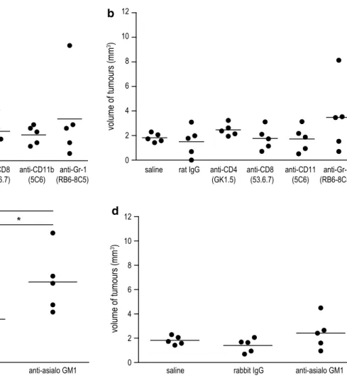
CD11b-positive cells nor depletion of neutrophils yielded enhanced tumour growth compared to saline-treated mice or to mice which received the respective control Igg (Fig. 1a, b). Only the treatment with anti-asialo gM1, an antibody which has been widely used for the depletion of nK cells, evoked a significant effect on tumour growth. While there was no difference between saline-treated mice and mice treated with rabbit Igg, nK cell-depleted mice showed a significantly increased tumour number com-pared to either of these control groups (n = 5; P = 0.047 saline vs asialo gM1 and P = 0.039 rabbit Igg vs anti-asialo gM1, Fig. 1c). no difference was found between the groups when analysing the tumour volume (Fig. 1d). How-ever, the missing difference in tumour volume might be a result of the chosen time point for killing of the animals
and would have been different most likely to a later time point.
Death-inducing cytokines of the tnF family are not involved in nK cell-mediated tumour suppression in a/J mice
Providing a contribution of nK cells to the suppression of lung cancer in a/J mice, we next asked which mecha-nisms nK cells used to suppress tumour growth in these mice. Death-inducing cytokines of the tnF family, espe-cially tnF-related apoptosis-inducing ligand (traIl) and Fasl (CD95l), were demonstrated to be involved in nK cell-mediated cytotoxicity [34]. to explore whether and which of these cytokines might be responsible for the b
a
d c
Fig. 1 effect of immune cell depletion on lung tumour development in a/J mice induced with nnK. Depletion of CD4+ or CD8+ t cells,
macrophages, neutrophiles and nK cells was initiated in 6-week-old a/J mice using i.p. injections with 100 μg of anti-CD4, anti-CD8, anti-CD11b or anti-gr-1 mab or 300 μg of anti-asialo gM1 ab, respectively. two days after first application of depleting antibod-ies, mice were induced for lung cancer with 2 mg of nnK. treat-ment with antibodies or control Igg was performed twice a week for 20 weeks. the tumour burden was assessed in a blinded manner. a
tumour number and b tumour volume for mice depleted of CD4+ or
CD8+ t cells, macrophages or neutrophils demonstrated no
signifi-cant differences compared to saline- or control rat Igg-treated mice. c Depletion of nK cells with anti-asialo gM1 ab showed an increased tumour number compared to saline-treated mice or mice treated with control rabbit Igg (n = 5; P = 0.047 saline vs anti-asialo gM1 and P = 0.039 rabbit Igg vs anti-asialo gM1, Student’s t test). d Differ-ences in tumour volume of nK cell-depleted mice were not signifi-cant compared to control groups. *P < 0.05
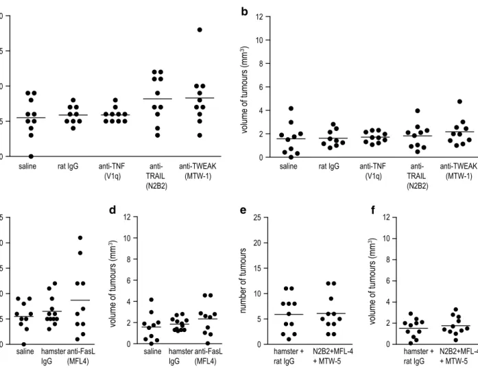
effects of nK cell-mediated tumour suppression, a/J mice were depleted of traIl, Fasl, tnF-α or tWeaK using specific neutralizing antibodies [27–30]. at 6 weeks of age, mice received a single dose of 2 mg nnK followed by application of 100 μg of neutralizing antibody. Con-trol groups were treated with saline, or rat or hamster Igg. treatment with antibodies was carried out twice weekly for 21 weeks. the subsequent analysis of tumour burden in the lung revealed no differences between groups neu-tralized for these cytokines and control groups (Fig. 2a– d). to exclude that this result was caused by redundancy of the neutralized cytokines, we next performed an exper-iment where traIl, Fasl and tWeaK were neutralized in combination. the control group received the respective amount of hamster and rat Igg. Similarly to the separate neutralization, the combined neutralization of traIl, Fasl and tWeaK had no effect on tumour growth,
neither on tumour number nor on tumour volume (Fig. 2e, f). Collectively, these data suggest that cytokines of the tnF family do not considerably contribute to the suppres-sion of lung cancer in a/J mice.
tumour suppression in a/J mice depends on perforin-mediated cytotoxicity
another mechanism for cell death induced by nK cells is represented by the perforin/granzyme pathway. While the function of certain members of the granzyme family for immunosurveillance of cancer is uncertain yet, it has been shown that nK cell activity against a number of tumours highly depends on an intact perforin molecule [35]. to answer the question whether perforin-mediated cytolysis is involved in nK cell-mediated tumour suppression in a/J mice, a/J mice deficient for perforin (referred as aJ.PKO)
b d c a f e
Fig. 2 neutralization of death-inducing tnF family ligands has no influence on tumour development in nnK-treated a/J mice. Six-week-old female a/J mice were exposed to a single dose of 2 mg nnK and subsequently treated with antibodies neutralizing tnF-α, traIl, tWeaK or Fasl. treatment with antibodies or control Igg was performed twice a week for 21 weeks. tumour number (a) and
(c) and tumour volume (b) and (d) of animals depleted of single tnF family ligands revealed no differences in tumour growth compared to saline- or Igg-treated groups. tumour number (e) and tumour volume (f) of mice depleted of traIl, Fasl and tWeaK in combination were statistically not different from those of the control Igg-treated group
were generated. these mice were induced for lung can-cer with 2 mg nnK at 6 weeks of age. twenty-two weeks later, mice were killed and the tumour burden of a/J mice heterozygously (+/−) or homozygously (−/−) deficient for
perforin were compared with wild-type (wt) a/J mice. We performed eight independent experiments with varying numbers of animals. From these eight experiments, one was excluded due to an exceptional high tumour number and volume in all three experimental groups. analysis of the remaining animals revealed that a/J mice deficient for perforin had a higher susceptibility for nnK-induced lung cancer. Both aJ.PKO+/− and aJ.PKO−/− showed a
signifi-cantly higher number of lung tumours compared with wt a/J mice (n = 17 for wt, n = 42 for aJ.PKO+/−, n = 26 for
aJ.PKO−/−; P = 0.031 for wt vs aJ.PKO+/−, P = 0.036 for
wt vs aJ.PKO−/−; Fig. 3a). the assessment of tumour
vol-ume demonstrated a significant difference for aJ.PKO+/−
compared to wt (P = 0.034; Fig. 3b) but not for aJ.PKO−/−
compared to wt (P = 0.16; Fig. 3b).
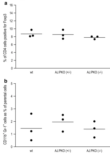
to rule out that immunosuppression other than decreased nK cell activity might play a role in aJ.PKO mice, we measured the level of tregs (CD4+Foxp3+ cells) and the
level of myeloid-derived suppressor (CD11+gr-1+) cells.
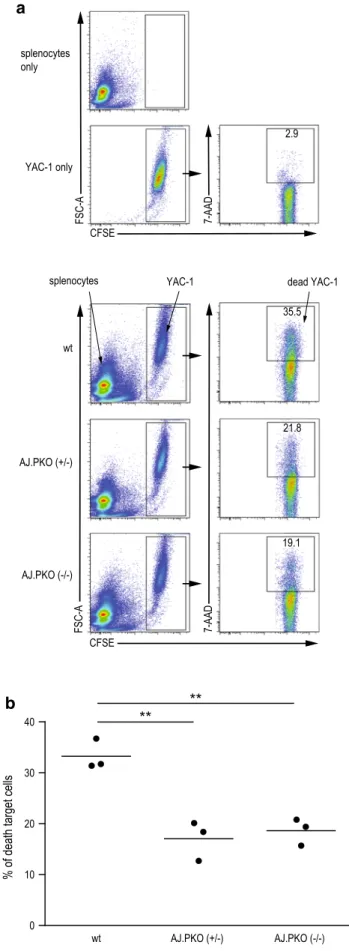
analysis of these cell types in the splenocyte population revealed no difference between wt a/J mice and aJ.PKO mice (Fig. 4a, b), indicating that decreased immunosur-veillance in perforin-deficient mice is due to an impaired function of nK cells. to provide more direct evidence for this suggestion, we subsequently measured in vitro cyto-toxic splenic nK cell activity of perforin-deficient mice. as shown in Fig. 5, the cytotoxic activity of both aJ.PKO−/+-
and aJ.PKO−/−-derived splenic nK cells was significantly
lower than the nK cell activity of wt a/J mice. these a
b
Fig. 3 Influence of perforin deficiency on lung tumour development. Six-week-old female aJ.PKO mice (heterozygous or homozygous) were exposed to 2 mg nnK. twenty-two weeks later, lung tissue was analysed. a aJ.PKO+/− and aJ.PKO−/− had a significantly higher
number of lung tumours compared with wt a/J mice (n = 17 for wt, n = 42 for aJ.PKO+/−, n = 26 for aJ.PKO−/−; P = 0.031 for wt vs
aJ.PKO+/−, P = 0.036 for wt vs aJ.PKO−/−). b the tumour volume
was significantly higher in aJ.PKO+/− compared to wt (P = 0.034)
but not in aJ.PKO−/− compared to wt (P = 0.16); Mann–Whitney U
test. *P < 0.05
a
b
Fig. 4 analysis of tregs (CD4+Foxp3+ cells) and myeloid-derived suppressor (CD11+gr-1+) cells in perforin-deficient a/J mice.
Sple-nocytes from wt a/J mice, aJ.PKO−/+ and aJ.PKO−/− mice were
stained with fluorochrome-labelled mabs and analysed by flow cytometry
findings propose that nK cells and the perforin-mediated cytolytic pathway indeed contribute to the prevention of chemically induced lung cancer.
Discussion
the immunogenicity of lung cancer remains an unsolved question until now. as referred above, most of the clinical trials for lung cancer using vaccination strategies failed to achieve their primary end points. this problem might have two reasons: One could be that the antigens used for vacci-nation were not ideal and the other could be that lung can-cer is in general not able to induce a sufficient reaction of the immune system. Our study was delineated to elucidate the second issue whether lung cancer has the ability to pro-voke an immune reaction or not.
In a first set of experiments, a/J mice which were induced for lung cancer with nnK were depleted of differ-ent types of immune cells. However, depletion of CD4+ t
cells, CD8+ t cells, CD11b+ macrophages or gr-1+
neu-trophils did not significantly affect the tumour burden in a/J mice. these results were not expected because the defi-ciency of Foxp3-positive regulatory t cells was shown to reduce tumour growth in a/J mice [36]. tregs are a subset of
CD4-positive t cells. Since we depleted in our experiments all CD4-positive cells, one might ask whether a proposed beneficial effect of losing these tregs had been negotiated by the loss of effector CD4+ t cells. therefore, we do not
exclude from our data that in wild-type a/J mice CD4-pos-itive cells other than tregs contribute to the suppression of lung cancer.
Our results further demonstrated that depletion of nK cells resulted in an increased tumour number, suggesting a pivotal role of nK cells for the control of tumour growth in this lung cancer model. nK cells execute their cyto-toxic effects either by direct target cell lysis or by secre-tion of cytokines such as interferon-γ, but without the
b a Fig. 5 Cytolytic nK cell activity in perforin-deficient a/J mice. nK cell-sensitive Yac-1 cells were used as target cells. Yac-1 cells were labelled with 5-(and-6)-carboxyfluorescein diacetate succinimydyl ester (CFSe). as effector cells, freshly isolated splenocytes from wt a/J mice, aJ.PKO+/− or aJ.PKO−/− were added at an
effector/tar-get ratio of 20:1 followed by an incubation for 4 h at 37 °C. Sam-ples were then stained with 7-amino-actinomycin D (7-aaD), and death of target cells was subsequently determined by flow cytometry analysing the number of CFSe+7/aaD+ events. a representative
dot plots for target and effector cells alone (upper panel) and when incubated together (lower panel). b Comparison of cytotoxic nK cell activity of the different groups of mice with n = 3 per group. P = 0.0046 wt a/J versus aJ.PKO+/− and P = 0.0031 wt a/J versus
aJ.PKO−/−, Student’s t test. **P < 0.01
need of antigen-specific recognition as required for cyto-toxic t lymphocytes. In this context, nK cell activity was shown to be important for the development or rejection of MHC class-I-deficient lymphomas [37], and depletion of nK cells was also found to promote tumour growth of fibrosarcomas [38]. Furthermore, there is evidence that lung cancer development depends on the function of nK cells. In a recent publication, Kreisel and colleagues dem-onstrated that depletion of nK cells promoted urethane-induced lung tumour growth in a mouse strain which is normally not susceptible to lung cancer [20]. Moreover, nK cells were shown to have lower activity in a/J mice [19], which are prone to develop lung cancer induced by certain chemicals such as the nitrosamine nnK. nnK itself was demonstrated to further reduce nK cell activ-ity in a/J mice [5], an effect which has been observed in human smokers as well [6]. Despite the assumption of lower nK cell activity in nnK-induced a/J mice, our data suggest that the remaining nK cell activity was suf-ficient to reduce lung tumour formation. However, once the lung tumours are established, another situation might arise comprising a direct inhibition of nK cell activity by lung cancer cells. thus, it has been shown that lung tumour cells from malignant pleural effusions inhibit nK cell activity [39], and it was further demonstrated that nK cells isolated from lung tumours exhibit decreased cyto-toxic activity [40].
the mechanisms how nK cells induce direct target cell lysis might vary; both the engagement of cytokines of the tnF family as well as the perforin-mediated granule exo-cytosis pathways are known [34]. Many studies were pub-lished showing a significant contribution of tnF family cytokines to the protection from different types of cancer. While the presence of traIl and Fasl was demonstrated to have anti-cancer attributes [41–43], the genetic abla-tion or antibody-mediated depleabla-tion of tnF-α inhibited the development of skin cancer [44, 45]. Similar to tnF-α, tWeaK seems to act in a pro-cancerogenic manner [46]. However, in our experimental setup, neither the neutraliza-tion of traIl, Fasl, tWeaK or tnF-α alone nor in com-bination had an influence on nnK-induced lung tumour development. this was somewhat surprising especially for traIl since our previous laboratory works provided promising results when treating lung cancer cells with this cytokine [47, 48]. as an alternative pathway, we found in vivo and in vitro evidence that the perforin-mediated path-way contributes to nK cell-mediated tumour suppression in a/J mice, as perforin-deficient a/J mice exhibited increased tumour development and reduced cytolytic nK cell activity. Moreover, the number of other immunosuppressive cells such as tregs was not different in perforin-deficient mice compared to their wild-type counterpart what might speak against a significant contribution. However, the effect of
PKO on tumour growth was less pronounced than the effect of nK cell depletion. therefore, the involvement of other factors such as IFn-γ or lymphotoxin-α which were shown before to be involved in nK cell-mediated cytotoxicity can-not be excluded [49, 50].
Collectively, our data suggest a significant contri-bution of nK cells to the suppression of lung cancer. this finding might have important consequences since nK cell activity can be stimulated using the glycolipid α-galactosylceramide (α-galCer), which activates invari-ant nKt cells and dendritic cells to produce IFn-γ and Il-12, respectively. Subsequent activation of nK cells by IFn-γ and Il-12 is again accompanied by profound pro-duction of IFn-γ [51]. the effects of α-galCer on nKt and nK cells were further explored in clinical trials. While systemic administration of α-galCer induced a prolonged depletion of nKt cells from peripheral blood [52], the ex vivo activation of respective nK cells using α-galCer is another promising approach. a phase I/II study includ-ing patients with advanced or recurrent nSClC has been published recently with encouraging results. a prolonged survival was observed in patients who showed increased IFn-γ production in peripheral blood mononuclear cells stimulated with α-galCer [53]. However, further studies are needed especially in an adjuvant setting of radically resected t1 and t2 nSClC in order to see whether stim-ulation of nK cells lowers the risk of local or metastatic recurrence.
Acknowledgments We thank Beatrice Zumkehr for technical assistance. We are also thankful to Hans Hengartner for providing a breeding pair of PKO mice on a C57Bl/6 background. this work was supported by the Bernische Krebsliga and by the Stiftung für Klinisch-experimentelle Krebsforschung Bern, both grants to Steffen Frese.
Conflict of interest none.
References
1. Jemal a, Bray F, Center MM, Ferlay J, Ward e, Forman D (2011) global cancer statistics. Ca Cancer J Clin 61(2):69–90. doi:10.3 322/caac.20107
2. Midthun De, Jett Jr (2008) lung tumors. In: albert rK, Spiro Sg, Jett Jr (eds) Clinical respiratory medicine, 3rd edn. Mosby elsevier, Philadelphia, p 605–632
3. Kantoff PW, Higano CS, Shore nD, Berger er, Small eJ, Penson DF, redfern CH, Ferrari aC, Dreicer r, Sims rB, Xu Y, Frohlich MW, Schellhammer PF, Investigators IS (2010) Sipuleucel-t immunotherapy for castration-resistant prostate cancer. n engl J Med 363(5):411–422. doi:10.1056/neJMoa1001294
4. Hodi FS, O’Day SJ, McDermott DF, Weber rW, Sosman Ja, Haanen JB, gonzalez r, robert C, Schadendorf D, Hassel JC, akerley W, van den eertwegh aJ, lutzky J, lorigan P, Vaubel JM, linette gP, Hogg D, Ottensmeier CH, lebbe C, Peschel C, Quirt I, Clark JI, Wolchok JD, Weber JS, tian J, Yellin MJ, nichol gM, Hoos a, Urba WJ (2010) Improved survival with
ipilimumab in patients with metastatic melanoma. n engl J Med 363(8):711–723. doi:10.1056/neJMoa1003466
5. rioux n, Castonguay a (1997) recovery from 4-(methylnitrosamino)-1-(3-pyridyl)-1-butanone-induced immunosuppression in a/J mice by treatment with nonsteroidal anti-inflammatory drugs. J natl Can-cer Inst 89(12):874–880
6. Holt Pg (1987) Immune and inflammatory function in cigarette smokers. thorax 42(4):241–249
7. gure aO, Chua r, Williamson B, gonen M, Ferrera Ca, gnjatic S, ritter g, Simpson aJ, Chen Yt, Old lJ, altorki nK (2005) Cancer-testis genes are coordinately expressed and are markers of poor outcome in non-small cell lung cancer. Clin Cancer res 11(22):8055–8062. doi:10.1158/1078-0432.CCr-05-1203
8. guddo F, giatromanolaki a, Koukourakis MI, reina C, Vignola aM, Chlouverakis g, Hilkens J, gatter KC, Harris al, Bon-signore g (1998) MUC1 (episialin) expression in non-small cell lung cancer is independent of egFr and c-erbB-2 expression and correlates with poor survival in node positive patients. J Clin Pathol 51(9):667–671
9. tyagi P, Mirakhur B (2009) MagrIt: the largest-ever phase III lung cancer trial aims to establish a novel tumor-specific approach to therapy. Clin lung Cancer 10(5):371–374. doi:10.38 16/ClC.2009.n.052
10. Butts C, Murray n, Maksymiuk a, goss g, Marshall e, Soulieres D, Cormier Y, ellis P, Price a, Sawhney r, Davis M, Mansi J, Smith C, Vergidis D, ellis P, Macneil M, Palmer M (2005) ran-domized phase IIB trial of BlP25 liposome vaccine in stage IIIB and IV non-small-cell lung cancer. J Clin Oncol 23(27):6674– 6681. doi:10.1200/JCO.2005.13.011
11. nemunaitis J, Dillman rO, Schwarzenberger PO, Senzer n, Cun-ningham C, Cutler J, tong a, Kumar P, Pappen B, Hamilton C, DeVol e, Maples PB, liu l, Chamberlin t, Shawler Dl, Fakhrai H (2006) Phase II study of belagenpumatucel-l, a transform-ing growth factor beta-2 antisense gene-modified allogeneic tumor cell vaccine in non-small-cell lung cancer. J Clin Oncol 24(29):4721–4730. doi:10.1200/JCO.2005.05.5335
12. Hecht SS, Morse Ma, amin S, Stoner gD, Jordan Kg, Choi CI, Chung Fl (1989) rapid single-dose model for lung tumor induc-tion in a/J mice by 4-(methylnitrosamino)-1-(3-pyridyl)-1-bu-tanone and the effect of diet. Carcinogenesis 10(10):1901–1904 13. Belinsky Sa, Stefanski Sa, anderson MW (1993) the a/J mouse
lung as a model for developing new chemointervention strategies. Cancer res 53(2):410–416
14. Hecht SS (1995) Chemoprevention by isothiocyanates. J Cell Biochem Suppl 22:195–209
15. takeuchi H, Saoo K, Yokohira M, Ikeda M, Maeta H, Miyazaki M, Yamazaki H, Kamataki t, Imaida K (2003) Pretreatment with 8-methoxypsoralen, a potent human CYP2a6 inhibitor, strongly inhibits lung tumorigenesis induced by 4-(methylnitrosamino)-1-(3-pyridyl)-1-butanone in female a/J mice. Cancer res 63(22): 7581–7583
16. lantry le, Zhang Z, Yao r, Crist Ka, Wang Y, Ohkanda J, Ham-ilton aD, Sebti SM, lubet ra, You M (2000) effect of farnesyl-transferase inhibitor FtI-276 on established lung adenomas from a/J mice induced by 4-(methylnitrosamino)-1-(3-pyridyl)-1-bu-tanone. Carcinogenesis 21(1):113–116
17. Belinsky Sa, Devereux tr, Maronpot rr, Stoner gD, ander-son MW (1989) relationship between the formation of promuta-genic adducts and the activation of the K-ras protooncogene in lung tumors from a/J mice treated with nitrosamines. Cancer res 49(19):5305–5311
18. Matzinger Sa, Crist Ka, Stoner gD, anderson MW, Pereira Ma, Steele Ve, Kelloff gJ, lubet ra, You M (1995) K-ras mutations in lung tumors from a/J and a/J x tSg-p53 F1 mice treated with 4-(methylnitrosamino)-1-(3-pyridyl)-1-butanone and phenethyl isothiocyanate. Carcinogenesis 16(10):2487–2492
19. Whyte al, Miller SC (1998) Strain differences in natural killer cell-mediated immunity among mice: a possible mechanism for the low natural killer cell activity of a/J mice. Immunobiology 199(1):23–38. doi:10.1016/S0171-2985(98)80061-2
20. Kreisel D, gelman ae, Higashikubo r, lin X, Vikis Hg, White JM, toth Ka, Deshpande C, Carreno BM, You M, taffner SM, Yokoyama WM, Bui JD, Schreiber rD, Krupnick aS (2012) Strain-specific variation in murine natural killer gene com-plex contributes to differences in immunosurveillance for urethane-induced lung cancer. Cancer res 72(17):4311–4317. doi:10.1158/0008-5472.Can-12-0908
21. Pepin P, Bouchard l, nicole P, Castonguay a (1992) effects of sulindac and oltipraz on the tumorigenicity of 4-(methylnitrosamino)1-(3-pyridyl)-1-butanone in a/J mouse lung. Carcinogenesis 13(3):341–348
22. Koebel CM, Vermi W, Swann JB, Zerafa n, rodig SJ, Old lJ, Smyth MJ, Schreiber rD (2007) adaptive immunity maintains occult cancer in an equilibrium state. nature 450(7171):903–907. doi:10.1038/nature06309
23. Crowe nY, Smyth MJ, godfrey DI (2002) a critical role for natu-ral killer t cells in immunosurveillance of methylcholanthrene-induced sarcomas. J exp Med 196(1):119–127
24. Stagg J, Sharkey J, Pommey S, Young r, takeda K, Yagita H, Johnstone rW, Smyth MJ (2008) antibodies targeted to traIl receptor-2 and erbB-2 synergize in vivo and induce an antitu-mor immune response. Proc natl acad Sci USa 105(42):16254– 16259. doi:10.1073/pnas.0806849105
25. takeda K, Yamaguchi n, akiba H, Kojima Y, Hayakawa Y, tan-ner Je, Sayers tJ, Seki n, Okumura K, Yagita H, Smyth MJ (2004) Induction of tumor-specific t cell immunity by anti-Dr5 antibody therapy. J exp Med 199(4):437–448. doi:10.1084/ jem.20031457
26. Hong F, Hansen rD, Yan J, allendorf DJ, Baran Jt, Ostroff gr, ross gD (2003) Beta-glucan functions as an adjuvant for mono-clonal antibody immunotherapy by recruiting tumoricidal granu-locytes as killer cells. Cancer res 63(24):9023–9031
27. Kayagaki n, Yamaguchi n, nakayama M, takeda K, akiba H, tsutsui H, Okamura H, nakanishi K, Okumura K, Yagita H (1999) expression and function of tnF-related apoptosis-inducing ligand on murine activated nK cells. J Immunol 163(4):1906–1913
28. nakayama M, Harada n, Okumura K, Yagita H (2003) Character-ization of murine tWeaK and its receptor (Fn14) by monoclonal antibodies. Biochem Biophys res Commun 306(4):819–825 29. Kayagaki n, Yamaguchi n, nagao F, Matsuo S, Maeda H,
Oku-mura K, Yagita H (1997) Polymorphism of murine Fas ligand that affects the biological activity. Proc natl acad Sci USa 94(8):3914–3919
30. echtenacher B, Falk W, Mannel Dn, Krammer PH (1990) requirement of endogenous tumor necrosis factor/cachectin for recovery from experimental peritonitis. J Immunol 145(11): 3762–3766
31. Kagi D, ledermann B, Burki K, Seiler P, Odermatt B, Olsen KJ, Podack er, Zinkernagel rM, Hengartner H (1994) Cyto-toxicity mediated by t cells and natural killer cells is greatly impaired in perforin-deficient mice. nature 369(6475):31–37. doi:10.1038/369031a0
32. Kagi D, Odermatt B, Seiler P, Zinkernagel rM, Mak tW, Hen-gartner H (1997) reduced incidence and delayed onset of dia-betes in perforin-deficient nonobese diabetic mice. J exp Med 186(7):989–997
33. Frese-Schaper M, Zbaeren J, gugger M, Monestier M, Frese S (2010) reversal of established lupus nephritis and prolonged survival of new Zealand black × new Zealand white mice treated with the topoisomerase I inhibitor irinotecan. J Immunol 184(4):2175–2182. doi:10.4049/jimmunol.0903153
34. Smyth MJ, Hayakawa Y, takeda K, Yagita H (2002) new aspects of natural-killer-cell surveillance and therapy of cancer. nat rev Cancer 2(11):850–861. doi:10.1038/nrc928
35. Voskoboinik I, Dunstone Ma, Baran K, Whisstock JC, tra-pani Ja (2010) Perforin: structure, function, and role in human immunopathology. Immunol rev 235(1):35–54. doi:10.1111/j.0105-2896.2010.00896.x
36. granville Ca, Memmott rM, Balogh a, Mariotti J, Kawabata S, Han W, lopiccolo J, Foley J, liewehr DJ, Steinberg SM, Fowler DH, Hollander MC, Dennis Pa (2009) a central role for Foxp3+ regulatory t cells in K-ras-driven lung tumorigenesis. PloS One 4(3):e5061. doi:10.1371/journal.pone.0005061
37. Karre K, ljunggren Hg, Piontek g, Kiessling r (1986) Selec-tive rejection of H-2-deficient lymphoma variants suggests alter-native immune defence strategy. nature 319(6055):675–678. doi:10.1038/319675a0
38. Smyth MJ, Crowe nY, godfrey DI (2001) nK cells and nKt cells collaborate in host protection from methylcholanthrene-induced fibrosarcoma. Int Immunol 13(4):459–463
39. Uchida a, Colot M, Micksche M (1984) Suppression of natural killer cell activity by adherent effusion cells of cancer patients. Suppression of motility, binding capacity and lethal hit of nK cells. Br J Cancer 49(1):17–23
40. Platonova S, Cherfils-Vicini J, Damotte D, Crozet l, Vieillard V, Validire P, andre P, Dieu-nosjean MC, alifano M, regnard JF, Fridman WH, Sautes-Fridman C, Cremer I (2011) Profound coordinated alterations of intratumoral nK cell phenotype and function in lung carcinoma. Cancer res 71(16):5412–5422. doi:10.1158/0008-5472.Can-10-4179
41. Cretney e, takeda K, Yagita H, glaccum M, Peschon JJ, Smyth MJ (2002) Increased susceptibility to tumor initiation and metas-tasis in tnF-related apoptosis-inducing ligand-deficient mice. J Immunol 168(3):1356–1361
42. roths JB, Murphy eD, eicher eM (1984) a new mutation, gld, that produces lymphoproliferation and autoimmunity in C3H/HeJ mice. J exp Med 159(1):1–20
43. takeda K, Smyth MJ, Cretney e, Hayakawa Y, Kayagaki n, Yagita H, Okumura K (2002) Critical role for tumor necrosis factor-related apoptosis-inducing ligand in immune surveillance against tumor development. J exp Med 195(2):161–169
44. Moore rJ, Owens DM, Stamp g, arnott C, Burke F, east n, Holdsworth H, turner l, rollins B, Pasparakis M, Kollias g, Balkwill F (1999) Mice deficient in tumor necrosis factor-alpha are resistant to skin carcinogenesis. nat Med 5(7):828–831. doi:10.1038/10552
45. Scott Ka, Moore rJ, arnott CH, east n, thompson rg, Scallon BJ, Shealy DJ, Balkwill Fr (2003) an anti-tumor necrosis fac-tor-alpha antibody inhibits the development of experimental skin tumors. Mol Cancer ther 2(5):445–451
46. Ho DH, Vu H, Brown Sa, Donohue PJ, Hanscom Hn, Win-kles Ja (2004) Soluble tumor necrosis factor-like weak inducer of apoptosis overexpression in HeK293 cells promotes tumor growth and angiogenesis in athymic nude mice. Cancer res 64(24):8968–8972. doi:10.1158/0008-5472.Can-04-1879
47. Frese S, Frese-Schaper M, andres aC, Miescher D, Zumkehr B, Schmid ra (2006) Cardiac glycosides initiate apo2l/traIl-induced apoptosis in non-small cell lung cancer cells by up-regu-lation of death receptors 4 and 5. Cancer res 66(11):5867–5874. doi:10.1158/0008-5472.Can-05-3544
48. Frese-Schaper M, Schardt Ja, Sakai t, Carboni gl, Schmid ra, Frese S (2010) Inhibition of tissue transglutaminase sen-sitizes traIl-resistant lung cancer cells through upregu-lation of death receptor 5. FeBS lett 584(13):2867–2871. doi:10.1016/j.febslet.2010.04.072
49. Smyth MJ, Johnstone rW, Cretney e, Haynes nM, Sedgwick JD, Korner H, Poulton lD, Baxter ag (1999) Multiple deficiencies underlie nK cell inactivity in lymphotoxin-alpha gene-targeted mice. J Immunol 163(3):1350–1353
50. Street Se, Cretney e, Smyth MJ (2001) Perforin and interferon-gamma activities independently control tumor initiation, growth, and metastasis. Blood 97(1):192–197
51. Smyth MJ, Crowe nY, Pellicci Dg, Kyparissoudis K, Kelly JM, takeda K, Yagita H, godfrey DI (2002) Sequential production of interferon-gamma by nK1.1(+) t cells and natural killer cells is essential for the antimetastatic effect of alpha-galactosylcera-mide. Blood 99(4):1259–1266
52. giaccone g, Punt CJ, ando Y, ruijter r, nishi n, Peters M, von Blomberg BM, Scheper rJ, van der Vliet HJ, van den eertwegh aJ, roelvink M, Beijnen J, Zwierzina H, Pinedo HM (2002) a phase I study of the natural killer t-cell ligand alpha-galactosyl-ceramide (Krn7000) in patients with solid tumors. Clin Cancer res 8(12):3702–3709
53. Motohashi S, nagato K, Kunii n, Yamamoto H, Yamasaki K, Okita K, Hanaoka H, Shimizu n, Suzuki M, Yoshino I, taniguchi M, Fujisawa t, nakayama t (2009) a phase I-II study of alpha-galactosylceramide-pulsed Il-2/gM-CSF-cultured peripheral blood mononuclear cells in patients with advanced and recurrent non-small cell lung cancer. J Immunol 182(4):2492–2501. doi:10. 4049/jimmunol.0800126