NON-THEMATIC REVIEW
GAP-independent functions of DLC1 in metastasis
David Barras&Christian Widmann# Springer Science+Business Media New York 2013
Abstract Metastases are responsible for most cancer-related deaths. One of the hallmarks of metastatic cells is increased motility and migration through extracellular matrixes. These processes rely on specific small GTPases, in particular those of the Rho family. Deleted in liver cancer-1 (DLC1) is a tumor suppressor that bears a RhoGAP activity. This protein is lost in most cancers, allowing malignant cells to proliferate and disseminate in a Rho-dependent manner. However, DLC1 is also a scaffold protein involved in alternative pathways lead-ing to tumor and metastasis suppressor activities. Recently, substantial information has been gathered on these mecha-nisms and this review is aiming at describing the potential and known alternative GAP-independent mechanisms allowing DLC1 to impair migration, invasion, and metastasis formation.
Keywords DLC1 . RhoGAP . GAP-independent . Migration . Invasion . Metastasis
1 Introduction
Cancer remains the second leading cause of death worldwide [1]. An almost irretrievable outcome of cancer is metastatic progression [2]. Initiation of the metastatic cascade starts with a cellular reprogramming called epithelial-to-mesenchymal transition (EMT) that prompts malignant cells to escape the primary tumor site by simultaneous loss of cell–cell contact and gain of motility and invasiveness [3]. Control of motility is therefore a crucial step for malignancy prevention. Migration is finely tuned by pathways that modulate actin dynamics and focal adhesion (FA) formation [4]. FAs are complex multiprotein structures that allow attachment of the cell with
the extracellular matrix (ECM) and that transduce various sig-naling pathways [5]. Small GTPases of the Rho family are major regulators of actin dynamics and FA formation [6]; their deregulation may therefore lead to tumorigenesis [7]. There are more than 60 members in the Rho family. Among these, Rho itself, Rac, and Cdc42 are the best characterized proteins that regulate the cytoskeleton during migration [8]. Rho controls the formation of actin stress fibers through actomyosin bundling and contraction [9]. Rac and Cdc42 are responsible for actin polymerization leading to formation of two kinds of protru-sions: filopodia and lamellipodia. Filopodia, which require the formation of long actin filaments, are controlled by Cdc42. These structures participate in environment sensing and direc-tional migration. Lamellipodia, which are large and branched actin network protrusions, are controlled by Rac [9].
Small GTPases, including those from the Rho family, cycle between an inactive GDP-bound state and an active GTP-bound conformation. In their active form, they modulate cell signaling by interacting with effector proteins [10]. Activation of small GTPases is performed by guanine nucleotide ex-change factors (GEFs) that catalyze the exex-change of GDP for GTP. Small GTPase inactivation, on the other hand, is performed by GTPase-activating proteins (GAPs) that in-crease the low intrinsic GTP-hydrolysis activity of the small GTPases. There are 67 GAPs that modulate the Rho family GTPase members [11]. Intuitively, it could have been antici-pated that loss of function mutations or epigenetic silencing of many of these GAPs would occur during carcinogenesis, yet only one of them, deleted in liver cancer-1 (DLC1), has been found to be widely downregulated in tumors [12].
DLC1 (also known as p122RhoGAP, ARHGAP7, and STARD12) is a RhoGAP protein with tumor and metastasis suppressor activities [13] that has been shown to be mutated almost as often as p53 in cancer [14]. DLC1 has been exten-sively investigated for its role as a tumor suppressor (reviewed in [13,15–17]) and during migration (reviewed in [18]). It is now known that DLC1 expression prevents cell migration [19–24], invasion [23, 24] and metastatic progression [25–27] in various cancer types. There is accumulating
D. Barras
:
C. Widmann (*)Department of Physiology, University of Lausanne, Bugnon 7, 1005 Lausanne, Switzerland
e-mail: [email protected] Published online: 15 December 2013
evidence that DLC1 exerts some of its functions in a GAP-independent manner by interacting with a series of newly identified partners. This review aims at describing the role of DLC1 during cell migration and metastatic pro-gression with a particular emphasis on its GAP-independent functions.
2 Deleted in liver cancer-1
The deleted in liver cancer (DLC) protein family is composed of DLC1, DLC2, and DLC3. Members of this small family share high similarity and functional redundancy [13]. They all are GTPase activating proteins. The DLC family members contain a steroidogenic acute regulatory protein-related lipid-transfer (StART) domain and hence belong to a larger group of proteins called the StART family [17].
DLC2 and DLC3 are thought to function similarly as DLC1; they remain however much less studied than DLC1 [13]. Yet the observation that DLC2-null mice are viable in opposition to DLC1-null mice that die in utero indicate that the different DLC isoforms are not performing fully overlap-ping functions [28].
DLC1 was first identified in 1995 in the rat and was named p122RhoGAP [29]. The human orthologue was cloned and described 3 years later by the laboratory of Nich-olas C. Popescu [30]. It has a wide expression in mouse and human (brain, heart, kidney, liver, lung and skin, etc.) [30,
31]. DLC1 harbors potent GAP activities towards RhoA, B and C, a more limited GAP activity towards Cdc42, but it does not regulate the GTPase activity of Rac1 [26,32]. Rho and Cdc42 inactivation limits cell survival responses and also prevents cell migration by inducing actin stress fiber disrup-tion [8]. DLC1 is also a platform protein that interacts with several partners, which modulates their activity in a GAP-independent manner.
First discovered as a tumor suppressor that was lost in hepatocarcinomas [30], DLC1 was later found to be down-regulated in several other malignancies including hematolog-ical malignancies, and lung, breast, prostate, kidney, colon, uterus, ovary, and stomach cancers [13, 33]. Importantly, DLC1 was found to be as frequently mutated as p53 in most aggressive cancers [12,14], which may not come as a surprise since Rho activation is required for tumor progression and for full transformation by oncogenes such as Ras [7,34]. Loss of DLC1 expression is associated with high metastatic potential [35]. In humans, deletion of the short arm of chromosome 8, which contains the dlc1 gene, is a frequent event associated with cancer development [14,36]. More targeted mutations of the dlc1 gene leading to reduced expression of the protein were also reported such as exonic missense mutations and intronic insertions/deletions in primary ovarian and colorectal
cancers [37]. DLC1 can also be epigenetically silenced [38,
39]. In liver, breast, colon and prostate cancers, this results from hypermethylation of the CpG island in the DLC1 pro-moter [38]. In some tumors, like those of the prostate, loss of DLC1 is mostly carried out by epigenetic silencing and not a consequence of gene mutations [39]. Acetylation also modu-lates DLC1 activity since treatment of several cancer types with the histone deacetylase inhibitor trichostatin A reactivates DLC1 expression [39]. DLC1 is also the target of several kinases including protein kinase A (PKA), Akt/PKB, PKC and PKD [40–43]. Phosphorylation of DLC1 on serine 567 by Akt and on serines 327 and 431 by PKC lead to inhibition of its GAP activity [40,42].
Initially, the tumor suppressor function of DLC1 was at-tributed to its GAP activity on Rho [12,14]. This notion was derived from observations such as that DLC1 silencing or expression of a constitutively active form of RhoA induced comparable accelerating effects on liver tumor growth [14]. However, GAP-independent mechanisms may also be in-volved in the tumor suppressive function of DLC1. Indeed, reintroduction of a GAP-dead DLC1 mutant in small cell lung cancer cells that did not express DLC1 impaired colony for-mation in soft agar, although to a lower extent than wild-type DLC1 [26]. Another observation supporting the GAP-independent tumor suppressive function of DLC1 is that ec-topic expression of a GAP-dead DLC1 mutant in NIH3T3 fibroblasts partially abrogated migration [44]. Also in this case, expression of the wild-type DLC1 led to a stronger inhibition of migration. It is therefore likely that DLC1 uses both GAP-dependent and GAP-independent mechanisms to exert its tumor suppressive effects.
The question as to why DLC1 is the only RhoGAP, out of 67, that is lost in malignancies has been raised [12]. Possibly, DLC1, but not other RhoGAPs, prevents the activation of a non-canonical Rho pathway that would be specifically re-quired for transformation. A more likely possibility to us is that it is the GAP-independent functions of DLC1 that strengthen its tumor suppressor activities. This does not mean that the other RhoGAPs do not have GAP-independent activ-ities, some in fact do [45,46], but presumably their GAP-independent activities are not involved in counteracting tumorigenesis.
2.1 DLC1 structures
DLC1 interacts with its partners through conserved domains: a sterile alpha motif (SAM) domain, a GAP domain, a StART domain and a serine-rich (SR) region (Fig.1). These domains are also found in the other members of the DLC family. SAM domains (amino acid 11 to 78 in human DLC1) are almost as frequently found are almost as frequently found in proteins as SH2 domains [45]. SAM domains display great functional
diversity. They can bind to one another, they can associate with non-SAM domains, and they can interact with RNA, lipid molecules, etc. [45]. SAM domains may be involved in the dimerization capacity of DLC1 [43]. The middle part of DLC1 contains a serine-rich unstructured and relatively poor-ly conserved region with numerous potential sites for tyrosine phosphorylation (amino acids 86 to 638). Within this central part lie two highly conserved motifs: (a) the LD motif (amino acids 469 to 476) that is known to be essential for DLC1 binding to FAK and talin, two proteins associated with focal adhesion [46], and (b) a binding site for tensin proteins (amino acids 440 to 445; more specifically tyrosine 442). The GAP domain (amino acids 639 to 847) is highly con-served between the three DLC isoforms and displays selec-tive GAP activity towards RhoA, B, and C, and Cdc42 [26]. Two arginine residues within the GAP domain, R677 and R718, are crucial for DLC1 GAP activity, most likely by acting as“arginine fingers” [46,47]. The C-terminal part of DLC1 is occupied by a StART domain (amino acids 878 to 1081). StART domains are conserved∼210 amino acid se-quence forming a hydrophobic pocket that can bind lipids such as cholesterol [48]. However, no role in lipid transport has been reported for the DLC family. The physiological functions of the DLC1 StART domain remain relatively elusive at this point. However, interaction partners have been identified for each domain/region of DLC1, including the StART domain (Fig.1), suggesting that these sequences play important roles in DLC1’s functions.
2.2 Differential DLC1 expression levels
Varying DLC1 expression has an impact on focal adhesions, actin dynamics and the migratory capacity of the cells. DLC1 is reduced, or even lost, in many tumors and this, based on the capacity of DLC1 to regulate cell migration, may affect the metastatic potential of malignant cells [49]. It is indeed well accepted that diminished DLC1 levels during tumorigenesis results in more motile and invasive cell behavior [14].
Accordingly, RNA interference-mediated DLC1 reduction in cancer cells promoted increased migration [19]. On the other hand, restoring DLC1 expression in cancer cells that had down-regulated DLC1 led to inhibition of migration and in v a si on , p ar ti cu l ar ly i n br e as t c a n ce r [2 5, 5 0] , hepatocarcinoma [23], multiple myeloma [24], and colon cancer [51]. Experimental overexpression of DLC1 resulted in disruption of focal adhesion, cell shrinkage and loss of stress fibers [22,23,47]. However, absence of DLC1 may also compromise cytoskeleton dynamics and migration. For example, silencing DLC1 in normal prostate epithelial cells inhibited their migration [52]. Additionally, DLC1 knock-out fibroblasts display disrupted stress fibers and fewer focal adhesions [53]. Therefore, overexpression and total absence of DLC1 may both compromise proper cytoskele-ton dynamics and migration, while reduced expression of DLC1 could favor migration. Consequently, inhibition of DLC1 in malignant cells with reduced expression of the protein may represent a therapeutical approach to prevent metastasis development.
3 Molecular basis of migration and metastasis
Cell migration is physiologically required during develop-ment, wound healing and immune responses, among others [54]. Its deregulation is involved in several pathological con-ditions such as metastatic progression and lissencephaly [54,
55]. Cell migration involves spatiotemporally regulated events that can be divided into four main steps [9]: (a) polar-ization of the cell, (b) formation of protrusion and adhesion structures, (c) contraction of the cell body, and (d) retraction of the cell rear. In order to move, cells have to create new actin filaments that push the plasma membrane forward and depo-lymerize actin filaments at the cell rear [56]. Actin dynamics is a highly complex phenomenon involving dozens of proteins. How actin polymerization and depolymerization is achieved has been reviewed in details elsewhere [4, 57]. The Rho
Fig. 1 DLC1 binding partners. The domains of DLC1 are represented by colored rectangles . Interactions with binding partners are indicated. DLC1 phosphorylation sites are indicated by P-containing circles and
labeled as follows: green circle for GAP-inhibiting phosphorylations, blue circles for GAP-activating phosphorylations, and red circles for phosphorylations that trigger DLC1 GAP-independent effects
GTPases, in particular Rho, Rac, and Cdc42, play pivotal roles during the four processes mentioned above. Cdc42 es-tablishes cell polarity by controlling the localization of the Golgi apparatus. Cdc42 also triggers filopodia formation [58]. Rac controls the generation of lamellipodia and is also respon-sible for early nascent focal contact formation [6]. Rho par-ticipates in the formation of FAs and stress fibers [59]. In addition, Rho triggers acto-myosin contraction that allows cell movement and tail retraction [8]. Inactivation of Rho, Rac, and Cdc42 abrogates migration [58,60].
Metastasis occurs when malignant cells, often in response to stresses such as those resulting from gamma irradiation [61], undergo epithelial-mesenchymal transition (EMT). This process consists in changes in cell morphology that will be required for a cell to migrate in its extracellular environment [62]. EMT is characterized by the loss of E-cadherins [63], and hence loss of cell-cell adhesion structures, but also by increased expression of proteins involved in cell migration such as the Rho GTPases. Studies performed with dominant negative mutants and constitutively active forms of Rho, Rac and Cdc42 revealed that all three proteins are indispensable for migration into the extracellular matrix [64,65]. Overex-pression of RhoA increases invasiveness and is often detected in metastatic tumors [66,67]. RhoB-null mouse embryonic fibroblasts display impaired motility and adhesion due to lower expression levels of theβ1-integrin adhesion receptor [68]. Studies on the RhoC knock-out mouse and analyses on highly metastatic melanoma cells revealed that RhoC is re-quired for the metastatic process, but it is not involved in cancer cell proliferation [69,70]. RhoC favors tumor dissem-ination through sequential activation of Pyk2, a focal adhesion tyrosine kinase, FAK, MAP kinases, Akt, and the upregulation of the MMP-2 and MMP-9 matrix metalloproteinases [71].
When Rho and Cdc42 are in the active GTP-bound form, they interact with several effectors known to directly affect cytoskeleton dynamics [9,72]. Rho in particular modulates actin dynamics through ROCK activation [73]. ROCK facil-itates acto-myosin contraction by inhibiting the myosin light chain (MLC) phosphatase and by directly phosphorylating MLC. Accumulation of phosphorylated MLC leads to actin bundling and contraction, and the formation of stress fibers. ROCK also has the ability to phosphorylate the LIM kinase (LIMK) that in turn phosphorylates and inactivates ADF/ cofilin, leading to actin filament stabilization [4]. Rho controls actin polymerization in at least two different ways. First, Rho-mediated activation of the mDia formins leads to actin nucle-ation and elongnucle-ation [74]. Second, activated Rho binds and activates phosphatidylinositol-4-phosphate 5-kinase (PIP ki-nase), which leads to accumulation of phosphatidylinositol 4,5-bisphosphate (PIP2) [9], which in turn promotes actin
polymerization (discussed in detail below).
When activated, Cdc42 binds and stimulates several effec-tors that positively regulate actin polymerization and hence
migration [75]. Cdc42 interacts with PAK1 and activates it leading to LIMK stimulation, cofilin inactivation and there-fore prevention of actin depolymerization [9]. In addition, Cdc42 has the ability to stimulate WASP, which results in actin polymerization via activation of the Arp2/3 nucleator [75]. Cdc42 controls the orientation of the microtubule-organizing center (MTOC) that defines the positioning of the Golgi apparatus that is required for directed migration [76]. Cdc42 has recently been shown to increase trans-endothelial migration by modulating β1-integrin expression level and activity [77].
4 DLC1 role in migration and invasion
4.1 What is regulated by the GAP activity of DLC1? As discussed before, DLC1 has GAP activity towards RhoA, B, and C, and, albeit with lower potency, towards Cdc42 [26]. The negative effect of DLC1 on Cdc42, initially demonstrated in vitro [26], was recently corroborated in vivo [32]. The physiological consequences of this modulation still remain to be established though. Inactivation of RhoA by DLC1 results in loss of stress fibers and focal adhesions [22]. This can negatively impact on the ROCK pathway as shown in hepatocellular carcinoma where DLC1 prevents phosphoryla-tion of MLC [78]. As indicated previously, DLC1 silencing increases Rho activity and favors migration. However, ROCK is not necessarily involved in this response. Indeed, DLC1 silencing in breast cancer increases migration in a mDia1-dependent but ROCK-inmDia1-dependent manner [50]. Yet, the tu-mor suppressor activity of DLC1 still appears to depend on ROCK since colony formation induced by DLC1 silencing is abrogated upon treatment with the Y-27632 ROCK inhibitor [14]. Participation of other Rho effectors that are inhibited by Y-27632, like citron kinase and PKN [79], cannot be excluded however. Possibly, ROCK inhibition by DLC1 contributes to its tumor growth suppressive function, while inhibition of mDia formins by DLC1 leads to a migration suppressor activity.
In addition to regulating the canonical Rho pathway, DLC1 is controlling adherens junction (AJ) stability [80]. AJs are structures that mediate cell-to-cell interactions and consist in complexes between the transmembrane E-cadherin protein and cytoplasmic p120 catenin andβ-catenin [81]. By binding toβ-catenin, α-catenin can be recruited to the AJ, which then leads to the attachment of the actin cytoskeleton to E-cadherins. This favors the maturation and the stability of AJs [82,83]. Loss of E-cadherin andα-catenin destabilizes AJs, a situation often documented during metastasis formation when tumor cells are no longer linked to one another, favoring their dissemination [63]. DLC1 binds toα-catenin and allows its interaction with AJs. This requires the GAP activity of DLC1 [80]. The presence of DLC1 in AJs contributes to its
oncosuppressive activity and reinforces AJ stability. DLC1-mediated inhibition of RhoA and RhoC has been shown to promote expression of E-cadherin, increasing the number of AJs and strengthening cell–cell interactions [84]. It is now well accepted that decreased AJ stability is associated with increased motility and dissemination during EMT [63]. The consequence of the DLC1-mediated effects on AJ remains however to be directly evaluated during cell migration and invasion. Nevertheless, it remains quite clear that DLC1, through its negative regulation of Rho and Cdc42, via its GAP domain, contributes to inhibition of cell migration and invasion [4].
DLC1 carries many potential phosphorylation sites in par-ticular in its serine-rich region. Akt- and PKD-mediated phos-phorylation of DLC1 can negatively affect its GAP activity [40,42]. PKD-mediated phosphorylation on serines 327 and 431 allows DLC1 to bind six out of the seven isoforms of the 14-3-3 scaffold protein family [40]. This binding sequesters DLC1 in the cytoplasm away from FAs, preventing it to fulfill its tumor and metastasis suppressor functions. Other phos-phorylation events induce reverse effects. Very recently, the cyclic AMP-dependent kinase PKA was shown to phosphor-ylate DLC1 on serines 431 and 549. This induced DLC1 dimerization and enhancement of its GAP activity, leading to increased oncosuppressive effects and inhibition of migra-tion, invasion and metastasis formation [43]. The GAP activ-ity of DLC1 can therefore be differentially tuned by phos-phorylation events.
The next section will discuss how DLC1 can also exert anti-metastatic activity independently of its GAP activity. 4.2 DLC1 GAP-independent signaling
Eleven DLC1 interacting proteins and four kinases that phos-phorylate DLC1 have been described so far; these are listed in Table1. Some of these interactions may modulate the GAP activity of DLC1 but may also result in DLC1 GAP-independent signaling and modulation of the DLC1-binding partner activity. The interaction map of DLC1 with other proteins is shown in Fig.1. The relevance of these interactions is discussed below.
4.2.1 PLCδ1 and phosphoinositide signaling
Phosphoinositides are phosphorylated forms of phos-phatidylinositol, a phospholipid found in cellular membranes. Phosphoinositides comprise phosphatidyl-inositol (PI) phos-phate (PIP), PI bisphosphos-phate (PIP2), or PI trisphosphate
(PIP3). These molecules act as secondary messengers, and as
scaffold and signaling molecules [92]. The phospholipase C proteins have the ability to hydrolyze the ester bond between the glycerol and the phosphate group and can therefore trans-form PIP2into inositol triphosphate (IP3) and diacyglycerol
(DAG) [92]. The first discovered binding partner of DLC1 was phospholipase lipase C delta 1 (PLCδ1). By binding to PLCδ1, DLC1 enhances its hydrolyzing activity resulting in
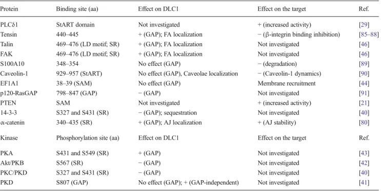
Table 1 DLC1-interacting partners
Protein Binding site (aa) Effect on DLC1 Effect on the target Ref.
PLCδ1 StART domain Not investigated + (increased activity) [29]
Tensin 440–445 + (GAP); FA localization − (β-integrin binding inhibition) [85–88]
Talin 469–476 (LD motif; SR) + (GAP); FA localization Not investigated [46]
FAK 469–476 (LD motif; SR) + (GAP); FA localization Not investigated [46]
S100A10 348–354 No effect (GAP) − (degradation) [89]
Caveolin-1 929–957 (StART) No effect (GAP), Caveolae localization − (Caveolin-1 dynamics) [90]
EF1A1 38–39 (SAM) No effect (GAP) Membrane recruitment [44]
p120-RasGAP 798–847 (GAP) − (GAP) Not investigated [91]
PTEN SAM Not investigated + (increased activity) [21]
14-3-3 S327 and S431 (SR) − (GAP); sequestration Not investigated [40]
α-catenin 340–435 (SR) + (GAP); AJ localization + (AJ stability) [80]
Kinase Phosphorylation site (aa) Effect on DLC1 Effect on the target Ref.
PKA S431 and S549 (SR) + (GAP) Not investigated [43]
Akt/PKB S567 (SR) − (GAP) Not investigated [42]
PKC/PKD S327 and S431 (SR) − (GAP) Not investigated [40]
PKD S807 (GAP) No effect (GAP); + (GAP-independent) Not investigated [41]
This table lists the DLC1-interacting partners and the effects of the indicated interaction on DLC1 and the concerned binding partner. Positive and negative modulation of DLC1 are indicated by the + and− symbols, respectively
increased IP3and DAG levels and a corresponding decrease of
PIP2. The consequence of this enzymatic reaction on the
cytoskeleton is complex (reviewed in [92]). PIP2establishes
a linkage between the actin cytoskeleton and the membrane and stimulates actin polymerization through several routes [92,93]. It primarily helps actin translocation to the plasma membrane where most actin polymerization proteins are lo-calized [93]. It stimulates the WASP protein that in turn activates Arp2/3, an actin nucleator that allows the formation of protrusions pushing the membrane forward [4]. PIP2binds
to several other actin regulators. By binding to the gelsolin capping protein, PIP2 disrupts the actin/gelsolin complex.
This leads to uncapping of the actin filament barbed end (also called the plus end), hence favoring actin polymerization [94]. Profilin is a protein that interacts with monomeric actin for delivery to the polymerizing barbed-ends of actin filaments [4]. PIP2then binds to profilin which dissociates from actin
allowing the latter to be incorporated on the growing actin fiber. PIP2is also required for the conformational activation of
vinculin and talin, two focal adhesion proteins the activation of which allows cell attachment, a step required for efficient migration.
Although PLCδ1 was the first identified DLC1 binding partner, the nature of their interaction has still not been fully determined although the GAP-StART domain region appears to be important (see Fig.1) [29]. The interaction of PLCδ1 and
DLC1 takes place at focal adhesions where DLC1 co-localizes with vinculin [95]. This localization is consistent with its potential role during migration. Interestingly, as DLC1, PLCδ1 has been defined as a tumor suppressor and inhibitor of migration [96]. Several approaches involving injection of PIP2or overexpression of negative regulators of PIP2have led
to the notion that increasing PIP2levels results in actin
poly-merization while decreasing PIP2levels leads to actin
depo-lymerization [92]. One can anticipate that the decrease of PIP2
levels through DLC1-mediated activation of PLCδ1 is a pos-sible Rho-independent mechanism allowing DLC1 to modu-late cytoskeleton dynamics and cell movement. The biological relevance of the DLC1/PLCδ1 interaction on migration re-mains however to be evaluated.
4.2.2 DLC1 and focal adhesion regulation
DLC1 can be found in several subcellular structures [80,90,95] but location at focal adhesions is the one most often reported [21,22,44,46,50,85–88,95,97–101]. What is commonly called focal adhesions actually comprise all types of anchorage contacts. These are, from smallest to biggest/longest, focal contacts (FXs), focal adhesions (FAs), and fibrillar adhesions (FBs) [102]. When integrins are engaged by their ECM ligands, talin and paxillin are quickly recruited to form the FXs, which are transient adhesive structures. FXs that are not turned over transform into FAs by recruiting proteins such as FAK,
vinculin,α-actinin, and zyxin, and this provides an anchorage point to link actin bundles. FBs are much longer and stronger adhesive structures that are localized in central areas under the cell and that are characterized by the presence of tensins. The tensin family is composed of four members, tensin-1, tensin-2, tensin-3, and C-terminal tensin-like (cten) [103]. Tensins are often downregulated in cancer, in particular in metastasis, sug-gesting that they play tumor and metastasis suppressor func-tions [103]. By linking the actin cytoskeleton and most of the integrin cytoplasmic tails (β1, β3, β5, and β7-integrins), tensins provide a crucial function required for cell migration [103–106].
DLC1 was reported to interact with each tensin family member [85–88]. The binding mode of DLC1 with the tensin proteins is however complex as it seems to vary between different tensin isoforms. The first identified binding mode involves the DLC1 SH2-binding motif containing serine 440 and tyrosine 442 (Fig.1) and the SH2 domain of tensin-1 and cten. This binding does not require phosphorylation of tyro-sine residues though [87]. Other binding modes have been unraveled recently, including interaction of the 374 to 388 DLC1 sequence with the phosphotyrosine-binding (PTB) do-main of tensin-2 [99] and the actin-binding motif of tensin-3 [88]. The latter contains a region, lacking in cten, that binds the DLC1 SAM domain [88]. DLC1-tensin binding occurs at focal adhesions where DLC1 most likely acts as a tumor [86,
87] and migration [98] suppressor. Interestingly, mutational-driven disruption of this binding prevents DLC1 FA localiza-tion and abrogates its oncosuppressive activity, without neg-atively impinging on its GAP activity [46,87]. When fused to a FA binding domain, the DLC1 mutant that is unable to bind tensins relocalizes to FBs but only partially recovers its tumor suppressor activity. Thus, the GAP activity at FBs does not appear to fully mediate some given DLC1 biological activi-ties. DLC1 was reported to compete with β3-integrin for binding with tensin-1 [86]. As a result, DLC1 engagement to tensin-1 prevents the connection between integrins and tensins. Since localization of tensins to FBs, where they interact with integrins, is crucial for establishing the anchorage points used by cells to move [105], this inhibitory function of DLC1 would be one of the means it can use to impede cell migration. Thus DLC1 can negatively regulate migration by competing with tensins for integrin binding independently of its Rho GAP activity. How this competition is regulated is only partially understood.
FAK and talin are two other FA proteins that can interact with DLC1 [46]. Talin, like tensins, can directly interact with integrins, while FAK mediates the link between talin and the actin cytoskeleton [5]. An eight amino acid motif within DLC1, called LD-motif (amino acids 469 to 476), is respon-sible for this binding. DLC1 can interact simultaneously with talin and FAK [46]. Potentially, this increases the capacity of DLC1 to translocate to FAs, which, as mentioned earlier, is the
place where DLC1 is supposed to exert its oncosuppressive functions. Paxillin is another focal adhesion protein that binds to FAK through its fourth LD motif [107]. DLC1 was shown to displace paxillin from FAK [46]. Interestingly, cells over-expressing a truncated version of paxillin that lacks the fourth LD motif display dramatically decreased migration [108]. While the effects of DLC1 binding on the structure of FAs have not yet been evaluated, one could predict that DLC1 binding to FAK negatively affects focal adhesion turnover and therefore migration by displacing paxillin from FAK. This represents yet another GAP-independent mechanism by which DLC1 inhibits migration and invasion.
4.2.3 The plasminogen connection
The urokinase plasminogen activator (uPA) and tissue plas-minogen activator (tPA) are activated upon binding to the uPAR membrane receptors. They then convert plasminogen into plasmin, a serine protease that degrades the ECM. They also stimulate matrix metalloproteinases (MMPs) that partic-ipate in ECM degradation [109]. The plasminogen activator inhibitor-1 (PAI-1) is a serine protease inhibitor that negative-ly regulates uPA and tPA. PAI-1 binds to uPA/tPA and leads to their internalization, preventing uPA/tPA to convert plasmin-ogen into plasmin [110,111]. PAI-1 also directly inhibits the MMP activity, as well as the αvβ3-integrin binding to
vitronectin, two crucial processes during invasion [112]. The S100A10 protein is a cell surface receptor and a plasminogen activator. It competes with PAI-1 to bind to uPA/tPA. This results in augmented plasmin production and promotion of invasiveness. S100A10 has also the ability to directly bind plasmin and activate its auto-proteolysis.
DLC1 modulates the plasminogen pathway to prevent dis-semination. DLC1 binds to S100A10, increasing its suscepti-bility to ubiquitin-dependent degradation and this potently re-duces, in a GAP-independent manner, S100A10-mediated plas-minogen activation [89]. The binding to S100A10 occurs through the 348-354 DLC1 sequence. RNAi-mediated silenc-ing of S100A10 in lung cancer cells leads to reduced colony formation and impaired invasiveness, as DLC1 does [89]. Another study investigating the role of DLC1 on the plasmin-ogen pathway reported an unexpected effect of DLC1 on migration [52]. In this study, silencing DLC1 in human prostate epithelial cells led to increased PAI-1 expression that resulted in migration inhibition. Conversely DLC1 overexpression de-creased PAI-1 expression levels. The reason of this discrepancy (i.e., DLC1/PAI-1 favors migration in lung cancer while it inhibits it in normal prostate cells) is unclear but may depend on the transformation status of a cell or possible differences in DLC1 residual expression after silencing.
ECM integrity and composition are often affected in meta-static contexts [113,114]. While certain ECM components are upregulated to favor motility, others are lost or degraded by
MMPs allowing dissemination [114, 115]. A study revealed that DLC1-mediated inhibition of liver metastasis formation was associated with the downregulation of the ECM compo-nent osteopontin, and the MMP-9 matrix metalloproteinase [27]. Osteopontin and MMP-9 have been associated with inva-sive phenotypes [115,116]. Therefore, this is a potential alter-native way for DLC1 to prevent metastasis formation. DLC1 has been shown to shuttle between the nucleus and the cyto-plasm [40]. Although its function in the nucleus is not known, it is possible that DLC1 regulates the activity of transcription factors. This could be the basis of the DLC1-mediated repres-sion of MMP-9 and osteopontin transcription.
4.2.4 DLC1 in caveolae
Caveolae are specialized plasma membrane subdomains that are involved in vesicular transport and compartmentalization of signaling proteins. Caveolin-1 is a major caveolae compo-nent, driving their formation and linking them to the cytoskel-eton [117]. These organelles play an important role during adhesion and migration in several ways [117]. Caveolin-1 binds to filamin, an actin-binding protein linking stress fibers at integrin sites and controling their stability. Caveolae pro-mote adhesion, migration and invasion by interacting with and stabilizing uPAR andαV-integrin [118] (see previous section).
Stable knock-down of caveolin-1 induces decreased β1-integrin-dependent adhesion and disruption of the caveolin-1/β1-integrin complex inhibits migration [118]. Caveolae also play a role during invasion by favoring plasminogen activa-tion and by hosting the MMP-2 [119]. Another evidence for the involvement of caveolae during migration is their ability to control calcium release at the cell rear which is required for calcium-dependent protease-mediated cleavage of focal adhe-sion proteins [120].
In 2004, the rat DLC1 orthologue and caveolin-1 were reported to colocalize in caveolae and to interact through the DLC1 GAP domain [121]. DLC1 interaction with caveolin-1 leads to both internalization of DLC1 and caveolae. The physiological importance of caveolin-1-DLC1 interaction was recently evaluated and the mapping of this interaction was revisited. It was found that the human DLC1 StART domain was responsible for this interaction rather than the previously reported rat DLC1 GAP domain [90]. Stable ex-pression of a DLC1 mutant that was unable to bind caveolin-1 in DLC1-negative lung cancer cells failed, unlike the wild-type DLC1, to inhibit colony formation, tumor migration into transwells, and in vivo tumor growth. Importantly, this DLC1 mutant retained a full GAP activity on RhoA, demonstrating that the migration inhibitory effect of DLC1 was either GAP-independent or that the DLC1 GAP activity needs to be brought to caveolae. How the DLC1/caveolin-1 interaction inhibits migration is however not fully understood. One can hypothesize however that DLC1 and caveolae internalization
prevents DLC1 from exerting a Rho GAP activity at the leading edge of the cell. Together with a decreased number of caveolae structures at the plasma membrane, this would be expected to markedly hamper cell migration.
4.2.5 The SAM binders
In contrast to most DLC1 partners that interact with the SR and C-terminal DLC1 regions, EF1A1 and PTEN bind to the N-terminal SAM domain (Fig.1). EF1A1 (for eukaryotic elongation factor 1A1) is involved in protein translation. EF1A1 also binds to the actin cytoskeleton and regulates its dynamics through several mechanisms, independently from its role in protein synthesis, by (a) it can bind to and bring β-actin mRNA at protrusion sites (i.e., plasma membrane sec-tions that are pushed out by growing actin fibers) [122], (b) through its actin-bundling activity [123], and (c) by binding to G-actin and possibly sequestering it [124]. EF1A1 binds to the 38–40 sequence of DLC1 within the SAM domain [44]. This interaction seems to be an important GAP-independent event controlling cell migration since transfection of a DLC1 mutant unable to bind EF1A1 but with an unaffected GAP activity induces a weaker migration inhibitory effect than the wild-type construct in fibroblasts. An interpretation that cannot be excluded though is that the DLC1 GAP activity needs to be located at the exact same place where EF1A1 lies. It was suggested that the SAM domain and the GAP domain of DLC1 can interact and this is believed to generate an auto-inhibitory signal. Indeed, the GAP domain of DLC1 is 20 times more active when isolated than when embedded in full-length DLC1 [22,26]. Consistent with these observations is that overexpression of the SAM domain induces a higher motility rate than full-length DLC1. Instead, overexpression of a SAM construct that does not bind EF1A1 did not stimu-late migration [44]. This indicates that modulation of migra-tion by the DLC1 SAM domain depends on its ability to bind EF1A1, possibly in a GAP-independent manner.
The phosphatase and tensin homolog (PTEN), a tumor suppressor, also binds to the DLC1 SAM domain [21]. This binding was proposed to mediate PTEN-induced dephosphor-ylation of FAK, leading to migration inhibition. However this hypothesis remains to be formally proven. Tensin-3 binds the DLC1 SAM domain through its actin-binding domain, the consequence of which is thought to release the SAM auto-inhibitory effect on GAP activity. The GAP-independent im-plications of tensin-DLC1 interaction are discussed above. 4.2.6 GAP-to-GAP
GAPs and GEFs rarely modulate a single GTPase. This is the case with DLC1 that regulate both Rho and Cdc42. Moreover, different GAPs and GEFs can modulate the same GTPase. This considerably increases the complexity of small GTPase
regulation [10]. Additionally, binding between different GAPs has been reported. For instance, p120 RasGAP and p190 RhoGAP interact and this is believed to control, on the one hand, the accessibility of the RasGAP SH3 domain and, on the other hand, the GAP activity of p190 [125,126]. Interestingly the RasGAP SH3 domain was shown to bind the DLC1 GAP domain resulting in inactivation of the latter’s GAP activity [91] and consequently this should increase cell motility. We recently discovered that the 317–326 amino acid sequence within the SH3 of RasGAP mediates its binding to DLC1 [134]. Treatment of cells with a cell-permeable version of the peptide corresponding to these 10 amino acids, the so-called TAT-RasGAP317–326 peptide, resulted in increased adhesion
associated with a potent inhibition of migration and invasion. This was an unexpected observation because, assuming that this peptide functions as the SH3 domain of RasGAP, it should also inhibit the GAP activity of DLC1 and hence favors migration. However, treatment with the C3 exoenzyme and Y-27632, which are inhibitors of Rho and ROCK, respective-ly, did not reverse this phenotype, suggesting that the RasGAP peptide exerts its activity in a DLC1 GAP-independent man-ner. Although the mechanisms induced by the RasGAP pep-tide downstream of DLC1 remain to be elucidated, this re-flects once more the importance of GAP-independent events in the DLC1-mediated control of migration.
4.2.7 DLC1 phosphorylation
The serine/threonine kinase PKD phosphorylates DLC1 on serine 807 that lies within its GAP domain. This phosphoryla-tion was reported to increase the inhibitory effect of DLC1 on colony formation and cell migration without modulating the GTP-bound state of RhoA [41]. Phosphorylation of DLC1 can therefore potentially inhibit dissemination and tumor growth in a GAP-dependent as well as in a GAP-independent manner. The in vivo relevance of PKD-mediated S807 phosphorylation remains however to be investigated.
5 Discussion
DLC1 is convincingly more than just a GAP for small GTPases of the Rho family; it is also a scaffolding protein that triggers a wide range of signaling responses affecting cell survival and controlling cell motility. Specific down-regulation of DLC1 may be used by malignant cells to survive and become metastatic. Together with the two well-established cancer associated proteins BRCA1 and HIF1-α, loss of DLC1 has recently been recognized as a biomarker for early stage lung adenomarcinomas [127]. Therapeutic strate-gies aimed at restoring DLC1 expression represent an attrac-tive anti-cancer strategy and this indeed has already been attempted. Using demethylation-inducing agents or dietary
flavones, DLC1 expression could be restored in cell lines in which it was epigenetically silenced and this hampered pro-liferation and induced cell cycle arrest [128]. In vivo antican-cer responses resulting from epigenetic restoration were how-ever not reported [128]. Rho isoforms are often overexpressed in cancer. Reactivating DLC1 in cancers would be interesting in this context since two distinct anticancer signaling would be triggered; those that dependent on the GAP activity of DLC1 and those that do not. These two effects would hopefully synergize or at least be additive. Our recent data obtained with TAT-RasGAP317–326 [134] indicate that DLC1-targeted
druggable compounds with anti-metastatic potential can be generated.
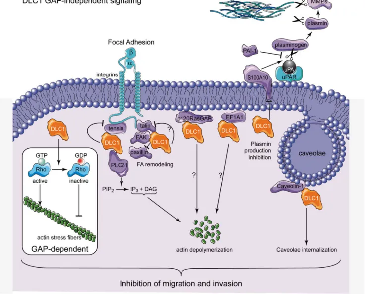
Figure2summarizes schematically the potential different GAP-independent mechanisms presented in this review. The-se include actin depolymerization (via PLCδ1 inhibition), FA remodeling (via interaction with tensins, FAK and talin),
plasminogen production inhibition (via S100A10 degrada-tion), caveolin internalization, and RasGAP and EF1A1-dependent anti-metastatic signaling. Such responses, in par-ticular if happening simultaneously, could induce potent inhi-bition of migration and invasion.
Although not discussed in this review, DLC2 and DLC3 can add an additional layer of complexity to this picture. Overexpression of DLC2 was shown to induce loss of stress fibers and inhibit cancer cell growth [129, 130]. Similarly, DLC2 loss is associated with enhanced migration [131]. DLC2 associates with mitochondria [132] and interacts with α-tubulin [129], two features that are not shared with DLC1. Interestingly, DLC2 seems to bear some activity towards Rac and could therefore synergize with DLC1, which does not regulate Rac, in preventing migration [133]. Akin to DLC1, DLC3 is a focal adhesion tumor suppressor broadly lost in various malignancies (reviewed in [17]) but its functions are
Fig. 2 DLC1 GAP-independent anti-metastatic mechanisms. This scheme highlights the anti-metastatic mechanisms used by DLC1 with a special emphasis on the GAP-independent signaling events. See main text for details
not well understood at the moment. GAP-independent signal-ing has not been reported for DLC2 and 3 but it would not be surprising that the mechanisms mentioned in this review are also used by them.
There is a wealth of data supporting the notion that normal or increased DLC1 expression levels inhibit migration or invasion, while decreased DLC1 expression favors these pro-cesses. Intriguing data challenge however this simple view. For example DLC1-null fibroblasts display a complete loss of stress fibers [53]. As stress fibers are required for migration, cell movement should be inhibited, contradicting the idea that DLC1 suppresses invasion. In addition, DLC1 silencing in normal prostate cells leads to migration inhibition, the oppo-site of what is usually found in the literature [52]. Intriguingly, these two studies reporting a pro-migratory role of DLC1 were performed in non-malignant cells. Modulation of DLC1 ex-pression levels in normal and cancer cells may therefore not have the same consequence. We recently generated data that also challenge the simple view that DLC1 loss favors migra-tion. We found out that the TAT-RasGAP317–326 peptide
inhibited migration in a large panel of normal and malignant cells but failed to have this effect in DLC1-null mouse em-bryonic fibroblasts [134]. DLC1 was expressed to very low, sometimes undetectable, levels in some of the cancer cells used in this study but TAT-RasGAP317–326was nevertheless
still able to inhibit their migration. Here again, the conse-quence of DLC1 loss may differ between malignant cells and the normal mouse embryonic fibroblasts. Alternatively, complete (as in knock-out DLC1 cells) or partial (as in cancer cells) DLC1 loss may not necessarily lead to the same migra-tory phenotype. It will be very important in the future to solve this conundrum.
In conclusion, DLC1 contributes to growth and migratory suppressive effects through dependent and GAP-independent mechanisms. In contrast to what have been pro-posed previously, the GAP-independent mechanism may ful-fill important tumor suppressor functions and represent there-fore potential avenues of research to develop new anti-cancer therapies.
Acknowledgments We thank Lluis Fajas and Mathieu Heulot for valu-able comments on this review. We apologize to colleagues whose work was not highlighted owing to space limitations. C.W. is supported by grants from the Swiss National Science Foundation (no. 31003A_141242/1) and the Swiss Cancer League (no. KFS-02543-02-2010).
References
1. Jemal, A., Bray, F., Center, M. M., Ferlay, J., Ward, E., & Forman, D. (2011). Global cancer statistics. CA: A Cancer Journal for Clinicians, 61(2), 69–90.
2. Hanahan, D., & Weinberg, R. A. (2011). Hallmarks of cancer: the next generation. Cell, 144(5), 646–674.
3. Chiang, A. C., & Massague, J. (2008). Molecular basis of metasta-sis. New England Journal of Medicine, 359(26), 2814–2823. 4. Pollard, T. D., & Borisy, G. G. (2003). Cellular motility driven by
assembly and disassembly of actin filaments. Cell, 112(4), 453– 465.
5. Mitra, S. K., Hanson, D. A., & Schlaepfer, D. D. (2005). Focal adhesion kinase: in command and control of cell motility. Nature Reviews Molecular Cell Biology, 6(1), 56–68.
6. Geiger, B., Bershadsky, A., Pankov, R., & Yamada, K. M. (2001). Transmembrane crosstalk between the extracellular matrix –cyto-skeleton crosstalk. Nature Reviews Molecular Cell Biology, 2(11), 793–805.
7. Grise, F., Bidaud, A., & Moreau, V. (2009). Rho GTPases in hepatocellular carcinoma. Biochimica et Biophysica Acta, 1795(2), 137–151.
8. Etienne-Manneville, S., & Hall, A. (2002). Rho GTPases in cell biology. Nature, 420(6916), 629–635.
9. Ridley, A. J. (2001). Rho GTPases and cell migration. Journal of Cell Science, 114(15), 2713–2722.
10. Bos, J. L., Rehmann, H., & Wittinghofer, A. (2007). GEFs and GAPs: critical elements in the control of small G proteins. Cell, 129(5), 865–877.
11. Tcherkezian, J., & Lamarche-Vane, N. (2007). Current knowledge of the large RhoGAP family of proteins. Biology of the Cell, 99(2), 67–86.
12. Lahoz, A., & Hall, A. (2008). DLC1: a significant GAP in the cancer genome. Genes and Development, 22(13), 1724–1730. 13. Durkin, M. E., Yuan, B. Z., Zhou, X., Zimonjic, D. B., Lowy, D. R.,
Thorgeirsson, S. S., et al. (2007). DLC-1:a Rho GTPase-activating protein and tumour suppressor. Journal of Cellular and Molecular Medicine, 11(5), 1185–1207.
14. Xue, W., Krasnitz, A., Lucito, R., Sordella, R., Vanaelst, L., Cordon-Cardo, C., et al. (2008). DLC1 is a chromosome 8p tumor suppressor whose loss promotes hepatocellular carcinoma. Genes and Development, 22(11), 1439–1444.
15. Liao, Y. C., & Lo, S. H. (2008). Deleted in liver cancer-1 (DLC-1): a tumor suppressor not just for liver. International Journal of Biochemistry and Cell Biology, 40(5), 843–847.
16. Lukasik, D., Wilczek, E., Wasiutynski, A., & Gornicka, B. (2011). Deleted in liver cancer protein family in human malignancies (Review). Oncology Letters, 2(5), 763–768.
17. El-Sitt, S., & El-Sibai, M. (2013). The STAR of the DLC family. Journal of Receptor and Signal Transduction Research, 33(1), 10– 13.
18. Kim, T. Y., Vigil, D., Der, C. J., & Juliano, R. L. (2009). Role of DLC-1, a tumor suppressor protein with RhoGAP activity, in reg-ulation of the cytoskeleton and cell motility. Cancer and Metastasis Reviews, 28(1–2), 77–83.
19. Feng, X., Li, C., Liu, W., Chen, H., Zhou, W., Wang, L., et al. (2013). DLC-1, a candidate tumor suppressor gene, inhibits the proliferation, migration and tumorigenicity of human nasopharyn-geal carcinoma cells. International Journal of Oncology, 42(6), 1973–1984.
20. Wu, P. P., Jin, Y. L., Shang, Y. F., Jin, Z., Wu, P., & Huang, P. L. (2009). Restoration of DLC1 gene inhibits proliferation and migra-tion of human colon cancer HT29 cells. Annals of Clinical and Laboratory Science, 39(3), 263–269.
21. Heering, J., Erlmann, P., & Olayioye, M. A. (2009). Simultaneous loss of the DLC1 and PTEN tumor suppressors enhances breast cancer cell migration. Experimental Cell Research, 315(15), 2505–2514. 22. Kim, T. Y., Healy, K. D., Der, C. J., Sciaky, N., Bang, Y. J., &
Juliano, R. L. (2008). Effects of structure of Rho GTPase-activating protein DLC-1 on cell morphology and migration. Journal of Biological Chemistry, 283(47), 32762–32770.
23. Wong, C. M., Yam, J. W., Ching, Y. P., Yau, T. O., Leung, T. H., Jin, D. Y., et al. (2005). Rho GTPase-activating protein deleted in liver cancer suppresses cell proliferation and invasion in hepatocellular carcinoma. Cancer Research, 65(19), 8861–8868.
24. Ullmannova-Benson, V., Guan, M., Zhou, X., Tripathi, V., Yang, X. Y., Zimonjic, D. B., et al. (2009). DLC1 tumor suppressor gene inhibits migration and invasion of multiple myeloma cells through RhoA GTPase pathway. Leukemia, 23(2), 383–390.
25. Goodison, S., Yuan, J., Sloan, D., Kim, R., Li, C., Popescu, N. C., et al. (2005). The RhoGAP protein DLC-1 functions as a metastasis suppressor in breast cancer cells. Cancer Research, 65(14), 6042– 6053.
26. Healy, K. D., Hodgson, L., Kim, T. Y., Shutes, A., Maddileti, S., Juliano, R. L., et al. (2008). DLC-1 suppresses non-small cell lung cancer growth and invasion by RhoGAP-dependent and indepen-dent mechanisms. Molecular Carcinogenesis, 47(5), 326–337. 27. Zhou, X., Zimonjic, D. B., Park, S. W., Yang, X. Y., Durkin, M. E.,
& Popescu, N. C. (2008). DLC1 suppresses distant dissemination of human hepatocellular carcinoma cells in nude mice through reduc-tion of RhoA GTPase activity, actin cytoskeletal disrupreduc-tion and down-regulation of genes involved in metastasis. International Journal of Oncology, 32(6), 1285–1291.
28. Yau, T. O., Leung, T. H., Lam, S., Cheung, O. F., Tung, E. K., Khong, P. L., et al. (2009). Deleted in liver cancer 2 (DLC2) was dispensable for development and its deficiency did not aggravate hepatocarcinogenesis. PLoS One, 4(8), e6566.
29. Homma, Y., & Emori, Y. (1995). A dual functional signal mediator showing RhoGAP and phospholipase C-δ stimulating activities. EMBO Journal, 14(2), 286–291.
30. Yuan, B. Z., Miller, M. J., Keck, C. L., Zimonjic, D. B., Thorgeirsson, S. S., & Popescu, N. C. (1998). Cloning, character-ization, and chromosomal localization of a gene frequently deleted in human liver cancer (DLC-1) homologous to rat RhoGAP. Cancer Research, 58(10), 2196–2199.
31. Durkin, M. E., Yuan, B. Z., Thorgeirsson, S. S., & Popescu, N. C. (2002). Gene structure, tissue expression, and linkage mapping of the mouse DLC-1 gene (Arhgap7). Gene, 288(1–2), 119–127. 32. Qian, X., Durkin, M. E., Wang, D., Tripathi, B. K., Olson, L., Yang,
X. Y., et al. (2012). Inactivation of the Dlc1 gene cooperates with downregulation of p15INK4b and p16Ink4a, leading to neoplastic transformation and poor prognosis in human cancer. Cancer Research, 72(22), 5900–5911.
33. Yuan, B. Z., Zhou, X., Durkin, M. E., Zimonjic, D. B., Gumundsdottir, K., Eyfjord, J. E., et al. (2003). DLC-1 gene in-hibits human breast cancer cell growth and in vivo tumorigenicity. Oncogene, 22(3), 445–450.
34. Sahai, E., Olson, M. F., & Marshall, C. J. (2001). Cross-talk be-tween Ras and Rho signalling pathways in transformation favours proliferation and increased motility. EMBO Journal, 20(4), 755– 766.
35. Kang, Y., Siegel, P. M., Shu, W., Drobnjak, M., Kakonen, S. M., Cordon-Cardo, C., et al. (2003). A multigenic program mediating breast cancer metastasis to bone. Cancer Cell, 3(6), 537–549. 36. Matsuyama, H., Pan, Y., Oba, K., Yoshihiro, S., Matsuda, K.,
Hagarth, L., et al. (2001). Deletions on chromosome 8p22 may predict disease progression as well as pathological staging in pros-tate cancer. Clinical Cancer Research, 7(10), 3139–3143. 37. Wilson, P. J., McGlinn, E., Marsh, A., Evans, T., Arnold, J., Wright,
K., et al. (2000). Sequence variants of DLC1 in colorectal and ovarian tumours. Human Mutation, 15(2), 156–165.
38. Yuan, B. Z., Durkin, M. E., & Popescu, N. C. (2003). Promoter hypermethylation of DLC-1, a candidate tumor suppressor gene, in several common human cancers. Cancer Genetics and Cytogenetics, 140(2), 113–117.
39. Guan, M., Zhou, X., Soulitzis, N., Spandidos, D. A., & Popescu, N. C. (2006). Aberrant methylation and deacetylation of deleted in
liver cancer-1 gene in prostate cancer: potential clinical applications. Clinical Cancer Research, 12(5), 1412–1419.
40. Scholz, R. P., Regner, J., Theil, A., Erlmann, P., Holeiter, G., Jahne, R., et al. (2009). DLC1 interacts with 14-3-3 proteins to inhibit RhoGAP activity and block nucleocytoplasmic shuttling. Journal of Cell Science, 122(1), 92–102.
41. Scholz, R. P., Gustafsson, J. O., Hoffmann, P., Jaiswal, M., Ahmadian, M. R., Eisler, S. A., et al. (2011). The tumor suppressor protein DLC1 is regulated by PKD-mediated GAP domain phos-phorylation. Experimental Cell Research, 317(4), 496–503. 42. Ko, F. C., Chan, L. K., Tung, E. K., Lowe, S. W., Ng, I. O., & Yam,
J. W. (2010). Akt phosphorylation of deleted in liver cancer 1 abrogates its suppression of liver cancer tumorigenesis and metas-tasis. Gastroenterology, 139(4), 1397–1407.
43. Ko, F. C., Chan, L. K., Man-Fong, S. K., Yeung, Y. S., Yuk-Ting, T. E., Lu, P., et al. (2013). PKA-induced dimerization of the RhoGAP DLC1 promotes its inhibition of tumorigenesis and metastasis. Nature Communications, 41618.
44. Zhong, D., Zhang, J., Yang, S., Soh, U. J., Buschdorf, J. P., Zhou, Y. T., et al. (2009). The SAM domain of the RhoGAP DLC1 binds EF1A1 to regulate cell migration. Journal of Cell Science, 122(3), 414–424. 45. Kim, C. A., & Bowie, J. U. (2003). SAM domains: uniform
struc-ture, diversity of function. Trends in Biochemical Sciences, 28(12), 625–628.
46. Li, G., Du, X., Vass, W. C., Papageorge, A. G., Lowy, D. R., & Qian, X. (2011). Full activity of the deleted in liver cancer 1 (DLC1) tumor suppressor depends on an LD-like motif that binds talin and focal adhesion kinase (FAK). Proceedings of the National Academy of Sciences of the United States of America, 108(41), 17129–17134.
47. Sekimata, M., Kabuyama, Y., Emori, Y., & Homma, Y. (1999). Morphological changes and detachment of adherent cells induced by p122, a GTPase-activating protein for Rho. Journal of Biological Chemistry, 274(25), 17757–17762.
48. Clark, B. J. (2012). The mammalian START domain protein family in lipid transport in health and disease. Journal of Endocrinology, 212(3), 257–275.
49. Friedl, P., & Wolf, K. (2003). Tumour-cell invasion and migration: diversity and escape mechanisms. Nature Reviews Cancer, 3(5), 362–374.
50. Holeiter, G., Heering, J., Erlmann, P., Schmid, S., Jahne, R., & Olayioye, M. A. (2008). Deleted in liver cancer 1 controls cell migration through a Dia1-dependent signaling pathway. Cancer Research, 68(21), 8743–8751.
51. Jin, Y., Tian, X., Shang, Y., & Huang, P. (2008). Inhibition of DLC-1 gene expression by RNA interference in the colon cancer LoVo cell line. Oncology Reports, 19(3), 669–674.
52. Shih, Y. P., Takada, Y., & Lo, S. H. (2012). Silencing of DLC1 upregulates PAI-1 expression and reduces migration in normal prostate cells. Molecular Cancer Research, 10(1), 34–39. 53. Durkin, M. E., Avner, M. R., Huh, C. G., Yuan, B. Z., Thorgeirsson,
S. S., & Popescu, N. C. (2005). DLC-1, a Rho GTPase-activating protein with tumor suppressor function, is essential for embryonic development. FEBS Letters, 579(5), 1191–1196.
54. Franz, C. M., Jones, G. E., & Ridley, A. J. (2002). Cell migration in development and disease. Developmental Cell, 2(2), 153–158. 55. Pilz, D., Stoodley, N., & Golden, J. A. (2002). Neuronal migration,
cerebral cortical development, and cerebral cortical anomalies. Journal of Neuropathology and Experimental Neurology, 61(1), 1–11. 56. Cramer L.P. (2013). Mechanism of cell rear retraction in migrating
cells. Current Opinion in Cell Biology 25:591–599.
57. Pollard, T. D., & Cooper, J. A. (2009). Actin, a central player in cell shape and movement. Science, 326(5957), 1208–1212.
58. Nobes, C. D., & Hall, A. (1999). Rho GTPases control polarity, protrusion, and adhesion during cell movement. Journal of Cell Biology, 144(6), 1235–1244.
59. Ridley, A. J., & Hall, A. (1992). The small GTP-binding protein rho regulates the assembly of focal adhesions and actin stress fibers in response to growth factors. Cell, 70(3), 389–399.
60. Takaishi, K., Kikuchi, A., Kuroda, S., Kotani, K., Sasaki, T., & Takai, Y. (1993). Involvement of rho p21 and its inhibitory GDP/ GTP exchange protein (rho GDI) in cell motility. Molecular and Cellular Biology, 13(1), 72–79.
61. Monnier, Y., Farmer, P., Bieler, G., Imaizumi, N., Sengstag, T., Alghisi, G. C., et al. (2008). CYR61 andαVβ5integrin cooperate
to promote invasion and metastasis of tumors growing in preirradiated stroma. Cancer Research, 68(18), 7323–7331. 62. Yilmaz, M., & Christofori, G. (2009). EMT, the cytoskeleton, and
cancer cell invasion. Cancer and Metastasis Reviews, 28(1–2), 15– 33.
63. Perl, A. K., Wilgenbus, P., Dahl, U., Semb, H., & Christofori, G. (1998). A causal role for E-cadherin in the transition from adenoma to carcinoma. Nature, 392(6672), 190–193.
64. Banyard, J., Anand-Apte, B., Symons, M., & Zetter, B. R. (2000). Motility and invasion are differentially modulated by Rho family GTPases. Oncogene, 19(4), 580–591.
65. Sahai, E., & Marshall, C. J. (2002). RHO-GTPases and cancer. Nature Reviews Cancer, 2(2), 133–142.
66. Yoshioka, K., Nakamori, S., & Itoh, K. (1999). Overexpression of small GTP-binding protein RhoA promotes invasion of tumor cells. Cancer Research, 59(8), 2004–2010.
67. Wheeler, A. P., & Ridley, A. J. (2004). Why three Rho proteins? RhoA, RhoB, RhoC, and cell motility. Experimental Cell Research, 301(1), 43–49.
68. Liu, A. X., Rane, N., Liu, J. P., & Prendergast, G. C. (2001). RhoB is dispensable for mouse development, but it modifies susceptibility to tumor formation as well as cell adhesion and growth factor signaling in transformed cells. Molecular and Cellular Biology, 21 (20), 6906–6912.
69. Hakem, A., Sanchez-Sweatman, O., You-Ten, A., Duncan, G., Wakeham, A., Khokha, R., et al. (2005). RhoC is dispensable for embryogenesis and tumor initiation but essential for metastasis. Genes and Development, 19(17), 1974–1979.
70. Clark, E. A., Golub, T. R., Lander, E. S., & Hynes, R. O. (2000). Genomic analysis of metastasis reveals an essential role for RhoC. Nature, 406(6795), 532–535.
71. Iiizumi, M., Bandyopadhyay, S., Pai, S. K., Watabe, M., Hirota, S., Hosobe, S., et al. (2008). RhoC promotes metastasis via activation of the Pyk2 pathway in prostate cancer. Cancer Research, 68(18), 7613–7620.
72. Bishop, A. L., & Hall, A. (2000). Rho GTPases and their effector proteins. Biochemistry Journal, 348(2), 2241–2255.
73. Matsui, T., Amano, M., Yamamoto, T., Chihara, K., Nakafuku, M., Ito, M., et al. (1996). Rho-associated kinase, a novel serine/ threonine kinase, as a putative target for small GTP binding protein Rho. EMBO Journal, 15(9), 2208–2216.
74. Chesarone, M. A., DuPage, A. G., & Goode, B. L. (2010). Unleashing formins to remodel the actin and microtubule cytoskeletons. Nature Reviews Molecular Cell Biology, 11 (1), 62–74.
75. Raftopoulou, M., & Hall, A. (2004). Cell migration: Rho GTPases lead the way. Developments in Biologicals, 265(1), 23–32. 76. Palazzo, A. F., Joseph, H. L., Chen, Y. J., Dujardin, D. L., Alberts,
A. S., Pfister, K. K., et al. (2001). Cdc42, dynein, and dynactin regulate MTOC reorientation independent of Rho-regulated micro-tubule stabilization. Current Biology, 11(19), 1536–1541. 77. Reymond, N., Im, J. H., Garg, R., Vega, F. M., Borda, D. B., Riou,
P., et al. (2012). Cdc42 promotes transendothelial migration of cancer cells throughβ1 integrin. Journal of Cell Biology, 199(4), 653–668.
78. Wong, C. C., Wong, C. M., Ko, F. C., Chan, L. K., Ching, Y. P., Yam, J. W., et al. (2008). Deleted in liver cancer 1 (DLC1)
negatively regulates Rho/ROCK/MLC pathway in hepatocellular carcinoma. PLoS One, 3(7), e2779.
79. Ishizaki, T., Uehata, M., Tamechika, I., Keel, J., Nonomura, K., Maekawa, M., et al. (2000). Pharmacological properties of Y-27632, a specific inhibitor of rho-associated kinases. Molecular Pharmacology, 57(5), 976–983.
80. Tripathi, V., Popescu, N. C., & Zimonjic, D. B. (2012). DLC1 interaction with α-catenin stabilizes adherens junctions and en-hances DLC1 antioncogenic activity. Molecular and Cellular Biology, 32(11), 2145–2159.
81. Harris, T. J., & Tepass, U. (2010). Adherens junctions: from mole-cules to morphogenesis. Nature Reviews Molecular Cell Biology, 11(7), 502–514.
82. Desai, R., Sarpal, R., Ishiyama, N., Pellikka, M., Ikura, M., & Tepass, U. (2013). Monomericα-catenin links cadherin to the actin cytoskeleton. Nature Cell Biology, 15(3), 261–273.
83. Kobielak, A., & Fuchs, E. (2004).α-catenin: at the junction of intercellular adhesion and actin dynamics. Nature Reviews Molecular Cell Biology, 5(8), 614–625.
84. Tripathi V., Popescu N.C. & Zimonjic D.B. (2013). DLC1 induces expression of E-cadherin in prostate cancer cells through Rho pathway and suppresses invasion. Oncogene. doi:10.1038/onc. 2013.7.
85. Yam, J. W., Ko, F. C., Chan, C. Y., Jin, D. Y., & Ng, I. O. (2006). Interaction of deleted in liver cancer 1 with tensin2 in caveolae and implications in tumor suppression. Cancer Research, 66(17), 8367– 8372.
86. Qian, X., Li, G., Asmussen, H. K., Asnaghi, L., Vass, W. C., Braverman, R., et al. (2007). Oncogenic inhibition by a deleted in liver cancer gene requires cooperation between tensin binding and Rho-specific GTPase-activating protein activities. Proceedings of the National Academy of Sciences of the United States of America, 104(21), 9012–9017.
87. Liao, Y. C., Si, L., deVere White, R. W., & Lo, S. H. (2007). The phosphotyrosine-independent interaction of DLC-1 and the SH2 domain of cten regulates focal adhesion localization and growth suppression activity of DLC-1. Journal of Cell Biology, 176(1), 43–49.
88. Cao, X., Voss, C., Zhao, B., Kaneko, T., & Li, S. S. (2012). Differential regulation of the activity of deleted in liver cancer 1 (DLC1) by tensins controls cell migration and transformation. Proceedings of the National Academy of Sciences of the United States of America, 109(5), 1455–1460.
89. Yang, X., Popescu, N. C., & Zimonjic, D. B. (2011). DLC1 inter-action with S100A10 mediates inhibition of in vitro cell invasion and tumorigenicity of lung cancer cells through a RhoGAP-independent mechanism. Cancer Research, 71(8), 2916–2925. 90. Du, X., Qian, X., Papageorge, A., Schetter, A. J., Vass, W. C., Liu,
X., et al. (2012). Functional interaction of tumor suppressor DLC1 and caveolin-1 in cancer cells. Cancer Research, 72(17), 4405– 4416.
91. Yang, X. Y., Guan, M., Vigil, D., Der, C. J., Lowy, D. R., & Popescu, N. C. (2009). p120Ras-GAP binds the DLC1 Rho-GAP tumor suppressor protein and inhibits its RhoA GTPase and growth-suppressing activities. Oncogene, 28(11), 1401–1409.
92. Yin, H. L., & Janmey, P. A. (2003). Phosphoinositide regulation of the actin cytoskeleton. Annual Review of Physiology, 65(65), 761–789. 93. Raucher, D., Stauffer, T., Chen, W., Shen, K., Guo, S., York, J. D.,
et al. (2000). Phosphatidylinositol 4,5-bisphosphate functions as a second messenger that regulates cytoskeleton-plasma membrane adhesion. Cell, 100(2), 221–228.
94. Toker, A. (1998). The synthesis and cellular roles of phos-phatidylinositol 4,5-bisphosphate. Current Opinion in Cell Biology, 10(2), 254–261.
95. Kawai, K., Yamaga, M., Iwamae, Y., Kiyota, M., Kamata, H., Hirata, H., et al. (2004). A PLCδ1-binding protein, p122RhoGAP,
is localized in focal adhesions. Biochemical Society Transactions, 32(6), 1107–1109.
96. Xiang, T., Li, L., Fan, Y., Jiang, Y., Ying, Y., Putti, T. C., et al. (2010). PLCD1 is a functional tumor suppressor inducing G(2)/M arrest and frequently methylated in breast cancer. Cancer Biology and Therapy, 10(5), 520–527.
97. Liao, Y. C., Shih, Y. P., & Lo, S. H. (2008). Mutations in the focal adhesion targeting region of deleted in liver cancer-1 attenuate their expression and function. Cancer Research, 68(19), 7718–7722. 98. Kawai, K., Iwamae, Y., Yamaga, M., Kiyota, M., Ishii, H., Hirata,
H., et al. (2009). Focal adhesion-localization of START-GAP1/ DLC1 is essential for cell motility and morphology. Genes to Cells, 14(2), 227–241.
99. Chan, L. K., Ko, F. C., Ng, I. O., & Yam, J. W. (2009). Deleted in liver cancer 1 (DLC1) utilizes a novel binding site for Tensin2 PTB domain interaction and is required for tumor-suppressive function. PLoS One, 4(5), e5572.
100. Chan, L. K., Ko, F. C., Sze, K. M., Ng, I. O., & Yam, J. W. (2011). Nuclear-targeted deleted in liver cancer 1 (DLC1) is less efficient in exerting its tumor suppressive activity both in vitro and in vivo. PLoS One, 6(9), e25547.
101. Chen, L., Liu, C., Ko, F. C., Xu, N., Ng, I. O., Yam, J. W., et al. (2012). Solution structure of the phosphotyrosine binding (PTB) domain of human tensin2 protein in complex with deleted in liver cancer 1 (DLC1) peptide reveals a novel peptide binding mode. Journal of Biological Chemistry, 287(31), 26104–26114. 102. Zaidel-Bar, R., Cohen, M., Addadi, L., & Geiger, B. (2004).
Hierarchical assembly of cell-matrix adhesion complexes. Biochemical Society Transactions, 32(3), 416–420.
103. Lo, S. H. (2004). Tensin. International Journal of Biochemistry and Cell Biology, 36(1), 31–34.
104. Chen, H., Duncan, I. C., Bozorgchami, H., & Lo, S. H. (2002). Tensin1 and a previously undocumented family member, tensin2, positively regulate cell migration. Proceedings of the National Academy of Sciences of the United States of America, 99(2), 733–738.
105. Chen, H., & Lo, S. H. (2003). Regulation of tensin-promoted cell migration by its focal adhesion binding and Src homology domain 2. Biochemistry Journal, 370(3), 1039–1045.
106. Calderwood, D. A., Fujioka, Y., de Pereda, J. M., Garcia-Alvarez, B., Nakamoto, T., Margolis, B., et al. (2003). Integrinβ cytoplasmic domain interactions with phosphotyrosine-binding domains: a structural prototype for diversity in integrin signaling. Proceedings of the National Academy of Sciences of the United States of America, 100(5), 2272–2277.
107. Schaller, M. D. (2001). Paxillin: a focal adhesion-associated adaptor protein. Oncogene, 20(44), 6459–6472.
108. West, K. A., Zhang, H., Brown, M. C., Nikolopoulos, S. N., Riedy, M. C., Horwitz, A. F., et al. (2001). The LD4 motif of paxillin regulates cell spreading and motility through an interaction with paxillin kinase linker (PKL). Journal of Cell Biology, 154(1), 161–176.
109. Li, Y., & Cozzi, P. J. (2007). Targeting uPA/uPAR in prostate cancer. Cancer Treatment Reviews, 33(6), 521–527.
110. Czekay, R. P., & Loskutoff, D. J. (2009). Plasminogen activator inhibitors regulate cell adhesion through a uPAR-dependent mech-anism. Journal of Cellular Physiology, 220(3), 655–663. 111. Czekay, R. P., Aertgeerts, K., Curriden, S. A., & Loskutoff, D. J.
(2003). Plasminogen activator inhibitor-1 detaches cells from extra-cellular matrices by inactivating integrins. Journal of Cell Biology, 160(5), 781–791.
112. Czekay, R. P., Wilkins-Port, C. E., Higgins, S. P., Freytag, J., Overstreet, J. M., Klein, R. M., et al. (2011). PAI-1: an integrator of cell signaling and migration. International Journal of Cell Biology, 2011562481.
113. Paszek, M. J., Zahir, N., Johnson, K. R., Lakins, J. N., Rozenberg, G. I., Gefen, A., et al. (2005). Tensional homeostasis and the malignant phenotype. Cancer Cell, 8(3), 241–254.
114. Stewart, D. A., Cooper, C. R., & Sikes, R. A. (2004). Changes in extracellular matrix (ECM) and ECM-associated proteins in the metastatic progression of prostate cancer. Reproductive Biology and Endocrinology, 22.
115. Deryugina, E. I., & Quigley, J. P. (2006). Matrix metalloproteinases and tumor metastasis. Cancer and Metastasis Reviews, 25(1), 9–34. 116. Wai, P. Y., & Kuo, P. C. (2004). The role of Osteopontin in tumor
metastasis. Journal of Surgical Research, 121(2), 228–241. 117. Navarro, A., Anand-Apte, B., & Parat, M. O. (2004). A role for
caveolae in cell migration. FASEB Journal, 18(15), 1801–1811. 118. Wei, Y., Yang, X., Liu, Q., Wilkins, J. A., & Chapman, H. A. (1999).
A role for caveolin and the urokinase receptor in integrin-mediated adhesion and signaling. Journal of Cell Biology, 144(6), 1285– 1294.
119. Puyraimond, A., Fridman, R., Lemesle, M., Arbeille, B., & Menashi, S. (2001). MMP-2 colocalizes with caveolae on the surface of endothelial cells. Experimental Cell Research, 262(1), 28–36.
120. Isshiki, M., Ando, J., Yamamoto, K., Fujita, T., Ying, Y., & Anderson, R. G. (2002). Sites of Ca(2+) wave initiation move with caveolae to the trailing edge of migrating cells. Journal of Cell Science, 115(3), 475–484.
121. Yamaga, M., Sekimata, M., Fujii, M., Kawai, K., Kamata, H., Hirata, H., et al. ( 200 4). A PLCδ1-binding protein, p122/RhoGAP, is localized in caveolin-enriched membrane do-mains and regulates caveolin internalization. Genes to Cells, 9(1), 25–37.
122. Liu, G., Grant, W. M., Persky, D., Latham, V. M., Jr., Singer, R. H., & Condeelis, J. (2002). Interactions of elon-gation factor 1α with F-actin and β-actin mRNA: implica-tions for anchoring mRNA in cell protrusions. Molecular Biology of the Cell, 13 (2), 579–592.
123. Gross, S. R., & Kinzy, T. G. (2005). Translation elongation factor 1α is essential for regulation of the actin cytoskeleton and cell morphology. Nature Structural and Molecular Biology, 12 (9), 772–778.
124. Zhang, J., Guo, H., Mi, Z., Gao, C., Bhattacharya, S., Li, J., et al. (2009). EF1A1-actin interactions alter mRNA stability to determine differential osteopontin expression in HepG2 and Hep3B cells. Experimental Cell Research, 315(2), 304–312.
125. Hu, K. Q., & Settleman, J. (1997). Tandem SH2 binding sites mediate the RasGAP-RhoGAP interaction: a conforma-tional mechanism for SH3 domain regulation. EMBO Journal, 16 (3), 473–483.
126. Bradley, W. D., Hernandez, S. E., Settleman, J., & Koleske, A. J. (2006). Integrin signaling through Arg activates p190RhoGAP by promoting its binding to p120RasGAP and recruitment to the mem-brane. Molecular Biology of the Cell, 17(11), 4827–4836. 127. Akagi, I., Okayama, H., Schetter, A. J., Robles, A. I., Kohno, T.,
Bowman, E. D., et al. (2013). Combination of Protein Coding and Noncoding Gene Expression as a Robust Prognostic Classifier in Stage I Lung Adenocarcinoma. Cancer Research, 73(13), 3821– 3832.
128. Ullmannova, V., & Popescu, N. C. (2007). Inhibition of cell proliferation, induction of apoptosis, reactivation of DLC1, and modulation of other gene expression by dietary flavone in breast cancer cell lines. Cancer Detection and Prevention, 31 (2), 110–118.
129. Nagaraja, G. M., & Kandpal, R. P. (2004). Chromosome 13q12 encoded Rho GTPase activating protein suppresses growth of breast carcinoma cells, and yeast two-hybrid screen shows its interaction with several proteins. Biochemical and Biophysical Research Communications, 313(3), 654–665.
130. Leung, T. H., Ching, Y. P., Yam, J. W., Wong, C. M., Yau, T. O., Jin, D. Y., et al. (2005). Deleted in liver cancer 2 (DLC2) suppresses cell transformation by means of inhibition of RhoA activity.
Proceedings of the National Academy of Sciences of the United States of America, 102(42), 15207–15212.
131. Lin, Y., Chen, N. T., Shih, Y. P., Liao, Y. C., Xue, L., & Lo, S. H. (2010). DLC2 modulates angiogenic responses in vascular endo-thelial cells by regulating cell attachment and migration. Oncogene, 29(20), 3010–3016.
132. Ng, D. C., Chan, S. F., Kok, K. H., Yam, J. W., Ching, Y. P., Ng, I. O., et al. (2006). Mitochondrial targeting of growth suppressor protein DLC2 through the START domain. FEBS Letters, 580(1), 191–198.
133. Ching, Y. P., Wong, C. M., Chan, S. F., Leung, T. H., Ng, D. C., Jin, D. Y., et al. (2003). Deleted in liver cancer (DLC) 2 encodes a RhoGAP protein with growth suppressor function and is underexpressed in hepatocellular carcinoma. Journal of Biological Chemistry, 278(12), 10824–10830.
134. Barras, D., Lorusso, G., Rüegg, C., & Widmann, C. (2013). Inhibition of cell migration and invasion by the TAT-RasGAP317-326 peptide requires the DLC1 tumor suppressor. Oncogene. doi: 10.1038/onc.2013.465.