Contents lists available atScienceDirect
Developmental Biology
journal homepage:www.elsevier.com/locate/developmentalbiology
Original research article
Dynamics of actinotrichia regeneration in the adult zebra
fish fin
Désirée König, Lionel Page, Bérénice Chassot, Anna Ja
źwińska
⁎Department of Biology, University of Fribourg, Chemin du Musée 10, 1700 Fribourg, Switzerland
A R T I C L E I N F O
Keywords: Actinodin Blastema Lepidotrichia Wound epithelium Osteoblasts Extracellular matrixA B S T R A C T
The skeleton of adult zebrafish fins comprises lepidotrichia, which are dermal bones of the rays, and actinotrichia, which are non-mineralized spicules at the distal margin of the appendage. Little is known about the regenerative dynamics of the actinotrichia-specific structural proteins called Actinodins. Here, we used immunofluorescence analysis to determine the contribution of two paralogous Actinodin proteins, And1/2, in regeneratingfins. Both proteins were detected in the secretory organelles in the mesenchymal cells of the blastema, but only And1 was detected in the epithelial cells of the wound epithelium. The analysis of whole mountfins throughout the entire regenerative process and longitudinal sections revealed that And1-positive fibers are complementary to the lepidotrichia. The analysis of another longfin fish, a gain-of-function mutation in the potassium channel kcnk5b, revealed that the long-fin phenotype is associated with an extended size of actinotrichia during homeostasis and regeneration. Finally, we investigated the role of several signaling pathways in actinotrichia formation and maintenance. This revealed that the pulse-inhibition of either TGFβ/ Activin-βA or FGF are sufficient to impair deposition of Actinodin during regeneration. Thus, the dynamic turnover of Actinodin duringfin regeneration is regulated by multiple factors, including the osteoblasts, growth rate in a potassium channel mutant, and instructive signaling networks between the epithelium and the blastema of the regeneratingfin.
1. Introduction
The zebrafish caudal fin provides a valuable model system to study mechanisms of adult organ regeneration in vertebrates. After fin amputation, the zebrafish is able to swim and to restore the original size and shape of the appendage within approximately three weeks. This phenomenon depends on appropriate wound healing, creation of blastema progenitor cells and on their progressive redifferentiation, as recently reviewed (Jazwinska and Sallin, 2016; Tornini and Poss, 2014; Wehner and Weidinger, 2015).
Thefin is a non-muscularized flattened appendage, which is used for propulsion while swimming. As opposed to the tetrapod limb, which is entirely supported by the endoskeleton, thefin fold is stabilized by skeletal elements of dermal origin, called rays, whereas the endoske-leton is confined only to the base of the fin (Grandel and Schulte-Merker, 1998; Thorogood, 1991; Witten and Huysseune, 2007). The zebrafish caudal fin typically contains 16–18 main occasionally bifur-cated rays, which are connected by soft interray tissue (Akimenko et al., 2003; Mari-Beffa and Murciano, 2010; Pfefferli and Jaźwińska, 2015). Each ray contains a pair of segmented parenthesis-shaped bones, called lepidotrichia, which are deposited by osteoblasts underneath the stratified epidermis. The tips of the rays lack bones and are solely
supported by unsegmented brush-like spicules, named actinotrichia (Becerra et al., 1983; Witten and Huysseune, 2007). While lepido-trichia are dermal structures, actinolepido-trichia are considered to be of ectodermal origin, as they are formed in the early embryonicfin fold, before mesenchyme recruitment, and they precede the differentiation of rays in variousfish species (Dane and Tucker, 1985; Feitosa et al., 2012; Geraudie, 1977; Grandel and Schulte-Merker, 1998; Heude et al., 2014; Kemp and Park, 1970; Thorogood, 1991; Wood and Thorogood, 1984; Zhang et al., 2010). In the mature zebrafish fin, both skeletal structures occupy the epidermal-mesenchymal interface along the proximal-distal axis of the appendage, whereas the interior space of thefin is filled with vascularized and innervated mesenchymal tissue (Akimenko et al., 2003; Mari-Beffa and Murciano, 2010; Pfefferli and Jaźwińska, 2015). After adultfin amputation, the stump forms a new outgrowth, which is initially supported by actinotrichia before lepido-trichia formation (Duran et al., 2011; Mari-Beffa et al., 1989). Actinotrichia play at least two functions in thefin: providing mechan-ical support to thefin fold during swimming and acting as a substrate for the migration of mesenchymal cells during morphogenesis (Bhadra and Iovine, 2015; Duran et al., 2011; van den Boogaart et al., 2012; Wood and Thorogood, 1984; Zhang et al., 2010). The attachment of mesenchymal cells to the actinotrichial migratory substrate requires
http://dx.doi.org/10.1016/j.ydbio.2017.07.024
Received 19 May 2017; Received in revised form 25 July 2017; Accepted 27 July 2017
⁎Corresponding author.
E-mail address:[email protected](A. Jaźwińska).
Available online 29 July 2017
0012-1606/ © 2017 The Authors. Published by Elsevier Inc. This is an open access article under the CC BY-NC-ND license (http://creativecommons.org/licenses/BY-NC-ND/4.0/).
Hemicentin 2 and Fibulin 1 matrix proteins (Feitosa et al., 2012). Actinotrichia are composed of multiple collagenous and non-collagenous matrix proteins, which form a supramolecular aggregate,
historically referred to as elastoidin (Galloway, 1985; Geraudie and Meunier, 1980; Mari-Beffa et al., 1989). By electron microscopy, actinotrichia exhibit a regular cross-banding, with a periodicity varying
D. König et al. Developmental Biology 433 (2018) 416–432
from 49 to 65 nm, depending on the species (Witten and Huysseune, 2007). Their unique architecture combines the rigidity of collagen fibers and the flexibility of elastic fibers, which might be advantageous for maintaining the structural integrity and the hydrodynamic function of the dermal fold (Galloway, 1985; van den Boogaart et al., 2012). Indeed, the biomechanical properties of actinotrichia are thought to be specific for swimming and irrelevant for walking on land, as the limb skeleton of terrestrial vertebrates lacks any actinotrichia-like structures (Lalonde et al., 2016; Sordino et al., 1995; Zhang et al., 2010). The unique components of these structures, called Actinodin (And) pro-teins, are fish-specific and were lost in the evolution of tetrapods (Zhang et al., 2010). Accordingly, Actinodin proteins are not involved in the support of thefin fold in aquatic tadpoles of amphibians.
In addition to the Actinodin proteins, fibrillar collagens, such as Collagen I and II, have been described to contribute to the composition of actinotrichia during development and regeneration (Bhadra and Iovine, 2015; Duran et al., 2015, 2011). In addition, afibril-associated collagen, Collagen IX, has been shown to be required for actinotrichia integrity (Huang et al., 2009). Fish that carry a mutation in the col9a1 gene, called prp mutants, develop shortened and deformed actinotri-chiafibers. Based on the known function of Collagen IX in bridging Collagen type IIfibrils (Huang et al., 2009), the prp phenotype suggests that supramolecular aggregation of fibrillar bundles is essential for shaping of actinotrichia.
Recent studies using transgenic reporterfish lines and functional assays have provided new insights into osteoblast de- and redi ffer-entiation during lepidotrichia regeneration (Blum and Begemann, 2015; Brown et al., 2009; Knopf et al., 2011; Sousa et al., 2011; Stewart et al., 2014; Stewart and Stankunas, 2012; Thorimbert et al., 2015; Tu and Johnson, 2011; Wehner et al., 2014). Regeneration of actinotrichia has been studied using ultrustructural, immunohisto-chemical and gene expression approaches onfin sections (Duran et al., 2011; Govindan and Iovine, 2015; Mari-Beffa et al., 1989; Mari-Beffa and Murciano, 2010; Zhang et al., 2010). The regulation of actino-trichia formation in the regenerating fin has been addressed using chemical inhibition of Sonic Hedgehog signaling, Bone Morphogenetic Protein (BMP) receptors and Histone deacetylase 1 (Hdac1), which did not interfere with initial blastema formation, but led to a blockage of actinotrichia formation and progression of regeneration (Armstrong et al., 2017; Pfefferli et al., 2014; Quint et al., 2002; Thorimbert et al., 2015). However, the dynamic configuration of Actinodin-containing fibers during the entire fin restoration has not been investigated in detail. Moreover, the secretion of Actinodin proteins has not yet been assessed at the subcellular level. Finally, it remains unknown, whether the early blastema signals regulate actinotrichia formation and turn-over during the outgrowth phase.
Here, we examined the temporal and spatial dynamics of actino-trichia regeneration by means of immunofluorescence analysis of Actinodin-1 in combination with lepidotrichial markers on whole-mountfins. We used alf mutants, which have longer and irregularly segmented lepidotrichia due to a potassium channel gain-of-function (Perathoner et al., 2014), to examine a correlation between enhanced fin growth and lengthened actinotrichia. We applied electron micro-scopy to detect fibrils assembling into thicker actinotrichia fibers specifically in the distal-most blastema, the site of their polymerization. Finally, we sought to characterize the importance of blastemal signal-ing pathways dursignal-ing actinotrichia regeneration, namely Fibroblast Growth Factor (FGF), Transforming Growth Factor β (TGFβ)/
Activin-βA and Insulin-like Growth Factor (IGF). These analyses provide new insights into the dynamics of actinotrichia reestablish-ment during zebrafish fin regeneration.
2. Results
2.1. Two paralogous Actinodin proteins colocalize in the actinotrichia fibers of the regenerating fin
Four actinodin genes have been identified in the embryonic fin fold and were examined in adultfish (Nolte et al., 2015; Zhang et al., 2010). To compare the expression of all four actinodin transcripts in the regenerative outgrowth, we performed in-situ hybridization of long-itudinalfin sections at 3 dpa, when actinotrichia massively emerge in the new tissue. Consistent with previous studies (Thorimbert et al., 2015; Zhang et al., 2010), and1 was detected in the lateral mesench-yme of the blastema and in the basal layer of the wound epithelium (Fig. 1A, B). Interestingly, the and1 probe labeled the basal wound epithelium more extensively in the interrays than in the rays. In the rays, the epithelial expression of this gene was restricted to the distal-most region, whereas it expanded further proximally in the interrays (Fig. 1A′, B′). By contrast, and2/3/4 transcripts were detected in the distal blastemal mesenchyme, but not in the epidermis (Fig. 1C-F). The weakest staining was obtained for and3, which has not been detected by in-situ hybridization on whole-mountfins (Nolte et al., 2015). Thus, among all actinodin genes, only and1 was present in the wound epithelium.
The localization of extracellular proteins cannot be solely inferred from the transcript distribution. Therefore, we raised antibodies against And1 and And2 unique peptides (less than 50% identity of amino acid sequences) in distinct species. First, we validated these antibodies on Western blot offin regenerates at 3 dpa. And1 and And2 antibodies detected different proteins at relative molecular weight of approx. 75 kDa and 57 kDa, respectively (Fig. S1). Then, we used both antibodies for double immunofluorescence analysis of fin regenerates. The examination of longitudinal ray sections revealed that And1 and And2 co-localized in extracellularfibers in the regenerative outgrowth at 3 dpa (Fig. 1G-J). We concluded that both proteins jointly contribute to actinotrichia, despite the differences in gene expression patterns.
As previously reported (Duran et al., 2011; Thorimbert et al., 2015; Zhang et al., 2010), actinotrichiafibers were predominantly deposited along the lateral sides of the regenerative outgrowth between the wound epidermis and the blastema. This localization was particularly well established in the interrays (Fig. 1I, J). In the rays, which were recognized by the presence of Zns5-positive osteoblasts, actinotrichia fibers were longitudinally organized underneath the wound epithelium mainly in the distal-most part of the outgrowth (Fig. 1H′). In the distal and proximal part, which contained osteoblasts underneath the lateral epidermis, the And1-positivefibers were displaced into the mesench-ymal compartment, which was often associated with a disruption of their longitudinal alignment.
In addition to the fibrillar pattern, both Actinodin antibodies recognized a dotty pattern laterally to thefibers, which might represent intermediate forms of actinotrichiafibers that undergo formation or turnover (Fig. 1H′, J′). We concluded that Actinodin 1 and Actinodin 2 proteins jointly contribute to the actinotrichiafibers in the regenerating zebrafish fin.
Fig. 1. Analysis of the actinotrichia-specific components in the regenerative outgrowth. (A-F) In-situ hybridization with probes against four actinodin genes on longitudinal fin sections at 3 dpa. N≥ 4 fins; ≥ 3 sections per fin. (A, B) and1 mRNA is detected in the mesenchyme (m) of the blastema and in the basal wound epithelium (bwe). No expression is detected in the basal wound epithelium adjacent to osteoblasts (ob) in the ray. (C, D) and2 (E) and3 (F) and4 genes are co-expressed in the mesenchyme of the blastema, but not in the epidermis (e). (G-J) Immunofluorescence staining of longitudinal fin sections of ray (G, H) and interray (I, J) at 3 dpa. And1 (green) and And2 (red) proteins are co-localized in actinotrichia fibers. Rays and interrays are identified by the presence versus absence of Zns5-positive osteoblasts (H, J, blue). Actinotrichia fibers are predominantly found in the subepithelial position with the exception of osteoblast-containing regions. N = 4fins; 3 sections per fin. Higher magnifications of framed areas are labeled with the same letter with a prime symbol. Fin amputation planes are shown with a dashed line. The same rules apply to all subsequentfigures.
D. König et al. Developmental Biology 433 (2018) 416–432
2.2. Osteoblasts displace And1-positivefibers into the mesenchymal compartment
In-situ hybridization with the and1 probe revealed a difference in the extent of the labeled basal wound epithelium between ray and interray (Fig. 1A, B). To further investigate this observation, we analyzed transversal sections of 3 dpa fin regenerates, in order to compare tissues of rays and interrays at the same proximo-distal level. As expected, and2 mRNA was restricted to the mesenchyme (Fig. 2C, D), whereas and1 expression in the basal wound epidermis was more complex (Fig. 2A, B). The sections through the distal-most regenerate displayed a continuous labeling with the and1 probe in the basal wound epidermis of the rays and interrays (Fig. 2A), whereas in distal regenerate sections, a conspicuous gap in and1 expression was detected in the wound epithelium of the rays (Fig. 2B). Thus, in situ hybridization on cross-sections clearly demonstrated that the epider-mal expression of and1 becomes downregulated in the distal and proximal part of the rays, without being changed in the adjacent interrays.
One of the structural differences between rays and interrays is the presence of the regenerating osteoblasts underneath the wound epidermis (Akimenko et al., 2003; Mari-Beffa and Murciano, 2010; Pfefferli and Jaźwińska, 2015). To determine the position of and1 expressing cells in relation to osteoblasts, we performed in situ hybridization combined with a subsequent immunofluorescence stain-ing onfin sections of osterix(Sp7):nlsGFP transgenic fish. Indeed, the absence of and1 expression in the wound epithelium correlated with underlying GFP-positive osteoblasts, as examined on transversal and longitudinal sections (Fig. 2E, F). This finding suggests a possible negative regulation between osteoblasts and epithelial and1 expres-sion.
To determine whether the regenerating bones affect the organiza-tion of the And1-positive actinotrichia fibers at a more advanced regeneration stage, we examined fins at 5 dpa. At this stage, the proximal outgrowth undergoes differentiation, which coincided with a degradation of actinotrichia (Fig. 2G-J). To detect all osteoblasts, we applied the Zns5 antibody, whereas to detect bone matrix deposition, we visualized chondroitin sulfate (Montes et al., 1982). In the rays, the presence of the leading edge of Zns5-positive osteoblasts coincided with the displacement of actinotrichia from the subepithelial position into the mesenchymal compartment, even before chondroitin sulfate had been deposited (Fig. 2G, H). Thus, migrating osteoblasts and not deposited bone matrix seemed to be the primary structure associated with the perturbation of the alignment of actinotrichiafibers. Solely the distal-most tissue of rays displayed the organized and well-aligned actinotrichia underneath the wound epithelium. In the interrays, most of actinotrichia retained the longitudinal arrangement (Fig. 2I, J). These findings suggest that regenerating osteoblasts might interfere with the subepithelial organization of actinotrichiafibers, resulting in their misalignment. Thus, aligned actinotrichia fibers and lepidotri-chia-forming cells are non-overlapping complementary skeletal ele-ments of the regenerative outgrowth.
2.3. Actinotrichia formation in the distal-most outgrowth at the subcellular level
The distal-most part of the outgrowth does not comprise
bone-forming cells, but it contains longitudinally organized actinotrichia. To characterize Actinodin-secreting cells, we performed high-resolution imaging of the epithelial-mesenchymal junctional region of the distal-most blastema on cross sections at 3 dpa (Fig. 3). In the transversal plane, Actinodin-labeled fibers appeared as circumferences of rods, indicating that the And1 and And2 antibodies detected predominantly the surface of actinotrichia (Fig. 3A-B). The visualization of the cytoplasm usingα-Tubulin immunostaining showed that mesenchymal cells appeared to partially engulf the adjacent actinotrichia fibers (Fig. 3B′, B″). Thus, blastema cells display a marked physical contact to Actinodin-coated actinotrichia, consistent with a function of these fibers as a substrate for guided cell migration during regeneration (Wood and Thorogood, 1984, 1987).
Next, we focused on intracellular distribution of Actinodin proteins. Both And1 and And2 were detected in the cytoplasm in a dotted pattern, suggesting a vesicular transport in the secretory pathway (Fig. 3C-E). In the blastemal mesenchyme, And1 and And2 formed together a granular aggregate in the peri-nuclear region of mesench-ymal cells. These aggregates colocalized with a cis-Golgi marker, GM130 (Fig. 3F). Consistent with the in-situ hybridization analysis, only And1, but not And2, was detected in the cells of the basal layer of the wound epidermis (Fig. 3C, D). These data provide evidence that in the distal-most outgrowth, epidermal cells secrete And1, whereas mesenchymal cells produce both And1 and And2.
The ultrastructure and properties of elastoidin, the actinotrichial substance, have previously been characterized by electron microscopy, X-ray diffraction studies and biophysical and chemical analysis (Galloway, 1985). In zebrafish, de-novo formation of actinotrichia fibers during regeneration has not yet clearly been demonstrated. To observe the fibrogenesis of actinotrichia, we performed electron microscopy analysis specifically of the distal-most outgrowth at 3 dpa. To detect And1, wefirst applied immunohistochemical antibody staining using the DAB-Peroxidase reaction on longitudinal sections. As DAB is electron dense, the DAB-labeled tissue is expected to be darker than in control samples, which were treated with the same protocol but without the primary And1 antibody. As previously reported (Becerra et al., 1996; Bechara et al., 2003; Huang et al., 2009; Mari-Beffa et al., 1989; van den Boogaart et al., 2012; Wood and Thorogood, 1987), the electron microscopy images of actinotrichia displayed collagen-like striatedfibers with the main periodic striation of approx. 60 nm (Fig. S2A, A′). The samples that were exposed to the And1 antibody appeared darker than control samples, validating the antibody immunoreactivity in these structures (Fig. S2B). We found that the extracellular matrix of the distal-most blastema contained abundant 20 nmfibrils that were arranged nearly parallel to each other (Fig. S2A, A″). Notably, these fibrils appeared to aggregate through parallel contacts into thicker higher-order fibers, suggesting actino-trichia polymerization in width and in length (Fig. S2A″, B′). These data suggest that actinotrichia assemble from preformed intermedi-ates, a process reminiscent of collagenfibrogenesis, which is consistent with ultrastructure analysis during zebrafish development (Birk and Brückner, 2011; Dane and Tucker, 1985). From the structural view-point, it is remarkable that despite the presence of non-collagenous components, such as Actinodin proteins, thefibers display clean cross-striations in a collagen-like configuration.
Fig. 2. Regenerating lepidotrichia replace actinotrichia in the subepidermal space. (A-D) In-situ hybridization on cross sections of distal-most and distalfin outgrowth at 3 dpa. N = 4 fins. (A, B) In the distal blastema, and1 is differentially expressed in the basal wound epithelium of the rays and interrays. Arrowheads indicate the positions of the interrupted and1 expression in the basal epithelium. m, mesenchyme; bwe, basal wound epithelium. (C, D) and2 is absent from the wound epidermis. (E, F) In-situ hybridization against and1 on transversal (E) and longitudinal sections (F) of osterix:nlsGFP transgenicfins. A lack of and1 expression in the basal wound epithelium of rays is associated with the presence of underlying osteoblasts (green). Arrowheads indicate the positions of the interrupted and1 expression in the basal epithelium. N = 6fins, 2 sections per fin. (G-J) Immunofluorescence staining for And1 (green), Zns5 (red) and Chondroitin sulfate (H, J, blue) at 5 dpa. (G, H) In the ray, the leading edge of osteoblasts (arrow) replaces actinotrichia in the subepidermal compartment. In the mesenchyme, actinotrichia are disrupted and misaligned. (I, J) In the interrays, no Zns5 and chondroitin sulfate is detected. Actinotrichia remain longitudinally organized in the junctional region between the epidermis and the mesenchyme. The green signal at the base of the outgrowth corresponds to blood autofluorescence. N = 4 fins; 2 sections perfin.
Fig. 3. The subcellular distribution of And1 and And2 suggests their vesicular transport in mesenchymal cells. (A-F) Immunofluorescence staining of transversal sections of the distal-most outgrowth at 3 dpa (A-E) Quadruple staining for And1, And2, the cytoplasmic markerα-Tubulin and nuclear DAPI. (A, B) Overview of the tissue and magnification of two cells in the blastema. And1/2 antibodies detect ring-like structures, which are cross-sections of actinotrichiafibers. Actinotrichia are partially engulfed by mesenchymal cells, but remain extracellular. (C-E) The same specimen of the distal-most outgrowth presented in a different combination of stainings and colors. Pink dashed lines indicate the position of the basement membrane. And1 and And2 colocalize at the circumference of actinotrichiafibers (white arrows). Dotted distribution of both proteins in the α-Tubulin-labeled cytoplasm (dashed outline) indicates a subcellular localization in the vesicular system. And1 is detected in the basal wound epithelium and mesenchyme, while And2 is detected only in the mesenchyme. N = 4fins. (F) Immunofluorescence for Golgi matrix protein GM130 (green) and And1 (red), showing colocalization of both markers in a form of perinuclear aggregates. m: mesenchyme, we: wound epidermis, bwe: basal wound epithelium. N = 4fins.
D. König et al. Developmental Biology 433 (2018) 416–432
Fig. 4. Dynamics of actinotrichia regeneration correlate with lepidotrichia regrowth. (A-P) Immunofluorescence staining for And1 (green) in osterix:nlsGFP fish (osteoblasts; red) at different time points after amputation (dpa). Bone matrix/tissue autofluorescence shown in blue. (Q) Quantification of the length of And1-positive regions at different time points during regeneration. Eachfin was represented by the measurements of the second, third, and fourth ray relative to the edge of both lobes. N ≥ 4 fins for each time point. 6 rays per fin. Error bars represent SEM. (J′) Magnification of a single ray at 5 dpa, showing inverse correlation between And1 deposition and osterix:nlsGFP. The V-shaped tip of the regenerating lepidotrichium indicates an initiated ray bifurcation.
Fig. 5. Elongated uninjuredfins of alfdty86mutantfish contain extended actinotrichia. (A, B) Live-imaging of adult caudal fins in wild-type fish and alfdty86mutants. (C, D) Alcian blue
histological staining of uninjured caudalfins shows uncalcified lepidotrichia/cartilage (blue). Bundles of actinotrichia are unstained, but visible by a differential contrast at the tips of rays. Brown patches correspond to endogenousfin pigmentation. N = 4 fins. (E, F) Double immunostaining of wild-type and alfdty86mutantfins using And1 (green) and osteoblast
marker Zns5 (red) antibodies. Tissue autofluorescence shown in blue. N = 4 fins. (G, H) In-situ hybridization with and1 probe on whole-mount fins labels uninjured fin margin of alf
dty86mutantfins, but not wild-type ones. N = 5 fins. (I) Quantification of length of actinotrichia in wild-type versus alfdty86uninjuredfins based on And1 staining (6 rays quantified for
eachfin, N = 4 fish per group. Error bars indicate SEM, ** P < 0.01).
D. König et al. Developmental Biology 433 (2018) 416–432
2.4. Dynamics of actinotrichia size throughout the regenerative process
Appendage regeneration involves re-establishment of the distal identity after amputation. As actinotrichia are considered the distal skeletal elements of thefin, we set out to determine the dynamics of actinotrichia reestablishment during the progressive reconstitution of the missing appendage parts. Specifically, we aimed to monitor the length of the actinotrichial tissue and its position relative to regenerat-ing lepidotrichia at different time points of regeneration. Therefore we performed And1 immunofluorescence analysis on whole-mount fins of osterix(Sp7):nlsGFPfish, which demarcate committed osteoblasts, at different time points (Fig. 4). After imaging, we performed histological Alizarin red/Alcian blue staining of the samefins to detect the level of bone matrix deposition and mineralization (Fig. S3). And1 immuno-fluorescence detected actinotrichia in uninjured fins at the ray tips (Fig. 4A, B). During the onset of blastema formation at 1 dpa, the stump did not show And1 expression, indicating that the activated pre-existing tissue is not capable of de-novo actinotrichia formation (Fig. 4C, D). However, already at 2 dpa, when the blastema starts to protrude beyond the amputation plane, And1-positive actinotrichia fibers emerged in the new tissue (Fig. 4E, F). At 3 dpa, actinotrichia massively populated the entire regenerative outgrowth (Fig. 4G, H). Thus, as previously reported (Duran et al., 2011; Zhang et al., 2010), during the early regenerate formation stage, actinotrichia are densely
synthesized exclusively in the newly formed tissue that protrudes beyond the injury plane. The absence of actinotrichia in the stump suggests that the ability of switching from the original proximal to the distal skeletal identity is restricted to the newly generated tissue.
During the advanced regenerative outgrowth stage, the blastema cells undergo progressive differentiation in the proximo-distal direc-tion (Pfefferli and Jaźwińska, 2015; Tornini and Poss, 2014; Wehner and Weidinger, 2015). At 5, 9 and 15 dpa, the highest accumulation of And1-positive actinotrichia was consistently observed at the apical part of the regenerate, while the lowest amounts of these structures remained at the proximal position of the outgrowth (Fig. 4I-N). This observation suggests that And1-immunoreactivefibers are maintained in undifferentiated tissue of the fin regenerate. In the rays, the proximal part of actinotrichia fibers, which can be observed by histological staining of the same specimens (Fig. S3), did not display And1-immunoreactivity, due to the limited penetrance of antibodies through the bone layers. After thefin re-growth reached termination, at 30 dpa, And1-labeled actinotrichia nearly recapitulated the pattern and size observed in uninjured fins (Fig. 4O, P). In the rays, these fibers inversely correlated with the regenerating osteoblasts (Fig. 4J′). To quantify the And1-positive actinotrichia, we measured the length of the And1-positivefibers laterally to bones in the interray. Remarkably, the length of the And1 positive tissue was dynamic throughout regenera-tion, reaching the maximal length of 550 µm at 5 dpa, and progres-sively diminishing at subsequent time points until the original size was
Fig. 6. Acceleratedfin regeneration in alfdty86mutantfish is accompanied by elongated actinotrichia. (A, C, E, G) Live-imaging of wild-type and alfdty86mutantfins at 3 and 6 dpa. The
regenerative outgrowth is more advanced in alfdty86mutantfins as compared to control. (B, D, F, H) Whole-mount immunofluorescence staining against And1 (green) and Zns5 (red).
Bone/tissue autofluorescence shown in blue. And1-positive tissue is elongated in the regenerative outgrowth of alfdty86mutantfins. N ≥ 4 fish per group. (I, J) Quantification of the
Fig. 7. Modulation of and1 and and2 expression in response to pulse inhibition of TGFβ/Activin-βA and IGF signaling. (A) Experimental design for inhibition of TGFβ/Activin-βA signaling by SB431542 and of IGF signaling by AEW541. (B) Quantification of length of regenerate as the average length of the second, third, and fourth rays from the lateral edge of the fin. N ≥ 4 for each group. Error bar indicates SEM, *P < 0.05, ** P < 0.01. (C-H) Whole-mount in-situ hybridization for and1 and and2 on 4 dpa fins. Inhibition of TGFβ/Activin-βA (SB431542) blocked expression of both genes, whereas inhibition of IGF (AEW541) had no effect. (I-K) Whole-mount immunofluorescence staining for And1 (green) on 4 dpa fins. TGFβ/Activin-βA inhibition (K) caused a loss of actinotrichia as compared to control (I) and IGF inhibition (J). (L) qRT-PCR analysis of and1 expression after 6 h or 16 h of treatment with the pharmacological inhibitors AEW541 (IGF pathway) and SB431542 (TGFβ/Activin-βA pathway). Treatments started at 2 dpa. The relative expression was normalized to control fins at 2 dpa. N = 3 (9 fins each). Error bars represent SEM, *** P < 0.001.
D. König et al. Developmental Biology 433 (2018) 416–432
reached (Fig. 4Q). This observation supports the notion that during the entire regenerative process, actinotrichia undergo a dynamic turnover and are confined to the distal fin skeleton (Bhadra and Iovine, 2015; Mari-Beffa et al., 1989).
To further visualize the arrangement of actinotrichiafibers in the outgrowth, we performed a 3-D reconstruction of the And1 immuno-fluorescence staining in whole mount fins. The analysis of projections revealed a bilateral distribution of parallel fibers along the proximo-distal axis in uninjured and 3 dpafins (Fig. S4and Movie 1 and 2). A dense network of And1-positive actinotrichia underneath the lateral epidermal surfaces appeared to encase the medially located mesench-yme.
2.5. alf mutantfins develop abnormally elongated actinotrichia during homeostasis and regeneration
Our analysis revealed that the distribution of And1-positive actino-trichia and lepidoactino-trichia were complementary during regeneration. To further determine whether the size determination of lepidotrichial segments and actinotrichia fibers are co-regulated in the fin, we analyzed another longfin (alfdty86) mutantfish, which carry a gain-of-function mutation in kcnk5b, a gene encoding a potassium channel (Perathoner et al., 2014; van Eeden et al., 1996). This mutation causes elongatedfins in proportion to the body size, due to the enlarged and irregular length of lepidotrichial segments (Fig. 5A, B) (Perathoner et al., 2014). It has been also reported that the organization and diameter of actinotrichia are partially affected in the fin regenerate of alfdty86(Bhadra and Iovine, 2015). Here, we examined whole-mount fins to determine the length of actinotrichia. To visualize actinotrichia in uninjuredfins, we performed Alcian blue histological staining and immunofluorescence analysis with And1 and Zns5 antibodies. Interestingly, we found that uninjured alfdty86fins contained approxi-mately 3-times longer actinotrichia in comparison to wild type fins (527 µm ± 36 vs. 172 µm ± 7) (Fig. 5C-F, I). Moreover, whole-mount in-situ hybridization detected enhanced expression of and1 along the fin margin of alfdty86mutantfish, suggesting an enhanced synthesis of actinotrichia during homeostasis (Fig. 5G, H). These data indicate that the activity of potassium channels directly or indirectly regulates the length determination of both skeletal structures, not only lepidotrichial segments, but also actinotrichia.
Previous studies demonstrated that alf dty86 mutantfish display accelerated regeneration due to elevated cell proliferation (Govindan and Iovine, 2014; Perathoner et al., 2014). To determine whether this phenotype is associated with changes in the actinotrichia length, we analyzed alf dty86 mutant fins at 3 and 6 dpa (Fig. 6A-H). Double staining of whole mountfins with And1 and Zns5 antibodies revealed that at both time points, the regenerative outgrowth and the And1-labeledfibers were significantly extended in comparison to wild type regenerates (Fig. 6I, J). Thus, the phenotype of alfdty86 mutants is associated not only with the overgrowth of lepidotrichial segments, but also with the prominent elongation of actinotrichiafibers during both homeostasis and regeneration.
2.6. Actinotrichia formation is regulated by TGFβ/Activin-βA and FGF signaling duringfin regeneration
The description of molecular mechanisms regulating actinotrichia regeneration is still incomplete. We used a candidate approach to examine the impact of several signaling pathways on actinotrichia duringfin regeneration. To investigate whether the IGF and the TGFβ/ Activin-βA signaling pathway are required for actinotrichia regenera-tion, we applied previously characterized pharmacological inhibitors NVP-AEW541 and SB431542, respectively, which block blastema formation (Chablais and Jazwinska, 2010; Jazwinska et al., 2007). In the absence of the blastema, actinotrichia formation was not detected (data not shown). Therefore, we designed a drug-shift experiment, in
which the treatment with inhibitors was performed for 2 days starting at 2 dpa, after the initial formation of the blastema (Fig. 7A). Consistently with previous studies, the exposure to NVP-AEW541 or SB431542 resulted in a shorter outgrowth as compared to control, indicating the impaired progression of regeneration (Fig. 7B). However, only the inhibition of TGFβ/Activin-βA signaling, but not IGF signaling, reduced and1 and and2 mRNA expression and And1 immunoreactivity in the impaired outgrowth (Fig. 7C-K). In order to determine how fast TGFβ/Activin-βA inhibition affects and1 expres-sion, qRT-PCR analysis was conducted onfin regenerates after 6 and 16 h of drug treatment. After 6 h exposure to NVP-AEW541 or SB431542, we did not observe any difference in the expression of and1, suggesting that TGFβ/Activin-βA may not directly regulate actinotrichia formation (Fig. 7L). By contrast, 16 h exposure to SB431542, but not NVP-AEW541 reduced and1 expression (Fig. 7L). These results suggest that actinotrichia formation and maintenance are regulated by the TGFβ/Activin-βA signaling pathway, however, prob-ably in an indirect manner. Moreover, a prolonged exposure during 14 days of uninjured fins to SB431542 did not affect the pre-existing actinotrichia (data not shown). Thus, TGFβ/Activin-βA signaling is not essential for the maintenance of actinotrichia during homeostasis, but only during regeneration.
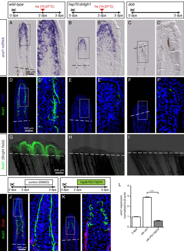
The FGF signaling pathway has been known for a long time as a major regulator of zebrafish regeneration (Lee et al., 2005; Poss et al., 2000; Whitehead et al., 2005). To investigate the role of the FGF pathway in actinotrichia regeneration, we used the transgenic fish hsp70:dnfgfr1-egfp, which overexpress a dominant negative form of the FGF receptor type 1 after heat shock induction (Lee et al., 2005). The heat shock was induced at 2 dpa, after the establishment of the normal blastema and the beginning of actinotrichia formation. The examination offins on the subsequent day, at 3 dpa, showed a reduced expression of and1 mRNA and severe suppression of the And1 deposition (Fig. 8A, B, D, E, G, H). A similar phenotype was observed after a pharmacological inhibition of the FGF signaling pathway using 10 µM PD173074. After one day of treatment, at 3 dpa, the blastema retained only rudimentary And1-positive actinotrichia in the impaired regenerate (Fig. 8J, K). A drastic reduction of and1 expression was also observed in qRT-PCR analysis after 6 h of treatment with PD173074, indicating that FGF pathway might directly regulate actinotrichia (Fig. 8L). The decrease in And1-labeled fibers after transgenic and pharmacological inhibition indicates that FGF signaling is not only necessary for de novo formation of actinotrichia, but also for the maintenance of pre-existingfibers.
To further determine the role of FGF in actinotrichia regeneration, we applied a genetic approach by using fgf20a homozygous mutantfish called devoid of blastema (dob), which fail to regenerate fins (Whitehead et al., 2005). At 3 dpa, we identified a few dob mutant fins that displayed rudimentary outgrowths. Nevertheless, the wound epidermis and mesenchyme of the protruding tissue lacked and1 expression above the amputation plane (Fig. 8C, F, I). The examination of uninjuredfins in this mutant revealed normal actinotrichia length and morphology (data not shown). Thus, fgf20a is not required for actinotrichia formation during normalfin growth, but it is essential for and1 gene expression after amputation. Taken together, the transgenic, pharmacological and genetic assays consistently demonstrate the essential role of FGF signaling for actinotrichia regeneration.
Finally, we investigated the ability offins to resume actinotrichia regeneration after transient inhibition. For the pharmacological ex-periments, thefish were exposed to the inhibitors of TGFβ/Activin-βA, IGF and FGF signaling for 2 days starting at 2 dpa, followed by another 6 days of recovery in normal conditions (Fig. S5A-C). For the transgenic overexpression of the dominant negative FGF receptor, hsp70:dnfgfr1-egfpfish were exposed to a heat shock at 2 dpa and subsequently left to regenerate without any further disturbance until 10 dpa (Fig. S5D-F). In all cases, actinotrichia resumed regeneration during the recovery period, and were similar to those of controlfins at
Fig. 8. FGF signaling is essential for actinotrichia formation and maintenance. (A-C) In-situ hybridization for and1 on 3 dpa sections in control, hsp70:dnfgfr1-egfp and dobfish. Control and dnfgfr1fish underwent heat shock (hs) at 2 dpa. N = 4 fins per group. (D-F) Immunofluorescence for And1 (green) on longitudinal sections at 3 dpa in control, hsp70:dnfgfr1-egfp, and dobfish. N = 4 fins per group. (G-I) Whole-mount immunofluorescence staining for And1 (green) overlaid with bright field images for all experimental groups. N = 3fins per group. Both, transgenic expression of dnfgfr1 and dob mutation cause a drastic decrease in and1 expression (B-C) and And1 deposition (E-F, H-I). (J, K) Immunofluorescence staining with And1 (green) and Zns5 (red) on 3 dpa longitudinal sections of fins of control fish and fish treated with PD173074 for 1 day. One day of treatment with the inhibitor of FGF signaling starting at 2 dpa is sufficient to disrupt actinotrichia in the blastema. N = 4 fins. (L) qRT-PCR analysis of and1 expression after 6 h of treatment with 10 μM of the pharmacological inhibitor of the FGF pathway, PD173074. Treatment started at 2 dpa. The relative expression was normalized to controlfins at 2 dpa. N = 3 (9 fins each). Error bars represent SEM. *** P < 0.001.
D. König et al. Developmental Biology 433 (2018) 416–432
10 dpa. We concluded that actinotrichia can be reestablished after their degradation due to transient inhibition of inductive pathways. 3. Discussion
Actinotrichia arefin-specific skeletal structures that arise by aggre-gation of collagens with other associated proteins. The unique compo-nents of these elements are Actinodin proteins, which have not been shown in any other tissue and are not conserved in tetrapods (Lalonde et al., 2016; Zhang et al., 2010). Thus, Actinodin proteins may determine the distinctive biophysical properties of actinotrichia, which are specia-lized for supporting theflattened extremity during swimming (van den Boogaart et al., 2012). Using specific antibodies, we demonstrated that two paralogous Actinodin proteins, epithelial-mesenchymal And1 and mesenchymal And2, jointly contribute to actinotrichia fibers. We identified that beside the well-aligned arrays of actinotrichia in the
subepidermal space in the distal-most ray blastema, the distal and proximal ray blastema also contains And1-labeled actinotrichia, how-ever, in a more disorganized configuration underneath the regenerating osteoblasts (Fig. 9). Interestingly, only the surface of the fibrils was immunoreactive for And1/2 antibodies. This localization suggests that actinodins are deposited on the surface of the rods, where they could play an essential role for linking mesenchymal cells to actinotrichia. On the other hand, however, it is possible that the dense molecular packing within the fibrillar core hinders the accessibility of the epitopes for immunodetection (Galloway, 1985; Hulmes et al., 1995). In addition to the extracellular localization of Actinodin, we detected these proteins in the intracellular secretory pathway. Further studies will be essential to understand the mechanisms regulating assembly of actinodins with collagens and their interaction with cell surfaces.
Previous studies of fins in various fish species suggested that structural interactions may occur between actinotrichia and
lepido-Fig. 9. Schematic representation of actinotrichia localization infins at 3 dpa. (A) Regenerating stump of a caudal zebrafish fin at 3 days post amputation (dashed line). Lepidotrichia of the stump contain bone matrix (dark brown lines), whereas regenerating bones in the outgrowth consists of activated osteoblasts (light brown) with no or little bone matrix at this time point. Actinotrichiafibers (green) are oriented longitudinally to the proximo-distal axis in the new tissue (B) Transversal (cross) sections at different proximo-distal levels of the regenerate. (C) Longitudinal sections through ray and interray tissue. Actinotrichia are located between epidermis and mesenchyme in the distal-most blastema of the rays and in the entire distal blastema of the interrays. In the presence of regenerating bones in the rays, actinotrichia are displaced from their subepidermal position into the mesenchymal compartment where they become disrupted and misaligned in the proximal part of the regenerates.
trichia (Becerra et al., 1983; Geraudie, 1988; Geraudie and Landis, 1982; Geraudie and Meunier, 1982). Here, we aimed to determine the dynamics of actinotrichia in relation to lepidotrichia during the entire fin regenerative process in the zebrafish caudal fin. Using whole-mount analysis of transgenic fish with an osteoblast reporter, we visualized that actinotrichia are the first skeletal elements that become re-established in the regenerate before bone regrowth. This observation is consistent with previous studies performed onfin sections (Duran et al., 2011; Thorimbert et al., 2015; Zhang et al., 2010). Since in the intact adult fin, the actinotrichia form the apical skeleton of the appendage, their early re-establishment in the regenerative outgrowth can be viewed as a re-establishment of the distal-most positional identity at the onset of regeneration. According to this observation, the activation of cells of the stump may involve a switch of the positional value from proximal to distal. Based on thisfinding, we propose that the emerging blastema at least partially reproduces structural features of the intactfin margin. Moreover, the examination of fins at 3 dpa revealed that the early regenerative outgrowth contains actinotrichia across the entire new tissue, including interrays, revealing a similarity to the embryonicfin fold, which is uniformly supported with parallel fibers (Dane and Tucker, 1985; Geraudie, 1977; Thorogood, 1991; van den Boogaart et al., 2012; Wood and Thorogood, 1984; Zhang et al., 2010). This correlation suggests that the initiation of adult fin regeneration might at least partially recapitulate certain developmental processes used for extremity formation during embryogenesis.
The relationship between growth and pattern formation of a vertebrate limb is a long-standing question in developmental and regenerative biology (Bryant and Gardiner, 2016; Makanae et al., 2014; Marrero et al., 2017; McCusker et al., 2015; Mescher, 2017; Towers et al., 2012). Although the size of actinotrichia dynamically varied during regeneration, the whole-mount analysis revealed a strong morphometric and spatial complementarity between both skeletal structures – actinotrichia and lepidotrichia – throughout the entire regeneration process. The correlation between both skeletal elements was also observed in another longfin (alfdty86) mutants, which contain irregular and long bony segments due to a gain-of-function mutation in the potassium channel kcnk5b (Perathoner et al., 2014). We showed that in addition to the defective lepidotrichia, the uninjured mutants displayed highly elongated actinotrichia. Moreover, the accelerated regeneration of alfdty86mutantfins was also associated with markedly elongated actinotrichia. Interestingly, disrupted actinotrichia were reported in alf dty86 and in short fin (sof b123) mutants during regeneration, probably due to alteration in the Connexin 43-dependent gene network (Bhadra and Iovine, 2015). It will be interesting to examine whether the phenotype is directly caused by the hyperpolar-ization of mutant cells or whether it arises as a secondary consequence of defective lepidotrichia.
The closer examination of longitudinalfin sections revealed that the epithelial-mesenchymal interface of the regenerating rays contains either actinotrichia or lepidotrichia-producing cells. The presence of osteoblasts was associated with a shift of actinotrichia from the subepithelial into the mesenchymal location. It is possible that regenerating osteoblasts actively change the configuration of pre-existing actinotrichia through a physical displacement and/or degrada-tion of pre-existing fibers. Moreover, osteoblasts might affect actino-trichia at the level of gene expression, namely through suppression of and1 in the lateral wound epithelium. In thefin, bone-forming cells are known to be tightly guided underneath of the basal wound epithelium (Quint et al., 2002; Smith et al., 2006; Stewart et al., 2014; Thorimbert et al., 2015; Zhang et al., 2012). The and1 transcription was down-regulated in the epithelial cells adjacent to osteoblasts, and it extended further proximally in the interray than in the ray. This observation can be explained by a potential requirement for paracrine signals from the mesenchyme, such as Fgf20a and Activin-βA (Jazwinska et al., 2007; Whitehead et al., 2005), to induce and1 expression in the overlying epithelium. A layer of bone-forming cells may form a barrier for the
penetration of inductive signals from the mesenchyme. Indeed, regen-erating osteoblasts are closely interconnected by N-cadherin-positive adherens junctions (Mateus et al., 2015), which might prevent di ffu-sion of molecules between the overlying and underlying tissues. Thus, the layer of osteoblasts may physically interrupt epithelial-mesenchy-mal signaling and extracellular matrix-cell interactions that are re-quired for the organization of actinotrichia.
The blocking of either TGFβ/Activin-βA or FGF signaling during the regenerative outgrowth phase inhibited not only de-novo synthesis of actinotrichia, but also impaired the maintenance of the pre-existing fibers. This finding supports previous observations of a high turnover of actinotrichia during regeneration (Mari-Beffa et al., 1989). In a similar experimental design, the inhibition of IGF signaling did not affect actinotrichia formation and maintenance, despite disrupting the re-growth process. This result indicates that the lack of actinotrichia formation after the inhibition of TGFβ/Activin-βA and FGF signaling are not merely side effects of the regenerative failure. This is consistent with previous studies, in which blocking of BMP signaling or Hdac1 impaired actinotrichia regeneration despite initiated dedifferentiation and partial outgrowth formation (Pfefferli et al., 2014; Thorimbert et al., 2015). Thus, actinotrichia deposition is not a default response to fin fold expansion, but it requires induction by signaling networks at the permissive epigenetic state of the cells. A recent study has reported an interesting approach to understanding the evolutionary and genetic principles of and1 gene expression, using identification of tissue-specific cis-acting regulatory sequences of this gene (Lalonde et al., 2016). The characterization of essential enhancer elements of the and1 gene opens a new perspective for studying the regulation of actino-trichia. The precise role of the signaling pathways, chromatin remodel-ing factors, matrix and cellular components durremodel-ing formation and turnover of actinotrichia provides a focus for future investigation.
4. Experimental procedures 4.1. Fish strains andfin amputations
For this study the following strains were used: AB strain (Oregon), hsp70:dnfgfr1-egfp (Lee et al., 2005), dob (devoid of blastema) (Whitehead et al., 2005), osterix:nlsGFP (OlSp7:nlsGFPzf132) (Spoorendonk et al., 2008), alf dty86 (another longfin dty86) (van Eeden et al., 1996). Adultfish were used at ages 12–36 months. For heat-shock experiments, the animals were placed in a water-bath regulated by a thermostat equipped with a cooling system (Lauda Eco), which was programed to increase the temperature from RT to 37 °C within 20 min, to maintain 37 °C for 1 h, and then to cool down to RT within 20 min. For fin amputation, fish were anesthetized in 0.6 mM tricaine (MS-222 ethyl-m-aminobenzoate, Sigma-Aldrich), and fins were amputated with a razor blade proximal to the first bone bifurcation point. Animals were kept at 27 °C for various durations beforefin collection. Fins were fixed in 2% paraformaldehyde (PFA) overnight before subsequent procedures. Live images of regenerating fins were taken with a Leica AF M205 FA stereomicroscope. Animal experimentation was approved by the Cantonal Veterinary office of Fribourg, Switzerland.
4.2. Inhibitor treatments
Drug treatments were performed in 100 mL coplin jars (up to 3fish per coplin jar). The following drugs were used: SB431542 (10 µM; Tocris), a TGFβ/Activin-βA receptor inhibitor; NVP-AEW541 (5 µM; Novartis Pharma AG), an IGF receptor inhibitor; PD173074 (10 µM; Selleckchem), an FGF receptor inhibitor. Drugs were dissolved in DMSO at 10 mM stock solution. Controlfish were kept in 100 mL coplin jars containing 0.1% DMSO.
D. König et al. Developmental Biology 433 (2018) 416–432
4.3. Immunofluorescence staining of fin sections
Fins were harvested at different time-points after amputation and fixed in 2% PFA in eppendorf tubes overnight at 4 °C. They were washed in PBS after fixation (3 × 10 min each) and transferred into Petri-dish containing a warm solution of 1.5% agarose/5% sucrose dissolved in water. After the gel solidified at RT, small blocks were cut around the embeddedfins and equilibrated in 30% sucrose solution overnight at 4 °C. Then, the blocks were mounted in tissue freezing media (Tissue-Tek O.C.T.; Sakura), cryosectioned at 16 µm thickness using a Hyrax C50 cryostat, collected on Superfrost Plus slides (Fisher) and allowed to air dry for approx. 1 h at RT. Sections were stored in tight boxes at−20 °C for future use. Before use, slides were brought to room temperature for 10 min, the area with sections was encircled with ImmEdge Pen (Vector Laboratories) to keep liquid on the slides, and left for another 10 min at RT to dry. Then, the slides were transferred to Coplin jars containing 0.3% Triton-X in PBS (PBST) for 10 min at RT. The slides were transferred to a box lined with water-soaked paper towels to make a humid chamber. Blocking solution (5% goat serum in PBST) was applied on the sections for 1 h at RT. Subsequently, the sections were covered with approx. 200 µl of primary antibody diluted in blocking solution and incubated overnight at 4 °C in the humid chamber. They were washed in PBST in coplin jars for 1 h at RT and again transferred to the humid chamber for incubation with secondary antibodies diluted in blocking solution. The slides were washed in PBST for 1 h at RT and mounted in 90% glycerol in 20 mM Tris pH 8 with 0.5% N-propyl gallate.
The following primary antibodies were used: Rabbit anti-And1 (1:2000, custom-made against the C-terminus peptide of And1 CGQDDHLAYNGDYRKK, Eurogentec), Rat anti-And2 (1:10, custom-made against the C-terminus peptide of And2 CRFEDFLRAYMGGYKK, Eurogentec), Mouse Zns5 (1:250, Zebrafish International Resource Center), Mouse α-Tubulin (1:1000, Sigma-Aldrich), Mouse anti-GM130 (1:200, BD Transduction Labs), Mouse IgM anti-chondroitin sulfate (1:2000, Sigma-Aldrich). Fluorescent dye-coupled secondary antibodies (Jackson Immunological) were used at 1:500 DAPI (Sigma-Aldrich) was used to label nuclei. Immunofluorescence and corre-sponding brightfield were imaged using a Leica SP5 confocal micro-scope. Images were analyzed and processed using ImageJ and Adobe Photoshop. Imaris was used for movies and 3D projections.
4.4. Western blot
Western blots were performed on proteins of 3 dpafin regenerates. Briefly, the regenerates from 20 fish were collected on dry ice, and homogenized in RIPA Lysis buffer (50 mM Tris–HCl pH = 8, 150 mM NaCl, 1% Triton X-100, Complete protease inhibitors (Roche)). Lysates were centrifuged to remove insoluble debris and the extracted soluble proteins were quantified with BCA kit against BSA standards, and denatured by boiling 5 min at 95 °C in sample buffer (final concentra-tions (1×): 12 mM Tris–HCl pH = 6.8, 2% SDS, 2% glycerol, 25 mM DTT). Serial dilutions of the soluble proteins (5, 15 and 45μg) were loaded twice and resolved side-by-side onto one 8% SDS/PAGE gel. Proteins were transferred onto a 0.45 µm nitrocellulose membranes (GE Healthcare), which were then cut. After blocking in a 5% milk in TBST (1xTBS, 0.1% Tween-20) solution, one half membrane was incubated with Rabbit anti-And1 antibody (1:2000 in 5% milk-TBST) overnight; the second half membrane was incubated in Rat anti-And2 antibody (1:250 in 5% milk-TBST). The next day, both membranes were washed with TBST (3 × 10 min) and incubated with secondary antibodies: HRP anti-Rabbit (1:10,000 in 5% milk-TBST, Jackson Immunoresearch) and HRP anti-Rat (1:5000 in 5% milk-TBST, Thermo Scientific) respectively. After a second series of washes, the SuperSignal™ West Femto Maximum Sensitivity Substrate revelation kit (Thermo Scientific) was applied and chemiluminescence images were acquired using a Li-cor Odyssey reader. To allow visualization of
the ladder, the bands were traced with a pen and imaged at 700 nm. Images of the two membranes were overlapped in Adobe photoshop. 4.5. Immunofluorescence staining of whole mount fins
Fins were harvested at different time-points after amputation and fixed in 2% PFA in eppendorf tubes overnight at 4 °C. They were washed in phosphate-buffered saline (PBS) after fixation, transferred to blocking solution containing 5% goat serum (Jackson Immunoresearch) in 0.3% Triton-X in PBS (PBST) for an hour at room temperature (RT). Then they were incubated in a primary antibody diluted in blocking solution overnight at 4 °C. On the next day,fins were washed in PBST (5 min, then 1 h) and incubated with a secondary antibody diluted in blocking solution for 2 h at room temperature. All the steps were done on a rocking platform. Thefins were washed again and mounted in 90% glycerol in 20 mM Tris pH 8 with 0.5% N-propyl gallate (Sigma-Aldrich).
4.6. Morphometric analysis and statistics
The software ImageJ (NIH) was used for measurements of the regenerative outgrowth and actinotrichia length on projected Z-stacks of whole mount stainings. Eachfin was represented by the average of the lengths obtained for the second, third and forth ray relative to the ventral and dorsal edges of thefin. Length of regenerates was measured by tracing and measuring a line from the amputation plane to the tip of the ray. Length of actinotrichia was measured by tracing a line from the most proximal And1-positive signal adjacent to a ray to the distal edge of the And1-positive signal. The values were then plotted in Graphpad prism. Student T-tests were performed comparing experimental groups to control group.
4.7. Histological staining of whole mountfins
Alcian Blue-Alizarin Red staining was performed according to previously published protocols (Laforest et al., 1998). Fins werefixed in 2% PFA and washed in PBS (3 × 10 min). Thefins were first stained with Alcian Blue for 6 h (0.1% Alcian blue diluted in 30% acetic acid/ 70% ethanol andfiltered). The fins were then dehydrated in a water-ethanol series (from 70% to 100% water-ethanol) and kept in water-ethanol at 4 °C overnight. On the next day, thefins were rehydrated in an ethanol-water series. They were then incubated 10 min at 37 °C in 0.5% trypsin in 2% sodium borate in water. After 5–6 rinses in water, they were then incubated 4 h in 0.1% Alizarin Red S in a 0.5% KOH solution for staining of bones. Thefins were then transferred through a 0.5% KOH-glycerol series (up to 100% KOH-glycerol). The fins were left in glycerol overnight at 4 °C and mounted in glycerol. To avoid damaging thefins, all steps were performed in glass staining plates with shallow rounded wells.
4.8. Immunohistochemistry for electron microscopy
Distalfin regenerates were harvested at 3 dpa and fixed in 4% PFA overnight at 4 °C. They were washed in PBS (3 × 5 min each), embedded in warm solution of 1.5% agarose/5% sucrose dissolved in water. After agarose solidified at RT, small blocks with fins were cut and equilibrated in 30% sucrose solution overnight at 4 °C. Then, the blocks were embedded in tissue freezing media (TBS), cryosectioned at 80 µm thickness and collected in eppendorf tubes with PBS. The sections were blocked in 5% goat serum in PBS (GS/PBS) in PBS and incubated with Rabbit anti-And1 antibody diluted at 1:2000 in 5% GS/ PBS at 4 °C overnight. The control sections were incubated with 5% GS/PBS without primary antibodies. On the next day, the sections were washed in PBS and incubated with Horse anti-Rabbit Biotinylated antibodies (Vector Laboratories) at 1:200 in GS/PBS for 2 h at room temperature. Then, the sections were washed in PBS and processed
using VECTASTAIN® ABC Kit (Vector Laboratories). The staining was performed using 3,3′-Diaminobenzidine (DAB) (Sigma-Aldrich) and H2O2 solution in Tris-buffered saline pH 7.6 solution. The sections were rinsed in PBS,fixed in 2% Glutaraldehyde in Cacodylate buffer, and processed for electron microscopy, according to the standard protocol.
4.9. In situ-hybridization
Primers for the generation of the template for the probes were as following:
and1 (NM_001197254): Fw 5′-caagacaggccttgaggaag-3′; Rev: 5′-gattgggaacttagtgggatgc-3′
and2 (NM_001111190): Fw cctgcagaacaagcacagag-3′; Rev: 5′-tggaactctgggagaactgg-3′
and3 (NM_001025511): Fw 5′-gtcacaggccacatctcttg-3′; Rev: 5′-atctgggagctctgatgacg-3′
and4 (NM_001136244): Fw 5′-tgtgtatgctgtccgtcctg-3′; Rev: 5′-gcgggtcgtagagttcagac-3′
To synthesize the anti-sense probes, a sequence of T3 RNA polymerase promoter (attaaccctcactaaagggaga) was added to the 5′ end of the reverse primers. The digoxigenin-RNA labeling mix (Roche) was used to generate the probe, which after purification was dissolved in hybridization solution and stored at−20 °C.
In-situ hybridization on wholefins and sections were performed as previously described (Chassot et al., 2016; Pfefferli and Jazwinska, 2017). Fin regenerates werefixed overnight at 4 °C in 4% PFA, then washed twice in PBS, once in PBS/methanol and in methanol for 10 min each, andfinally transferred to methanol. Fixed fins were stored at−20 °C until processed for whole-mount in-situ hybridization or for cryosectioning.
For cryosectioning,fin regenerates were rehydrated in a methanol-PBS series and embedded using the same procedure as for immuno-fluorescence. Before use, slides were brought to room temperature for 10 min, and incubated in hybridization solution (50% formamide, 5 x SSC, 1 x Denhardt's solution, 10% dextran sulfate, 0.1 mg/mL yeast tRNA; all from Sigma-Aldrich) for 1 h at RT and for 1 h at 60 °C. Then, they were placed into tight boxes with wet paper towels, and 200 µl of diluted probe in hybridization solution (1:200) was applied on each slide that was then covered with a plastic coverslip (ApopTag, Merck Millipore) and placed in an incubator at 60 °C overnight. On the next day, slides were transferred to a coplin jar and soaked for 5 min with prewarmed 5 × SSC (Saline-sodium citrate) to allow the removal of cover slips, and washed twice with 5 × SSC for 30 min and once with 0.2 × SSC for 1 h, each step at 60 °C. Thefinal wash was done with 0.2 × SSC for 5 min at RT. Slides were allowed to dry for a few minutes at RT and the area with sections was encircled with the ImmEdge Pen, and left for 15 min for drying. The slides were placed into humidified boxes, and 1× blocking reagent solution (Roche) in maleic acid buffer (100 mM Maleic Acid, 150 mM NaCl, 0.2% Tween-20, pH 7.5; all from Sigma-Aldrich) was applied on the sections for 1 h at RT. Subsequently, 250 µl of anti-dig-alkaline phosphatase (AP) antibody (Roche; diluted 1:4000 in blocking reagent solution) was applied on the sections for 2 h at RT. The slides were rinsed and washed twice for 30 min in maleic acid buffer in coplin jars. Then, the slides were washed twice in AP-buffer (100 mM Tris HCL pH 9.5, 100 mM NaCl, 50 mM MgCl2, and 0.2% Tween 20) for 5 min. To prepare the staining reagents, NBT and BCIP compounds (Roche) were diluted in AP-buffer according to manufacturer's instruction with addition of 10% Polyvinyl alcohol (Sigma-Aldrich). The staining reaction was performed in a humidified box at 37 °C. The reaction was monitored under the microscope, and was stopped by washes in coplin jars with PBS for 15 min, 70% ethanol for 1 h, and 15 min in PBS. The slides were mounted in Aquatex mounting medium (Merck Millipore).
For thefins of transgenic osterix(sp7):nlsEGFP fish, after comple-tion of in-situ hybridizacomple-tion, we continued with the standard
immuno-fluoresce staining using chicken antibodies against GFP at 1:1000 (GFP-1010, Aves Labs). Immunofluorescence and corresponding brightfield images were taken using a Leica SP5 confocal microscope and a Zeiss microscope, respectively, and were superimposed using Adobe Photoshop.
4.10. Quantitative gene expression analyses
Quantitative RT-PCR reactions were performed as previously described (Chablais and Jazwinska, 2010). The values were normalized for β-actin. The expression ratios were calculated using the Pfaffl method. The following primers were used:
and1: Fw 5′-ccgaacccgtcagtatttcc-3′; Rev: 5′-gcgtcatattgctgctggtc-3′ β-actin: Fw 5′-ttggcaatgagaggttcagg-3′; Rev 5′-tggagtt-gaaggtggtctcg-3′
Acknowledgement
We are grateful to Prof. Marco Celio (Department of Medicine, University of Fribourg), in whose lab this work was started. Special thanks to B. Scolari for electron microscopy imaging, S. Käser-Pébernard for help with the Western blot, V. Zimmermann for technical help, C. Pfefferli for critical reading of the manuscript, F. Hofmann, Novartis Pharma AG for providing NVP-AEW541, C. Bono Mestre and F. Chablais and V. Thorimbert for experimental contribu-tion. This work was supported by the Swiss National Science Foundation, grant numbers: 310030_159995 and CRSII3_147675. Competing interest
The authors declare nofinancial or non-financial competing inter-ests.
Appendix A. Supporting information
Supplementary data associated with this article can be found in the online version atdoi:10.1016/j.ydbio.2017.07.024.
References
Akimenko, M.A., Mari-Beffa, M., Becerra, J., Geraudie, J., 2003. Old questions, new tools, and some answers to the mystery offin regeneration. Dev. Dyn. 226, 190–201.
Armstrong, B.E., Henner, A., Stewart, S., Stankunas, K., 2017. Shh promotes direct interactions between epidermal cells and osteoblast progenitors to shape regenerated zebrafish bone. Development 144, 1165–1176.
Becerra, J., Junqueira, L.C., Bechara, I.J., Montes, G.S., 1996. Regeneration offin rays in teleosts: a histochemical, radioautographic, and ultrastructural study. Arch. Histol. Cytol. 59, 15–35.
Becerra, J., Montes, G.S., Bexiga, S.R., Junqueira, L.C., 1983. Structure of the tailfin in teleosts. Cell Tissue Res. 230, 127–137.
Bechara, J., Böckelmann, P., Santiago Montes, G., da Cruz-Höfling, M.A., 2003. Inhibiiton of caudalfin actinotrichia regeneration by acetylsalicylic acid (Aspirin) in teleosts. Braz. J. Morphol. Sci. 20, 67–74.
Bhadra, J., Iovine, M.K., 2015. Hsp47 mediates Cx43-dependent skeletal growth and patterning in the regeneratingfin. Mech. Dev. 138 (Pt 3), 364–374.
Birk, D.E., Brückner, P., 2011. Collagens, Suprastructures, and Collagen Fibril Assembly. In: Mecham, R.P. (Ed.), The Extracellular Matrix: an Overview. Springer-Verlag Berlin Heidelberg, Berlin Heidelberg, 77–115.
Blum, N., Begemann, G., 2015. Osteoblast de- and redifferentiation are controlled by a dynamic response to retinoic acid during zebrafish fin regeneration. Development 142, 2894–2903.
Brown, A.M., Fisher, S., Iovine, M.K., 2009. Osteoblast maturation occurs in overlapping proximal-distal compartments duringfin regeneration in zebrafish. Dev. Dyn. 238, 2922–2928.
Bryant, S.V., Gardiner, D.M., 2016. The relationship between growth and pattern formation. Regeneration.
Chablais, F., Jazwinska, A., 2010. IGF signaling between blastema and wound epidermis is required forfin regeneration. Development 137, 871–879.
Chassot, B., Pury, D., Jazwinska, A., 2016. Zebrafish fin regeneration after cryoinjury-induced tissue damage. Biol. Open 5, 819–828.
Dane, P.J., Tucker, J.B., 1985. Modulation of epidermal-cell shaping and extracellular-matrix during caudalfin morphogenesis in the zebra fish Brachydanio-rerio. J. Embryol. Exp. Morphol. 87, 145–161.
D. König et al. Developmental Biology 433 (2018) 416–432