HAL Id: hal-02982518
https://hal.archives-ouvertes.fr/hal-02982518
Submitted on 2 Nov 2020
HAL is a multi-disciplinary open access
archive for the deposit and dissemination of
sci-entific research documents, whether they are
pub-lished or not. The documents may come from
teaching and research institutions in France or
abroad, or from public or private research centers.
L’archive ouverte pluridisciplinaire HAL, est
destinée au dépôt et à la diffusion de documents
scientifiques de niveau recherche, publiés ou non,
émanant des établissements d’enseignement et de
recherche français ou étrangers, des laboratoires
publics ou privés.
β2-adrenergic signals downregulate the innate immune
response and reduce host resistance to viral infection
Elisabeth Wieduwild, Mathilde Girard-Madoux, Linda Quatrini, Caroline
Laprie, Lionel Chasson, Rafaëlle Rossignol, Claire Bernat, Sophie Guia,
Sophie Ugolini
To cite this version:
Elisabeth Wieduwild, Mathilde Girard-Madoux, Linda Quatrini, Caroline Laprie, Lionel Chasson,
et al.. β2-adrenergic signals downregulate the innate immune response and reduce host resistance
to viral infection. Journal of Experimental Medicine, Rockefeller University Press, 2020, 217 (4),
�10.1084/jem.20190554�. �hal-02982518�
BRIEF DEFINITIVE REPORT
β2-adrenergic signals downregulate the innate
immune response and reduce host resistance to viral
infection
Elisabeth Wieduwild1, Mathilde J. Girard-Madoux1, Linda Quatrini1,2, Caroline Laprie1, Lionel Chasson1, Rafa¨elle Rossignol1, Claire Bernat1,
Sophie Guia1, and Sophie Ugolini1
In humans, psychological stress has been associated with a higher risk of infectious illness. However, the mechanisms by
which the stress pathway interferes with host response to pathogens remain unclear. We demonstrate here a role for the
β2-adrenergic receptor (
β2-AR), which binds the stress mediators adrenaline and noradrenaline, in modulating host response to
mouse cytomegalovirus (MCMV) infection. Mice treated with a
β2-AR agonist were more susceptible to MCMV infection. By
contrast,
β2-AR deficiency resulted in a better clearance of the virus, less tissue damage, and greater resistance to MCMV.
Mechanistically, we found a correlation between higher levels of IFN-
γ production by liver natural killer (NK) cells and
stronger resistance to MCMV. However, the control of NK cell IFN-
γ production was not cell intrinsic, revealing a cell-extrinsic
downregulation of the antiviral NK cell response by adrenergic neuroendocrine signals. This pathway reduces host immune
defense, suggesting that the blockade of the
β2-AR signaling could be used to increase resistance to infectious diseases.
Introduction
Protection against infection is thought to be ensured principally by the host’s immune system. However, several studies have revealed the importance of neuroimmune regulation in host resistance to infections (Quatrini et al., 2018a;Rankin and Artis, 2018). Receptors for neurohormones, such as glucocorticoids, adrenaline, and noradrenaline, regulate immune cell functions in infectious diseases (Moriyama et al., 2018;Quatrini et al., 2018b;Quatrini et al., 2017). Adrenaline and noradrenaline are produced upon activation of the sympathetic nervous system and transmit signals from the brain to the peripheral tissues. They bind to adrenergic receptors (ARs) expressed by many cell types, including immune cells (Elenkov et al., 2000). Adrenergic signals can have pleiotropic effects. They have been shown to control myeloid cell migration into tissues by controlling adhesion molecule and chemoattractant expression by vascular endothelial cells (Scheiermann et al., 2012). In adaptive lymphocytes, signals mediated byβ2-ARs control lymphocyte dynamics by altering the responsiveness of chemoattractant receptors (Nakai et al., 2014). After stroke or cerebral artery occlusion, high levels of sympa-thetic activity can induce changes in the behavior of invariant natural killer (NK) T cells in the liver or NK cell counts in the spleen (Wong et al., 2011;Liu et al., 2017). Theβ2-AR pathway is
also a cell-intrinsic negative regulator of type 2 innate lymphoid cell (ILC) responses in the intestine, acting through the inhibi-tion of effector funcinhibi-tion and cell proliferainhibi-tion (Moriyama et al., 2018). However, the role of theβ2-AR pathway in viral infections in vivo is poorly understood.
Here, we dissect the role of theβ2-AR pathway in controlling early immune responses and resistance to mouse CMV (MCMV). MCMV is commonly used as a model of human CMV infection. The initial cytokine response to MCMV infection includes type 1 IFNs, IL-12, TNF-α, IL-6, and IL-18, which are produced prin-cipally by myeloid cells. These proinflammatory cytokines me-diate various antiviral effects, including NK cell activation (Biron and Tarrio, 2015). NK cells play a major role in the early innate immune response to MCMV (Lam and Lanier, 2017). Type 1 IFN enhances NK cell–mediated killing, whereas IL-12 induces IFN-γ production by these cells (Biron and Tarrio, 2015). In C57BL/6 mice, NK cells can also be directly activated through recognition of MCMV-infected cells by the activating receptor Ly49H (Dokun et al., 2001;Daniels et al., 2001;Arase et al., 2002;
Smith et al., 2002). It has recently been shown that liver-resident ILC1s also confer early host protection against MCMV infection through their IFN-γ production (Weizman et al., 2017).
...
1Aix Marseille University, Centre National de la Recherche Scientifique, Institut National de la Sant´e et de la Recherche M´edicale, Centre d’Immunologie de Marseille-Luminy, Marseille, France; 2Department of Immunology, Istituto di Ricovero e Cura a Carattere Scientifico Bambino Ges`u Children’s Hospital, Rome, Italy.
Correspondence to Sophie Ugolini:ugolini@ciml.univ-mrs.fr.
© 2020 Wieduwild et al. This article is available under a Creative Commons License (Attribution 4.0 International, as described athttps://creativecommons.org/licenses/ by/4.0/).
Here, we investigated the role of the β2-AR pathway in controlling the host response to MCMV. We found that mice treated with aβ2-AR agonist were more susceptible to MCMV infection. By contrast, β2-AR–deficient mice (Adrb2−/− mice)
produced higher levels of inflammatory cytokines and were more resistant to MCMV infection than their littermate con-trols. This phenotype was associated with a better clearance of the virus and less tissue damage in the spleen of infected mice. We analyzed the underlying regulatory mechanisms using ge-netic dissection, including conditional β2-AR depletion in lymphoid or myeloid cell subsets and bone marrow (BM) chi-mera experiments.
Results and discussion
Theβ2-AR pathway regulates host resistance to MCMV infection
Psychological distress, which is associated with the production of adrenaline and noradrenaline, has been linked to a higher risk of developing acute infectious diseases (Cohen et al., 1991;Glaser and Kiecolt-Glaser, 2005;Irwin and Cole, 2011). We assessed the potential contribution of theβ2-AR pathway to this process in a mouse model of acute MCMV infection. We first treated WT C57BL/6J mice with theβ2-AR agonist Clenbuterol for 7 d before and during the course of MCMV infection. WT mice treated with Clenbuterol in drinking water were more susceptible to MCMV infection than their untreated littermates (survival rate of 10% vs. 50%, respectively;Fig. 1 A). We investigated the importance of theβ2-AR pathway in a more physiological setting without the addition of exogenous stress hormones by comparing host resistance to MCMV inβ2-AR–deficient (Adrb2−/−) and control
(Adrb2+/+) littermates infected with a LD
50. Adrb2−/−mice were
significantly more resistant to MCMV than control Adrb2+/+mice
(survival rate of 80% vs. 40%, respectively;Fig. 1 B). These data demonstrate that theβ2-AR pathway has a deleterious effect on host resistance to acute MCMV infection.
We investigated the mechanisms involved by measuring the viral load over the course of MCMV infection in the spleen and the liver, the principal organs in which the virus replicates (Biron and Tarrio, 2015). Liver viral loads were similar in the two genotypes at 44 h, 3.5 d, and 4.5 d post-infection (pi;Fig. S1 A). In addition, a histological analysis of tissue damage 4.5 d pi revealed no difference between the livers of Adrb2−/−and control mice (Fig. S1, C and D). Consistent with this result, an analysis of serum alanine aminotransferase (ALT) activity levels, an indicator of liver disease, revealed no difference between β2-AR–deficient and WT animals (Fig. S1 E). The viral loads were also similar in the spleens of Adrb2−/−and control mice 44 h and 3.5 d pi (Fig. S1 B). However, 4.5 d pi, just before the first animals began to succumb to the disease (Fig. 1 B), we were able to identify two groups of mice: those with high viral loads (>104
copies of the viral genome/50 ng mRNA) and mice with lower viral loads (<104copies of the viral genome/50 ng mRNA;Fig. 1
C). This dichotomy is consistent with the use of an infectious dose close to the LD50to generate infections from which only
some of the mice would be expected to recover (Fig. 1 B). In the group of mice with lower viral loads, viral clearance in the
spleen was more efficient in Adrb2−/−than in control Adrb2+/+
mice (Fig. 1 C). During MCMV infection, the viral load is cor-related with the severity of tissue damage and subsequent host survival (Bukowski et al., 1984). We performed histological analyses on the spleens of these animals to determine whether these differences in viral load could explain the higher survival of mice lacking theβ2-AR. The severity of spleen lesions was scored from 0 to 4 (Fig. 1 D). In the group of mice with low viral loads, the control mice had a higher frequency of high-grade lesions, most of the scores obtained being between 2 and 4, whereas Adrb2−/− mice had scores between 0 and 1 (Fig. 1 E). Collectively, these data suggest that, in response to acute MCMV infection, theβ2-AR pathway reduces the ability of mice to clear the virus, increasing the severity of virus-induced lesions in the spleen and reducing host resistance to the disease.
Theβ2-AR signaling pathway downregulates the inflammatory cytokine response to MCMV
We then decided to analyze the mechanisms involved in the greater resistance of Adrb2−/−mice to MCMV infection, by an-alyzing their immune system at steady state and after infection. We first investigated the role of theβ2-AR pathway in hema-topoietic development and homeostasis by analyzing the dis-tribution of major immune cell subsets in the spleen, liver, and blood ofβ2-AR–deficient (Adrb2−/−) and control (Adrb2+/+) mice
at steady state. Adrb2+/+and Adrb2−/−mice had similar numbers
of neutrophils, monocytes, and T, B, and NK cells, in the blood (data not shown). Adrb2+/+ and Adrb2−/− mice also had similar
frequencies of NK, B, T, and NK T cells, eosinophils, neutrophils, monocytes, and dendritic cells (DCs) in the spleen, as well as NK, B, and T cells in the liver (Fig. S1, F and G). These results are consistent with previous studies (Sanders et al., 2003) and show thatβ2-AR deficiency does not impair the development or homeostasis of major immune cell subsets in homeostatic conditions.
We then investigated whetherβ2-AR signaling regulated the host immune response to MCMV infection. Depending on the cell type and the pathological context, theβ2-AR pathway can regulate immune cell trafficking, survival, or proliferation (Wong et al., 2011;Nakai et al., 2014;Liu et al., 2017;Moriyama et al., 2018). We thus analyzed the distribution of immune cells in the spleen, liver, and blood of infected mice. We detected no differences between Adrb2−/−and Adrb2+/+control mice in terms
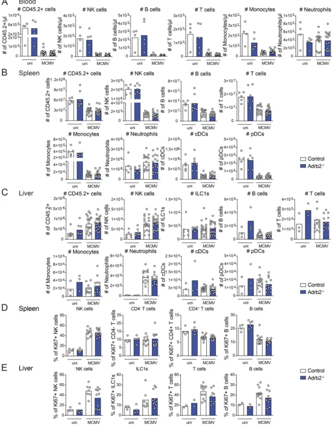
of the distribution of the major subsets of immune cells in these tissues upon MCMV infection (Fig. S2, A–C). Moreover, Ki67 expression revealed no difference in the proliferation of B, T, and NK cells and ILC1s between Adrb2−/− mice and Adrb2+/+
control mice (Fig. S2, D and E).β2-AR signaling is, therefore, not required for the early modulation of immune cell trafficking or proliferation of the main immune cell subsets in the context of MCMV infection. We then investigated whether immune cell functions were regulated by this pathway. We monitored the early cytokine response induced by MCMV infection in these animals at 44 h, 3.5 d, and 4.5 d pi. The levels of CCL3, CCL2, and IL-12p70 increased similarly in the blood of Adrb2−/−mice and Adrb2+/+control mice, peaking at 44 h pi (Fig. S3 A). By contrast,
levels of CXCL1, TNF-α, IL-6, and IFN-γ production were higher
in the bloodstream of infected Adrb2−/−mice than in that of their control Adrb2+/+littermates 44 h pi (Fig. 2 AandFig. S3 A). IL-10
levels, which are usually low at 44 h pi, were also slightly higher at this time point (Fig. 2 AandFig. S3 A).
These data show that the β2-AR signaling pathway is re-quired to control the magnitude of the early innate inflamma-tory response to MCMV, particularly in terms of the production of CXCL1, TNF-α, IL-6, and IFN-γ.
β2-AR expression in LysM+myeloid cells is not required for
control of the inflammatory cytokine response to MCMV Early in MCMV infection, TNF-α, IL-6, IL-10, and CXCL1 are produced, principally by myeloid cells (Biron and Tarrio, 2015). Previous in vitro studies suggested a potential role of β-AR pathways in polarizing BM-derived macrophages toward an
anti-inflammatory phenotype (Lamkin et al., 2016). We inves-tigated whether TNF-α, IL-6, CXCL1, and IL-10 levels were modified by the intrinsic regulation of the adrenergic pathway in myeloid cell subsets in vivo in the context of MCMV infection. We studied LysMCre/+Adrb2flx/flx mice (hereafter referred to as
Adrb2LysMCremice), in which the Adrb2 gene is selectively deleted
in LysM+cells, including neutrophils, macrophages, monocytes,
and DC subsets (Abram et al., 2014) and their littermate controls (LysM+/+Adrb2flx/flx). MCMV infection induced similar increases
in CXCL1, IL-6, TNF-α, IL-10, IL-12p70, CCL2, and CCL3 levels in the blood of Adrb2LysMCreand control mice at 44 h pi (Fig. 2 Band
Fig. S3 B). The stronger chemokine and cytokine responses to MCMV infection observed inβ2-AR–deficient mice (Fig. 2 A) are not, therefore, due to an intrinsic regulation of LysM+myeloid cell
functions by β2-AR. In addition, resistance to MCMV infection
Figure 1. β2-AR signaling regulates viral clearance and resistance to MCMV infection. (A) Survival rate of WT mice infected with MCMV at LD50. Mice were treated (filled brown circles) or not (empty black circles) with Clenbuterol in drinking water during 7 d before infection and throughout the experiment (pool of two independent experiments; Mantel-Cox test, *, P < 0.05). (B) Survival rate of Adrb2−/−mice (filled blue circles) and control Adrb2+/+littermates (empty black circles) after infection with MCMV at LD50(pool of three independent experiments; Mantel-Cox test, *, P < 0.05). (C) Viral titers in the spleens of Adrb2−/−mice (filled blue bars) and control Adrb2+/+littermates (empty bars) at 4.5 d pi. Mice were grouped according to high (left) and low (right) viral loads (pool of two independent experiments; each point represents one mouse; unpaired t test, *, P < 0.05). (D) H&E staining of spleen sections after MCMV infection at LD50. Histopathological lesions were scored from grade 0 to 4, and a color code was attributed to each score. Scale bars = 50 µm. (E) Histo-pathological analysis of the spleens of infected Adrb2−/−and control Adrb2+/+littermates 4.5 d pi with MCMV at LD
50. Scoring is based on the grading and color code shown in D. The mice were divided into two groups according to their viral loads as shown in B. The frequency of mice with a given pathological score is shown for each group of mice (pool of two independent experiments; n = 12–14 per group).
was similar in Adrb2LysMCreand control mice (Fig. 2 C), showing
thatβ2-AR signals in LysM+cells are not required for the
modu-lation of host resistance to infection.
β2-AR controls IFN-γ production by NK cells in an organ-specific manner
NK cells and liver-resident ILC1s play a major role in the response to MCMV infection through their effector functions, which in-clude cytotoxicity and early IFN-γ production (Vivier et al., 2008;
Biron and Tarrio, 2015;Weizman et al., 2017). IFN-γ was among
the cytokines upregulated in Adrb2−/− mice relative to their Adrb2+/+littermates at 44 h pi (Fig. 2 A). Transcriptomic analysis
showed that Adrb2 was expressed in NK cells and ILC1s, both at steady state and 1 d after MCMV infection (see Immunological
Genome Project database:http://www.immgen.org/databrowser). Moreover, in this issue, Diaz-Salazar et al. found that Adrb2 expression in NK cells was upregulated after MCMV infection and revealed that NK cells localize near splenic adrenergic neurons during infection. Previous studies have suggested that signaling via β-AR can regulate both the trafficking and ac-tivity of NK cells (Kradin et al., 2001;Pedersen et al., 2016;De Lorenzo et al., 2015;Tarr et al., 2012). However, most of these studies used a pharmacological blockade of the adrenergic pathway, making it difficult to determine whether the effects observed were cell intrinsic or extrinsic. We assessed the functionality of β2-AR in NK cells by studying the effect of noradrenaline on NK cell activation in vitro. We found that noradrenaline inhibited the NK cell degranulation (CD107a
Figure 2. β2-AR signals downregulate the cytokine and chemokine responses to MCMV. (A) Concentrations of CXCL1, TNF-α, IL-6, IL-10, MIP-1a (CCL3), MCP1 (CCL2), IL-12p70, and IFN-γ in the serum of Adrb2−/−(filled blue circles) and control Adrb2+/+littermates (empty circles) at 44 h pi (pool of three in-dependent experiments; each point represents one mouse; unpaired t test, *, P < 0.05). (B) Concentrations of CXCL1, TNF-α, IL-6, and IL-10 in the serum of Adrb2LysMCremice (filled green circles) and controls (empty circles) at 44 h pi (pool of three independent experiments; each point represents one mouse). (C) Survival of Adrb2LysMCremice (filled green circles) and control littermates (empty circles) after infection with MCMV at LD
50(pool of three independent experiments). uni, uninfected mice; n.s., not significant.
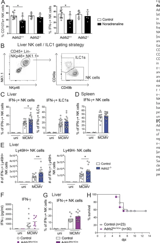
expression) and IFN-γ production induced by stimulation with plate-bound anti-NK1.1 antibodies (Fig. 3 A). This inhibition was strictlyβ2-AR dependent, as it was not observed when NK cells were isolated from Adrb2−/−mice. We then investigated the in vivo role of this pathway in the context of MCMV in-fection. The cellular source of the higher levels of IFN-γ pro-duction observed in Adrb2−/− mice was investigated by intracellular staining on innate lymphocytes from infected mice. The frequency of IFN-γ–producing NK cells in the liver
was higher in Adrb2−/−mice (32%) than in their Adrb2+/+
lit-termates (24%;Fig. 3 C). This regulation of IFN-γ production by theβ2-AR pathway was both cell type and organ specific, as it was not observed for liver ILC1s, liver NK T cells, or spleen NK cells (Fig. 3, C and DandFig. S3 C). MCMV infection also induced a similar increase in granzyme B (GzB) levels in NK cells and ILC1s from the spleen and liver of Adrb2−/−mice and control Adrb2+/+littermates (Fig. S3, C and D), suggesting that
β2-AR does not regulate the cytotoxic functions of NK cells.
Figure 3. β2-AR signals regulate IFN-γ pro-duction by liver NK cells. (A) Splenocytes from Adrb2+/+ and Adrb2−/− littermates were stimu-lated in vitro with anti-NK1.1 mAb-coated plates in the presence (filled black bars) of noradrena-line (10 µM) or diluent (control, empty bars). The frequency of CD107a (left) and IFN-γ–producing NK cells (right) 4 h after stimulation is shown (each point represents one mouse; means ± SEM are shown; unpaired t test, *, P < 0.05). (B) Gating strategy used to identify NK cells (CD45+, CD3−, CD8−, CD19−, Ly6G−, NKp46+, NK1.1+, CD49a−, CD49b+) and ILC1s (CD45+, CD3−, CD8−, CD19−, Ly6G−, NKp46+, NK1.1+, CD49a+, CD49b−) in the liver. (C and D) Frequency of IFN-γ–producing NK cells and ILC1s in the liver (C) and spleen (D) of Adrb2−/−mice (filled blue bars) and control Adrb2+/+littermates (empty bars) at 44 h pi (pool of three independent experiments; each point represents one mouse; unpaired t test, *, P < 0.05). (E) Frequency of IFN-γ–producing Ly49H+ (left) and Ly49H− (right) NK cells in the liver of Adrb2−/−mice (filled blue bars) and control Adrb2+/+ littermates (empty bars) at 44 h pi (pool of three independent ex-periments; each point represents one mouse; unpaired t test, *, P < 0.05; **, P < 0.005). (F) Concentration of IFN-γ in the serum of Adrb2Ncr1iCremice (filled purple circles) and con-trol littermates (empty circles) at 44 h pi (pool of three independent experiments; each point represents one mouse). (G) Frequency of IFN-γ–producing NK cells in the liver of Adrb2Ncr1iCre mice (filled purple bars) and control littermates (empty bars) at 44 h pi (pool of three independent experiments; each point represents one mouse). (H) Survival rate of Adrb2Ncr1iCremice (filled purple cir-cles) and control littermates (empty circir-cles) after infection with MCMV at LD50(pool of four inde-pendent experiments). uni, uninfected.
In C57BL/6J mice, host protection against MCMV involves recognition of the virus-encoded glycoprotein m157 by the Ly49H molecule, which is expressed on the surface of a subset of NK cells (Dokun et al., 2001;Daniels et al., 2001;Arase et al., 2002;Smith et al., 2002). We analyzed the role of the β2-AR pathway in the activation program of this subset. The Ly49H+
NK cell counts were similar in the spleen and liver of Adrb2−/− and control Adrb2+/+littermates at 44 h after MCMV infection
(Fig. S3 E). Moreover, the higher frequency of IFN-γ–producing
NK cells in the liver of Adrb2−/−mice at 44 h pi involved both the Ly49H-positive and -negative subsets (Fig. 3 E). These data show that β2-AR signaling does not selectively affect m157-Ly49H–mediated NK cell activation during the early stages of MCMV infection. However, in adoptive transfer experiments,
Diaz-Salazar et al. (2020)demonstrated that this pathway could be important to elicit robust adaptive NK cell responses at later time points.
The differential effect ofβ2-AR on the control of IFN-γ pro-duction in spleen and liver NK cells raised questions concerning the intrinsic or extrinsic nature of this regulation. We analyzed the MCMV response in Ncr1Cre/+Adrb2flx/flx mice (hereafter
re-ferred to as Adrb2Ncr1iCremice), in which the Adrb2 gene is deleted
selectively in NCR1+cells, including NK cells and ILC1s (
Narni-Mancinelli et al., 2011). By contrast to the phenotype observed in Adrb2−/−mice, no differences in serum IFN-γ levels or the fre-quency of IFN-γ–producing NK cells were observed between Adrb2Ncr1iCremice and their littermate controls (Ncr1Cre/+Adrb2+/+;
Fig. 3, F and G). Furthermore, the survival rates were similar for infected Adrb2Ncr1iCre and control mice (Fig. 3 H). Thus, upon
MCMV infection,β2-AR selectively downregulates IFN-γ pro-duction by liver NK cells but not by spleen NK cells. This organ-specific regulation is NK cell extrinsic and results in a systemic decrease in IFN-γ levels in the serum, potentially affecting host resistance to infection.
β2-AR signaling in hematopoietic and nonhematopoietic cells differentially regulates the cytokine response to MCMV We found thatβ2-AR signaling downregulated the early cyto-kine and chemocyto-kine response to MCMV (Fig. 2 A). However, the conditional deletion of Adrb2 in LysM+ or NCR1+cells was not
sufficient to modulate the production of IL-6, TNF-α, CXCL1, IFN-γ, or IL-10 or to affect mouse susceptibility to MCMV in-fection (Fig. 2 Band Fig. 3, F–H). The early host response to MCMV involves both hematopoietic and nonhematopoietic cells (Loewendorf and Benedict, 2010). To further investigate the regulatory mechanisms underlying the enhanced immune re-sponse and resistance of MCMV-infected Adrb2−/− mice, we
performed BM chimera experiments. First, CD45.1-WT BM cells were transplanted into lethally irradiated CD45.2-Adrb2+/+
or -Adrb2−/− recipients from the same litter. Chimeric (WT→Adrb2+/+) and (WT→Adrb2−/−) mice were infected, and
their production of chemokines and cytokines was monitored by analyzing blood samples collected 44 h after MCMV infec-tion. The production of CXCL1, TNF-α, IL-10, and IL-6 increased similarly in both types of chimera, indicating that β2-AR expression in nonhematopoietic radioresistant cells is not sufficient to modulate the expression of these cytokines and
chemokines (Fig. 4 A). By contrast, a defect ofβ2-AR signaling in nonhematopoietic radioresistant cells was sufficient to en-hance the systemic production of IL-12p70 and IFN-γ (Fig. 4 A). We then generated chimeras in which BM cells from CD45.2-Adrb2+/+or -Adrb2−/−littermates were transplanted into
irra-diated CD45.1-WT hosts. After MCMV infection, the levels of TNF-α and IL-6, but not of IL-10, CXCL1, IL-12p70, or IFN-γ, were higher in the blood of mice with BM-derived cells lacking β2-AR (Adrb2−/−→WT mice) than in control (Adrb2+/+→WT)
mice (Fig. 4 B). These BM chimera experiments show that the deletion ofβ2-AR in BM-derived hematopoietic cells is neces-sary and sufficient to upregulate TNF-α and IL-6 production upon MCMV infection. We found no difference in TNF-α and IL-6 levels in the Adrb2LysMCre mice (Fig. 2 B), demonstrating
that the regulation of cytokine production byβ2-AR is not in-trinsic to LysM+cells. LysM-cre promotes significant deletion in
macrophages and neutrophils but not in DC subsets, including plasmacytoid DCs (pDCs;Abram et al., 2014). One of the earliest sources of TNF-α during MCMV infection is the pDCs (Biron and Tarrio 2015). A role of the β2-AR on LysM− DCs might,
therefore, account for the higher levels of IL-6 and TNF-α production in (Adrb2−/−→WT) chimeras and in Adrb2−/−
mice. By contrast, the regulation of CXCL1 and IL-10 required the expression of β2-AR in both hematopoietic and non-hematopoietic cells (Fig. 2 AandFig. 4, A and B). We also found that the regulation of IFN-γ levels required the expression of β2-AR on nonhematopoietic cells (Figs. 2 Aand4, A and B). In BM-chimera settings, the stronger IFN-γ response was associ-ated with higher levels of IL-12p70 production. IL-12p70 was not significantly upregulated in conditions of complete Adrb2 knockout, although there was a trend in this direction (Fig. 2 A). Therefore, we cannot exclude a role of IL-12 in the upre-gulation of IFN-γ levels in the absence of β2-AR regulation. Nevertheless, our data suggest that theβ2-AR pathway modu-lates the expression/production of other, still unidentified, factors in nonhematopoietic cells that are involved in upregu-lating the IFN-γ response.
Collectively, these data show that β2-AR signals down-regulate the early inflammatory cytokine response to MCMV through a combination of pleiotropic effects in both BM-derived hematopoietic cells and nonhematopoietic cells.
The higher resistance ofβ2-AR–deficient mice to MCMV is associated with higher IFN-γ levels and is partially NK cell dependent
We investigated the mechanism by which theβ2-AR pathway regulates host susceptibility to MCMV by monitoring mouse survival in BM-chimera animals displaying differential regula-tion of the inflammatory cytokines TNF-α, IL-6, IL-12p70, and IFN-γ (Fig. 4). The upregulation of IL-6 and TNF-α levels in (Adrb2−/−→WT) chimeras was not associated with greater re-sistance to MCMV infection, as the survival of these animals was similar to that of (Adrb2+/+→WT) mice (Fig. 5 A). By contrast, the
deletion ofβ2-AR in nonhematopoietic recipient cells, enhanc-ing the production of IL-12p70 and IFN-γ (Fig. 4 A), was suffi-cient to increase host resistance to MCMV infection, as the survival of (WT→Adrb2−/−) chimeras (82%) was greater than
that of the corresponding (WT→Adrb2+/+) controls (20%;Fig. 5
B). Together, these data show that the higher levels of IFN-γ
production in Adrb2−/−mice and BM chimeras were selectively associated with stronger resistance to MCMV (Fig. 1 B,Fig. 2 A,
Fig. 4, A and B; andFig. 5, A and B). The levels of the other in-flammatory cytokines/chemokines (CXCL1, IL-10, IL-6, and TNF-α) for which increases were observed in Adrb2−/− mice
(Fig. 2 A) were not regulated by the same mechanisms and were not associated with different survival rates in BM chimeras (Fig. 1 B,Fig. 2 A,Fig. 4 A, andFig. 5, A and B).
These results suggest that the control of IFN-γ levels by β2-AR signals may affect host survival in this model. As liver NK cells were the main source of this increase in IFN-γ production in Adrb2−/−mice (Fig. 3, C and DandFig. S3 C), we investigated the possibility of NK cells being responsible for this greater re-sistance to infection. Adrb2−/−mice were treated with an NK1.1-depleting mAb 2 d before MCMV infection (Fig. 5 C). 2 d after anti-NK1.1 mAb treatment, the frequency of NK cells had de-creased by 93%, 76%, and 88% in the blood, spleen, and liver, respectively. Anti-NK1.1 mAb treatment greatly decreased the resistance of Adrb2−/− mice to MCMV, as the survival of NK
cell–depleted Adrb2−/− mice (40%) was lower than that of NK
cell–sufficient Adrb2−/−mice (70%;Fig. 5 C). However, NK cells
were not the only cell type contributing to the higher resistance of Adrb2−/− mice to infection, as NK cell–depleted control (Adrb2+/+) animals remained more susceptible to MCMV than
did NK cell–depleted Adrb2−/−mice (Fig. 5 C). The frequency of
ILC1 in the liver also decreased by 96% upon NK1.1 depletion. We cannot, therefore, rule out a role for this cell type in the phe-notype observed upon anti-NK1.1 mAb treatment. However, the observation of a specific increase in the number of IFN-γ–producing liver NK cells but not ILC1s in infected Adrb2−/−
mice relative to their control littermates (Fig. 3) favors a model in which NK cells play a major role.
β2-AR signals controlling IFN-γ production and host susceptibility to MCMV are not induced through catecholaminergic innervation
We investigated whether the host response to MCMV was controlled by regulation of theβ2-AR pathway by noradrenergic neurons in tissue or systemically by blood catecholamine levels. Mice were systemically treated with the catecholaminergic neurotoxin 6-hydroxydopamine (6-OHDA) for this purpose (Fig. 5 D). 6-OHDA treatment is used to selectively target nor-adrenergic neurons without affecting the adrenal medulla or plasma adrenaline levels (Clark et al., 1972;Tsunokuma et al., 2017). As expected, 6-OHDA treatment induced the ablation of tyrosine hydroxylase (TH)+ sympathetic fibers in both the
spleen and the liver (Fig. 5 E; data not shown). Control (PBS-treated) and 6-OHDA–treated mice then received injections of anti-NK1.1 antibody or an isotypic control and were infected with MCMV (Fig. 5 D). In mice treated with the isotype control antibody, sympathetic denervation did not affect IFN-γ levels in the bloodstream 44 h after MCMV infection (Fig. 5 F). By con-trast, NK1.1 depletion induced a large decrease in IFN-γ levels, confirming that NK1.1+cells are the main source of systemic
IFN-γ at this time point (Fig. 5 F). Thus, catecholaminergic neurons are not involved in the regulation of IFN-γ production by NK1.1+
cells in MCMV-infected mice.
Consistent with a major role of IFN-γ in this model, sympa-thetic denervation did not affect survival in mice treated with the isotype control antibody (Fig. 5 G, black lines). Moreover, NK1.1+cell depletion increased susceptibility to infection
simi-larly in PBS-treated and 6-OHDA–treated mice (Fig. 5 G, purple
Figure 4. Differential regulation of the inflammatory cytokine response byβ2-AR signaling in hematopoietic and nonhematopoietic cells. (A) Concentration of CXCL1, TNF-α, IL-6, IL-10, IL-12p70, and IFN-γ in the serum of (WT→Adrb2−/−) BM-chimeras (filled red circles) and (WT→Adrb2+/+) BM-chimeras (empty red circles) at 44 h pi (pool of three independent experiments; each point represents one mouse; unpaired t test, *, P < 0.05; **, P < 0.005). (B) Concentration of CXCL1, TNF-α, IL-6, IL-10, IL-12p70, and IFN-γ in the serum of (Adrb2−/−→WT) BM-chimeras (filled orange circles) and (Adrb+/+→WT) BM-chimeras (empty orange circles) at 44 h pi (pool of three independent experiments; each point represents one mouse; unpaired t test, *, P < 0.05; **, P < 0.005). Data obtained for uninfected mice (uni) are shown.
Figure 5. β2-AR signaling in nonhematopoietic cells regulates resistance to MCMV infection independently of catecholaminergic innervation. (A) Survival rate of (Adrb2−/−→WT; filled orange circles), (Adrb2+/+→WT; empty orange circles), and (Adrb2−/−→Adrb2−/−; filled black circles) BM chimeras after MCMV infection at LD50(pool of one to two experiments). (B) Survival rate of (WT→Adrb2−/−; filled red circles), (WT→Adrb2+/+; empty red circles), and (Adrb2−/−→Adrb2−/−; filled black circles) BM chimeras after MCMV infection at LD
50(pool of one to two experiments; Mantel-Cox test, *, P < 0.05). (C) Survival rate of Adrb2−/−mice (filled circles) and control littermates (empty circles) with (blue circles) or without (black circles) NK cell depletion with anti-NK1.1 mAb treatment 2 d before MCMV infection (pool of three independent experiments; Mantel-Cox test, *, P < 0.05; **, P < 0.005). (D) Experimental design for the experiments presented in E, F, and G. (E) Immunofluorescence analysis of liver sections from mice treated with 6-OHDA or PBS as control. Endothelial cells from blood vessels were stained with anti-CD31 antibody (left, white staining; right, green staining in overlaid images). TH+nerves were stained with anti-TH antibodies (middle, white staining; right, red staining in overlaid images; scale bars = 50 µm). (F) Serum IFN-γ concentrations of 6-OHDA–treated mice (filled
lines). Thus, sympathetic innervation does not contribute to the increased host resistance to MCMV infection observed in Adrb2−/−mice.
Collectively, these data support a model (Fig. 5 H) in which catecholamines produced by the adrenal gland and released systemically in the bloodstream act on nonhematopoietic cells in tissues viaβ2-AR to modulate proinflammatory signals. These proinflammatory signals are important for NK cell activation, and their modulation affects the NK cell IFN-γ response, which is necessary for efficient viral clearance. The modulation of the systemic IFN-γ response by this adrenergic pathway reduces the control of viral replication and increases the severity of tissue lesions, especially in the spleen, decreasing host resistance to infection.
Concluding remarks
This study highlights the mechanisms by which the stress pathway can increase host susceptibility to viral infection. Stimulation of theβ2-AR pathway was found to be detrimental for host survival to MCMV infection, suggesting that the stress mediators adrenaline and noradrenaline have a negative impact on host resistance to infection. Consistent with this hypothesis, β2-AR deficiency resulted in a higher resistance to infection, which was associated with stronger IFN-γ responses in liver NK cells. This stronger response inβ2-AR–deficient mice was as-sociated with a better control of viral replication and less severe tissue damage. Moreover, NK cell depletion reduced the survival of β2-AR–deficient mice. However, the regulation of IFN-γ production in liver NK cells was not cell intrinsic and involved β2-AR expression in radio-resistant nonhematopoietic cells. These results are consistent with previous studies showing that host susceptibility to infection involves, not only the host im-mune system, but also the ability of parenchymal tissues to tol-erate or to react to pathogen-induced dysfunctions (Medzhitov et al., 2012;Soares et al., 2017). Further studies are required to investigate in greater detail the contribution of β2-AR in non-hematopoietic cell types, particularly in the liver.
The role ofβ2-AR appears to be different at different stages of MCMV infection. Indeed, we show that this pathway is a cell-extrinsic negative regulator of NK cell IFN-γ production at early stages of infection, with a cost in terms of host resistance. By contrast, at later stages, cell-intrinsic adrenergic signaling can be protective and promotes the adaptive response and expansion of the NK cell population in the spleen (Diaz-Salazar et al., 2020). This secondary level of regulation may account, at least in part, for the maintenance of this pathway during evolution.
Interestingly,Diaz-Salazar et al. (2020)observed a modest intrinsic role of β2-AR in modulating early IFN-γ production upon MCMV infection. This role was revealed in the context of
mixed-BM chimera experiments in whichβ2-AR–deficient NK cells are in competition with WT NK cells for their development and activation. This phenotype was associated with a defect in the maturation status of β2-AR–deficient NK cells compared withβ2-AR–sufficient NK cells present in the same recipient. Such functional and maturation defects were not observed in Adrb2Ncr1iCremice (Fig. 3 G;Diaz-Salazar et al., 2020), suggesting
that the role ofβ2-AR signaling in NK cells is context dependent. Consistent with this hypothesis, the β2-AR can be coupled to different intracellular pathways depending on its state when activated, inducing different intracellular responses upon ligand binding (Matera et al., 2018). For example, the engagement of theβ2-AR on T and B lymphocytes regulates their function ac-cording to the molecular signaling pathway activated, the cy-tokine microenvironment, and the time of receptor engagement in relation to the activation and differentiation state of the cell (Sanders, 2012). This complexity may explain some of the con-troversies in the literature suggesting apparently conflicting functions ofβ2-ARs in immune cells (Sanders, 2012;Wu et al., 2018).
Clinical studies revealed that psychological stress is associ-ated with a higher risk of developing acute infectious illness (Cohen et al., 1991;Glaser and Kiecolt-Glaser, 2005;Irwin and Cole, 2011). It will be important to determine whether these effects are at least partly mediated byβ2-AR signals and to de-termine whether β-blocker treatment might be beneficial in some circumstances.
This study expands our understanding of host protection from infectious diseases by showing that the stress pathway, by triggeringβ2-AR signals, downregulates the innate inflamma-tory response, affecting host fitness.
Materials and methods
Mice
C57BL/6J Ly5.2 mice were purchased from Janvier Labs; C57BL/6J Ly5.1 mice were purchased from Charles River; and Adrb2LoxP/LoxP
mice and Adrb2−/−mice (Hinoi et al., 2008;Chruscinski et al.,
1999) were kindly provided by Nicolas Glaichenhaus (Institut de Pharmacologie Mol´eculaire et Cellulaire, Nice, France). Ncr1iCremice (Narni-Mancinelli et al., 2011) were obtained from
Eric Vivier (Centre d’Immunologie de Marseille Luminy, Mar-seille, France). LysMCremice (Clausen et al., 1999) were kindly
provided by Toby Lawrence (Centre d’Immunologie de Mar-seille Luminy, MarMar-seille, France). All the mice were bred and maintained under specific pathogen–free conditions at the Centre d’Immunophenomique in Marseille and the Centre d’Immunologie de Marseille Luminy. Mice were housed under a standard 12 h/12 h light-dark cycle with food and water ad
circles) and control mice (empty circles) with NK1.1 cell depletion (purple circles) or without NK1.1 cell depletion (black circles) after infection with MCMV at LD50(pool of two independent experiments; each point represents one mouse; Mann-Whitney U test, *, P < 0.05; **, P < 0.05). (G) Survival rate of 6-OHDA treated mice (filled circles) and control mice (empty circles) with NK1.1 cell depletion (purple circles) or without NK1.1 cell depletion (black circles) after infection with MCMV at LD50(pool of two independent experiments; Mantel-Cox test, *, P < 0.05). (H) Model:β2-AR signaling induced by catecholamines produced systemically in the blood circulation act on nonhematopoietic cells to modulate proinflammatory signals. This modulation downregulates the NK cell IFN-γ response, which is necessary for efficient viral clearance. This pathway reduces the control of viral replication, increases the severity of spleen lesions, and dampens host resistance to infection. uni, uninfected.
libitum. Age matched (8–12-wk-old) female mice were used. All experiments were conducted in accordance with institutional committee recommendations (Comit´e d’Ethique de Marseille no. 14-APAFiS; no. 14260) and French and European guidelines for animal care.
Clenbuterol treatment
Clenbuterol (catalog no. C5423; Sigma-Aldrich) was added at 9 µg/ml in the drinking water of mice for 7 d before and during MCMV infection. Controls were kept on water alone.
Organ preparation
Blood was taken through the retro-orbital sinus. The mice were then euthanized, perfused with 10–20 ml PBS 1×, and the spleen and liver were taken. The spleen and liver were smashed through 70-µm cell strainers. Red blood cell lysis was performed with the Red Blood Cell Lysis Buffer from eBioscience on spleen suspensions. Liver lymphocytes were isolated on a 37.5%–67.5% Percoll gradient. Blood was used for Trucount (BD Biosciences) according to the manufacturer’s protocol.
Flow cytometry
Single-cell suspensions were incubated with the Fc blocking antibody (2.4G2) and with fixable blue dead cell stain kit (In-vitrogen). To stain surface molecules the following antibodies were used: anti-CD3 (145-2C11), anti-CD11b (M1/70), anti-CD19 (1D3), anti-CD45.1 (A20), anti-CD45.2 (104), anti-CD49a (Ha31/ 8), anti-Ly6C (AL-21), anti-Ly6C/Ly6G (RB6-8C5), anti-Ly49H (3D10), MHCII (M5/114.15.2), NK1.1 (PK136), and anti-TCRβ (H57-597) from BD Biosciences; NKp46 (29A1.4), anti-CD49b (DX5 or HMa2), anti-F4/80 (BM8), and anti-Ly49H (3D10) from eBioscience; and anti-CD11c (N418) and anti-Ly6G (1A8) from Biolegend. For intracellular staining, the cells were fixed and permeabilized with an intracellular staining kit (eBioscience), and the following antibodies were used: anti-IFN-γ (XMG1.2) from Biolegend, anti-Ki67 (B56) from BD Biosciences, and anti-GzB (GB12) from Life Technologies. Analysis was per-formed with FlowJo Software.
Cytokine analysis
For serum, blood was collected from the retro-orbital sinus of MCMV-infected mice under low stress conditions (i.e., within 2 min of handling). The concentration of IL-6, IL-10, IL-12p70, MIP-1α, MCP-1, KC, TNF-α, and IFN-γ were assessed by cyto-metric bead array according to the manufacturer’s protocol (BD Biosciences).
MCMV infection
MCMV (Smith strain) was diluted in DMEM and injected in-traperitoneally in female mice at the LD50. DMEM only was
injected in the uninfected control group. All MCMV injections were done between 2 pm and 4 pm. For survival experiments, the mice were weighed every 24 h and checked for signs of distress. For the time point experiments, blood was taken via the retro-orbital sinus, mice were euthanized and perfused, and organs were harvested for further analysis. For the NK cell de-pletion experiments, 100 µg of anti-NK1.1 (PK136) from bioXcell
or vehicle were injected via the retro-orbital sinus 2 d before MCMV infection.
In vitro NK cell activation
Splenocyte suspensions were distributed in a 96-well 2HB Im-mulon plate precoated with antibody against NK1.1 (PK136; 27 µg/ml) in the presence of noradrenaline (10 µM) or diluent (control). Cells were activated in the presence of monensin (GolgiStop; BD Biosciences) in complete medium RPMI 1640 (Gibco/Invitrogen) supplemented with 10% fetal calf serum, 1 mM sodium pyruvate, 10 mM Hepes, penicillin (100 U/ml), and streptomycin (100 mg/ml). After 4 h at 37°C, cell-surface staining was performed. For intracellular IFN-γ staining, cells were fixed with 2% paraformaldehyde and permeabilized with Perm/Wash solution (BD PharMingen).
Viral titer and quantitative real-time PCR
Organs were kept in RNAlater (Qiagen) after harvesting. RNA was extracted from organ homogenates with the RNeasy Fibrous Tissue Mini Kit (Qiagen) and reverse transcribed with the iScript cDNA Synthesis kit (Bio-Rad Laboratories). Viral titers were determined, by quantitative PCR, as absolute levels of the Ie1 gene (forward: 59-GAGTCTGGAACCGAAACCGT-39; reverse: 59-GTCGCTGTTATCATTCCCCAC-39; Sigma-Aldrich) using the SYBR Green Master Mix (Takara).
Histology
For immunofluorescence staining, tissues were fixed in Anti-genfix (Diapath) for 3–4 h, dehydrated in 30% sucrose overnight at 4°C, and embedded in TissueTek optimal cutting temperature compound (Sakura). Sections of 8 µm were cut using a Cryostat Leica 3050s and mounted on slides. The sections were rehy-drated with PBS, blocked with 2% BSA, permeabilized with 0.3% X100-Triton, and stained with anti-CD31 (553370; BD Phar-Mingen) and anti-TH (AB152; Millipore) antibodies overnight at 4°C. For histological analysis, tissues were fixed in 10% neutral buffered formalin, dehydrated, and embedded in paraffin. Sec-tions of 3.5 µm were cut using the microtome Leica RM2245. H&E staining was effectuated automatically with Leica autos-tainer XL, and slides were mounted with Entellan and kept at room temperature. Histological slides of spleen and liver tissue were assessed by an anatomopathologist in a blinded way. For spleen inflammation grading, a score was assigned based on the severity: 0 for a normal spleen, 1 for mild (multifocal pyogra-nulomas in marginal zones), 2 for moderate (locally coalescing pyogranulomas in marginal zones with small necrotic foci), 3 for marked (large and coalescing pyogranulomas throughout the splenic parenchyma with extensive necrotic foci, the peri-arteriolar lymphoid sheaths are preserved), and 4 for severe (extensive necrotic and pyogranulomatous foci; periarteriolar lymphoid sheaths are partially replaced by necrotic and granu-lomatous inflammation). For liver inflammation grading, a score was assigned based on the severity: 0 for normal, 1 for mild (multifocal pyogranulomatous hepatitis with scattered single necrotic hepatocytes), 2 for moderate (multifocal to coalescing necrotic and pyogranulomatous hepatitis with intranuclear in-clusions in hepatocytes), and 3 for marked (coalescing necrotic
and pyogranulomatous hepatitis with intranuclear inclusions in hepatocytes). Analysis was performed on random fields, chosen on digitally scanned spleen and liver sections (Case Viewer Software, 3Dhistech).
ALT activity assay
An ALT activity assay was done on serum samples with the ALT Activity Assay Kit from Sigma-Aldrich (I0634).
Generation of BM chimeras
Before treatment, donor and recipient mice were kept on Bac-trim in the drinking water for 1 wk. Recipient mice at the age of 6–7 wk were irradiated once with 5.5 Gy. 1 d later, 5 × 10⁶BM
cells of donor mice were transferred via injection in the retro-orbital sinus. Mice were then kept on Bactrim in the drinking water for≤1 mo after irradiation. Experiments were performed 8–9 wk after BM transfer. All mice showed a chimerism of a minimum of 85% in the liver and spleen (data not shown). Statistical analysis
Statistical analysis was achieved with GraphPad Prism Software. Data were considered statistically significant when the P value was < 0.05 (*, P < 0.05; **, P < 0.01). Data were compared by an unpaired Student’s t test when values followed a Gaussian dis-tribution with similar variances or with the Mann-Whitney U test. For multigroup comparisons, we applied one-way ANOVA or multiple t test. Differences in survival were evaluated with the Mantel-Cox test.
Online supplemental material
Fig. S1shows that viral clearance and tissue damage in the liver of Adrb2−/−mice after MCMV infection is unaffected compared
with their littermate controls.Fig. S2shows thatβ2-AR defi-ciency does not alter the trafficking of major immune cell sub-sets upon MCMV infection. Fig. S3 shows the inflammatory cytokine and innate immune responses in MCMV infected Adrb2−/−and Adrb2LysMCremice.
Acknowledgments
We thank Eric Vivier for providing the Ncr1Cre mice and for
providing insightful comments and helpful discussions. We thank Nicolas Glaichenhaus for providing the Adrb2−/−and the Adrb2flx/flxmouse models and for helpful discussions. We thank
Toby Lawrence for providing the LysMCremice. We thank Justine
Galluso for mouse breeding and genotyping. We thank Carlos Diaz-Salazar and Joseph C. Sun for sharing their data and manuscript with us before publication. We thank the Centre d’Immunologie de Marseille-Luminy mouse house and core cy-tometry facilities.
This project received funding from the European Research Council under the European Union’s Horizon 2020 research and innovation program, under grant agreement 648768; the Agence Nationale de la Recherche (grant ANR-14-CE14-0009-01); the ARC Foundation (grant PGA120140200817); and Fon-dation pour la Recherche M´edicale (grant FDT201805005824). This work was also supported by institutional grants from
Institut National de la Sant´e et de la Recherche M´edicale, Centre National de la Recherche Scientifique, Aix-Marseille University, and Marseille-Immunopole to the Centre d ’Immu-nologie de Marseille-Luminy.
Author contributions: E. Wieduwild designed and performed experiments and analyzed data. C. Laprie is the pathologist who analyzed MCMV-induced tissue lesions. M.J. Girard-Madoux, C. Bernat, L. Quatrini, R. Rossignol, L. Chasson, and S. Guia per-formed experiments. S. Ugolini conceived, designed, and di-rected the study. E. Wieduwild and S. Ugolini wrote the manuscript. All authors reviewed and provided input on the manuscript.
Disclosures: The authors declare no competing interests exist. Submitted: 27 March 2019
Revised: 28 October 2019 Accepted: 13 January 2020
References
Abram, C.L., G.L. Roberge, Y. Hu, and C.A. Lowell. 2014. Comparative analysis of the efficiency and specificity of myeloid-Cre deleting strains using ROSA-EYFP reporter mice. J. Immunol. Methods. 408:89–100.https://doi .org/10.1016/j.jim.2014.05.009
Arase, H., E.S. Mocarski, A.E. Campbell, A.B. Hill, and L.L. Lanier. 2002. Direct recognition of cytomegalovirus by activating and inhibitory NK cell receptors. Science. 296:1323–1326.https://doi.org/10.1126/science .1070884
Biron, C.A., and M.L. Tarrio. 2015. Immunoregulatory cytokine networks: 60 years of learning from murine cytomegalovirus. Med. Microbiol. Im-munol. (Berl.). 204:345–354.https://doi.org/10.1007/s00430-015-0412-3 Bukowski, J.F., B.A. Woda, and R.M. Welsh. 1984. Pathogenesis of murine cytomegalovirus infection in natural killer cell-depleted mice. J. Virol. 52:119–128.https://doi.org/10.1128/JVI.52.1.119-128.1984
Chruscinski, A.J., D.K. Rohrer, E. Schauble, K.H. Desai, D. Bernstein, and B.K. Kobilka. 1999. Targeted disruption of the beta2 adrenergic receptor gene. J. Biol. Chem. 274:16694–16700.https://doi.org/10.1074/jbc.274.24.16694 Clark, D.W., R. Laverty, and E.L. Phelan. 1972. Long-lasting peripheral and
central effects of 6-hydroxydopamine in rats. Br. J. Pharmacol. 44: 233–243.https://doi.org/10.1111/j.1476-5381.1972.tb07259.x
Clausen, B.E., C. Burkhardt, W. Reith, R. Renkawitz, and I. F¨orster. 1999. Con-ditional gene targeting in macrophages and granulocytes using LysMcre mice. Transgenic Res. 8:265–277.https://doi.org/10.1023/A:1008942828960 Cohen, S., D.A. Tyrrell, and A.P. Smith. 1991. Psychological stress and sus-ceptibility to the common cold. N. Engl. J. Med. 325:606–612.https://doi .org/10.1056/NEJM199108293250903
Daniels, K.A., G. Devora, W.C. Lai, C.L. O’Donnell, M. Bennett, and R.M. Welsh. 2001. Murine cytomegalovirus is regulated by a discrete subset of natural killer cells reactive with monoclonal antibody to Ly49H. J. Exp. Med. 194:29–44.https://doi.org/10.1084/jem.194.1.29
Diaz-Salazar, C., R.B. Puerto, A.M. Mujal, C.M. Lau, M. von Hoesslin, D. Zehn, and J.C. Sun. 2020. Cell-intrinsic adrenergic signaling controls the adaptive NK cell response to viral infection. J. Exp. Med.https://doi.org/ 10.1084/jem.20190549
De Lorenzo, B.H., L. de Oliveira Marchioro, C.R. Greco, and D. Suchecki. 2015. Sleep-deprivation reduces NK cell number and function mediated by β-adrenergic signalling. Psychoneuroendocrinology. 57:134–143.https:// doi.org/10.1016/j.psyneuen.2015.04.006
Dokun, A.O., S. Kim, H.R. Smith, H.S. Kang, D.T. Chu, and W.M. Yokoyama. 2001. Specific and nonspecific NK cell activation during virus infection. Nat. Immunol. 2:951–956.https://doi.org/10.1038/ni714
Elenkov, I.J., R.L. Wilder, G.P. Chrousos, and E.S. Vizi. 2000. The sympathetic nerve--an integrative interface between two supersystems: the brain and the immune system. Pharmacol. Rev. 52:595–638.
Glaser, R., and J.K. Kiecolt-Glaser. 2005. Stress-induced immune dysfunction: implications for health. Nat. Rev. Immunol. 5:243–251.https://doi.org/10 .1038/nri1571
Hinoi, E., N. Gao, D.Y. Jung, V. Yadav, T. Yoshizawa, M.G. Myers Jr., S.C. Chua Jr., J.K. Kim, K.H. Kaestner, and G. Karsenty. 2008. The sympathetic tone mediates leptin’s inhibition of insulin secretion by modulating osteocalcin bioactivity. J. Cell Biol. 183:1235–1242. https://doi.org/10 .1083/jcb.200809113
Irwin, M.R., and S.W. Cole. 2011. Reciprocal regulation of the neural and innate immune systems. Nat. Rev. Immunol. 11:625–632.https://doi.org/ 10.1038/nri3042
Kradin, R., G. Rodberg, L.H. Zhao, and C. Leary. 2001. Epinephrine yields translocation of lymphocytes to the lung. Exp. Mol. Pathol. 70:1–6. https://doi.org/10.1006/exmp.2000.2342
Lam, V.C., and L.L. Lanier. 2017. NK cells in host responses to viral infections. Curr. Opin. Immunol. 44:43–51.https://doi.org/10.1016/j.coi.2016.11.003 Lamkin, D.M., H.Y. Ho, T.H. Ong, C.K. Kawanishi, V.L. Stoffers, N. Ahlawat, J.C.Y. Ma, J.M.G. Arevalo, S.W. Cole, and E.K. Sloan. 2016. β-Adrenergic-stimulated macrophages: Comprehensive localization in the M1-M2 spectrum. Brain Behav. Immun. 57:338–346.https://doi.org/10.1016/j.bbi .2016.07.162
Liu, Q., W.N. Jin, Y. Liu, K. Shi, H. Sun, F. Zhang, C. Zhang, R.J. Gonzales, K.N. Sheth, A. La Cava, and F.D. Shi. 2017. Brain ischemia suppresses im-munity in the periphery and brain via different neurogenic in-nervations. Immunity. 46:474–487. https://doi.org/10.1016/j.immuni .2017.02.015
Loewendorf, A., and C.A. Benedict. 2010. Modulation of host innate and adaptive immune defenses by cytomegalovirus: timing is everything. J. Intern. Med. 267:483–501.https://doi.org/10.1111/j.1365-2796.2010.02220.x
Matera, M.G., C. Page, and B. Rinaldi. 2018.β2-Adrenoceptor signalling bias in asthma and COPD and the potential impact on the comorbidities associated with these diseases. Curr. Opin. Pharmacol. 40:142–146. https://doi.org/10.1016/j.coph.2018.04.012
Medzhitov, R., D.S. Schneider, and M.P. Soares. 2012. Disease tolerance as a defense strategy. Science. 335:936–941.https://doi.org/10.1126/science .1214935
Moriyama, S., J.R. Brestoff, A.L. Flamar, J.B. Moeller, C.S.N. Klose, L.C. Rankin, N.A. Yudanin, L.A. Monticelli, G.G. Putzel, H.R. Rodewald, and D. Artis. 2018.β2-adrenergic receptor-mediated negative regulation of group 2 innate lymphoid cell responses. Science. 359:1056–1061.https:// doi.org/10.1126/science.aan4829
Nakai, A., Y. Hayano, F. Furuta, M. Noda, and K. Suzuki. 2014. Control of lymphocyte egress from lymph nodes throughβ2-adrenergic receptors. J. Exp. Med. 211:2583–2598.https://doi.org/10.1084/jem.20141132 Narni-Mancinelli, E., J. Chaix, A. Fenis, Y.M. Kerdiles, N. Yessaad, A.
Rey-nders, C. Gregoire, H. Luche, S. Ugolini, E. Tomasello, et al. 2011. Fate mapping analysis of lymphoid cells expressing the NKp46 cell surface receptor. Proc. Natl. Acad. Sci. USA. 108:18324–18329.https://doi.org/10 .1073/pnas.1112064108
Pedersen, L., M. Idorn, G.H. Olofsson, B. Lauenborg, I. Nookaew, R.H. Han-sen, H.H. JohanneHan-sen, J.C. Becker, K.S. PederHan-sen, C. DethlefHan-sen, et al. 2016. Voluntary running suppresses tumor growth through epineph-rine- and IL-6-dependent NK cell mobilization and redistribution. Cell Metab. 23:554–562.https://doi.org/10.1016/j.cmet.2016.01.011 Quatrini, L., E. Wieduwild, S. Guia, C. Bernat, N. Glaichenhaus, E. Vivier, and
S. Ugolini. 2017. Host resistance to endotoxic shock requires the
neuroendocrine regulation of group 1 innate lymphoid cells. J. Exp. Med. 214:3531–3541.https://doi.org/10.1084/jem.20171048
Quatrini, L., E. Vivier, and S. Ugolini. 2018a. Neuroendocrine regulation of innate lymphoid cells. Immunol. Rev. 286:120–136.https://doi.org/10 .1111/imr.12707
Quatrini, L., E. Wieduwild, B. Escaliere, J. Filtjens, L. Chasson, C. Laprie, E. Vivier, and S. Ugolini. 2018b. Endogenous glucocorticoids control host resistance to viral infection through the tissue-specific regulation of PD-1 expression on NK cells. Nat. Immunol. 19:954–962.https://doi.org/ 10.1038/s41590-018-0185-0
Rankin, L.C., and D. Artis. 2018. Beyond host defense: emerging functions of the immune system in regulating complex tissue physiology. Cell. 173: 554–567.https://doi.org/10.1016/j.cell.2018.03.013
Sanders, V.M. 2012. The beta2-adrenergic receptor on T and B lymphocytes: do we understand it yet? Brain Behav. Immun. 26:195–200.https://doi .org/10.1016/j.bbi.2011.08.001
Sanders, V.M., D.J. Kasprowicz, M.A. Swanson-Mungerson, J.R. Podojil, and A.P. Kohm. 2003. Adaptive immunity in mice lacking the beta(2)-ad-renergic receptor. Brain Behav. Immun. 17:55–67. https://doi.org/10 .1016/S0889-1591(02)00056-9
Scheiermann, C., Y. Kunisaki, D. Lucas, A. Chow, J.E. Jang, D. Zhang, D. Hashimoto, M. Merad, and P.S. Frenette. 2012. Adrenergic nerves govern circadian leukocyte recruitment to tissues. Immunity. 37: 290–301.https://doi.org/10.1016/j.immuni.2012.05.021
Smith, H.R., J.W. Heusel, I.K. Mehta, S. Kim, B.G. Dorner, O.V. Naidenko, K. Ii-zuka, H. Furukawa, D.L. Beckman, J.T. Pingel, et al. 2002. Recognition of a virus-encoded ligand by a natural killer cell activation receptor. Proc. Natl. Acad. Sci. USA. 99:8826–8831.https://doi.org/10.1073/pnas.092258599 Soares, M.P., L. Teixeira, and L.F. Moita. 2017. Disease tolerance and
im-munity in host protection against infection. Nat. Rev. Immunol. 17:83–96. https://doi.org/10.1038/nri.2016.136
Tarr, A.J., N.D. Powell, B.F. Reader, N.S. Bhave, A.L. Roloson, W.E. Carson III, and J.F. Sheridan. 2012.β-Adrenergic receptor mediated increases in activation and function of natural killer cells following repeated social disruption. Brain Behav. Immun. 26:1226–1238.https://doi.org/10.1016/j .bbi.2012.07.002
Tsunokuma, N., T. Yamane, C. Matsumoto, M. Tsuneto, K. Isono, K. Imanaka-Yoshida, and H. Yamazaki. 2017. Depletion of neural crest-derived cells leads to reduction in plasma noradrenaline and alters B lymphopoiesis. J. Immunol. 198:156–169.https://doi.org/10.4049/jimmunol.1502592 Vivier, E., E. Tomasello, M. Baratin, T. Walzer, and S. Ugolini. 2008.
Func-tions of natural killer cells. Nat. Immunol. 9:503–510.https://doi.org/10 .1038/ni1582
Weizman, O.E., N.M. Adams, I.S. Schuster, C. Krishna, Y. Pritykin, C. Lau, M.A. Degli-Esposti, C.S. Leslie, J.C. Sun, and T.E. O’Sullivan. 2017. ILC1 confer early host protection at initial sites of viral infection. Cell. 171: 795–808.e12.https://doi.org/10.1016/j.cell.2017.09.052
Wong, C.H.Y., C.N. Jenne, W.-Y. Lee, C. L´eger, and P. Kubes. 2011. Functional innervation of hepatic iNKT cells is immunosuppressive following stroke. Science. 334:101–105.https://doi.org/10.1126/science.1210301 Wu, L., Y. Tai, S. Hu, M. Zhang, R. Wang, W. Zhou, J. Tao, Y. Han, Q. Wang, and
W. Wei. 2018. Bidirectional role ofβ2-adrenergic receptor in autoimmune diseases. Front. Pharmacol. 9:1313.https://doi.org/10.3389/fphar.2018.01313
Supplemental material
Figure S1. Viral clearance and tissue damage in the liver of Adrb2−/−mice after MCMV infection. (A) Viral titer in the liver of Adrb2−/−mice (filled blue bars) and control Adrb2+/+littermates (empty bars) at 44 h, 3.5 d, and 4.5 d pi (pool of two independent experiments per time point; each point represents one mouse). (B) Viral titer in the spleen of Adrb2−/−mice (filled blue bars) and control Adrb2+/+littermates (empty bars) at 44 h and 3.5 d (pool of two independent experiments per time point; each point represents one mouse). (C) H&E staining of liver sections after MCMV infection at LD50. Shown are examples for the grading used to determine tissue damage and the color code. Scale bars = 100 µm. (D) Histological inflammatory scores of livers from Adrb2−/−mice and control Adrb2+/+littermates 4.5 d pi with MCMV at LD
50. Scoring was based on the grading shown in C. (E) ALT activity in the serum of Adrb2−/−mice (filled blue circles) and control Adrb2+/+littermates (empty circles) at 4.5 d pi (one experiment). (F) Frequency of NK, B, T, and NK T cells, eosinophils, neutrophils, monocytes, macrophages, pDCs, and classical DCs (cDCs) in the spleen of Adrb2−/−mice (filled blue bars) and control littermates (empty bars) at steady state (pool of two independent experiments; each point represents one mouse). (G) Frequency of NK, B, and T cells among total CD45.2+cells in the liver of Adrb2−/− mice (filled blue bars) and control littermates (empty bars) at steady state (pool of two independent experiments; each point represents one mouse). uni, uninfected.
Figure S2. β2-AR deficiency does not alter the trafficking of major immune cell subsets upon MCMV infection. (A–C) Immune cell subsets were analyzed in the (A) blood, (B) spleen, and (C) liver of Adrb2−/−mice (filled blue bars) and control littermates (empty bars) at 44 h pi (pool of three independent experiments; each point represents one mouse). (A) Total number of CD45.2+cells, B cells (CD45.2+, CD19+), monocytes (CD45.2+, TCRβ−, NKp46−, CD19−, GR-1+), neutrophils (CD45.2+, GR-1high), NK cells (CD45.2+, TCRβ−, CD19−, NKp46+, NK1.1+) and T cells (CD45.2+, CD19−TCRβ+). (B) Total number of CD45.2+cells, NK cells (CD45.2+, TCRβ−, CD19−, Ly6G−, NKp46+, NK1.1+), B cells (CD45.2+, TCRβ−, NKp46−, Ly6G−, CD19+), T cells (CD45.2+, TCRβ+, CD19−, NKp46−, Ly6G−), monocytes (CD45.2+, TCRβ−, NKp46−, Ly6G−, CD19−, CD11c−, CD11bhigh, Ly6Chigh), neutrophils (CD45.2+, TCRβ−, NKp46−, CD19−, Ly6G+), classical DCs (cDCs; CD45.2+, TCRβ−, NKp46−, CD19−, Ly6G−, CD11blow, CD11c+, MHC-II+), and pDCs (CD45.2+, TCRβ−, NKp46−, CD19−, Ly6G−, CD11b−, CD11c+, Ly6C+). (C) Total number of CD45.2+cells, ILC1s (CD49a+, CD49b−), NK cells (CD49a−, CD49b+), B cells, T cells, monocytes, neutrophils, cDCs, and pDCs. (D and E) Frequency of Ki67+cells among total B, T, and NK cells and ILC1s in the spleen (D) and liver (E) of Adrb2−/−mice (filled blue bars) and control littermates (empty bars) at 44 h pi (pool of two to three independent experiments; each point represents one mouse). uni, uninfected.
Figure S3. Inflammatory cytokine response in MCMV-infected Adrb2−/−and Adrb2LysMCremice. (A) Kinetics of CXCL1, TNF-α, IL-6, IL-10, MIP-1α (CCL3), MCP-1 (CCL2), IL-12p70, and IFN-γ production in the serum of Adrb2−/−mice (filled blue circles) and control littermates (empty circles) after infection with MCMV at LD50(pool of two to three experiments per time point). (B) Concentrations of IL-12p70, MCP-1 (CCL2), and MIP-1α (CCL3) in the serum of Adrb2LysMCre mice (filled green circles) and control littermates (empty circles) 44 h pi (pool of three independent experiments; each point represents one mouse). (C) Total number of IFN-γ–producing NK T cells in the liver (left) and spleen (right) of Adrb2−/−mice (filled blue bars) and control littermates (empty bars) at 44 h pi (pool of three independent experiments; each point represents one mouse). (D) Frequency of GzB expressing NK cells in the spleen and liver and frequency of GzB-expressing ILC1s in the liver of Adrb2−/−mice (filled blue bars) and control littermates (empty bars) at 44 h pi (pool of three independent experiments; each point represents one mouse). (E) Total number of Ly49H+NK cells in the spleen (left) and liver (right) of Adrb2−/−mice (filled blue bars) and control littermates (empty bars) at 44 h pi (pool of three independent experiments; each point represents one mouse). uni, uninfected.