HAL Id: hal-02622895
https://hal.inrae.fr/hal-02622895
Submitted on 26 May 2020
HAL is a multi-disciplinary open access
archive for the deposit and dissemination of
sci-entific research documents, whether they are
pub-lished or not. The documents may come from
teaching and research institutions in France or
abroad, or from public or private research centers.
L’archive ouverte pluridisciplinaire HAL, est
destinée au dépôt et à la diffusion de documents
scientifiques de niveau recherche, publiés ou non,
émanant des établissements d’enseignement et de
recherche français ou étrangers, des laboratoires
publics ou privés.
Audrey Zamora, Melinda Alves, Charlotte Chollet, Nicole Therville, Tiffany
Fougeray, Florence Tatin, Camille Franchet, Anne Gomez-Brouchet, Charlotte
Vaysse, Laurent O. Martinez, et al.
To cite this version:
Audrey Zamora, Melinda Alves, Charlotte Chollet, Nicole Therville, Tiffany Fougeray, et al..
Pacli-taxel induces lymphatic endothelial cells autophagy to promote metastasis. Cell Death and Disease,
Nature Publishing Group, 2019, 10 (12), 12 p. �10.1038/s41419-019-2181-1�. �hal-02622895�
A R T I C L E
O p e n A c c e s s
Paclitaxel induces lymphatic endothelial cells
autophagy to promote metastasis
Audrey Zamora
1, Melinda Alves
1, Charlotte Chollet
2, Nicole Therville
3, Tiffany Fougeray
4, Florence Tatin
1,
Camille Franchet
5,6, Anne Gomez-Brouchet
5,6, Charlotte Vaysse
2, Laurent O. Martinez
1, Souad Najib
1,
Julie Guillermet-Guibert
3, Eric Lacazette
1, Anne-Catherine Prats
1and Barbara Garmy-Susini
1Abstract
Cytotoxic therapy for breast cancer inhibits the growth of primary tumors, but promotes metastasis to the sentinel
lymph nodes through the lymphatic system. However, the effect offirst-line chemotherapy on the lymphatic
endothelium has been poorly investigated. In this study, we determined that paclitaxel, the anti-cancer drug approved for the treatment of metastatic or locally advanced breast cancer, induces lymphatic endothelial cell (LEC) autophagy to increase metastases. While paclitaxel treatment was largely efficacious in inhibiting LEC adhesion, it had no effect on cell survival. Paclitaxel inhibited LEC migration and branch point formation by inducing an autophagy mechanism independent of Akt phosphorylation. In vivo, paclitaxel mediated a higher permeability of lymphatic endothelium to tumor cells and this effect was reversed by chloroquine, an autophagy-lysosome inhibitor. Despite a strong effect on reducing tumor size, paclitaxel significantly increased metastasis to the sentinel lymph nodes. This effect was restricted
to a lymphatic dissemination, as chemotherapy did not affect the blood endothelium. Taken together, ourfindings
suggest that the lymphatic system resists to chemotherapy through an autophagy mechanism to promote malignant progression and metastatic lesions. This study paves the way for new combinative therapies aimed at reducing the number of metastases.
Introduction
Breast cancer remains the leading cause of cancer mortality worldwide, despite a significant decline in death rates due to early detection. The majority of cancer mortalities are due to the metastasis of tumor cells to
other organs. Despite well-established benefits of
che-motherapy on tumor growth, metastasis remains the major risk of death from this disease. Importantly, recent
evidence revealed that paclitaxel, first-line chemotherapy
for breast and lung cancer, increases the process of intravasation of tumor cells into the blood and lymphatic
vasculature, in addition to killing tumor cells1. It enhances
liver metastasis in murine model of breast cancer by acting directly on tumor cell invasion or by activating
their immune microenvironment2,3.
For many carcinomas, dissemination of tumor cells via lymphatic system is the most common metastatic route. Lymphatic vessels encircle solid tumors and enhance metastasis by improving the capillary high permeability
and the collecting vessels dilatation4,5. Nonetheless, very
little is known regarding the molecular mechanisms governing cancer invasion into the lymphatic system in response to chemotherapy.
The lymphatic system comprises a network of blind-ended, thin walled lymphatic capillaries and collecting vessels. The main function of the lymphatic vasculature is
to return fluid, fat, macromolecules and cells, such as
leukocytes, back to the circulating blood through the
lymphatico–venous junctions in the jugular area6
. Lym-phangiogenesis, the growth of new lymphatic vessels, is
© The Author(s) 2019
Open Access This article is licensed under a Creative Commons Attribution 4.0 International License, which permits use, sharing, adaptation, distribution and reproduction in any medium or format, as long as you give appropriate credit to the original author(s) and the source, provide a link to the Creative Commons license, and indicate if changes were made. The images or other third party material in this article are included in the article’s Creative Commons license, unless indicated otherwise in a credit line to the material. If material is not included in the article’s Creative Commons license and your intended use is not permitted by statutory regulation or exceeds the permitted use, you will need to obtain permission directly from the copyright holder. To view a copy of this license, visithttp://creativecommons.org/licenses/by/4.0/.
Correspondence: Barbara Garmy-Susini (barbara.garmy-susini@inserm.fr)
1
UMR1048-I2MC, Université de Toulouse, Inserm, UT3, Toulouse, France
2Department of Gynecology Surgery, University Hospital Centre—Toulouse,
IUCT-Oncopole, Toulouse, France
Full list of author information is available at the end of the article. Edited by B. Zhivotovsky 1234567890() :,; 1234567890( ):,; 1234567890() :,; 1234567890( ):,;
induced by lymphangiogenic growth factors VEGF-C and VEGF-D that can bind their receptors on lymphatic
endothelial cells VEGFR2 and VEGFR-37,8. It is crucially
involved in the pathological conditions such as tumor metastasis, lymphedema, and various inflammatory
dis-eases9. Increased expression of the lymphangiogenic
fac-tors VEGF-C and VEGF-D in tumors closely correlates with increased incidence of regional lymph node metas-tases in both humans and animals. VEGF-C-mediated signaling stimulates lymphatic endothelial cell (LEC) invasion and survival during lymphangiogenesis, as
VEGFR-3 activates PI3K/Akt pathway10.
Recent studies demonstrate that chemotherapy induces
lymphatic function disorders11. Lymph node dissection,
radiation therapy, and the use of taxane were significant risk factors for lymphedema. Also, paclitaxel directly
alters the VEGF-C/VEGFR-3 signaling3. Despite these
studies suggesting an effect of chemotherapy on the lymphatic system, the mechanistic is still poorly
descri-bed. Paclitaxel is one of the first-line therapy in various
cancers, including breast cancer12; however, toxicity,
resistance, and treatment failure limit its clinical use13.
Also, recent evidence suggests that cytotoxic therapy may promote drug resistance and metastasis while inhibiting the growth of primary tumors. In that context, the widely used paclitaxel has been described to promote breast
cancer metastases to the lymph nodes14and it has been
proposed to combine paclitaxel with anti-angiogenic
therapy to reduce metastases15. Paclitaxel is a widely
used anti-cancer drug with a well-defined mechanism of action in normal and transformed epithelial cells. How-ever, its effect on endothelial cells is largely unknown. The emergence of drug resistance is a major limitation of the clinically success of chemotherapies. Evidence has shown that paclitaxel resistance is a process with multifactorial participation that may originate from a series of
mod-ifications including autophagy.
Autophagy is a cytoprotective function that also leads to one of the forms of cell death, by which cytoplasmic cargo sequestered inside double-membrane vesicles is delivered
to the lysosome for degradation16,17. This process not only
discards intracellular damaged organelles and misfolded or long-lived proteins, but also recycles them to provide nutrients and energy to cells exposed to various stresses. Thus, the predominant role of autophagy is considered to confer a cytoprotective function to maintain cell
survi-val16. A number of studies have revealed that autophagy,
which has been found to protect cancer cells from anti-cancer drug-induced death, may contribute to the
devel-opment of drug resistance18.
Recently, many groups have independently reported that the autophagic response in blood endothelial cells is
regulated by shear stress. Autophagy in endothelial cells is stimulated by cardiovascular risk factors and works as an adaptative response mediating cardiovascular protective
effects19. In contrast, the autophagy mechanisms of
endothelial cells in a tumor context have been poorly investigated. Importantly, nothing is known about the autophagic response of lymphatic endothelial cells. Here, we demonstrate that paclitaxel promotes autophagy of LECs. We have shown that paclitaxel interferes with lymphatic vessel recruitment and activation in tumors to promote metastases. Our data suggest that chemotherapy induces LECs autophagy to induce vessel survival and to stimulate metastases while tumor growth is inhibited.
Materials and methods
Reagents
Rabbit anti-mouse lyve-1 antibody (RDI-103PA50) was from Research Diagnostics Incorporated (Concord, MA). Donkey anti-rabbit and rat IgGs conjugated with alexa 488, 594 were from TebuBio (TebuBio, Le Perray en Yvelines, France). Anti-pSer473Akt, anti-Akt, anti-pErk, anti-Erk1/2 are from Cell Signalling Technology. Anti-Phalloidin was from Cytoskeleton (actin-stain phalloidin 488, cat PHDG1). Anti-phospho-VEGFR-3 is from Cell applications Inc. Rabbit anti-human LC3B, anti-human Notch/NCID and anti-p62 were from Cell Signaling (#2775), Goat anti-human VE-Cadherin from Santa-Cruz ((C19):SC6458) and Rabbit anti-human ATG5 and ATG7 from Sigma. Anti-human active beta-catenin was from Milliopore.
Small-interference RNAs
Human ATG5 SMART Pool siRNA (E-004374) and human ATG7 SMART Pool siRNA (E-020112) and Non-targeting pool control (D-001910) were from Dharmacon. Human dermal lymphatic endothelial cell (HDLEC) were transfected with 30 nM and 1 nM of ATG5 and ATG7 siRNA, respec-tively, using Lipofectamine 2000 (Invitrogen) according to the
manufacter’s recommendations. Cells were incubated at 37 °C
for 48 h before experimentation.
Cell culture
HDLECs (Promocell, Heidelberg, Germany) were cul-tured in endothelial growth medium MV2 (EGM-MV2) containing 5% FBS (Promocell). Proliferation, migration, branching assays were performed as previously
descri-bed20. Positive control consists in 5% FBS, negative
con-trol consists in 0.5% serum. HDLEC cells were pre-treated with CQ (Chloroquine diphosphate salt, Sigma-C6628,
10μM) for 1 h, after washing with phosphate-buffered
saline (PBS) the cells were treated with or without PTX (Sigma- T4702, 10 nM) for 30 min, 1, 4, and 24 h.
Mouse model of skinflap
Animal experiments were conducted in accordance with recommendations of the European Convention for the Protection of Vertebrate Animals used for experi-mentation. All animal experiments were performed according to the INSERM IACUC guidelines for labora-tory animals' husbandry and have been approved by the local branch Inserm Rangueil-Purpan of the
Midi-Pyr-énées ethics committee. Skin flap was established in the
left upper limbs of 6 weeks old C57Bl/6 female mice (n = 10). Surgically induced dermal lymphatic dissection in
mice was spontaneous regenerated across skinflap
inci-sions after 4 weeks.
Tumor studies
To test the effect of PTX in an experimental lymph node tumor metastasis, mice (n = 10) were injected IP with PTX (10 mg/kg or chloroquine (50 mg/kg) 24 h before intralymphatic tumor cells injection. Anesthetized
mice were inoculated with 106 CMTMR 4T1 cells in a
volume of 50μL saline by intradermal injection in the left
footpad, with gentle massage of the footpad. Mice were
sacrificed 1 day after cell injection. Bilateral inguinal
lymph nodes were removed from all mice and embedded
in OCT for cryosectioning and immunofluorescence. Red
fluorescent tumor cells and Lyve-1 + pixels were quanti-fied on five different fields per lymph node cryosection.
To study orthotopic model of breast carcinoma, 50.000 syngeneic Balb/c 4T1 cells were injected in the fourth mammary fatpad. Mice (n = 7) were treated IP with PTX (10 mg/kG) or chloroquine (50 mg/kg) 3, 6, 9, and 12 days after tumor injection. Animals were sacrificed after 14 days, tumor and inguinal lymph nodes were excised and embedded in OCT.
Whole-mount immunostaining
For whole-mount immunostaining, samples werefixed
in 4% formaldehyde in PBS for 2 h on ice. After washing twice in PBS, sample were permeabilized in PBS 0.1% TritonX-100 (PBST) and saturated for 2 h in PBS con-taining 0.1% TritonX-100, 3% milk (PBSMT) at room temperature (RT). After washing twice in PBS, samples were incubated with primary antibodies: anti-mouse podoplanin from developmental studies hybridoma bank
at 0.3μg/mL; rabbit anti-mouse Lyve-1 (Interchim
70R-LR005) at 2μg/mL and rat anti-mouse CD31 (BD 553370)
at 2.5μg/mL in PBST overnight at 4 °C with gentle
agi-tation. Samples were washed every 15 min during 3 h in PBST and incubated overnight at 4 °C with secondary antibodies in PBST. Sample were then washed as descri-bed for the primary antibodies and mounted in Mowiol
(Mowiol 4–88, Hoechst) supplemented with 2.5%
anti-bleaching agent DABCO (Sigma-Aldrich, France). Images were recorded using an LSM780 laser scanning confocal
microscope (Zeiss). Negative controls were performed with no primary antibody.
Statistical analysis
All statistical analyses were performed using either a two-tailed Student's t-test for analyses of two groups, or one-way analysis of variance (ANOVA) for analyses of three or more groups. One-way ANOVA was followed by post-hoc test of Bonferroni. All experiments were
per-formed three times, where the quantification is reported
as the average+ /− SEM of three separate animal
experiments.
Immunocytochemistry
Cells were cultured as a confluent monolayer on
cov-erslip, treated as described, washed with PBS and fixed
with 4% paraformaldehyde for 10 min. Subsequently, the cells were permeabilized with 0.3% Triton for 1 min, and washed with PBS. ECs were incubated for 5 min in blocking buffer (1% bovine serum albumin (BSA)-2% fetal bovine serum (FBS) in PBS) before exposure to rabbit anti-LC3 antibody (1:200) overnight at 4 °C. Cells were
then incubated with anti-rabbit antibody at 7.5μg/mL for
1 h. Goat anti-VE-cadherin was used at 1μg/mL and
anti-goat antibody at 4.6μg/mL. For actin staining phalloidin
was diluted 1:200. Nuclei were counterstained with 4-6-diamidino-2-phenylindole (DAPI).
Image analysis
The number of LC3 punctae positives cells was assessed
under a fluorescence microscope (Leica DMi8) at 40x
objective lens magnification, from at least 15 fields. Representative images of LC3B quantification were taken on a confocal microscope (LSM780) at 63x objective magnification. Maximum intensity projection images were exported from Zen software as full-resolution tagged
image file format (TIFF) images and opened using
Pho-toshop CS6 software (Adobe Systems, Inc., San Jose, CA).
Gaps quantification were done manually using Image J
software (http://imagej.nih.gov/ij/; provided in the public
domain by the National Institutes of Health, Bethesda,
MD, USA), fromfive random fields using a 40x objective.
Results
Paclitaxel inhibits lymphatic healing
The lymphatic system, acting in concert with the blood vascular system, is maintaining tissue homeostasis. The taxane chemotherapeutic agents are most often associated with hypersensitivity reactions, encountered either during
or shortly after infusion, leading to edema21–23. With
current treatment strategies aimed at multimodal proto-cols, including surgery, radiation, and chemotherapeutic regimens, it is not surprising that wound healing becomes a matter of critical importance.
To study the effect of paclitaxel (PTX) on lymphatic
system in vivo, we first performed a pectoral skin flap
model to examine wound-healing lymphangiogenesis
(Fig. 1). Mice were treated with three intraperitoneal
injections of paclitaxel and dermal lymphatic and blood
vasculature were analyzed 4 weeks after surgery (Fig.1a).
We observed a delay in lymphangiogenesis in the scar area from PTX-treated mice compared to
adjuvant-treated mice (Fig. 1b, c). Surprisingly, paclitaxel, had no
effect on lymphatic and blood vessel shape and diameter
(Fig. 1d, e and Supplementary Fig. 1), but we observed
that the inhibition of lymphangiogenesis was correlated
with an increase of the marker of autophagy LC3
expression in lymphatic endothelial cells (Fig.1f, g).
PTX inhibits lymphatic endothelial cells (LEC) functions in vitro
To determine how paclitaxel modulates the lymphatic system, we tested its effect in vitro on human dermal
lymphatic endothelial cells (HDLEC) (Fig. 2). Cortical
actin was found to be organized in a jagged fashion 1 h post-exposure with PTX compared to untreated cells, which displayed a linear distribution. Alteration of cortical actin preceded retraction of the cellular membranes
Fig. 1 PTX reduces lymphatic healing. a Schematic representation of the skinflap experimental procedure. b Representative fluorescent images of Lyve-1-positive regenerated lymphatic vessels from PTX-treated mice 4 weeks after skinflap. Scale bar: 50 μm. c Quantification of the lymphatic vessel density in PTX-treated mice, *p < 0.05. d Immunostaining for Lyve-1 shows no difference in lymphatic vessel size in PTX-treated mice. Scale bar: 200μm. e Quantification of the lymphatic vessel diameter in PTX-treated mice skin. f Immunostaining for Lyve-1 (green) and LC3 (red) shows increase of LC3 expression in lymphatic vessels from PTX-treated mice. Scale bar: 50μm. g Quantification of the percentage of LC3-positive lymphatic vessels in PTX-treated mice skin.
observed after 4 h of PTX treatment (Fig. 2a) and was accompanied by a marked decrease of membrane-distributed VE-cadherin, which no longer co-localized with cortical actin. However, after 24 h of incubation of the HDLEC with PTX, linear organization of cortical actin and continuous VE-cadherin staining along the cell bor-der were recovered. PTX-induced actin cytoskeleton polymerization was associated with decrease in LEC
adhesion (Fig. 2b) without any effect on cell survival
(Fig.2c). In parallel, we found that PTX inhibited HDLEC
migration and tube formation in the presence of 5%
serum (Fig.2d, e).
PTX induces autophagy in lymphatic endothelial cells
We next analyzed the effect of paclitaxel on HDLEC autophagy. Microtubule-associated protein 1 light chain 3A/B (LC3A/B) was used as a marker of autophagy. LC3 is
a soluble protein that is proteolytically modified by a
C-terminal cleavage to generate a form (LC3-I) that is
subsequently conjugated to phosphatidylethanolamine
(PE) to produce LC3–II, which is recruited to phagophore
membranes. Meanwhile, the high conversion of LC3-I
into LC3-II reflects either a high-autophagic flux, or a
blockade in autolysosomal degradation24. The ratio of
LC3-II/GAPDH was higher in HDLECs treated for 30 min to 4 h with PTX (10 nM), suggesting that paclitaxel
induces autophagy in HDLECs (Fig.3a–c). To confirm the
HDLEC autophagy, we show that a key regulator of autophagy, the autophagy-related protein 5 (ATG5), was upregulated in PTX-stimulated HDLECs whereas P62
level is altered (Fig.3c). Notch pathways did not seem to
be modified by PTX treatment (Fig.3c). However, active
beta-catenin was upregulated after 30 min and 1 h of PTX stimulation, but decreased after 4 h, suggesting a decrease
of cell adhesion at this time point (Fig. 3c). Conversely,
the increase of autophagy markers ATG5 and LC3-II was associated with a transient dephosphorylation of
VEGFR-3 (Fig.3d), in line with the decrease of cell migration and
Fig. 2 PTX inhibits lymphatic endothelial cell function in vitro. a Phalloidin (green) and VE-cadherin (red) immunostaining on HDLEC stimulated by PTX (10 nM) for 30 min, 1 h, 4 h, and 24 h shows a downregulation of VE-cadherin expression after 1 h and 4 h associated with an alteration of cortical actin organization at cellular junctions as show with phalloidin immunostaining. Scale bar 50μm. b, c Increasing doses of PTX (0.1–100 nM) reduced HDLEC adhesion in the presence of 5% SVF without significant effect on HDLEC survival as shown by MTT assay. d Quantification of the percentage of HDLEC migration using scratch wound healing assay shows an inhibition of serum-induced (5%SVF) migration by PTX. **p < 0.01. e PTX antagonizes serum-induced (5%SVF) HDLEC branching in matrigel. **p < 0.01.
tube formation observed in Fig. 2d, e. The Akt
phos-phorylation was activated after 30 min, but significantly
decreased after 4 h treatment with PTX following the beta-catenin activation. In contrast, Erk phosphorylation
was not significantly modified by PTX in LEC (Fig.3d).
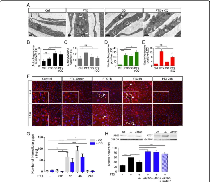
To confirm the induction of autophagy in LECs, Transmission Electron microscopy (TEM) has been per-formed to better detect autophagy/autophagosomes in
PTX-treated LEC (Fig.4a–c). Autophagosome and
auto-lysosome numbers have been quantified in PTX, CQ, and
CQ+ PTX-treated LEC. As expected, we observed that
CQ, known to inhibit the fusion between autophagosomes and autolysosomes, increases autophagosome number
(Fig. 4b). PTX induced an increase of autolysosome
for-mation that was reversed by CQ treatment (Fig.4c), which
demonstrate that PTX induces autophagy in LECs.
Finally, autophagy flux was measured by performing
mRFP-GFP-LC3 assay on LECs treated by PTX and CQ
(Fig. 4d, e and Supplementary Fig 2). We observed an
increase of autophagosome formation after PTX and CQ treatment and an increase of autolysosome formation
after PTX treatment that was reversed by CQ (Fig.4d, e).
PTX induced disruption of the endothelial barrier
Barrier function is largely determined by structural and junctional proteins. To assess the effect of PTX on endothelial barrier integrity, we analyzed the distribution of VE-cadherin in a confluent monolayer of HDLEC. Cell retraction with the concomitant loss of VE-cadherin from
the cell surface were associated with a significant increase
of intercellular gaps (Fig. 4f, g). In the regions of gap
formation slight traces of VE-cadherin were still visible at
Fig. 3 PTX induces HDLEC autophagy. a LC3 (green) and VE-cadherin (red) immunostaining on HDLEC stimulated by PTX (10μM) for 30 min, 1 h, 4 h, and 24 h shows an upregualtion of LC3 dots after 1 h, 4 h and 24 h of stimulation. b Quantification of the LC3-positive HDLEC treated with PTX. *p < 0.05. c Immunoblot analysis of LC3-I, LC3-II, Notch/NCID, beta-catenin, P62 and ATG5 in HDLECs stimulated by PTX (10 nM) for 30 min, 1 h, and 4 h. d Immunoblot analysis of P-VEGFR-3, pS473Akt, Akt, pERK, and ERK1/2 in HDLECs stimulated by PTX (10 nM) for 30 min, 1 h, and 4 h.
some remaining cell-cell contacts (Fig.4f). Gaps numbers appeared to be also significantly reduced 24 h following incubation with PTX. Furthermore, when PTX-treated cells were pre-treated with CQ, VE-cadherin staining was preserved and the formation of the gaps was limited
(Fig.4f, g), suggesting that autophagy could function as a
degradation mechanism for this protein. Then, we studied the effect of siRNA-mediated knockdown of ATG5 knockdown on the LEC monolayer. We observed a delay in the gap formation induced by PTX in comparison with
non-targeting control siRNA with a complete inhibition of gap formation following PTX treatment for 1 h (Sup-plementary Fig 3), which confirms the effect observed
with CQ (Fig.4g).
These results indicate that the loss of interaction between adjacent endothelial cells upon stabilization of microtubule by PTX, is dependent on actin disorganiza-tion and adherent juncdisorganiza-tion disassembly. Increase of
intercellular gaps in PTX-treated LEC may reflect barrier
dysfunction through increased vascular permeability.
Fig. 4 PTX-induced gap formation in HDLEC monolayer is reversed by Chloroquine. a Transmission electron microscopy representative images of PTX, CQ, PTX+ CQ-treated LEC. b Quantification of the lymphatic endothelial autophagosome. *p < 0.05; ***p < 0.001. c Quantification of the lymphatic endothelial autolysosomes. *p < 0.05; ***p < 0.001. d, e Quantification of autophagosomes (d) and autolysosomes (e) in PTX and CQ-treated LEC using mRFP-GFP-LC3 assay. f VE-cadherin immunostaining on HDLEC stimulated by PTX (10 nM) for 30 min, 1 h, 4 h, and 24 h shows gap between endothelial cells after 1 h and 4 h that is in part prevented by chloroquine (CQ). Scale bar 50μm. g Quantification of the lymphatic endothelial gap formation. *p < 0.05; ***p < 0.001.; ****p < 0.0001. h Knockdown of ATG5 and ATG7 reversed PTX inhibition of HDLEC branching in matrigel. **p < 0.01; ***p < 0.001.
PTX enhances tumor cell invasion into the lymph nodes
The lymphatic system serves as route for tumor metas-tasis to the lymph nodes. To determine whether PTX might promote tumor cells metastasis to lymph nodes, cell tracker (CMTMR) labeled 4T1 tumor cells were injected into the footpads of mice pre-treated with PTX. Treatment with PTX prior to injection into the footpad strongly suppressed tumor cell retention in lymph nodes without
affecting lymphatic vessel density (Fig.5a, b).
In contrast, inhibition of PTX-induced lymphatic autop-hagy by chloroquine decreased lymph node lymphatic vessel density and prevented 4T1 cell retention in vivo
(Fig. 5a, b), indicating that PTX resistance of lymphatic
vessels using autophagy-mediated lymphatic metastasis. Our studies suggest that PTX induced lymphatic permeability to
increase the tumor cell transendothelial migration into the lymph nodes. As lymphatic physiologic role is to transport immune cells, it was tempted to postulate that an increase in lymphatic permeability could also lead to an increase of immune cells infiltration. We then investigated whether lymphocyte trafficking was modified in metastatic breast cancer patients by measuring lymphocyte infiltration in ten neoadjuvant and ten adjuvant PTX-treated patients tissue biopsies obtained from the biology resource center of Ran-gueil hospital (Toulouse). We observed an increased CD3-positive lymphocyte infiltration in the vicinity of tumor lymphatic vessels from patients treated with neoadjuvant therapy (Supplementary Fig. 4), supporting the hypothesis that neoadjuvant PTX therapy increases the tumor lym-phatic vessels permeability.
Fig. 5 Chloroquine inhibits PTX-induced tumor cells extravasation. a Representativefluorescent images of “extravasated” CMTMR-labeled tumor cells (red) in the inguinal lymph node from PTX or CQ-treated mice. b Quantification of CMTMR-labeled tumor cells in the inguinal lymph node. *p < 0.05.
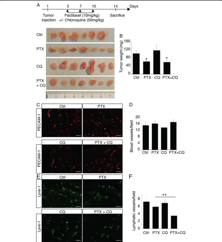
PTX inhibits tumor lymphangiogenesis but had no effect on angiogenesis
To study the effect of PTX on tumor lymphangiogenesis, we used chemotherapy in 4T1-bearing mice, a murine
syngeneic model of metastatic breast cancer. In this model, mice exhibit reproducible inguinal lymph nodes metastases after 2 weeks. Following tumor transplantation, mice were injected IP with PTX (10 mg/kg) at days 3, 6, and 9 and
Fig. 6 Chloroquine inhibits PTX-induced tumor lymphangiogenesis. a Schematic representation of the skinflap experimental procedure and images of 4T1 tumors from mice treated with PTX, CQ or PTX+ CQ. b Quantification of tumor weights. *p < 0.05. c Representative fluorescent images of PECAM-1-positive blood vessels from PTX- or CQ-treated tumor bearing mice. Scale bar: 50μm. d Quantification of the blood vessel density in 4T1 tumors from PTX- or CQ-treated mice. e Representativefluorescent images of Lyve-1-positive lymphatic vessels from PTX- or CQ-treated tumor bearing mice. Scale bar: 50μm. f Quantification of the lymphatic vessel density in 4T1 tumors from PTX- or CQ-treated mice, **p < 0.001.
tumor were harvested at day 14 (Fig.6a). As expected, PTX
reduced tumor weight (Fig.6a, b), but had no effect on other
organ weights (not shown). This effect on tumor size was attributed to the anti-mitogenic activity of chemotherapy on primary tumor as we did not observe any effect on tumor
angiogenesis (Fig. 6c, d). In parallel, we observed a slight
inhibition of tumor lymphangiogenesis induced by PTX that was dramatically increased by the association with chlor-oquine (50 mg/kg), suggesting that autophagy protects the lymphatic endothelium from the adverse effect of
che-motherapy (Fig.6e, f). These data suggest that an inhibition
of lymphatic resistance to chemotherapy can be hampered by the delivery of chloroquine, an autophagy inhibitor.
Chloroquine inhibits PTX-induced lymphatic metastasis to the lymph nodes
To further study the effect of PTX on lymphatic
metastasis, we analyzed the sentinel lymph nodes (Fig.7).
PTX and chloroquine alone had no effect on lymph node
lymphangiogenesis and angiogenesis (Fig. 7a–c). In
con-trast, the combination of these molecules strongly reduced lymph node lymphangiogenesis and metastases
to the lymph nodes and lymphoid tissues (Fig.7d, e and
Supplementary Fig. 5). This was associated with a strong upregulation of LC3 expression in lymphatic vessels from mice treated with PTX and CQ (Supplementary Fig. 6).
Discussion
Lymphatic vessels develop in solid tumors but also in the lymph nodes to provide an easier way for metastasis
dis-semination due to their high permeability10. Thus,
lym-phangiogenesis appears as a major therapeutic target in
treatments against cancer dissemination25. However, the
effect of cytotoxic agents on the proliferation of the lym-phatic vessels during tumor progression has been poorly understood. Here, we identify a mechanism of autophagy developed by the lymphatics in response to PTX che-motherapy to maintain routes for metastases to escape from
primary tumor. We recently identified stress-induced
molecular regulation of lymphangiogenic growth factors
VEGF-C and VEGF-D in tumors7,8. We found that hypoxia
strongly stimulates VEGF-C synthesis in breast cancer in
both primary tumors and in metastatic loci26. In parallel,
VEGF-D is induced by inflammatory stress in breast cancer to stimulate lymphatic vessel dilatation to increase
metas-tases26. These studies revealed that lymphangiogenesis, as
well as lymphatic vessel architecture are both key actors in tumor dissemination. Altogether, many targets and signal-ing pathways have been identified in the past decade to block the tumor lymphangiogenesis. In this study, we would like to identify whether chemotherapy could interfere with these processes. Despite its crucial role in tumor metastasis,
the effect offirst-line chemotherapy on the lymphatic
sys-tem has surprisingly never been investigated.
In the present study, we have found that paclitaxel, a drug that induces mitotic arrest due to activation of the mitotic checkpoint, could promote lymphatic endothelial cells autophagy. Despite ample evidence showing that autophagy plays an important role on blood endothelium, its role in tumoral lymphatic vasculature remains unclear. Chlor-oquine, an autophagy blocker, not only reduces tumor growth but also improves normalization and function of
tumor-associated blood vessels27. Recently, a study
sug-gested that photodynamic therapy of cancer could promote
lymphatic endothelial cells autophagy28. Here, we have
investigated the effect of chemotherapy on the lymphatic endothelium both in vitro and in vivo to better understand whether the lymphatic vessel resistance to chemotherapy could continue to provide route for metastases.
Autophagy has beenfirst described as negatively
regu-lated by Akt in response to mitogens via activation of
Target of Rapamycin (TOR)29. However, other studies
revealed an mTOR-independent mechanism by which Akt
can suppress autophagy30. Here, we report that Akt
reg-ulates autophagy in a TOR-independent manner in LEC in response to chemotherapy. Interestingly, this is associated with a dephosphorylation of VEGFR-3 and a decrease in adhesion molecule expression. The rearrangement of the actin cytoskeleton was followed by a transient lymphatic permeability increase generated by endothelial gap
for-mation induced by PTX. This was confirmed in vivo by an
experimental approach aiming at injecting tumor cells into the lymphatic system to promote metastasis independently of a primary tumor. Our studies indicate that systemic PTX might promote widespread of tumor metastases in the lymph node by modulating vessel permeability. This process was independent from lymph node lymphangio-genesis as we found that PTX strongly inhibits lym-phangiogenesis only in association with chloroquine, the autophagy inhibitor. The discordance with the inhibition
of lymphangiogenesis by PTX observed in the skin flap
model is probably attributed to a compensation induced by the secretion of lymphangiogenic factors VEGF-C and -D by the primary tumor.
Altogether, our results suggest that PTX increased lymphatic metastases by promoting LEC autophagy to maintain lymphatic vessel structure during chemotherapy, and by inducing vessel permeability. Also, intravenous chemotherapy has poor access to metastatic lymph nodes and is limited by short-lived drug concentrations. Therefore, the administration of chemotherapy via the lymphatic network appears as a new concept for the prevention and treatment of metastatic lymph nodes. Altogether, these data reveal the need for a better understanding of the action of chemotherapies on the lymphatic vascular endothelium. In conclusion, this study allows an advance in the fundamental knowledge of the lymphangiogenic process in breast cancer metastasis. We
have demonstrated the importance of the maintenance of a functional lymphatic architecture in tumorous lym-phangiogenesis after chemotherapy. Data from the lit-erature suggest that paclitaxel induces metastasis despite an inhibitory effect on tumor growth. Here, we highlight which mechanism is specifically activated in lymphatic endothelial cells to allow the tumor dissemination.
Acknowledgements
We thank J.J. Maoret and F. Martins (GenoToul platform) for their technical support as well the platform Anexplo Genotoul (Inserm US006, Toulouse, France) for their outstanding technical assistance. We thank Remy Flores from the imaging platform of I2MC institute and Vanessa Soldan from the electron
microscopy platform (ibcg, genotoul). We thank Lamia Ghezali for the technical assistance. This work has been supported by the Cancéropôle GSO, the Federal University of Toulouse Idex grant, the Ligue Régionale Contre le Cancer, the Foundation ARC pour la Recherche contre le Cancer, the foundation Toulouse Cancer Santé, the National Institute of Cancer (Inca), and the Region Midi-Pyrenees.
Author details
1UMR1048-I2MC, Université de Toulouse, Inserm, UT3, Toulouse, France. 2Department of Gynecology Surgery, University Hospital Centre—Toulouse,
IUCT-Oncopole, Toulouse, France.3UMR 1037-CRCT, Inserm, Université de
Toulouse, UT3, Toulouse, France.4INRA Toxalim, UT3, Toulouse, France.5UMR 5089-IPBS, CNRS, UPS, Toulouse, France.6Department of Pathology,
IUCT-Oncopole, Toulouse, France
Fig. 7 Chloroquine inhibits PTX-induced tumor metastasis. a Representativefluorescent images of Lyve-1-positive lymphatic vessels and PECAM-1-positive blood vessels in the inguinal lymph node from PTX- or CQ-treated tumor bearing mice. b, c Quantification of the lymphatic (b) and blood (c) vessel density in the inguinal lymph node from 4T1 tumors bearing mice treated with PTX or CQ, **p < 0.001. d Representativefluorescent images of Lyve-1-positive lymphatic vessels and cytokeratin-positive tumor metastases in the inguinal lymph node from PTX- or CQ-treated tumor bearing mice. e Quantification of the cytokeratin-positive tumor metastases in the inguinal lymph node from 4T1 tumors bearing mice treated with PTX or CQ, **p < 0.001.
Conflict of interest
The authors declare that they have no conflict of interest.
Publisher’s note
Springer Nature remains neutral with regard to jurisdictional claims in published maps and institutional affiliations.
Supplementary Information accompanies this paper at (https://doi.org/ 10.1038/s41419-019-2181-1).
Received: 10 May 2019 Revised: 1 October 2019 Accepted: 25 October 2019
References
1. Volk-Draper, L. et al. Paclitaxel therapy promotes breast cancer metastasis in a TLR4-dependent manner. Cancer Res. 74, 5421–5434 (2014).
2. Li, Q. et al. Low doses of paclitaxel enhance liver metastasis of breast cancer cells in the mouse model. FEBS J. 283, 2836–2852 (2016).
3. Alishekevitz, D. et al. Macrophage-Induced lymphangiogenesis and metastasis following paclitaxel chemotherapy is regulated by VEGFR3. Cell Rep. 17, 1344–1356 (2016).
4. Garmy-Susini, B. et al. Integrin alpha4beta1 signaling is required for lym-phangiogenesis and tumor metastasis. Cancer Res. 70, 3042–3051 (2010). 5. Garmy-Susini, B., Makale, M., Fuster, M. & Varner, J. A. Methods to study
lym-phatic vessel integrins. Methods Enzymol. 426, 415–438 (2007).
6. Alitalo, K., Tammela, T. & Petrova, T. V. Lymphangiogenesis in development and human disease. Nature 438, 946–953 (2005).
7. Morfoisse, F. et al. Hypoxia induces VEGF-C expression in metastatic tumor cells via a HIF-1alpha-independent translation-mediated mechanism. Cell Rep. 6, 155–167 (2014).
8. Morfoisse, F. et al. Nucleolin promotes heat shock-associated translation of VEGF-D to promote tumor lymphangiogenesis. Cancer Res. 76, 4394–4405 (2016).
9. Alitalo, K. The lymphatic vasculature in disease. Nat. Med 17, 1371–1380 (2011). 10. Garmy-Susini, B. et al. PI3Kalpha activates integrin alpha4beta1 to establish a metastatic niche in lymph nodes. Proc. Natl Acad. Sci. USA 110, 9042–9047 (2013).
11. Padera, T. P., Meijer, E. F. & Munn, L. L. The lymphatic system in disease processes and cancer progression. Annu. Rev. Biomed. Eng. 18, 125–158 (2016).
12. Perez, E. A. Paclitaxel in breast cancer. oncologist 3, 373–389 (1998). 13. Perez, E. A. & Buckwalter, C. A. Sequence-dependent cytotoxicity of etoposide
and paclitaxel in human breast and lung cancer cell lines. Cancer Chemother. Pharmacol. 41, 448–452 (1998).
14. Vyas, D., Laput, G. & Vyas, A. K. Chemotherapy-enhanced inflammation may lead to the failure of therapy and metastasis. OncoTargets Ther. 7, 1015–1023 (2014).
15. El-Araby, M. E., Omar, A. M., Khayat, M. T., Assiri, H. A. & Al-Abd, A. M. Molecular mimics of classic P-glycoprotein inhibitors as multidrug resistance suppressors and their synergistic effect on paclitaxel. PloS ONE 12, e0168938 (2017).
16. Moreau, K., Luo, S. & Rubinsztein, D. C. Cytoprotective roles for autophagy. Curr. Opin. Cell Biol. 22, 206–211 (2010).
17. Ravikumar, B., Moreau, K., Jahreiss, L., Puri, C. & Rubinsztein, D. C. Plasma membrane contributes to the formation of pre-autophagosomal structures. Nat. Cell Biol. 12, 747–757 (2010).
18. Li, Y. J. et al. Autophagy and multidrug resistance in cancer. Chin. J. cancer 36, 52 (2017).
19. Bharath, L. P. et al. Endothelial cell autophagy maintains shear stress-induced nitric oxide generation via glycolysis-dependent purinergic signaling to endothelial nitric oxide synthase. Arterioscler. Thromb. Vasc. Biol. 37, 1646–1656 (2017).
20. Prats, A. C. et al. CXCL4L1-fibstatin cooperation inhibits tumor angio-genesis, lymphangiogenesis and metastasis. Microvascular Res. 89, 25–33 (2013).
21. Caiado, J. & Picard, M. Diagnostic tools for hypersensitivity to platinum drugs and taxanes: skin testing, specific IgE, and mast cell/basophil mediators. Curr. Allergy Asthma Rep. 14, 451 (2014).
22. Lee, C., Gianos, M. & Klaustermeyer, W. B. Diagnosis and management of hypersensitivity reactions related to common cancer chemotherapy agents. Ann. Allergy, Asthma Immunol.: Off. Publ. Am. Coll. Allergy Asthma Immunol. 102, 179–187 (2009). quiz 187-179, 222.
23. Bronstad, A., Berg, A. & Reed, R. K. Effects of the taxanes paclitaxel and doc-etaxel on edema formation and interstitialfluid pressure. Am. J. Physiol. Heart Circulatory Physiol. 287, H963–H968 (2004).
24. Tanida, I., Ueno, T. & Kominami, E. LC3 and Autophagy. Methods Mol. Biol. 445, 77–88 (2008).
25. Avraamides, C. J., Garmy-Susini, B. & Varner, J. A. Integrins in angiogenesis and lymphangiogenesis. Nat. Rev. Cancer 8, 604–617 (2008).
26. Degenhardt, K. et al. Autophagy promotes tumor cell survival and restricts necrosis, inflammation, and tumorigenesis. Cancer Cell 10, 51–64 (2006).
27. Maes, H. et al. Tumor vessel normalization by chloroquine independent of autophagy. Cancer Cell 26, 190–206 (2014).
28. Wachowska, M. et al. Investigation of cell death mechanisms in human lymphatic endothelial cells undergoing photodynamic therapy. Photo-diagnosis Photodyn. Ther. 14, 57–65 (2016).
29. Janku, F., McConkey, D. J., Hong, D. S. & Kurzrock, R. Autophagy as a target for anticancer therapy. Nat. Rev. Clin. Oncol. 8, 528–539 (2011).
30. Wang, R. C. et al. Akt-mediated regulation of autophagy and tumorigenesis through Beclin 1 phosphorylation. Science 338, 956–959 (2012).