HAL Id: hal-01977794
https://hal.sorbonne-universite.fr/hal-01977794
Submitted on 11 Jan 2019HAL is a multi-disciplinary open access archive for the deposit and dissemination of sci-entific research documents, whether they are pub-lished or not. The documents may come from teaching and research institutions in France or abroad, or from public or private research centers.
L’archive ouverte pluridisciplinaire HAL, est destinée au dépôt et à la diffusion de documents scientifiques de niveau recherche, publiés ou non, émanant des établissements d’enseignement et de recherche français ou étrangers, des laboratoires publics ou privés.
c-MET Overexpression as a Poor Predictor of MET
Amplifications or Exon 14 Mutations in Lung
Sarcomatoid Carcinomas
Xavier Mignard, Anne-Marie Ruppert, Martine Antoine, Julie Vasseur,
Nicolas Girard, Julien Mazieres, Denis Moro-Sibilot, Vincent Fallet, Nathalie
Rabbe, Françoise Thivolet-Béjui, et al.
To cite this version:
Xavier Mignard, Anne-Marie Ruppert, Martine Antoine, Julie Vasseur, Nicolas Girard, et al.. c-MET Overexpression as a Poor Predictor of c-MET Amplifications or Exon 14 Mutations in Lung Sarcomatoid Carcinomas. Journal of Thoracic Oncology, Lippincott, Williams & Wilkins, 2018, 13 (12), pp.1962-1967. �10.1016/j.jtho.2018.08.008�. �hal-01977794�
c-MET Overexpression as a Poor Predictor of MET Amplifications or Exon 14 Mutations in Lung Sarcomatoid Carcinomas
Xavier Mignard1, Anne-Marie Ruppert1,2, Martine Antoine1,3, Julie Vasseur4, Nicolas Girard5,
Julien Mazières6, Denis Moro-Sibilot7, Vincent Fallet2, Nathalie Rabbe2, Françoise
Thivolet-Bejui8, Isabelle Rouquette9, Sylvie Lantuejoul10,11, Alexis Cortot12, Raphaël Saffroy4, Jacques
Cadranel1,2, Antoinette Lemoine4, and Marie Wislez1,2.
1 - Sorbonne Universités, UPMC Univ Paris 06, GRC n°04, Theranoscan, F-75252, Paris, France
2 - AP-HP, Hôpital Tenon, Service de Pneumologie, F-75970, Paris, France
3 - AP-HP, Hôpital Tenon, Service de Cytologie et Anatomie Pathologique, F-75970, France 4 - AP-HP, Hôpitaux Universitaires Paris-Sud, Service d'Oncogénétique - Oncomolpath, Vil-lejuif, France
5 - Respiratory Medicine and Thoracic Oncology Service, Hôpital Louis Pradel, Hospices Civils de Lyon, 69677 Lyon cedex, France
6 - Hôpital Larrey CHU Toulouse, Toulouse, France
7 - CHU De Grenoble, Hôpital A. Michalon, La Tronche, France
8 - Hospices Civils de Lyon, Centre de biologie et pathologie Est, Lyon, France
9 - Institut Universitaire du Cancer de Toulouse, Oncopôle, Service d'Anatomie Patholo-gique, Toulouse, France
10 – Département de Biopathologie, Centre Léon Bérard, Grenoble, France 11 - Grenoble Alpes Université, Grenoble, France
Corresponding author
Pr Marie Wislez, Service de Pneumologie, Hôpital Tenon, 4 rue de la Chine, F-75970 Paris Cedex 20, France. Email: [email protected]. Phone: +33 (0)1 56 01 65 15. Fax: +33 (0)1 56 01 72 48
Abstract
Background: Mesenchymal-to-epithelial transition (MET) abnormalities like amplification and exon 14 mutations may be responsive to targeted therapies. They are prevalent in lung sar-comatoid carcinomas (LSCs) and must be diagnosed as efficiently as possible. Hypothetical-ly, c-MET overexpression by immunohistochemistry (IHC) may prove effective as a screen-ing test for MET abnormalities. Material and methods: Tissue samples were obtained from consecutive patients with a resected LSC, in four oncologic centers. IHC was performed us-ing the SP44 antibody (Ventana) and evaluated usus-ing the MetMab score and H-score. Fluo-rescence in situ hybridization (FISH) was applied with the dual color probe set from Zyto-vision (Clinisciences). True MET amplification was diagnosed when MET gene copy number was ≥5 and the ratio between MET gene copy number and chromosome 7 number was >2. All MET exon 14 alterations including those affecting splice sites occurring within splice do-nor and acceptor sites were detected in the routine molecular testing on genetic platforms. Results: A total of 81 LSCs were included. 14 (17%) exhibited positive IHC using the MetMab score and 15 (18.5%) using the H-score. MET amplification was detected in six tu-mors (8.5%) and MET exon 14 mutation in five (6%). A weak positive correlation between IHC and FISH was found (r=0.27, p=0.0001). IHC sensitivity for MET amplification was 50%, with a specificity of 83%, positive predictive value of 21.4%, and negative predictive value of 94.7%. IHC sensitivity for MET exon 14 mutations was 20%, with a specificity of 83%, posi-tive predicposi-tive value of 7%, and negaposi-tive predicposi-tive value of 94%. Conclusion: IHC is not a relevant screening tool for MET abnormalities in LSC.
Keywords
Acknowledgements
The authors extend their thanks to Mrs. Gwenaëlle Pontdeme (Sorbonne Universités, UPMC Univ Paris 06, GRC n°04, Theranoscan, F-75252, Paris, France), Anita Rodenas, and Chris-tel Daubrosse (Pathology and Cytology Laboratory, Tenon University Hospital, AP-HP) for their technical assistance regarding IHC.
Author’s disclosures of potential conflicts of interest
Dr MORO-SIBILOT reports personal fees from Pfizer, Novartis, Roche, Lilly, Boehringer, Astra Zeneca, Amgen, during the conduct of the study; personal fees from Abbvie, Takeda, BMS, Msd, outside the submitted work.
Dr WISLEZ reports personal fees from Roche.
Introduction
The mesenchymal-to-epithelial transition (MET) pathway includes the tyrosine kinase recep-tor (TKR) c-MET, its ligand, hepatocyte growth facrecep-tor (HGF), as well as downstream path-ways involved in tumor growth, cell survival, invasion, and cell migration (1). MET pathway activation involves different mechanisms, such as amplification of the MET gene, exon 14 splicing site mutations, activating point mutations, or HGF-dependent upregulation.
Amplification of the MET gene, characterized by an increased MET gene copy number measured by FISH (fluorescence in situ hybridization), is found in 1 to 4% of non-small-cell lung cancers (NSCLCs) (2).
Mutations in MET exon 14 and its flanking introns can induce exon 14 splicing that is thought to cause loss of the intracellular juxta-membrane domain, whilst increasing c-MET expres-sion at the membrane. The diversity of MET exon 14 alterations, including base substitutions or indels that disrupt the branch point of intron 13, the 3’ splice site of intron 13, or the 5’ splice site of intron 14, requires improved technologies such as next-generation sequencing (NGS) to cover the whole c.2942-64 to c.3082+42 region (3).
Recently, MET tyrosine kinase inhibitors (TKIs), such as crizotinib and capmatinib, have proven their efficacy on tumors harboring MET mutations or amplifications, suggesting onco-gene addiction, therefore rendering MET screening highly relevant (4), (5).
Lung sarcomatoid carcinomas (LSCs) are rare tumors, accounting for less than 3% of all NSCLCs. They are more prevalent among males and smokers and characterized by both high resistance to platinum-based chemotherapy and poor prognosis (6). A higher frequency of MET exon 14 mutations has been reported in LSCs (7), with a prevalence ranging be-tween 4.9% and 31.8% (8).
Techniques for the diagnosis of MET amplification and exon 14 mutations are not readily accessible in routine practice. FISH is expensive and time-consuming, whereas NGS panels
require sufficient tumor material and high DNA quality, although samples usually are needle-biopsied formalin-fixed paraffin-embedded (FFPE) tumors.
IHC could prove effective as a screening test in molecular abnormalities such as ALK rear-rangements. While FISH remains the gold standard, ALK IHC is highly sensitive (100%), FISH being only performed in ambiguous IHC cases (9).
The aim of this study was to compare MET IHC, FISH, and exon 14 mutation detection in a series of LSC in order to evaluate IHC as a screening test.
Patients and methods
Patients and tissue tumor collection
Tissue samples were obtained from surgical lung biopsies of all consecutive patients with LSC diagnosed between 2005 and 2012 in four referral thoracic oncology centers. Clinical data and tumor characteristics were recorded as previously described (8). Each patient signed an informed consent form as required by national guidelines and samples were col-lected in line with current legislation.
Immunohistochemistry (IHC)
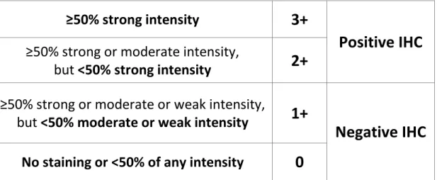
In brief, c-MET protein expression was assessed by IHC on 3µm-thick sections of FFPE tu-mor samples, using the rabbit monoclonal primary antibody SP44 (Ventana, Arizona, USA). The MetMab score was calculated using the method detailed in Table 1 (10). The H-score was obtained by multiplying the intensity (from 0 to 3) by the percentage of positive cells (from 0 to 100%), thus obtaining a scale ranging between 0 and 300. The H-score positivity threshold was 150 (11). IHC was conducted by a referent pathologist (Dr. M. Antoine).
Fluorescence in situ hybridization (FISH)
MET gene amplification was assessed using the MET/CEP7 dual color probe set from Zyto-vision (Clinisciences, France), according to the manufacturer’s instructions. MET gene ampli-fication was defined as a MET gene copy number ≥5 and a MET/CEP7 ratio >2 (2), per-formed by the same referent pathologist.
Molecular testing for MET gene sequence abnormalities
Overall, 10μm-thick sections were cut from the paraffin blocks. Tumor enrichment was per-formed through selection and macro-dissection of areas with at least 50% tumor cells. Total DNA was extracted and purified after paraffin removal, as previously described (8). All MET alterations from c.2942-64 to c.3082+42 affecting exon 14 splice sites that are indels occurring within splice donor and acceptor sites were detected in the routine molecular testing on ISO15189 certified genetic platforms. Mutations analysis methods were previously reported (8). All tumor samples were tested by a combined strategy of High Resolution Melting (HRM) assay using Lightcycler 480 system (Roche Diagnostics) in order to screen all gene se-quence abnormalities of exon 14 of the MET gene c3082, c3082+1, c3082+2, and c3082+3, confirmed using MassARRAY iPLEX technology (Agena Bioscience). Samples for which material was available (n=40) were also tested by NGS using the solid tumor solution by So-phia Genetics® based on the xGen Lockdown IDT® probe-based capture technology.
Molecular screening for other mutations
Mass spectrometry was employed to test 214 mutations affecting 26 oncogenes and tumor suppressor genes (Panel Lungcarta©MassARRAY iPLEX genotyping technology [Agena Bioscience, San Diego, USA]), as previously described (8).
Statistical analyses
Continuous variables were expressed as medians with [min, max] intervals. Categorical vari-ables were expressed as percentages. Comparisons between non-parametric continuous variables were conducted via Mann-Whitney test, and those between categorical variables with the Chi-squared test, or the Fisher’s exact test when n <5. Correlation between non-normally distributed continuous variables was calculated using Spearman’s correlation coef-ficient. All the tests were two-sided, with results considered significant when p <0.05. Anal-yses were performed using GraphPad Prism (GraphPad Software Inc, California, USA).
Results
General characteristics
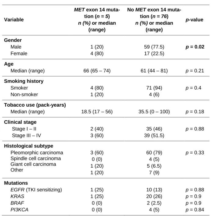
Between 2005 and 2012, 81 patients with LSC were included, of whom 60 (74%) were male, and 75 (94%) were smokers (Table 2). The median age was 62 years. At diagnosis, 37 pa-tients (46%) were at stage I-II disease, and 42 (52%) Stage III-IV disease. The most common histological subtype consisted of pleomorphic carcinoma (n=63, 77.5%). The most commonly detected mutations were KRAS (n=21, 26%), and EGFR (n=11, 13.5%). Positive c-MET IHC was observed in 15 (18.5%) tumors using the H-score (median 30 [0 - 260]), and in 14 (17%) using the MetMab score (median 0 [0 - 3]). True MET amplification was found in six patients (8.5%) and MET exon 14 mutation in five (6%). High MET amplification (MET/CEP7 ratio ≥5) was found in two patients.
Characteristics of patients with true MET amplification (MET gene copy number ≥5 and ratio MET/CEP7 >2)
Men represented 100% of patients with MET amplification, vs 71% (n=46) of patients without amplification (p=0.18, Table SI). Tumors with MET amplification all displayed a pleomorphic histological subtype, vs 50 (77%) without amplification (p=0.33). Patients with MET amplifica-tion exhibited less KRAS mutaamplifica-tions, yet the difference did not reach statistical significance. Patients with polysomy were not considered (n=8, 11%).
Characteristics of patients with MET exon 14 mutations
MET exon 14 mutations were detected in five patients (6%). They were more often women (80% vs 22.5%, p=0.02). No other significant differences were found according to MET muta-tion status (Table SII).
Correlation between IHC and MET FISH / MET exon 14 mutation
In total, 15 patients (18.5%) had a positive IHC using the H-score and 14 (17%) using the MetMab score. Among MET amplifications (n=6), three (50%) had a positive c-MET IHC. No MET exon 14 mutations were associated with MET amplification. One tumor sample out of five (20%) with exon 14 mutation exhibited c-MET positive IHC (Figure 1).
Among chromosome 7 polysomies (n=8), one (12.5%) had a positive IHC, with no significant association (p=1.0). Two (25%) had a MET exon 14 mutation (versus 3% MET exon 14 mu-tations in patients without polysomy) with a significant association (p=0.02).
The correlation between IHC H-score and FISH proved weakly positive (R coefficient be-tween 0 and 0.5) (MET gene copy number ≥5 regardless of MET/CEP7 ratio, i.e. including polysomies, in Figure 2, then MET gene copy number ≥5 and MET/CEP7 >2, i.e. without polysomies, in Figure 3) .
Considering FISH as the gold standard for MET amplification, IHC sensitivity was 50%, spec-ificity 83%, positive predictive value 21.4%, and negative predictive value 94.7%. For high MET amplifications, IHC sensitivity was also 50%. IHC sensitivity for MET exon 14 mutations was 20%, with a specificity of 83%, positive predictive value of 7%, and negative predictive value of 94%.
Discussion
In this cohort of surgically resected LSCs, c-MET IHC overexpression, as well as MET ampli-fication and MET exon 14 mutations were analyzed, to determine whether IHC could be a screening test for amplification or mutation. Altogether, 14 tumors (17%) had positive c-MET IHC, six had a MET amplification (8.5%), and five a MET exon 14 mutation (6%).
As MET amplification and exon 14 mutations are potential targets for TKIs (4) (5) in NSCLCs, and particularly in LSCs, these abnormalities must be diagnosed as efficiently as possible.
In this study, c-MET IHC could not be considered as a screening test either for MET amplifi-cation or MET exon 14 mutations, as sensitivity (50% and 20% respectively) and correlation (r=0.27) proved poor, with similar results found for MetMab and H-score. Current data re-garding correlation between IHC and MET molecular alterations are discordant. Watermann et al. found a weak correlation (r=0.06, p >0.05) between MET IHC and FISH in 214 NSCLC samples (12). Casadevall et al. showed no association between IHC and FISH in a non-squamous NSCLC cohort (13). Conversely, Park et al. reported a significant association be-tween IHC score and MET amplification (p<0.001) using the Chi-squared test on a large se-ries of 316 adenocarcinomas, with neither sensitivity nor correlation reported (14). Additional-ly, Tong et al. revealed in 687 NSCLCs a significant association between IHC and amplifica-tion, and between IHC and exon 14 mutation (p <0.001), based on Chi-squared test anal-yses. Nevertheless, the correlation using Spearman’s test proved to be weak (3). Further-more, whether significant association between IHC and amplification/mutation does exist or not, sensitivity is a better parameter for diagnosis purposes.
For several other molecular abnormalities, such as ALK rearrangement, IHC proves to be an effective screening option. ALK IHC and FISH are highly correlated, with a sensitivity for IHC approaching 100% (9). FISH is performed only in ambiguous IHC cases. For ROS1
rear-rangements, IHC sensitivity compared to FISH is reported close to 100% with a specificity of 97% (15).
As MET exon 14 mutations induce loss of ubiquitination and increased c-MET membrane presence, their lack of association with c-MET overexpression remains to understand. One explanation is that the oncogenic properties of MET exon 14 mutations involve other mecha-nisms (loss of serine 985 with increased kinase activity, loss of aspartate 1002 resulting in loss of pro-apoptotic signals) (1). Another explanation is that LSCs have a different genomic background compared to NSCLCs, which might influence these results. Indeed, MET gene abnormalities occur in a context of other oncogenic and tumor suppressive genes abnormali-ties involving TP53, LKB1 or EGFR point mutations, or gene copy number variations that can influence the c-MET protein expression.
Regarding MET exon 14 mutation detection, we used a combined technique (HRM + Mas-sARRAY) for all tumor samples (n=81). This allows detection of point mutations, and indels that are deleterious with a higher sensitivity than NGS. Nevertheless, the MassARRAY tech-nology can miss some large deletions. Therefore, NGS was performed on tumor samples with sufficient material (n=40) to increase detection sensitivity. It is worth noticing that NGS allowed detection of one exon 14 mutation that was not diagnosed by HRM + MassARRAY, but also that MassARRAY allowed diagnosis of one more exon 14 mutation, undetected by NGS (data not shown).
Few studies focus specifically on LSC. The main cohorts investigating MET pathway abnor-malities in LSCs are summed up in Table SIII. Discrepancies exist in the prevalence of posi-tive IHC, amplification or exon 14 mutations, between those cohorts. Our results tend to be on the low end of each one of those abnormalities. It is however worth noticing that patients with exon 14 mutations and negative IHC exist in other series. These discrepancies can be explained by the small number of positive cases, which also reduces the power of the given sensitivity and specificity values.
Our findings suggest that MET IHC may not be employed as a screening tool. MET FISH and molecular biology techniques (NGS, WES, and fragment analysis) are recommended for the detection of amplifications or exon 14 mutations in NSCLC in routine practice.
References
1. Cortot AB, Kherrouche Z, Descarpentries C, Wislez M, Baldacci S, Furlan A, et al. Exon 14 Deleted MET Receptor as a New Biomarker and Target in Cancers. J Natl Cancer Inst. 2017 01;109(5).
2. Cappuzzo F, Marchetti A, Skokan M, Rossi E, Gajapathy S, Felicioni L, et al. In-creased MET gene copy number negatively affects survival of surgically resected non-small-cell lung cancer patients. J Clin Oncol Off J Am Soc Clin Oncol. 2009 Apr 1;27(10):1667–74. 3. Tong JH, Yeung SF, Chan AWH, Chung LY, Chau SL, Lung RWM, et al. MET Ampli-fication and Exon 14 Splice Site Mutation Define Unique Molecular Subgroups of Non-Small Cell Lung Carcinoma with Poor Prognosis. Clin Cancer Res Off J Am Assoc Cancer Res. 2016 Jun 15;22(12):3048–56.
4. Frampton GM, Ali SM, Rosenzweig M, Chmielecki J, Lu X, Bauer TM, et al. Activation of MET via diverse exon 14 splicing alterations occurs in multiple tumor types and confers clinical sensitivity to MET inhibitors. Cancer Discov. 2015 Aug;5(8):850–9.
5. Ou S-HI, Kwak EL, Siwak-Tapp C, Dy J, Bergethon K, Clark JW, et al. Activity of cri-zotinib (PF02341066), a dual mesenchymal-epithelial transition (MET) and anaplastic lym-phoma kinase (ALK) inhibitor, in a non-small cell lung cancer patient with de novo MET am-plification. J Thorac Oncol Off Publ Int Assoc Study Lung Cancer. 2011 May;6(5):942–6. 6. Vieira T, Girard N, Ung M, Monnet I, Cazes A, Bonnette P, et al. Efficacy of first-line chemotherapy in patients with advanced lung sarcomatoid carcinoma. J Thorac Oncol Off Publ Int Assoc Study Lung Cancer. 2013 Dec;8(12):1574–7.
7. Liu X, Jia Y, Stoopler MB, Shen Y, Cheng H, Chen J, et al. Next-Generation Se-quencing of Pulmonary Sarcomatoid Carcinoma Reveals High Frequency of Actionable MET Gene Mutations. J Clin Oncol Off J Am Soc Clin Oncol. 2016 Mar 10;34(8):794–802.
8. Saffroy R, Fallet V, Girard N, Mazieres J, Sibilot DM, Lantuejoul S, et al. MET exon 14 mutations as targets in routine molecular analysis of primary sarcomatoid carcinoma of the lung. Oncotarget. 2017 Jun 27;8(26):42428–37.
9. McLeer-Florin A, Moro-Sibilot D, Melis A, Salameire D, Lefebvre C, Ceccaldi F, et al. Dual IHC and FISH testing for ALK gene rearrangement in lung adenocarcinomas in a rou-tine practice: a French study. J Thorac Oncol Off Publ Int Assoc Study Lung Cancer. 2012 Feb;7(2):348–54.
10. Spigel DR, Edelman MJ, O’Byrne K, Paz-Ares L, Mocci S, Phan S, et al. Results From the Phase III Randomized Trial of Onartuzumab Plus Erlotinib Versus Erlotinib in Pre-viously Treated Stage IIIB or IV Non-Small-Cell Lung Cancer: METLung. J Clin Oncol Off J Am Soc Clin Oncol. 2017 Feb;35(4):412–20.
11. Cappuzzo F, Hirsch FR, Rossi E, Bartolini S, Ceresoli GL, Bemis L, et al. Epidermal growth factor receptor gene and protein and gefitinib sensitivity in non-small-cell lung cancer.
J Natl Cancer Inst. 2005 May 4;97(9):643–55.
12. Watermann I, Schmitt B, Stellmacher F, Müller J, Gaber R, Kugler C, et al. Improved diagnostics targeting c-MET in non-small cell lung cancer: expression, amplification and acti-vation? Diagn Pathol. 2015 Jul 28;10:130.
13. Casadevall D, Gimeno J, Clavé S, Taus Á, Pijuan L, Arumí M, et al. MET expression and copy number heterogeneity in nonsquamous non-small cell lung cancer (nsNSCLC). Oncotarget. 2015 Jun 30;6(18):16215–26.
14. Park S, Koh J, Kim D-W, Kim M, Keam B, Kim TM, et al. MET amplification, protein expression, and mutations in pulmonary adenocarcinoma. Lung Cancer Amst Neth. 2015 Dec;90(3):381–7.
15. Mescam-Mancini L, Lantuéjoul S, Moro-Sibilot D, Rouquette I, Souquet P-J, Audigier-Valette C, et al. On the relevance of a testing algorithm for the detection of ROS1-rearranged lung adenocarcinomas. Lung Cancer Amst Neth. 2014 Feb;83(2):168–73.
Figures and tables
Table 1: IHC results according to the MetMab score
≥50% strong intensity
3+
Positive IHC
≥50% strong or moderate intensity,
but <50% strong intensity
2+
≥50% strong or moderate or weak intensity,
but <50% moderate or weak intensity
1+
Negative IHC
No staining or <50% of any intensity
0
Table 2: Characteristics of overall population (n=81)
Variable All patients (n = 81)
Gender Male Female 60 (74) 21 (26) Age Median (Range) 62 (44 – 81) Ethnicity Caucasian Asian Northern African Sub-Saharan African 65 (80) 0 (0) 10 (12.5) 1 (1.5) Smoking history Smoker Non-smoker 75 (94) 5 (6) Clinical stage Stage I – II Stage III – IV 37 (46) 42 (52) Histological subtype Pleomorphic carcinoma Spindle cell carcinoma Giant cell carcinoma Other 63 (77.5) 4 (5) 6 (7.5) 8 (10) Mutations MET exon 14
EGFR (TKI sensitizing) KRAS BRAF PI3KCA 5 (6) 11 (13.5) 21 (26) 2 (2.5) 4 (5) Positive IHC MetMab H-score 14 (17) 15 (18.5) Positive FISH Polysomy
True MET amplification
14 (20) 8 (11) 6 (8.5)
MET = Mesenchymal to Epithelial Transition; EGFR = Epidermal Growth Factor Receptor; TKI = tyrosine kinase inhibitor; KRAS
= Kirsten rat sarcoma oncogene; BRAF = B-raf proto-oncogene; PI3KCA = Phosphatidyl-inositol-3-kinase Catalytic Subunit Alpha; IHC = immunohistochemistry; FISH = Fluorescence in situ hybridization.
Table SI: Characteristics according to MET amplification status Variable MET amplification (n = 6) n (%) or median (range) No MET amplification (n = 65) n (%) or median (range) p-value Gender Male Female 6 (100) 0 (0) 46 (71) 19 (29) p = 0.18 Age Median (Range) 61.5 (57 – 80) 61.5 (44 – 81) p = 0.52 Ethnicity Caucasian Asian Northern African Sub-Saharan African 5 (83) 0 (0) 0 (0) 0 (0) 53 (81) 0 (0) 8 (12) 1 (1.5) p = 0.66 Smoking history Smoker Non-smoker 6 (100) 0 (0) 60 (92) 4 (6) p = 1.0
Tobacco use (pack-years)
Median (Range) 37.5 (12 – 40) 36 (0 – 100) p = 0.62 Occupational exposure 0 (0) 8 (12) p = 1.0 Clinical stage Stage I – II Stage III – IV 2 (33) 4 (67) 30 (46) 34 (52) p = 0.68 Histological subtype Pleomorphic carcinoma Spindle cell carcinoma Giant cell carcinoma Other 6 (100) 0 (0) 0 (0) 0 (0) 50 (77) 3 (4.5) 5 (8) 7 (10.5) p = 0.33 Mutations MET exon 14
EGFR (TKI sensitizing) KRAS BRAF PI3KCA TP53 NRAS 0 (0) 1 (17) 0 (0) 0 (0) 0 (0) 3 (50) 0 (0) 5 (7.5) 7 (11) 21 (32) 2 (3) 4 (6) 10 (15) 1 (1.5) p = 0.51 p = 0.52 p = 0.17 p = 1.0 p = 1.0 p = 0.07 p = 1.0
Table SII: Characteristics according to MET exon 14 mutation status
Variable
MET exon 14 muta-tion (n = 5) n (%) or median
(range)
No MET exon 14 muta-tion (n = 76) n (%) or median (range) p-value Gender Male Female 1 (20) 4 (80) 59 (77.5) 17 (22.5) p = 0.02 Age Median (range) 66 (65 – 74) 61 (44 – 81) p = 0.21 Smoking history Smoker Non-smoker 4 (80) 1 (20) 71 (94) 4 (6) p = 0.4
Tobacco use (pack-years)
Median (range) 18.5 (17 – 56) 35.5 (0 – 100) p = 0.18 Clinical stage Stage I – II Stage III – IV 2 (40) 3 (60) 35 (46) 39 (51.5) p = 0.88 Histological subtype Pleomorphic carcinoma Spindle cell carcinoma Giant cell carcinoma Other 3 (60) 0 (0) 1 (20) 1 (20) 60 (79) 4 (5) 5 (6.5) 7 (9) p = 0.33 Mutations
EGFR (TKI sensitizing) KRAS BRAF PI3KCA 1 (25) 1 (25) 0 (0) 0 (0) 10 (13) 20 (26) 2 (2.5) 4 (5) p = 0.88 p = 0.9 p = 0.9 p = 0.84
Table SIII: Comparison of MET pathway abnormalities prevalence among different cohorts of patients with LSC
Caption: LSC = Lung Sarcomatoid Carcinoma ; IHC+ = positive immunohistochemistry ; Amp+ = positive amplification ; ex 14+ = positive exon 14 mutation ; PCR = Polymerase chain reaction ; NGS = Next-Generation Sequencing ; CGP = Comprehensive genomic profiling ; HRM = High resolution melting ; NA = Not applicable ; * Percentage of patients harboring a MET exon 14 mutation, exhib-iting positive c-MET IHC ; ** Percentage of patients harboring a MET exon 14 mutation, exhibexhib-iting positive MET amplification
Study LSC (n) cMET IHC+ n (%) Primary antibody MET Amp+ n (%) MET ex 14+ n (%) MET ex 14 technique MET ex 14
muta-tions and c-MET IHC+ n (%)*
MET ex 14
muta-tions and MET Amp+ n (%)** Vieira et al, Lung Cancer, 2014 77 26 (34%) SP44 NA 2 (3%) Sizing analysis of PCR products (only 3’-splice site of MET ex 14
dele-tions)
1 (50%) NA
Liu et al, JCO
2015 36 NA NA NA 8 (22.2%)
Whole exome
sequen-cing NA NA
Awad et al,
JCO 2016 15 NA SP44 NA 4 (26.6%) NGS NA NA
Schrock et al,
JTO 2016 104 NA NA NA 8 (7.7%) NGS NA 1 (NA)
Tong et al, Clin
Can Res, 2016 22 9 (40.9%) SP44 3 (13.6%) 7 (31.8%)
Whole ex 14 + flanking intronic regions Sanger
sequencing 7 (100%) 2 (28.5%) Kwon et al, Lung Cancer 2017 45 NA SP44 NA 9 (20%) qRT-PCR 3 (33%) NA Schrock et al, JTO 2017 125 NA NA n=1 15 (12%) CGP NA NA Mignard et al, 2018 81 15 (18.5%) SP44 6 (8.5%) 4 (4.9%) HRM + MassArray + NGS 1 (20%) 0 (0%)
Figure 1
Title: Distribution of MET expression positivity by IHC, FISH and exon 14 mutations in the overall population
Caption: Positive c-MET IHC was found in 15 (18.5%) tumors using the H-score and in 14 (17%) using the MetMab score. Six patients (8.5%) had a true MET amplification (MET gene copy number ≥5 and ratio MET/CEP7 >2). Five patients (6%) had a MET exon 14 mutation.
Figure 2
Title: Correlation between H-score based IHC and MET gene copy number by FISH
Caption: The horizontal axe represents the H-score value (between 0 and 300) for c-MET IHC and the vertical axe represents the MET gene copy number measured by FISH. Spear-man’s rho coefficient was calculated (R=0.41, p=0.0006).
Figure 3
Title: Correlation between H-score based IHC and MET true amplification by FISH
Caption: The horizontal axe represents the H-score value (between 0 and 300) for c-MET IHC and the vertical axe represents the MET gene copy number/chromosome 7 ratio meas-ured by FISH. Spearman’s rho coefficient was calculated (R=0.27, p=0.0001).