HAL Id: hal-02950982
https://hal.archives-ouvertes.fr/hal-02950982
Submitted on 26 May 2021
HAL is a multi-disciplinary open access
archive for the deposit and dissemination of
sci-entific research documents, whether they are
pub-lished or not. The documents may come from
teaching and research institutions in France or
abroad, or from public or private research centers.
L’archive ouverte pluridisciplinaire HAL, est
destinée au dépôt et à la diffusion de documents
scientifiques de niveau recherche, publiés ou non,
émanant des établissements d’enseignement et de
recherche français ou étrangers, des laboratoires
publics ou privés.
life-threatening COVID-19
Qian Zhang, Paul Bastard, Zhiyong Liu, Jérémie Le Pen, Marcela
Moncada-Velez, Jie Chen, Masato Ogishi, Ira Sabli, Stephanie Hodeib, Cecilia
Korol, et al.
To cite this version:
Qian Zhang, Paul Bastard, Zhiyong Liu, Jérémie Le Pen, Marcela Moncada-Velez, et al.. Inborn errors
of type I IFN immunity in patients with life-threatening COVID-19. Science, American Association
for the Advancement of Science, 2020, 370 (6515), pp.eabd4570. �10.1126/science.abd4570�.
�hal-02950982�
RESEARCH ARTICLE SUMMARY
◥CORONAVIRUS
Inborn errors of type I IFN immunity in patients
with life-threatening COVID-19
Qian Zhang
et al.
INTRODUCTION:
Clinical outcomes of human
severe acute respiratory syndrome
corona-virus 2 (SARS-CoV-2) infection range from
silent infection to lethal coronavirus disease
2019 (COVID-19). Epidemiological studies have
identified three risk factors for severe disease:
being male, being elderly, and having other
medical conditions. However, interindividual
clinical variability remains huge in each
demo-graphic category. Discovering the root cause
and detailed molecular, cellular, and tissue- and
body-level mechanisms underlying life-threatening
COVID-19 is of the utmost biological and medical
importance.
RATIONALE:
We established the COVID Human
Genetic Effort (www.covidhge.com) to test
the general hypothesis that life-threatening
COVID-19 in some or most patients may be
caused by monogenic inborn errors of
immu-nity to SARS-CoV-2 with incomplete or
com-plete penetrance. We sequenced the exome or
genome of 659 patients of various ancestries
with life-threatening COVID-19 pneumonia
and 534 subjects with asymptomatic or benign
infection. We tested the specific hypothesis that
inborn errors of Toll-like receptor 3 (TLR3)
–
and interferon regulatory factor 7 (IRF7)
–
dependent type I interferon (IFN) immunity
that underlie life-threatening influenza
pneu-monia also underlie life-threatening COVID-19
pneumonia. We considered three loci identified
as mutated in patients with life-threatening
influenza:
TLR3, IRF7, and IRF9. We also
con-sidered 10 loci mutated in patients with other
viral illnesses but directly connected to the three
core genes conferring influenza susceptibility:
TICAM1/TRIF, UNC93B1, TRAF3, TBK1, IRF3,
and
NEMO/IKBKG from the TLR3-dependent
type I IFN induction pathway, and
IFNAR1,
IFNAR2, STAT1, and STAT2 from the
IRF7-and IRF9-dependent type I IFN amplification
pathway. Finally, we considered various modes
of inheritance at these 13 loci.
RESULTS:
We found an enrichment in variants
predicted to be loss-of-function (pLOF), with a
minor allele frequency <0.001, at the 13
can-didate loci in the 659 patients with
life-threatening COVID-19 pneumonia relative to
the 534 subjects with asymptomatic or benign
infection (
P = 0.01). Experimental tests for all
118 rare nonsynonymous variants (including
both pLOF and other variants) of these 13 genes
found in patients with critical disease identified
23 patients (3.5%), aged 17 to 77 years, carrying
24 deleterious variants of eight genes. These
variants underlie autosomal-recessive (AR)
defi-ciencies (
IRF7 and IFNAR1) and
autosomal-dominant (AD) deficiencies (
TLR3, UNC93B1,
TICAM1, TBK1, IRF3, IRF7, IFNAR1, and IFNAR2)
in four and 19 patients, respectively. These
patients had never been hospitalized for other
life-threatening viral illness. Plasmacytoid
den-dritic cells from IRF7-deficient patients produced
no type I IFN on infection with SARS-CoV-2, and
TLR3
−/−, TLR3
+/−, IRF7
−/−, and IFNAR1
−/−fibro-blasts were susceptible to SARS-CoV-2
infec-tion in vitro.
CONCLUSION:
At least 3.5% of patients with
life-threatening COVID-19 pneumonia had known
(AR IRF7 and IFNAR1 deficiencies or AD TLR3,
TICAM1, TBK1, and IRF3 deficiencies) or new
(AD UNC93B1, IRF7, IFNAR1, and IFNAR2
deficiencies) genetic defects at eight of the
13 candidate loci involved in the TLR3- and
IRF7-dependent induction and amplification
of type I IFNs. This discovery reveals essential
roles for both the double-stranded RNA sensor
TLR3 and type I IFN cell-intrinsic immunity in
the control of SARS-CoV-2 infection. Type I IFN
administration may be of therapeutic benefit
in selected patients, at least early in the course
of SARS-CoV-2 infection.
▪
The full author list and the list of affiliations is available in the full article online.
Corresponding author: Jean-Laurent Casanova (casanova@ rockefeller.edu)
This is an open-access article distributed under the terms of the Creative Commons Attribution license (https:// creativecommons.org/licenses/by/4.0/), which permits unrestricted use, distribution, and reproduction in any medium, provided the original work is properly cited.
Cite this article as Q. Zhanget al., Science 370, eabd4570
(2020). DOI: 10.1126/science.abd4570
READ THE FULL ARTICLE AT
https://doi.org/10.1126/science.abd4570
Type I IFNs
ISGs
Type I IFNs
Plasmacytoid
dendritic cells
Respiratory epithelial cells
Inborn errors of type I IFN immunity
SARS-COV-2
Asymptomatic/mild
Life-threatening
IFNAR1
IFNAR2
TLR3
IRF7
IRF7
IRF9
STAT1
STAT2
IRF7
IRF3
UNC93B1
TRIF
NEMO
TRAF3
TBK1
TLR3
IFNAR1
Inborn errors of TLR3- and IRF7-dependent type I IFN production and amplification underlie
life-threatening COVID-19 pneumonia. Molecules in red are encoded by core genes, deleterious variants of
which underlie critical influenza pneumonia with incomplete penetrance, and deleterious variants of genes
encoding biochemically related molecules in blue underlie other viral illnesses. Molecules represented in bold
are encoded by genes with variants that also underlie critical COVID-19 pneumonia.
on May 26, 2021
http://science.sciencemag.org/
RESEARCH ARTICLE
◥CORONAVIRUS
Inborn errors of type I IFN immunity in patients with
life-threatening COVID-19
Qian Zhang
1, Paul Bastard
2,3*, Zhiyong Liu
1*, Jérémie Le Pen
4*, Marcela Moncada-Velez
1*, Jie Chen
1*,
Masato Ogishi
1*, Ira K. D. Sabli
5*, Stephanie Hodeib
5*, Cecilia Korol
2*, Jérémie Rosain
2,3*,
Kaya Bilguvar
6*, Junqiang Ye
7*, Alexandre Bolze
8*, Benedetta Bigio
1*, Rui Yang
1*,
Andrés Augusto Arias
1,9,10*, Qinhua Zhou
1*, Yu Zhang
11,12*, Fanny Onodi
13, Sarantis Korniotis
13,
Léa Karpf
13, Quentin Philippot
2,3, Marwa Chbihi
2,3, Lucie Bonnet-Madin
14, Karim Dorgham
15,
Nikaïa Smith
16, William M. Schneider
4, Brandon S. Razooky
4, Hans-Heinrich Hoffmann
4,
Eleftherios Michailidis
4, Leen Moens
17, Ji Eun Han
1, Lazaro Lorenzo
2,3, Lucy Bizien
2,3, Philip Meade
18,
Anna-Lena Neehus
2,3, Aileen Camille Ugurbil
1, Aurélien Corneau
19, Gaspard Kerner
2,3, Peng Zhang
1,
Franck Rapaport
1, Yoann Seeleuthner
2,3, Jeremy Manry
2,3, Cecile Masson
20, Yohann Schmitt
20,
Agatha Schlüter
21, Tom Le Voyer
2,3, Taushif Khan
22, Juan Li
1, Jacques Fellay
23,24,25, Lucie Roussel
26,
Mohammad Shahrooei
27,28, Mohammed F. Alosaimi
29, Davood Mansouri
30,31,32, Haya Al-Saud
33,
Fahd Al-Mulla
34, Feras Almourfi
33, Saleh Zaid Al-Muhsen
35, Fahad Alsohime
29, Saeed Al Turki
36,37,
Rana Hasanato
29, Diederik van de Beek
38, Andrea Biondi
39, Laura Rachele Bettini
39,
Mariella D
’Angio’
39, Paolo Bonfanti
40, Luisa Imberti
41, Alessandra Sottini
41, Simone Paghera
41,
Eugenia Quiros-Roldan
42, Camillo Rossi
43, Andrew J. Oler
44, Miranda F. Tompkins
45, Camille Alba
45,
Isabelle Vandernoot
46, Jean-Christophe Goffard
47, Guillaume Smits
46, Isabelle Migeotte
48,
Filomeen Haerynck
49, Pere Soler-Palacin
50, Andrea Martin-Nalda
50, Roger Colobran
51,
Pierre-Emmanuel Morange
52, Sevgi Keles
53, Fatma Çölkesen
54, Tayfun Ozcelik
55,
Kadriye Kart Yasar
56, Sevtap Senoglu
56,
Şemsi Nur Karabela
56, Carlos Rodríguez-Gallego
57,58,
Giuseppe Novelli
59, Sami Hraiech
60, Yacine Tandjaoui-Lambiotte
61,62, Xavier Duval
63,64,
Cédric Laouénan
63,64,65, COVID-STORM Clinicians
†, COVID Clinicians†, Imagine COVID Group†,
French COVID Cohort Study Group
†, CoV-Contact Cohort†, Amsterdam UMC Covid-19 Biobank†,
COVID Human Genetic Effort
†, NIAID-USUHS/TAGC COVID Immunity Group†, Andrew L. Snow
66,
Clifton L. Dalgard
45,67, Joshua D. Milner
68, Donald C. Vinh
26, Trine H. Mogensen
69,70, Nico Marr
22,71,
András N. Spaan
1,72, Bertrand Boisson
1,2,3, Stéphanie Boisson-Dupuis
1,2,3, Jacinta Bustamante
1,2,3,73,
Anne Puel
1,2,3, Michael J. Ciancanelli
1,74, Isabelle Meyts
17,75, Tom Maniatis
7,76, Vassili Soumelis
13,77,
Ali Amara
14, Michel Nussenzweig
78,79, Adolfo García-Sastre
18,80,81,82, Florian Krammer
18,
Aurora Pujol
21, Darragh Duffy
16, Richard P. Lifton
83,84,85‡, Shen-Ying Zhang
1,2,3‡, Guy Gorochov
15‡,
Vivien Béziat
1,2,3‡, Emmanuelle Jouanguy
1,2,3‡, Vanessa Sancho-Shimizu
5‡, Charles M. Rice
4‡,
Laurent Abel
1,2,3‡, Luigi D. Notarangelo
11,12§, Aurélie Cobat
1,2,3§,
Helen C. Su
11,12§, Jean-Laurent Casanova
1,2,3,79,86§¶
Clinical outcome upon infection with severe acute respiratory syndrome coronavirus 2 (SARS-CoV-2)
ranges from silent infection to lethal coronavirus disease 2019 (COVID-19). We have found an enrichment
in rare variants predicted to be loss-of-function (LOF) at the 13 human loci known to govern Toll-like
receptor 3 (TLR3)
– and interferon regulatory factor 7 (IRF7)–dependent type I interferon (IFN) immunity
to influenza virus in 659 patients with life-threatening COVID-19 pneumonia relative to 534 subjects
with asymptomatic or benign infection. By testing these and other rare variants at these 13 loci, we
experimentally defined LOF variants underlying autosomal-recessive or autosomal-dominant deficiencies
in 23 patients (3.5%) 17 to 77 years of age. We show that human fibroblasts with mutations affecting
this circuit are vulnerable to SARS-CoV-2. Inborn errors of TLR3- and IRF7-dependent type I IFN
immunity can underlie life-threatening COVID-19 pneumonia in patients with no prior severe infection.
S
evere acute respiratory syndrome
coro-navirus 2 (SARS-CoV-2) has already
claimed at least 1 million lives, has been
detected in at least 20 million people,
and has probably infected at least
anoth-er 200 million. The clinical manifestations
range from silent infection to lethal disease,
with an infection fatality rate of 0.1 to 0.9%.
Three epidemiological factors increase the
risk of severity: (i) increasing age, decade by
decade, after the age of 50, (ii) being male,
and (iii) having various underlying medical
conditions (
1
). However, even taking these
factors into account, there is immense
inter-individual clinical variability in each
demo-graphic category considered. Following on
from our human genetic studies of other
severe infectious diseases (
2
,
3
), we established
the COVID Human Genetic Effort (
https://
www.covidhge.com
) to test the general
hy-pothesis that in some patients, life-threatening
coronavirus disease 2019 (COVID-19) may be
caused by monogenic inborn errors of
immu-nity to SARS-CoV-2 with incomplete or
com-plete penetrance (
4
). We enrolled 659 patients
(74.5% men and 25.5% women, 13.9% of whom
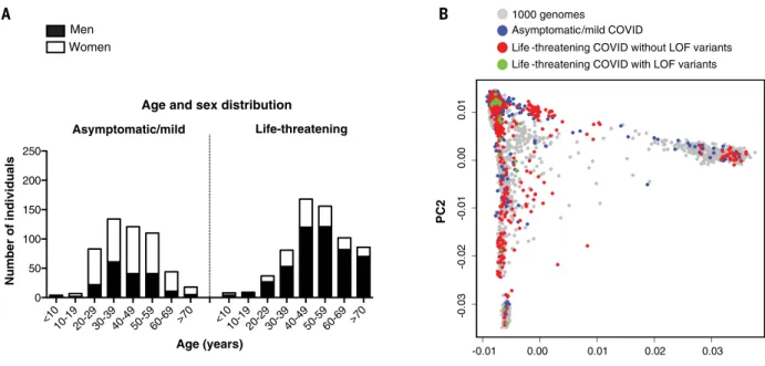
died) of various ancestries between 1 month
and 99 years of age (Fig. 1A). These patients
were hospitalized for life-threatening
pneumo-nia caused by SARS-CoV-2 (critical COVID-19).
We sequenced their whole genome (
N = 364)
or exome (
N = 295), and principal component
analysis (PCA) on these data confirmed their
ancestries (Fig. 1B).
Candidate variants at 13 human loci that
govern immunity to influenza virus
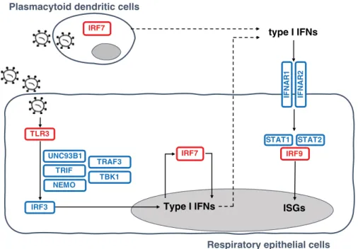
We first tested the specific hypothesis that
in-born errors of Toll-like receptor 3 (TLR3)
– and
interferon regulatory factor 7 (IRF7)
–dependent
type I interferon (IFN) immunity, which
un-derlie life-threatening influenza pneumonia,
may also underlie life-threatening COVID-19
pneumonia (
5
) (Fig. 2). We considered three
loci previously shown to be mutated in patients
with critical influenza pneumonia:
TLR3 (
6
),
IRF7 (
7
), and
IRF9 (
8
). We also considered
10 loci mutated in patients with other viral
illnesses but directly connected to the three
core genes conferring influenza susceptibility:
TICAM1/TRIF (
9
),
UNC93B1 (
10
),
TRAF3 (
11
),
TBK1 (
12
),
IRF3 (
13
), and
NEMO/IKBKG (
14
) in
the TLR3-dependent type I IFN induction
path-way, and
IFNAR1 (
15
),
IFNAR2 (
16
),
STAT1
(
17
), and
STAT2 (
18
) in the IRF7- and
IRF9-dependent type I IFN amplification pathway.
We collected both monoallelic and biallelic
nonsynonymous variants with a minor allele
frequency (MAF) <0.001 at all 13 loci. Twelve
of the 13 candidate loci are autosomal, whereas
NEMO is X-linked. For the latter gene, we
con-sidered only a recessive model (
19
).
Autosomal-dominant (AD) inheritance has not been proven
for six of the 12 autosomal loci (
UNC93B1, IRF7,
IFNAR1, IFNAR2, STAT2, and IRF9).
Never-theless, we considered heterozygous variants
because none of the patients enrolled had
been hospitalized for critical viral infections
before COVID-19, raising the possibility that
any underlying genetic defects that they might
have display a lower penetrance for influenza
and other viral illnesses than for COVID-19,
which is triggered by a more virulent virus.
Enrichment of variants predicted to be LOF
at the influenza susceptibility loci
We found four unrelated patients with
bial-lelic variants of
IRF7 or IFNAR1 (Table 1 and
table S1). We also found 113 patients carrying
113 monoallelic variants at 12 loci:
TLR3 (N = 7
patients/7 variants),
UNC93B1 (N = 10/9),
TICAM1 (N = 17/15), TRAF3 (N = 6/6), TBK1
(
N = 12/11), IRF3 (N = 5/5), IRF7 (N = 20/13),
IFNAR1 (N = 14/13), IFNAR2 (N = 17/15), STAT1
(
N = 4/4), STAT2 (N = 11/11), and IRF9 (N =
4/4). We detected no copy number variation
on May 26, 2021
http://science.sciencemag.org/
1St. Giles Laboratory of Human Genetics of Infectious Diseases, Rockefeller Branch, The Rockefeller University, New York, NY, USA.2Laboratory of Human Genetics of Infectious Diseases, Necker Branch,
INSERM U1163, Necker Hospital for Sick Children, Paris, France.3University of Paris, Imagine Institute, Paris, France.4Laboratory of Virology and Infectious Disease, The Rockefeller University, New York, NY,
USA. 5Department of Paediatric Infectious Diseases & Virology, Imperial College London, London, UK. 6Yale Center for Genome Analysis and Department of Genetics, Yale School of Medicine, New Haven, CT,
USA. 7Zukerman Mind Brain Behavior Institute, Columbia University, New York, NY, USA.8Helix, San Mateo, CA, USA.9Primary Immunodeficiencies Group, University of Antioquia UdeA, Medellin, Colombia.
10School of Microbiology, University of Antioquia UdeA, Medellin, Colombia.11Laboratory of Clinical Immunology and Microbiology, Division of Intramural Research, NIAID, NIH, Bethesda, MD, USA.12NIAID
Clinical Genomics Program, NIH, Bethesda, MD, USA.13Université de Paris, Institut de Recherche Saint-Louis, INSERM U976, Hôpital Saint-Louis, Paris, France. 14Laboratory of Genomes & Cell Biology of
Disease, INSERM U944, CNRS UMR 7212, Université de Paris, Institut de Recherche Saint-Louis, Hôpital Saint-Louis, Paris, France. 15Sorbonne Université, Inserm, Centre d’Immunologie et des Maladies
Infectieuses–Paris (CIMI PARIS), Assistance Publique-Hôpitaux de Paris (AP-HP) Hôpital Pitié-Salpêtrière, Paris, France. 16Translational Immunology Lab, Institut Pasteur, Paris, France.17Laboratory for Inborn
Errors of Immunity, Department of Microbiology, Immunology and Transplantation, Department of Pediatrics, University Hospitals Leuven, KU Leuven, Leuven, Belgium. 18Department of Microbiology, Icahn
School of Medicine at Mount Sinai, New York, NY, USA.19Sorbonne Université, UMS037, PASS, Plateforme de Cytométrie de la Pitié-Salpêtrière CyPS, Paris, France.20Bioinformatics Platform, Structure
Fédérative de Recherche Necker, INSERM UMR1163, Université de Paris, Imagine Institute, Paris, France.21Neurometabolic Diseases Laboratory, IDIBELL-Hospital Duran i Reynals, CIBERER U759, and Catalan
Institution of Research and Advanced Studies (ICREA), Barcelona, Spain.22Department of Immunology, Research Branch, Sidra Medicine, Doha, Qatar.23School of Life sciences, Ecole Polytechnique Fédérale
de Lausanne, Lausanne, Switzerland.24Precision Medicine Unit, Lausanne University Hospital and University of Lausanne, Lausanne, Switzerland.25Swiss Institue of Bioinformatics, Lausanne, Switzerland.
26Infectious Disease Susceptibility Program, Research Institute, McGill University Health Centre, Montréal, Québec, Canada.27Specialized Immunology Laboratory of Dr. Shahrooei, Sina Medical Complex, Ahvaz,
Iran.28Department of Microbiology and Immunology, Clinical and Diagnostic Immunology, KU Leuven, Leuven, Belgium.29Department of Pathology and Laboratory Medicine, College of Medicine, King Saud
University, Riyadh, Saudi Arabia.30Department of Clinical Immunology and Infectious Diseases, National Research Institute of Tuberculosis and Lung Diseases, Shahid Beheshti University of Medical Sciences,
Tehran, Iran.31The Clinical Tuberculosis and Epidemiology Research Center, National Research Institute of, Tuberculosis and Lung Diseases (NRITLD), Masih Daneshvari Hospital, Shahid Beheshti, University of
Medical Sciences, Tehran, Iran. 32Pediatric Respiratory Diseases Research Center, National Research Institute of Tuberculosis and Lung Diseases, Shahid Beheshti, Iran.33National Center of Genomics
Technology, King Abdulaziz City for Science and Technology, Riyadh, Saudi Arabia.34Dasman Diabetes Institute, Department of Genetics and Bioinformatics, Kuwait.35Immunology Research Laboratory,
Department of Pediatrics, College of Medicine and King Saud University Medical City, King Saud University, Riyadh, Saudi Arabia.36Translational Pathology, Department of Pathology and Laboratory Medicine,
King Abdulaziz Medical City, Misery of National Guard Health Affairs, Riyadh, Saudi Arabia.37Cancer & Blood Research, King Abdullah International Medical Research Center, Riyadh, Saudi Arabia.
38Amsterdam UMC, Department of Neurology, Amsterdam Neuroscience, Amsterdam, Netherlands. 39Pediatric Departement and Centro Tettamanti-European Reference Network PaedCan, EuroBloodNet,
MetabERN-University of Milano-Bicocca-Fondazione MBBM-Ospedale, San Gerardo, Monza, Italy. 40Department of Infectious Diseases, San Gerardo Hospital–University of Milano-Bicocca, Monza, Italy.41CREA
Laboratory, Diagnostic Laboratory, ASST Spedali Civili di Brescia, Brescia, Italy. 42Department of Infectious and Tropical Diseases, University of Brescia and ASST Spedali di Brescia, Brescia, Italy.43Chief
Medical Officer, ASST Spedali Civili di Brescia, Brescia, Italy. 44Bioinformatics and Computational Biosciences Branch, Office of Cyber Infrastructure and Computational Biology, NIAID, NIH, Bethesda, MD, USA.
45PRIMER, Uniformed Services University of the Health Sciences, Bethesda, MD, USA. 46Center of Human Genetics, Hôpital Erasme, Université Libre de Bruxelles, Brussels, Belgium.47Department of Internal
Medicine, Hôpital Erasme, Université Libre de Bruxelles, Brussels, Belgium.48Fonds de la Recherche Scientifique (FNRS) and Center of Human Genetics, Hôpital Erasme, Université Libre de Bruxelles, Brussels,
Belgium. 49Department of Paediatric Immunology and Pulmonology, Centre for Primary Immunodeficiency Ghent (CPIG), PID Research Lab, Jeffrey Modell Diagnosis and Research Centre, Ghent University
Hospital, Ghent, Belgium. 50Pediatric Infectious Diseases and Immunodeficiencies Unit, Hospital Universitari Vall d’Hebron, Vall d’Hebron Research Institute, Vall d’Hebron Barcelona Hospital Campus,
Universitat Autònoma de Barcelona (UAB), Barcelona, Catalonia, Spain. 51Immunology Division, Genetics Department, Hospital Universitari Vall d’Hebron, Vall d’Hebron Research Institute, Vall d’Hebron
Barcelona Hospital Campus, UAB, Barcelona, Catalonia, Spain.52Aix Marseille Univ, INSERM, INRAE, C2VN, CHU Timone, Marseille, France. 53Necmettin Erbakan University, Meram Medical Faculty, Division of
Pediatric Allergy and Immunology, Konya, Turkey.54Department of Infectious Diseases and Clinical Microbiology, Konya Training and Research Hospital, Konya, Turkey.55Department of Molecular Biology and
Genetics, Bilkent University, Bilkent-Ankara, Turkey. 56Departments of Infectious Diseases and Clinical Microbiology, Bakirkoy Dr. Sadi Konuk Training and Research Hospital, University of Health Sciences,
Istanbul, Turkey. 57Department of Immunology, Hospital Universitario de G.C. Dr. Negrín, Canarian Health System, Las Palmas de Gran Canaria, Spain.58University Fernando Pessoa Canarias, Las Palmas de
Gran Canaria, Spain. 59Department of Biomedicine and Prevention, University of Rome“Tor Vergata,” Rome, Italy. 60Intensive Care Unit, AP-HM, Marseille, France. 61Avicenne Hospital Intensive Care Unit,
APHP, Bobigny, INSERM U1272 Hypoxia & Lung, Paris, France. 62PH Réanimation CHU Avicenne, Bobigny, INSERM U1272 Hypoxie & Poumon, Paris, France.63Université de Paris, IAME UMR-S 1137, INSERM,
Paris, France. 64Inserm CIC 1425, Paris, France.65AP-HP, Département Epidémiologie Biostatistiques et Recherche Clinique, Hôpital Bichat, Paris, France.66Department of Pharmacology & Molecular
Therapeutics, Uniformed Services University of the Health Sciences, Bethesda, MD, USA. 67Department of Anatomy, Physiology & Genetics, Uniformed Services University of the Health Sciences, Bethesda,
MD, USA.68Division of Pediatric Allergy, Immunology and Rheumatology, Columbia University, New York, USA.69Department of Infectious Diseases, Aarhus University Hospital, Skejby, Denmark.
70Department of Biomedicine, Aarhus University, Aarhus, Denmark.71College of Health and Life Sciences, Hamad Bin Khalifa University, Doha, Qatar.72Department of Medical Microbiology, Utrecht UMC,
Utrecht, Netherlands. 73Study Center for Primary Immunodeficiencies, Necker Hospital for Sick Children, Paris, France.74Turnstone Biologics, New York, NY, USA.75Department of Pediatrics, University
Hospitals Leuven, KU Leuven, Leuven, Belgium. 76New York Genome Center, New York, NY, USA. 77AP-HP, Hôpital Saint-Louis, Laboratoire d’Immunologie, Paris, France.78Laboratory of Molecular
Immunology, Rockefeller University, New York, NY, USA.79Howard Hughes Medical Institute, New York, NY, USA.80Department of Medicine, Division of Infectious Diseases, Icahn School of Medicine at Mount
Sinai, New York, NY, USA.81Global Health and Emerging Pathogens Institute, Icahn School of Medicine at Mount Sinai, New York, NY, USA.82The Tisch Cancer Institute, Icahn School of Medicine at Mount
Sinai, New York, NY, USA.83Laboratory of Genetics and Genomics, The Rockefeller University, New York, NY, USA. 84Department of Genetics, Yale University School of Medicine, New Haven, CT, USA.85Yale
Center for Genome Analysis, Yale School of Medicine, New Haven, CT, USA. 86Pediatric Hematology and Immunology Unit, Necker Hospital for Sick Children, AP-HP, Paris, France.
*These authors contributed equally to this work.
†All collaborators and their affiliations appear at the end of this paper. ‡These authors contributed equally to this work.
§These authors contributed equally to this work. ¶Corresponding author. Email: [email protected]
<10 10-1920-2930-3940-4950-5960-69>7 0 <10 10-1 9 20-2 9 30-3940-4950-5960-6 9 >70 0 50 100 150 200 250
Numbe
r of indiv
idua
ls
Age and sex distribution
Asymptomatic/mild
Life-threatening
Men
Women
1000 genomes
Life -threatening COVID without LOF variants Asymptomatic/mild COVID
Life -threatening COVID with LOF variants
PC1
PC
2
-0 .0 3 -0 .0 2 -0 .0 1 0 .0 0 0 .0 1 -0.01 0.00 0.01 0.02 0.03Age (years)
A
B
Fig. 1. Demographic and genetic data for the COVID-19 cohort. (A) Age and sex distribution of patients with life-threatening COVID-19. (B) PCA of patient (with or
without LOF variants in the 13 candidate genes) and control cohorts (patients with mild or asymptomatic disease and individuals from the 1000 Genomes Project).
on May 26, 2021
http://science.sciencemag.org/
for these 13 genes. Unexpectedly, one of these
variants has been reported in patients with
life-threatening influenza pneumonia (
TLR3
p.Pro554Ser) (
6
,
20
) and another was shown
to be both deleterious and dominant-negative
(
IFNAR1 p.Pro335del) (
21
). Nine of the 118
biallelic or monoallelic variants were predicted
to be LOF (pLOF), whereas the remaining 109
were missense or in-frame indels (table S1). In
a sample of 534 controls with asymptomatic
or mild SARS-CoV-2 infection, we found only
one heterozygous pLOF variation with a MAF
<0.001 at the 13 loci (
IRF7 p.Leu99fs). A
PCA-adjusted burden test on the 12 autosomal
loci revealed significant enrichment in pLOF
variants in patients relative to controls [
P =
0.01; odds ratio (OR) = 8.28; 95% confidence
interval (CI) = 1.04 to 65.64] under an AD mode
of inheritance. The same analysis performed
on synonymous variants with a MAF <0.001
was not significant (
P = 0.19), indicating that
our ethnicity-adjusted burden test was well
calibrated.
Experimentally deleterious alleles at the
influenza susceptibility loci in 3.5%
of patients
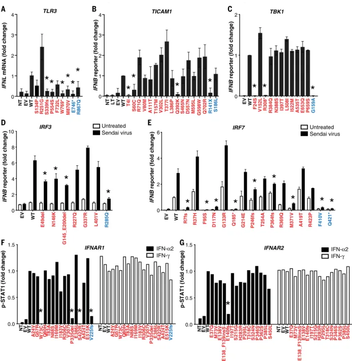
We tested these 118 variants experimentally in
ad hoc overexpression systems. We found that
24 variants of eight genes were deleterious
(including all the pLOF variants) because they
were loss-of-expression, LOF, or severely
hypo-morphic:
TLR3 (N = 4 variants), UNC93B1
(
N = 1), TICAM1 (N = 3), TBK1 (N = 2), IRF3
(
N = 2), IRF7 (N = 8), IFNAR1 (N = 3), and
IFNAR2 (N = 1) (table S1, Fig. 3, and figs. S1 to
S8). Consistently, heterozygous LOF variants
of
IRF3 and IRF7 were reported in single
pa-tients with life-threatening influenza
pneumo-nia (
22
,
23
). The remaining 94 variants were
biochemically neutral. Twenty-three patients
carried these 24 deleterious variants, resulting
in four autosomal-recessive (AR) deficiencies
(homozygosity or compound heterozygosity
IF NAR1 IF NAR2 STAT1 STAT2 IRF9 TLR3
type I IFNs
ISGs
Type I IFNs
IRF7 UNC93B1 TRAF3 TRIF NEMO IRF3 TBK1Plasmacytoid dendritic cells
Respiratory epithelial cells
IRF7Fig. 2. Illustration of TLR3- and IRF7-dependent type I IFN production and amplification circuit.
Molecules in red are encoded by core genes, deleterious variants of which underlie critical influenza
pneumonia with incomplete penetrance; deleterious variants of genes encoding biochemically related molecules in
blue underlie other viral illnesses. Type I IFNs also induce themselves. ISGs, interferon-stimulated genes.
Table 1. Disease-causing variants identified in patients with life-threatening COVID-19.
Gene
Inheritance
Genetic form
Genotype
Gender
Age [years]
Ancestry/residence
Outcome
TLR3
AD
Known
p.Ser339fs/WT
M
40
Spain
Survived
...
TLR3
AD
Known
p.Pro554Ser/WT
M
68
Italy
Survived
...
TLR3
AD
Known
p.Trp769*/WT
M
77
Italy
Survived
...
TLR3
AD
Known
p.Met870Val/WT
M
56
Colombia/Spain
Survived
...
UNC93B1
AD
New
p.Glu96*/WT
M
48
Venezuela/Spain
Survived
...
TICAM1
AD
Known
p.Thr4Ile/WT
M
49
Italy
Survived
...
TICAM1
AD
Known
p.Ser60Cys/WT
F
61
Vietnam/France
Survived
...
TICAM1
AD
Known
p.Gln392Lys/WT
F
71
Italy
Deceased
...
TBK1
AD
Known
p.Phe24Ser/WT
F
46
Venezuela/Spain
Survived
...
TBK1
AD
Known
p.Arg308*/WT
M
17
Turkey
Survived
...
IRF3
AD
Known
p.Glu49del/WT
F
23
Bolivia/Spain
Survived
...
IRF3
AD
Known
p.Asn146Lys/WT
F
60
Italy
Survived
...
IRF7
AR
Known
p.Pro364fs/p.Pro364fs
F
49
Italy/Belgium
Survived
...
IRF7
AR
Known
p.Met371Val/p.Asp117Asn
M
50
Turkey
Survived
...
IRF7
AD
New
p.Arg7fs/WT
M
60
Italy
Survived
...
IRF7
AD
New
p.Gln185*/WT
M
44
France
Survived
...
IRF7
AD
New
p.Pro246fs/WT
M
41
Spain
Survived
...
IRF7
AD
New
p.Arg369Gln/WT
M
69
Italy
Survived
...
IRF7
AD
New
p.Phe95Ser/WT
M
37
Turkey
Survived
...
IFNAR1
AR
Known
p.Trp73Cys/Trp73Cys
M
38
Turkey
Survived
...
IFNAR1
AR
Known
p.Ser422Arg/Ser422Arg
M
26
Pakistan/Saudi Arabia
Deceased
...
IFNAR1
AD
New
p.Pro335del/WT
F
23
China/Italy
Survived
...
IFNAR2
AD
New
p.Glu140fs/WT
F
54
Belgium
Survived
...
on May 26, 2021
http://science.sciencemag.org/
0 2 4 6 EV WT R7 fs R37H G133 R Q185* G214 E P364fs R369Q P246fs T254A M371V A419T F410V Q421* D117N R423P F95S IRF7 IFNB
reporter (fold change
) UntreatedSendai virus
*
* *
*
*
*
*
* *
IFNAR1 IFN-α2
IFN-γ IFN-α2IFN-γ
IFNAR2
A
0 1 2 EV WT F24S N388S I397 T I522M A535T R308* R384Q P659SV152L L508I E653Q G159A
TBK1
IFNB
reporter (fold change
)
* *
*
C
D
0 1 2 3 4 NT LT EV WT T4I S60C R71Q V80M T157M V302L T377I Q392K S465N D557N M595L G598 W Q702R R141X S186L TICAM1 IFNBreporter (fold change
) A111T L386P
*
*
*
*
B
E
F
G145_E200delG
0 2 4 6 8 10 EV E49del N146K WT R285Q R227Q G337R IRF3 IFNBreporter (fold change
) UntreatedSendai virus
*
L401V*
*
*
W769* 0 1 2 3 4 R251G NT EV WT M870V S339fs P554S F732L E746* R867Q S134P IFNLmRNA (fold change
) TLR3
** * *
*
*
p-S T A T 1 (f o ld c h a n ge ) NT EV WT E 138V E 37Q E 140f s H 283R I197T M 73V P 295L P 346S P 362S P385L S 215G S 324N S450L T 318C NT EV WT E 138V E 37Q E 140f s H 283R I197T M 73V P 295L P 346S P 362S P385L S 215G S 324N S450L T 318C p-S T A T 1 (f o ld c h a n ge ) 0.0 0.5 1.0 1.5 E 138_F 139in sS E 138_F 139in sS W 73C NT WT P 335d el EV S 422R A 24V Q 80H N 88S A 424T I183V E386L E515K I169M T 83A G 57R V 225f s 0.0 0.5 1.0 1.5 R 306C V307I W73C NT WT P 335d el EV S 422R A 24V Q 80H N 88S A 424T I183V E386L E515K I169M T 83A G 57R V 225f s R 306C V307IFig. 3. Impact of
TLR3, TICAM1, TBK1, IRF3, IRF7, IFNAR1, and IFNAR2
variants on type I IFN signaling. (A) TLR3-deficient P2.1 fibrosarcoma cells
were stably transfected with plasmids expressing WT or mutant forms of TLR3,
and IFNL1 mRNA levels were determined by reverse transcription quantitative
PCR. IFNL1 mRNA levels were expressed relative to the housekeeping gene GUS
and then normalized. IFNL1 was undetectable in unstimulated cells. The differences
between variants and WT were tested using one-way ANOVA (*P < 0.05). (B)
TICAM1-deficient SV40-Fib cells were transiently transfected with WT or mutant forms of
TICAM1, together with an IFN-b luciferase reporter and a constitutively expressed
reporter. Normalized luciferase induction was measured 24 hours after
transfection. The differences between variants and WT were tested using one-way
ANOVA (*P < 0.05). (C) HEK293T cells were transiently transfected with WT
and mutant forms of TBK1, together with an IFN-b luciferase reporter and a
constitutively expressed reporter. Normalized luciferase activity was measured
24 hours after transfection. The differences between variants and WT were tested
using one-way ANOVA (*P < 0.05). (D) IRF3-deficient HEK293T cells were
transiently transfected with WT and mutant forms of IRF3, together with an IFN-b
luciferase reporter and a constitutively expressed reporter. Cells were either
left untreated or infected with Sendai virus for 24 hours before the normalized
measurement of luciferase activity. The differences between variants and WT were
evaluated using two-way ANOVA (*P < 0.05). (E) HEK293T cells were transiently
transfected with WT and mutant forms of IRF7, together with an IFN-b luciferase
reporter and a constitutively expressed reporter. Cells were either left untreated
or infected with Sendai virus for 24 hours before the normalized measurement of
luciferase activity. The differences between variants and WT were tested using
two-way ANOVA (*P < 0.05). (F and G) IFNAR1- or IFNAR2-deficient SV40-Fib cells
were transiently transfected with WT or mutant forms of IFNAR1 for 36 hours,
and either left untreated or stimulated with IFN-a2 or IFN-g. Fluorescence-activated
cell sorting (FACS) staining with anti-p-STAT1 antibody and the z-score of the MFI
were assessed. Asterisks indicate variants with MFI <50% of WT. Variants in red were
identified in COVID-19 patients. Variants in blue are known deleterious variants and
served as negative controls. EV, empty vector; LT, lipofectamine. Three technical
repeats were performed for (A) to (E). Means and SD are shown in the columns and
horizontal bars when appropriate.
on May 26, 2021
http://science.sciencemag.org/
for
IRF7; homozygosity for IFNAR1) and 19 AD
deficiencies. These 23 patients did not carry
candidate variants at the other 417 loci known to
underlie inborn errors of immunity (table S2)
(
24
–
26
). These findings suggest that at least
23 (3.5%) unrelated patients of the 659 patients
tested suffered from a deficiency at one of eight
loci among the 13 tested: four patients with a
known AR disorder (
IRF7 or IFNAR1) (
7
,
15
),
11 with a known AD disorder (
TLR3, TICAM1,
TBK1, or IRF3) (
6
,
9
,
12
,
13
,
20
), and eight with
a previously unknown AD genetic disorder
(
UNC93B1, IRF7, IFNAR1, or IFNAR2).
Impaired TLR3- and IRF7-dependent type I
immunity in patient cells in vitro
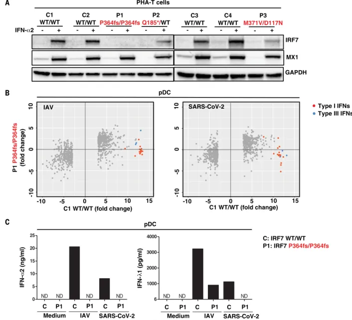
We tested cells from patients with selected
genotypes and showed that PHA-driven T cell
blasts (PHA-T cells) from patients with AR
or AD IRF7 deficiency had low levels of IRF7
expression (Fig. 4A). We then isolated
circulat-ing plasmacytoid dendritic cells (pDCs) from
a patient with AR IRF7 deficiency (fig. S9A)
(
7
). These cells were present in normal
pro-portions (fig. S9B), but they did not produce
any detectable type I or III IFNs in response to
SARS-CoV-2, as analyzed by cytometric bead
array (CBA), enzyme-linked immunosorbent
assay (ELISA), and RNA sequencing
(RNA-seq) (Fig. 4, B and C). We also showed that
PHA-T cells from a patient with AR IFN-a/b
receptor 1 (IFNAR1) deficiency had impaired
IFNAR1 expression and responses to IFN-a2 or
IFN-
b, and that the patient’s SV40-transformed
fibroblast (SV40-Fib) cells did not respond to
IFN-a2 or IFN-b (Fig. 5). We then infected
TLR3
−/−, TLR3
+/−, IRF7
−/−SV40-Fib cells, and
IRF7
−/−SV40-Fib cells rescued with wild-type
(WT) IRF7; IFNAR1
−/−SV40-Fib cells, and
IFNAR1
−/−SV40-Fib cells rescued with WT
-
+
-
+
-
+
-
+
IFN- 2
C1
WT/WT
C2
WT/WT
P1
P364fs/P364fs
P2
Q185*/
WT
IRF7
MX1
GAPDH
-
+
-
+
-
+
P3
M371V/D117N
C3
WT/WT
C4
WT/WT
PHA-T cells
%& '()*+ $% &',* -. /0% '()*+ /0% ',*-.1 345' ()*+ 2345' ,*-.1 0 5 10 15 20 25 ND ND ND ND ,-: 3 9 '; <=> & 0 ?Medium
IAV
C
P1
C
P1
C
P1
IFN-2
(ng
/ml)
SARS-CoV-2
%& '()*+ $% &' ,*-. /0%' ()*+ /0%' ,*-.1 345' ()*+ 345' ,*-.1 6 1000 2000 3000 4000 ND ND ND ,-;+ 7 '< =>? & 0 @Medium
IAV
C
P1
C
P1
C
P1
SARS-CoV-2
IFN-1
(pg
/ml)
C: IRF7 WT/WT
P1: IRF7
P364fs/P364fs
pDC
-10
-5
0
5
10
15
-1
0
-5
0
5
10
-10
-5
0
5
10
15
-1
0
-5
0
5
10
IAV
SARS-CoV-2
C1 WT/WT (fold change)
C1 WT/WT (fold change)
P1
P364f
s
/P364f
s
(fol
d
c
h
a
nge
)
pDC
Type I IFNs
Type III IFNs
A
B
C
Fig. 4. Type I IFN responses in patient cells defective for IRF7. (A) Levels
of the IRF7 protein in PHA-T cells from two patients with AR IRF7 deficiency
(P1 and P3), one patient with AD IRF7 deficiency (P2), and four healthy donors
(C1 to C4). Cells were either left untreated or stimulated with IFN-a2 for
24 hours, and protein levels were measured by Western blotting. MX1 was used
as a positive control for IFN-a2 treatment. (B) pDCs isolated from an AR
IRF7-deficient patient (P1) and a healthy donor (C1) were either left untreated or
infected with influenza A virus (IAV) or SARS-CoV-2, and RNA-seq was performed.
Genes with expression >2.5-fold higher or lower in C1 after infection are plotted
as the fold change in expression. Red dots are type I IFN genes; blue dots are type III
IFN genes. (C) pDCs isolated from healthy donor C and IRF7-deficient patient
(P1) were either left untreated (Medium) or infected with IAV or SARS-CoV-2,
and the production of IFN-a2 and IFN-l1 was measured by CBA and ELISA,
respectively, on the supernatant. ND, not detected.
on May 26, 2021
http://science.sciencemag.org/
IFNAR1, all of which were previously
trans-duced with angiotensin-converting enzyme 2
(ACE2) and transmembrane protease, serine 2
(TMPRSS2). SARS-CoV-2 infection levels were
higher in mutant cells than in cells from healthy
donors, and transduction of WT
IRF7 or IFNAR1
rescued their defects (Fig. 6). Collectively, these
findings showed that AR IRF7 deficiency
im-paired the production of type I IFN by pDCs
stimulated with SARS-CoV-2, whereas AR and
AD deficiencies of TLR3 or AR deficiency of
IFNAR1 impaired fibroblast-intrinsic type I
IFN immunity to SARS-CoV2. They also
sug-gest that heterozygosity for LOF variations at
the other five mutated loci also underlie
life-threatening COVID-19.
Impaired production of type I IFNs in
patients in vivo
We tested whether these genotypes impaired
the production of type I IFN in vivo during the
course of SARS-CoV-2 infection. We measured
the levels of the 13 types of IFN-a in the blood
of patients during the acute phase of COVID-19.
We found that 10 of the 23 patients with
mutations for whom samples were available
(one with AR IRF7 deficiency, four with AD
IRF7 deficiency, one with AD TLR3 deficiency,
two with AD TBK1 deficiency, one with AR
IFNAR1 deficiency, and one with AD TICAM1
deficiency) had serum IFN-a levels <1 pg/ml
(Fig. 7). By contrast, previously published
co-horts of patients hospitalized with unexplained,
severe COVID-19 had various serum IFN-a
levels, significantly higher than our 10 patients
[one-way analysis of variance (ANOVA),
P =
1.4 × 10
−7; Fig. 7] (
27
,
28
). Another 29 patients
from our cohort displaying auto-antibodies
(auto-Abs) against type I IFNs, reported in
an accompanying paper, had undetectable
levels of serum IFN-a (
29
). Moreover, none of
the 23 patients with LOF mutations of the
eight genes had detectable auto-Abs against
type I IFNs (
29
), strongly suggesting that the
two mechanisms of disease are similar but
independent. Excluding patients with
auto-Abs against type I IFN from the burden test
of pLOF variants at the 12 autosomal loci
strengthened the association signal (
P = 0.007;
OR = 8.97; 95% CI = 1.13 to 71.09).
Inborn errors of TLR3- and IRF7-dependent
type I immunity underlie critical COVID-19
Collectively, our data suggest that at least 23 of
the 659 patients with life-threatening COVID-19
pneumonia studied had known (six disorders)
or new (four disorders) genetic defects at eight
loci involved in the TLR3- and IRF7-dependent
induction and amplification of type I IFNs.
This discovery reveals the essential role of
both the double-stranded RNA sensor TLR3
and type I IFN cell-intrinsic immunity in the
control of SARS-CoV-2 infection in the lungs,
consistent with their previously documented
roles in pulmonary immunity to influenza
virus (
5
–
8
). These genotypes were silent until
infection with SARS-CoV-2. The most
thought-provoking examples are the AR deficiencies
of IRF7 and IFNAR1. AR IRF7 deficiency was
diagnosed in two individuals aged 49 and
50 years, and AR IFNAR1 deficiency was
diag-nosed in two individuals aged 26 and 38 years,
and none of the four patients had a prior
history of life-threatening infections (Table 1).
One patient with IRF7 deficiency was tested
and was seropositive for several common
ruses, including various influenza A and B
vi-ruses (figs. S10 and S11). These genetic defects
therefore display incomplete penetrance for
influenza respiratory distress and only
man-ifested clinically upon infection with the more
virulent SARS-CoV-2.
Conclusion
The AR form of IFNAR1 deficiency highlights
the importance of type I IFN production
rela-tive to type III IFN production, which is also
impaired by defects of TLR3, IRF7, and IRF9
(
5
). This conclusion is also supported by our
accompanying report of neutralizing auto-Abs
against type I IFNs, but not type III IFNs, in
other patients with life-threatening COVID-19
pneumonia (
29
). Inborn errors of TLR3- and
20
40
60
80
100
0
20
40
60
80
100
0
p-STAT1
% of Ma
x
IFN-IFN- 2
IL-27
0
0
0
0
0
0
0
10
210
310
410
210
310
410
210
310
4P5
W73C/W73C
IFN- 2
IFN-
IFN-100
80
60
40
20
0
10
210
110
310
110
210
310
110
210
3100
80
60
40
20
0
C3
WT/WT
PHA-T cells
SV40-Fib cells
Unstimulated
Stimulated
A
100
IFNAR1
% of Ma
x
C1
C2
P5
isotype mAb
anti -IFNAR1 mAb
C1, C2: IFNAR1 WT/WT
P5: IFNAR1
W73C/W73C
PHA-T cells
10
110
210
310
110
210
310
110
210
320
40
60
80
0
B
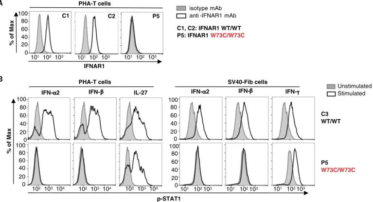
Fig. 5. Type I IFN responses in patient cells defective for IFNAR1. (A) FACS staining of IFNAR1 on the surface of PHA-T cells from a patient with AR IFNAR1
deficiency (P5) and healthy donors (C1 and C2). (B) PHA-T cells and SV40-Fib from a patient with AR IFNAR1 deficiency (P5) and a healthy donor (C3) were
stimulated with IFN-a2 or IFN-b, and p-STAT1 levels were determined by FACS. Interleukin-27 stimulation served as a positive control on PHA-T cells, whereas IFN-g
stimulation served as a positive control on SV40-Fib cells.
on May 26, 2021
http://science.sciencemag.org/
IRF7-dependent type I IFN immunity at eight
loci were found in as many as 23 patients (3.5%)
of various ages (17 to 77 years) and ancestries
(various nationalities from Asia, Europe, Latin
America, and the Middle East) and in patients
of both sexes (Table 1). Our findings suggest
that there may be mutations in other type I
IFN
–related genes in other patients with
life-threatening COVID-19 pneumonia. They also
suggest that the administration of type I IFN
may be of therapeutic benefit in selected
patients, at least early in the course of
SARS-CoV-2 infection.
Methods
Patients
We included in this study 659 patients with
life-threatening COVID-19 pneumonia, defined
as patients with pneumonia who developed
critical disease, whether pulmonary with
mech-anical ventilation (CPAP, BIPAP, intubation,
hi-flow oxygen), septic shock, or with any other
organ damage requiring admission to the
intensive care unit. Patients who developed
Kawasaki-like syndrome were excluded. The
age of the patients ranged from 0.1 to 99 years,
with a mean age of 51.8 years (SD 15.9 years),
and 25.5% of the patients were female. As
con-trols, we enrolled 534 individuals infected
with SARS-CoV-2 based on a positive
poly-merase chain reaction (PCR) and/or
serologi-cal test and/or the presence of typiserologi-cal symptoms
such as anosmia or ageusia after exposure to
a confirmed COVID-19 case, who remained
asymptomatic or developed mild, self-healing,
ambulatory disease.
Next-generation sequencing
Genomic DNA was extracted from whole blood.
For the 1193 patients and controls included,
the whole exome (
N = 687) or whole genome
(
N = 506) was sequenced. We used the
Ge-nome Analysis Software Kit (GATK) (version
3.4-46 or 4) best-practice pipeline to analyze
our whole-exome
–sequencing data (
30
). We
aligned the reads obtained with the human
reference genome (hg19) using the maximum
exact matches algorithm in Burrows
–Wheeler
Aligner software (
31
). PCR duplicates were
re-moved with Picard tools (
http://broadinstitute.
github.io/picard/
). The GATK base quality score
recalibrator was applied to correct sequencing
artifacts.
All of the variants were manually curated
using Integrative Genomics Viewer (IGV) and
confirmed to affect the main functional
pro-tein isoform by checking the propro-tein sequence
before inclusion in further analyzes. The main
functional protein isoforms were TLR3 (NM_
003265), UNC93B1 (NM_030930.4), TICAM1
(NM_182919), TRAF3 (NM_145725.2), TBK1
(NM_013254.4), IRF3 (NM_001571), IRF7 (NM_
001572.5), IFNAR1 (NM_000629.3), IFNAR2
(NM_001289125.3), STAT1 (NM_007315.4), STAT2
S-protein MFI (z-score)
S -prote in pos itiv e (%) -2 0 2 0 20 40 60 80 100 -2 0 2 0 20 40 60 80 100 -2 0 2 0 20 40 60 80 100 -2 0 2 0 20 40 60 80 100 -2 0 2 0 20 40 60 80 100 -2 0 2 0 20 40 60 80 100 -2 0 2 0 20 40 60 80 100 -2 0 2 0 20 40 60 80 100 -2 0 2 0 20 40 60 80 100 -2 0 2 0 20 40 60 80 100 -2 0 2 0 20 40 60 80 100 -2 0 2 0 20 40 60 80 100 Mock SARS-CoV-2 untreated IFN-untreated IFN-untreated IFN-TLR3 WT/WT TLR3 P554S/WT TLR3 P554S/E746* IRF7 F410V/Q421* IRF7 F410V/Q421* + LUC IRF7 F410V/Q421* + WT IFNAR1 WT/WT + EV IFNAR1 WT/WT + WT IFNAR1 V225fs/V225fs + EV IFNAR1 V225fs/V225fs + WT Mock SARS-CoV-2 Mock SARS-CoV-2