The MIT Faculty has made this article openly available. Please share
how this access benefits you. Your story matters.
Citation Kruszka, Paul et al. "Circle of Willis anomalies in Turner syndrome: Absent A1 segment of the anterior cerebral artery." Birth Defects Research 111, 19 (October 2019): 1584-1588 © 2019 Wiley
Periodicals
As Published http://dx.doi.org/10.1002/bdr2.1609
Publisher Wiley
Version Original manuscript
Citable link https://hdl.handle.net/1721.1/126421
Terms of Use Creative Commons Attribution-Noncommercial-Share Alike
For Peer Review
Circle of Willis Anomalies in Turner Syndrome: Absent A1 Segment of
the Anterior Cerebral Artery
Paul Kruszka1, Ashley Buscetta1, Maria T. Acosta1,2, Nicole Banks1, Yonit A.
Addissie1, Camilo Toro2, Marie Luby3, Lawrence Latour3, Gilbert Vezina4, David C.
Page5,Maximilian Muenke1
1. Medical Genetics Branch, National Human Genome Research Institute, National Institutes of Health, Bethesda, MD
2. Undiagnosed Disease Network, National Human Genome Research Institute, National Institutes of Health, Bethesda, MD
3. Stroke Branch, National Institute of Neurological Disorders and Stroke, National Institutes of Health, Bethesda, MD
4. The Children’s Research Institute, Children’s National Health System, Washington, D.C.
5. Whitehead Institute, Cambridge, MA; Dept. of Biology, Massachusetts Institute of Technology, Cambridge, MA; Howard Hughes Medical Institute, Whitehead Institute, Cambridge, MA
Correspondence: Paul Kruszka, paul.kruszka@nih.gov; 301-402-9654 and Maximilian Muenke, mamuenke@mail.nih.gov; 301-402-8167
John Wiley & Sons, Inc.
3 4 5 6 7 8 9 10 11 12 13 14 15 16 17 18 19 20 21 22 23 24 25 26 27 28 29 30 31 32 33 34 35 36 37 38 39 40 41 42 43 44 45 46 47 48 49 50 51 52 53 54 55 56 57 58 59
For Peer Review
ABSTRACT
Purpose: Turner syndrome is the most common sex chromosome disorder in
women and is associated with a higher than expected death rate secondary to cerebrovascular disease, including stroke. This study evaluates the cerebral vascular anatomy of individuals with Turner syndrome.
Methods: Twenty-one women with Turner syndrome had brain magnetic
resonance angiography (MRA). These MRAs were evaluated in a blinded manner with a control group of 25 men and 25 women who had MRA imaging for multiple indications including migraine headaches, psychiatric disorders, and seizures.
Results: Twenty-nine percent of women with Turner syndrome were missing an A1
segment of the anterior cerebral artery compared to 0% in the control group
(p<0.001). There were no other significant differences in the circle of Willis (COW) in women with Turner syndrome compared to the control group. A complete COW was found in 3/21 (14%) of women with Turner syndrome and 12/47 (26%) controls (p=0.36).
Conclusion: Women with Turner syndrome have a significantly different intracranial
vascular anatomy, specifically the absence of the A1 segment of the anterior
cerebral artery when compared to male and female controls. More research in brain imaging in women with Turner syndrome and stroke and other cerebrovascular disease is needed to determine the clinical significance of this anomaly.
Keywords: Turner syndrome; monosomy X; circle of Willis; magnetic resonance
angiography; cerebrovascular disease
John Wiley & Sons, Inc.
3 4 5 6 7 8 9 10 11 12 13 14 15 16 17 18 19 20 21 22 23 24 25 26 27 28 29 30 31 32 33 34 35 36 37 38 39 40 41 42 43 44 45 46 47 48 49 50 51 52 53 54 55 56 57 58 59
For Peer Review
INTRODUCTION
Turner syndrome (TS) occurs in 1.0-1.5% of clinically recognized conceptuses, but only 1 in 2000 females have TS, due to a high percentage of embryonic and fetal loss.(Hook & Warburton, 2014; Stochholm, Juul, Juel, Naeraa, & Gravholt, 2006) TS is the result of complete or partial absence of the second sex chromosome. The classic phenotype is short stature, infertility secondary to ovarian dysgenesis,
congenital heart disease, and exam findings that include low hair line and
lymphedema. Mortality in TS is three-fold higher than in the general population and cerebrovascular disease plays a role in this statistic.(Schoemaker et al., 2008) Multiple epidemiological studies have documented higher than expected deaths due to cerebrovascular disease in TS.(Gravholt, Juul, Naeraa, & Hansen, 1998;
Schoemaker et al., 2008; Stochholm et al., 2006) Although structural anomalies of the heart and aorta have been thoroughly evaluated in TS, variants in the cerebral vascular anatomy are not well documented.
The circle of Willis (COW) is a cerebral vascular anastomosis at the base of the brain that provides for collateral circulation to the brain (Figure 1A). Blood supply to the brain is primarily divided into the anterior circulation and the posterior circulation. The anterior circulation originates from the internal carotid arteries (ICA) which
supply the anterior cerebral arteries (ACA) and middle cerebral arteries (MCA); the posterior circulation originates from the vertebral arteries, which join to form the basilar artery, and again bifurcate to form the posterior cerebral arteries (PCA) (Figure 1B). The posterior communicating arteries (Pcom) function as anastomoses joining the anterior to the posterior circulation, and the anterior communicating artery (Acom) joins the left and right anterior cerebral arteries.
John Wiley & Sons, Inc.
3 4 5 6 7 8 9 10 11 12 13 14 15 16 17 18 19 20 21 22 23 24 25 26 27 28 29 30 31 32 33 34 35 36 37 38 39 40 41 42 43 44 45 46 47 48 49 50 51 52 53 54 55 56 57 58 59
For Peer Review
The circle consists of the left and right A1 segments of the ACAs originating from the ICAs, the Acom, the left and right P1 segments of the PCAs originating from the basilar artery, and the left and right Pcoms joining the PCAs to the ICAs (Figure 1B). Normal variants in the COW, such as a “fetal” PCA with an absent P1 segment, are not uncommon and can result in an incomplete circle. In a large autopsy series of normal brains, a complete COW was found in only 52% of individuals, with the most common anomalies occurring in the Pcoms and Acoms.(Alpers, Berry, & Paddison, 1959) In a more recent study of 111 healthy volunteers, only 42% of individuals had a complete COW.(Krabbe-Hartkamp et al., 1998) Variants in the COW limit proximal pathways for collateral flow and may increase the propensity for infarction due to a vascular occlusion.
In this study we use cerebral magnetic resonance angiography (MRA) to compare the anatomy of the circle of Willis (COW) of TS patients to an age matched control cohort of patients evaluated to rule-out acute stroke.
MATERIALS AND METHODS
Twenty-one individuals with TS were evaluated at the Clinical Center at the National Institutes of Health (NIH) as part of the Sex Chromosome Disorders Study (12-HG-0000). The evaluation included a physical exam (P.K., A.B., M.T.A., N.B.), a 200 metaphase karyotype, echocardiography, and 3-Dimensional time-of-flight MRA of the cerebral vasculature.
John Wiley & Sons, Inc.
3 4 5 6 7 8 9 10 11 12 13 14 15 16 17 18 19 20 21 22 23 24 25 26 27 28 29 30 31 32 33 34 35 36 37 38 39 40 41 42 43 44 45 46 47 48 49 50 51 52 53 54 55 56 57 58 59
For Peer Review
A control cohort of 50 age-matched patients (25 males and 25 females) also underwent time-of-flight MRA as part of an evaluation to rule-out acute stroke and were selected from an existing de-identified, delinked dataset accessed under a “not human subjects” research determination. Both males and females were used as controls the for purpose of varying the chromosome X dosage. The etiology and final diagnosis for the control cohort was not stroke and is summarized in Table S1.
Multiple different 3T MRI systems were used (See Table S2 for cases and Table S3 for controls for model, manufacturer, and field strength used for each individual in this study); however, the parameters used to acquire the MRA were comparable. All identifiers were removed from the MRA images and replaced with randomly
assigned new identification numbers, blinding TS vs control, and inferior to superior coverage of MRA images was standardized to maintain the blind. For all study participants, the field of view was set from the vertebrobasilar junction to the pons curve (approximately 6cm) and the image slice thickness was set at 0.78 millimeters (mm). The blinded evaluators (G.V. and M.T.A.) were given four files for each participant: 1. 76 axial slices at a thickness of 0.78 mm; 2. A reformatted axial view with a maximum intensity projection (MIP) of 10 mm and 29 slices; 3. Spin view with maximum MIP with 80 frames for a 180 degree horizontal rotational view; 4. Tumble view with maximum MIP, 80 frames and 180 degrees of vertical rotation.
In this study, two scores were possible for cerebral arteries: “absent” or “present”. In previous studies the category of hypoplastic cerebral arteries has been used for diameters less than 0.8 mm-1.0 mm (Alpers & Berry, 1963; Krabbe-Hartkamp et al., 1998). Krabbe-Hartkamp et al. considered hypoplastic cerebral arteries to be in the
John Wiley & Sons, Inc.
3 4 5 6 7 8 9 10 11 12 13 14 15 16 17 18 19 20 21 22 23 24 25 26 27 28 29 30 31 32 33 34 35 36 37 38 39 40 41 42 43 44 45 46 47 48 49 50 51 52 53 54 55 56 57 58 59
For Peer Review
same category as absent arteries for study classification purposes; whereas,
Shaban et al. grouped hypoplastic and intact arteries in the same category (Shaban et al., 2015). Similar to the Shaban study which studied acute ischemic stroke and its association with the A1 segment of the anterior cerebral artery, the present study considered hypoplastic arteries as intact. Statistical analysis was completed using Microsoft Excel and Fisher exact test.
RESULTS
Twenty-one women with TS were recruited for this study and underwent brain MRA and clinical evaluation (Table S1 summarizes cohort and Table S2 characterizes the participants individually). The average age of these women was 45 years with a range of 27 to 71 years, and the majority of women were Caucasian (Table S2). Table S1 shows a summation of characteristics of cases and controls. For women with TS, 38% had bicuspid aortic valve and 33% had coarctation of the aorta. The most common risk factor for atherosclerotic disease was hypertension in 38% of the women with TS. Table S2 lists each woman with TS individually, her karyotype, and echocardiography results; 19/21 (90%) of the women with TS were 45,X
(non-mosaic based on analysis of 1,000 metaphase karyotypes). Three controls were discarded due to excess motion artifacts of the brain MRAs, leaving 47 for analysis. Migraine headache and psychiatric indications were the most common diagnoses in the control group (Table S1 and Table S3). The controls were age matched
resulting in similar mean ages and age ranges for cases and controls (Table S1).
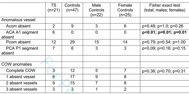
Table 1 shows the summation of MRA analysis. The most significant finding is absence of the A1 segment of the anterior cerebral artery in 6/21 (29%) women with
John Wiley & Sons, Inc.
3 4 5 6 7 8 9 10 11 12 13 14 15 16 17 18 19 20 21 22 23 24 25 26 27 28 29 30 31 32 33 34 35 36 37 38 39 40 41 42 43 44 45 46 47 48 49 50 51 52 53 54 55 56 57 58 59
For Peer Review
TS and 0/47 of the controls (p<0.001) (see Figure 1C shows a schematic of the missing A1 segment and Figure 1D shows an MRA with the A1 segment absent. However, the proportion of subjects with variants in the communicating arteries did not differ between TS and control. The anterior communicating artery was absent in 2/21 (10%) women with TS and 9/47 (19%) of controls (p=0.48); and, the posterior communicating artery was absent in similar fractions of TS cases and controls: 12/21 (57%) and 29/47 (62%), respectively. Every woman with TS and a missing A1 segment of the anterior cerebral artery had an intact anterior communicating artery, allowing for collateral circulation to the contralateral COW (Table S2). As seen in Table 1, there were no statistical differences in fraction of complete COWs or the number of anomalies between women with TS and total controls, male controls or female controls. In women with TS, 3/21 (14%) had a complete COW compared with 26% of controls (p=0.36). Tables S2 and S3 list the results for each case and control, respectively.
Table S2 shows the chest MRA and echocardiogram results for each individual with TS. Evaluating for correlation between heart malformations and COW anomalies, 3 of 6 individuals with missing A1 segments had bicuspid aortic valves compared to 5 of 9 individuals with intact A1 segments (P=0.64; Fisher exact test). For coarctation of the aorta, 3 of 6 individuals with missing A1 segments had coarctation of the aorta compared to 4 of 9 individuals with intact A1 segments (P=0.62: Fisher exact test). Interestingly, only two individuals had intact COWs and both of these individuals have tricuspid aortic valves and did not have coarctation of the aorta (Table S2).
John Wiley & Sons, Inc.
3 4 5 6 7 8 9 10 11 12 13 14 15 16 17 18 19 20 21 22 23 24 25 26 27 28 29 30 31 32 33 34 35 36 37 38 39 40 41 42 43 44 45 46 47 48 49 50 51 52 53 54 55 56 57 58 59
For Peer Review
Two of the individuals with TS (number 4036 and 4077 in Table S2) are sisters, both with 45,X and cleft palate. However, their COW anatomies are not similar; Individual 4036 has an absent A1 segment on the left side and her sister has both A1 segments present.
DISCUSSION
In this study, we document the anatomy of the circle of Willis (COW) using 3D time-of-flight MRA of 21 women with TS and compare to 47 age-matched control males and females. As noted in previous studies, the anterior and posterior
communicating arteries were the most commonly missing or dysplastic.(Alpers et al., 1959) Here we find that a high proportion of women with TS, 6/21 (29%), were missing an A1 segment of the anterior cerebral artery compared to none of 47 controls (p<0.001).
In the general population, an absent or dysplastic COW artery is common, especially the posterior communicating arteries.(Alpers et al., 1959; Krabbe-Hartkamp et al., 1998) In the present study, only 26% of controls and 14% of women with TS had complete COWs, with the most common anomaly being an absent posterior communicating artery (57% and 62% in cases and controls, respectively) . However, missing the A1 segment of the ACA is unusual. In 350 normal brain autopsies, Alpers et al. found no absent A1 segments and found “string-like” vessels (less than 1 mm in external diameter) in 8 (2%) individuals.(Alpers et al., 1959) In another study of 150 healthy volunteers, 6.7% of the cohort had dysplastic or missing (a distinction between hypoplasia and absent arteries was not made) A1 segments of the anterior cerebral artery.(Krabbe-Hartkamp et al., 1998)
John Wiley & Sons, Inc.
3 4 5 6 7 8 9 10 11 12 13 14 15 16 17 18 19 20 21 22 23 24 25 26 27 28 29 30 31 32 33 34 35 36 37 38 39 40 41 42 43 44 45 46 47 48 49 50 51 52 53 54 55 56 57 58 59
For Peer Review
The significance of an absent A1 segment is not clear. It has been known for decades that an incomplete COW is associated with ischemic stroke. Alpers and Berry demonstrated that there was an increase in COW anomalies in 194 individuals with ischemic stroke compared to 350 normal brains at autopsy: normal circles were found in 52% of unaffected brains in contrast to 33% with ischemia.(Alpers & Berry, 1963) In 75 cases and 100 controls using transcranial color-coded duplex
ultrasonography, Hoksbergen et al. showed that a nonfunctional anterior collateral pathway in the COW was found in 33% of cases and 6% of controls
(P<0.001).(Hoksbergen et al., 2003) More specific to this study, in individuals with ischemic stroke and patent internal carotid arteries, Chuang et al found absent or hypoplastic A1 segments in 15% of cases, compared to 4.3% of controls.(Chuang, Liu, Pan, & Lin, 2007) In this study, the majority of individuals with A1 segment interruptions (71.4%) had ipsilateral striatal lacunar infarctions.(Chuang et al., 2007) In contrast, in a study of 1146 patients with acute ischemic stroke, 5.6% has absent A1 segments, but compared to the others, there was no difference in stroke severity or side of stroke.(Shaban et al., 2015)
As noted earlier, there is some evidence that COW anomalies, especially the
anterior circulation anomalies, are associated with increased risk of ischemic stroke in the general population. Although in our small cohort of 21 women with TS, there were no reported ischemic strokes, larger studies have shown an increased risk of cerebrovascular events in women with TS. In a study of 594 women with TS over a period of 10 years, there were 7 cases of cerebrovascular disease when only 2.71 were expected (95%CI 1.04-5.33).(Gravholt et al., 1998) In a larger Denmark
John Wiley & Sons, Inc.
3 4 5 6 7 8 9 10 11 12 13 14 15 16 17 18 19 20 21 22 23 24 25 26 27 28 29 30 31 32 33 34 35 36 37 38 39 40 41 42 43 44 45 46 47 48 49 50 51 52 53 54 55 56 57 58 59
For Peer Review
analysis (n=781), there were 4 deaths due to cerebrovascular disease when only 1.8 were expected (95%CI 0.6-5.66).(Stochholm et al., 2006) In a study of 3439
individuals with TS in Great Britain, the standardized mortality ratio (ratio of observed to expected deaths ) for cerebrovascular disease was 3.9 (95% CI: 2.6-5.9) (P<0.001) and absolute excess risk (expected subtracted from the observed number of deaths divided by person years at risk and then multiplying by 100,000 ) was 33.6.(Schoemaker et al., 2008)
A recent case report described a woman with TS and moyamoya and aortopathy, raising the question whether there is an association between cardiovascular and cerebrovascular disease (Jagannath, Rastogi, Spooner, Lin, & Agnihotri, 2010). As noted above, 50% of individuals with TS who were missing an A1 segment had bicuspid aortic valve and 50% of individuals with TS who were missing the A1 segment had coarctation of the aorta (Table S2). However, there was no significant differences between those missing an A1 segment compared to those with intact A1 segments, possibly due to insufficient numbers in this study.
There are a number of limitations to this study, including the use of MRA versus contrast enhanced angiography. MRA only detects blood vessels with blood flow. Thus, there is the potential that a false positive could occur in the case of reduced blood flow in the communicating arteries. However, for the A1 segment of the anterior cerebral artery, MRA has been found to be both sensitive and specific. In a study which evaluated 427 stroke patients with both computed tomographic
angiography (CTA) and MRA, MRA detected 25/25 absent A1 segments (100% sensitivity) and ruled out absent A1 segments in 399/402 (99% specificity).(Shaban
John Wiley & Sons, Inc.
3 4 5 6 7 8 9 10 11 12 13 14 15 16 17 18 19 20 21 22 23 24 25 26 27 28 29 30 31 32 33 34 35 36 37 38 39 40 41 42 43 44 45 46 47 48 49 50 51 52 53 54 55 56 57 58 59
For Peer Review
et al., 2015) A second limitation is the small study size. Ideally, the study would be large enough, especially to include women who have experienced brain pathology such as cerebral vascular disease to better understand the significance of COW variants.
In summary, we screened a cohort of women with TS with brain MRA as part of a NIH natural history study and found that a significant fraction had missing A1
segments of the anterior cerebral artery when compared to controls. There were no other differences between women with TS and controls with respect to the circle of Willis. Further research will be needed to determine the clinical significance of this anomaly.
ACKNOWLEDGMENTS
This study was supported by the Intramural Research Program and grant 1U01HG007587-01A1 from the National Human Genome Research Institute.
REFERENCES
Alpers, B. J., & Berry, R. G. (1963). Circle of Willis in cerebral vascular disorders. The anatomical structure. Arch Neurol, 8, 398-402.
Alpers, B. J., Berry, R. G., & Paddison, R. M. (1959). Anatomical studies of the circle of Willis in normal brain. AMA Arch Neurol Psychiatry, 81(4), 409-418.
Chuang, Y. M., Liu, C. Y., Pan, P. J., & Lin, C. P. (2007). Anterior cerebral artery A1 segment hypoplasia may contribute to A1 hypoplasia syndrome. Eur Neurol,
57(4), 208-211. doi:10.1159/000099160
John Wiley & Sons, Inc.
3 4 5 6 7 8 9 10 11 12 13 14 15 16 17 18 19 20 21 22 23 24 25 26 27 28 29 30 31 32 33 34 35 36 37 38 39 40 41 42 43 44 45 46 47 48 49 50 51 52 53 54 55 56 57 58 59
For Peer Review
Gravholt, C. H., Juul, S., Naeraa, R. W., & Hansen, J. (1998). Morbidity in Turner syndrome. J Clin Epidemiol, 51(2), 147-158.
Hoksbergen, A. W., Legemate, D. A., Csiba, L., Csati, G., Siro, P., & Fulesdi, B. (2003). Absent collateral function of the circle of Willis as risk factor for ischemic stroke. Cerebrovasc Dis, 16(3), 191-198. doi:10.1159/000071115 Hook, E. B., & Warburton, D. (2014). Turner syndrome revisited: review of new data
supports the hypothesis that all viable 45,X cases are cryptic mosaics with a rescue cell line, implying an origin by mitotic loss. Hum Genet, 133(4), 417-424. doi:10.1007/s00439-014-1420-x
Jagannath, A. D., Rastogi, U., Spooner, A. E., Lin, A. E., & Agnihotri, A. K. (2010). Aortic dissection and moyamoya disease in Turner syndrome. Am J Med
Genet A, 152A(8), 2085-2089. doi:10.1002/ajmg.a.33539
Krabbe-Hartkamp, M. J., van der Grond, J., de Leeuw, F. E., de Groot, J. C., Algra, A., Hillen, B., . . . Mali, W. P. (1998). Circle of Willis: morphologic variation on three-dimensional time-of-flight MR angiograms. Radiology, 207(1), 103-111. doi:10.1148/radiology.207.1.9530305
Schoemaker, M. J., Swerdlow, A. J., Higgins, C. D., Wright, A. F., Jacobs, P. A., & United Kingdom Clinical Cytogenetics, G. (2008). Mortality in women with turner syndrome in Great Britain: a national cohort study. J Clin Endocrinol
Metab, 93(12), 4735-4742. doi:10.1210/jc.2008-1049
Shaban, A., Albright, K., Gouse, B., George, A., Monlezun, D., Boehme, A., . . . Martin-Schild, S. (2015). The impact of absent A1 segment on ischemic stroke characteristics and outcomes. J Stroke Cerebrovasc Dis, 24(1), 171-175. doi:10.1016/j.jstrokecerebrovasdis.2014.08.001
John Wiley & Sons, Inc.
3 4 5 6 7 8 9 10 11 12 13 14 15 16 17 18 19 20 21 22 23 24 25 26 27 28 29 30 31 32 33 34 35 36 37 38 39 40 41 42 43 44 45 46 47 48 49 50 51 52 53 54 55 56 57 58 59
For Peer Review
Stochholm, K., Juul, S., Juel, K., Naeraa, R. W., & Gravholt, C. H. (2006).
Prevalence, incidence, diagnostic delay, and mortality in Turner syndrome. J
Clin Endocrinol Metab, 91(10), 3897-3902. doi:10.1210/jc.2006-0558
FIGURE LEGEND
Figure 1. A) the Circle of Willis is located at the base of the brain. B) Arteries comprising the Circle of Willis. C) Schematic of a Circle of Willis with a missing A1 segment of the anterior cerebral artery. D) magnetic resonance angiography (MRA) of an individual with Turner syndrome and a missing A1 segment of the anterior cerebral artery (yellow arrow).
John Wiley & Sons, Inc.
3 4 5 6 7 8 9 10 11 12 13 14 15 16 17 18 19 20 21 22 23 24 25 26 27 28 29 30 31 32 33 34 35 36 37 38 39 40 41 42 43 44 45 46 47 48 49 50 51 52 53 54 55 56 57 58 59
For Peer Review
Table 1. MRA FindingsTS (n=21) Controls (n=47) Male Controls (n=22) Female Controls (n=25)
Fisher exact test (total; males; females) Anomalous vessel Acom absent 2 9 3 6 p=0.48; p=1.0; p=0.26 ACA A1 segment absent 6 0 0 0 p<0.01; p=0.01; p=0.01 Pcom absent 12 29 15 14 p=0.79; p=0.54; p=1.00 PCA P1 segment absent 7 6 3 3 p=0.09; p=0.16; p=0.15 COW anomalies Complete COW 3 12 5 7 p=0.36; p=0.70; p=0.31 1 absent vessel 6 17 9 8 2 absent vessels 9 15 7 8 3 absent vessels 3 3 1 2
TS indicates Turners syndrome; Acom, anterior communicating artery; ACA, anterior cerebral artery; Pcom, posterior communicating artery; PCA, posterior cerebral artery; and COW, circle of Willis.
John Wiley & Sons, Inc.
3 4 5 6 7 8 9 10 11 12 13 14 15 16 17 18 19 20 21 22 23 24 25 26 27 28 29 30 31 32 33 34 35 36 37 38 39 40 41 42 43 44 45 46 47 48 49 50 51 52 53 54 55 56 57 58 59
For Peer Review
A B Anterior Communicating Artery
Posterior Communicating Artery Anterior Cerebral Artery (A1 Segement)
Anterior Cerebral Artery (A1 Segement)
Middle Cerebral Artery
Posterior Cerebral Artery Superior Cerebral Artery Basilar Artery
Internal Carotid Artery
C D
John Wiley & Sons, Inc.
3 4 5 6 7 8 9 10 11 12 13 14 15 16 17 18 19 20 21 22 23 24 25 26 27 28 29 30 31 32 33 34 35 36 37 38 39 40 41 42 43 44 45 46 47 48 49 50 51 52 53 54 55 56 57 58 59