Inflamm. res. 55 (2006) 469–475 1023-3830/06/110469-07
DOI 10.1007/s00011-006-5196-x
Inflammation Research
The active metabolite of leflunomide, A77 1726, increases
proliferation of human synovial fibroblasts in presence of
IL-1b and TNF-a
D. Magne1,2, F. Mézin1, G. Palmer1and P.-A. Guerne1
1 Division of Rheumatology, University Hospital, Geneva, Switzerland and Department of Pathology and Immunology, University of Geneva School
of Medicine, Geneva, Switzerland, Fax: ++41 22 38 23530, e-mail: [email protected]
2 Present address: Biomaterials and Biotechnologies, Boulogne/mer, France
Received 6 December 2005; returned for revision 30 May 2006; accepted by J. Di Battista 3 June 2006
Abstract. Objective and design: Excessive synovial
fibrob-last (SF) proliferation is detrimental in rheumatoid arthritis. We therefore sought to determine the effects of A77 1726, the active metabolite of leflunomide, on SF proliferation.
Methods: Human SFs were used. Cell proliferation was in-vestigated using MTS assay, by 3H-thymidine incorporation
and cell counts.
Results: Whereas A77 1726 alone had no effects, it signifi-cantly increased the mitogenic effects of interleukin-1b (IL-1b) and tumor necrosis factor-a (TNF-a). Cyclooxygenase inhibition might be at least partly involved, since indometh-acin displayed similar effects, and since prostaglandin E2 inhibited SF proliferation. In contrast, the effect of A77 1726 did not appear to be mediated through depletion of the pyri-midine pool or inhibition of tyrosine kinases.
Conclusion: A77 1726 displays proliferative effects in pres-ence of IL-1b and TNF-a. Further elucidation of involved mechanisms may prove useful for the utilization of lefluno-mide, the development of related compounds or elaboration of new therapeutic strategies.
Key words: Leflunomide – Rheumatoid arthritis – Synovial
fibroblast – Proliferation – Cyclooxygenase
Introduction
Leflunomide is a new oral disease-modifying anti-rheumatic drug (DMARD) approved for the treatment of rheumatoid arthritis (RA). The FDA approval was based on the results of three large phase III clinical trials that showed efficacy and safety to be nearly identical to methotrexate and significantly superior to placebo in particular for retardation of joint
dam-age (for review see [1]). Moreover, leflunomide significantly improved health-related quality of life and functional ability as compared with methotrexate [2].
Leflunomide is an immunomodulatory and anti-inflam-matory agent that acts through its biologically active me-tabolite, A77 1726. After oral administration, leflunomide is rapidly, non-enzymatically and completely converted into its long-acting, active metabolite A77 1726. Serum levels of A77 1726 in patients with rheumatoid arthritis treated with leflunomide shows large variability in the range of 3–176 mg/L (15–750 µM) [3, 4]. A77 1726 is known to ex-ert three distinct effects. At low doses, it reversibly inhibits dihydroorotate dehydrogenase (DHODH), which catalyzes the rate-limiting step in the de novo synthesis of pyrimidines (for review see [5]). This effect is likely responsible for the immunomodulatory effect of leflunomide in vivo. Indeed, unlike most other cells, activated T lymphocytes expand their pyrimidine pool by approximately eightfold during proliferation [6]. To meet this demand, lymphocytes use both salvage and de novo synthesis pathways. Inhibition of DHODH by A77 1726 thus prevents the accumulation of sufficient pyrimidines to support DNA synthesis. This effect of leflunomide can be reversed in vitro by addition of uridine to the culture medium [7]. The second known effect of leflunomide is inhibition of cyclooxygenase (COX) activ-ity, both in vitro and in vivo [8]. Finally, at higher doses, leflunomide also inhibits tyrosine kinases. Indeed, A77 1726 has been shown to inhibit the activities of the src related ty-rosine kinases p56lck and p59fyn in Jurkat T cells [9], and interleukin-2 signaling through blockage of Jak1 and Jak3 phosphorylation [10].
RA is characterized by the thickening of synovial tissue due to increased proliferation of synovial cells and infiltra-tion of inflammatory cells such as lymphocytes and macro-phages. Synovial fibroblasts (SFs) and macrophages secrete proteases and factors that are responsible for the degradation of articular cartilage and bone erosion [11]. Many studies indicate that SFs play a major role in cartilage destruction,
Materials and methods Materials
A77 1726 (HMR1726), the active metabolite of leflunomide, was kindly provided by Aventis Pharma (Frankfurt am Main, Germany). A77 1726 was dissolved as a 10 mM stock solution in phosphate buffered saline. Cell culture reagents were obtained from Gibco (Life Technologies AG, Basel, Switzerland). Recombinant human IL-1b and TNF-a were pur-chased from R&D Systems (Abington, United Kingdom). Uridine and prostaglandin E2 (PGE2) were obtained from Sigma Fine Chemicals (St. Louis, MO). The tyrosine kinase inhibitors genistein and PP1 were purchased from Calbiochem (Lucerne, Switzerland). 3H-thymidine was
obtained from Amersham International (Little Chalfont, United King-dom). The Cell Titer 96 Aqueus Non-Radioactive cell proliferation as-say was obtained from Promega (Wallisellen, Switzerland).
Culture conditions
Synovium was obtained from patients undergoing joint replacement (knee or hip prosthetic surgery) for osteoarthritis (OA) or RA. A proto-col for the use of synovial fibroblasts from patients undergoing articular replacement is currently submitted to the Ethics Committee of the Uni-versity Hospital of Geneva. SFs were isolated by collagenase digestion as reported previously [18], and cultured in DMEM supplemented with L-glutamine, streptomycin, penicillin and 10 % heat-inactivated fetal calf serum at 37 °C in a humidified atmosphere containing 5 % CO2.
Cells were used between passages 1 and 10, since in past studies and in this work, we did not observe differences in cell responsiveness in these conditions [18–20]. To first estimate cell proliferation with the Cell Titer 96 Aqueus Non-Radioactive Cell Proliferation Assay, 40,000 cells per well were grown in 96-well plates. Cell number was studied after trypan blue staining of cells grown in 24-well plates (12,500 cells per well) and 3H-thymidine incorporation was measured on cells in 96-well
plates (5,000 cells per well). Cells were treated with IL-1b and/or TNF-a for four dTNF-ays TNF-and, to investigTNF-ate the effects of A77 1726 on stimulTNF-ated SFs, cells were treated for 2 h with various concentrations of A77 1726 before addition of cytokines. Since we previously observed that at 100 µM, A77 1726 had maximal beneficial effects on IL-1Ra production with 1 ng/ml IL-1b and 10 ng/ml TNF-a levels [19], we used the same concentrations, which are in the range of concentrations used in in vitro experiments on synovial fibroblast biology. Moreover, to decipher the mechanisms involved in the effects of A77 1726, we also treated cells with the COX inhibitor indomethacin (5 µg/ml), and the tyrosine kinase inhibitors genistein (0.1 to 50 µM) and PP1 (1 to 25 µM) 2 h before IL-1b and/or TNF-a treatment.
In order to check the bioactivity of A77 1726, we measured its ef-fects on Jurkat T cell proliferation, as previously reported [21]. Briefly, Jurkat T cells were grown in RPMI 1640 containing 10 % heat-inacti-vated fetal calf serum at 37 °C in a humidified atmosphere containing 5 % CO2. To analyze cell proliferation in response to A77 1726, 20,000
cells per well were incubated in presence of 0.5 % FCS in 96-well plates.
by adding 100 µl NaOH 0.1M into each well, and the cell lysates were transferred into liquid scintillation vials. Non-adherent Jurkat cells were grown for 2 days before addition of 3H-Thymidine (1 µCi per well) for
6 h as previously described. Jurkat cells were then harvested on glass fil-ter paper using a multi-well automated cell harvesfil-ter (Skatron, Sfil-terling, VA) and the filters were placed into vials. Radioactivity was quantified by liquid scintillation counting.
Statistical analysis
Significance of differences was calculated by ANOVA. A difference between experimental groups was considered significant when the p value was <0.05.
Results
In the present experiments, we observed no differences related to synovial fibroblast source, i.e. osteoarthritis or rheumatoid arthritis, or to cell passage.
A77 1726 stimulates SF proliferation in presence of IL-1b and/or TNF-a
In a first effort to investigate the effects of A77 1726 on SF behavior, we measured MTS conversion after 4 days of treatment in presence of IL-1b and TNF-a. The results showed that IL-1b and TNF-a increased MTS activity in cultures of SFs (Fig. 1A), according to their known mitogenic effect on these cells. A77 1726 inhibited both the baseline MTS activity and the activity stimulated by cytokines when added at a final concentration of 100 µM, which is in the range of plasma concentrations in humans at a steady-state under daily doses of 20 mg [4, 22–24]. We then performed cell counts to examine whether this decrease in MTS activity was accompanied by a decrease in cell number. The results indicated that the cell number was elevated after 4 days of treatment with IL-1b and TNF-a, but surprisingly was further increased by 100 µM A77 1726 (Fig. 1B). Therefore, to ascertain whether these effects of A77 1726 on cell counts were due to a stimulation of proliferation, we next investigated the effects of A77 1726 on 3H-thymidine incorporation by SFs. We observed that
A77 1726 increased the stimulatory effects of IL-1b and TNF-a on SF 3H-thymidine incorporation (Fig. 1C). The
with 25 and 50 µM A77 1726, and this effect was significantly blocked by addition of 5 µM uridine (Fig. 3).
The mitogenic effect of A77 1726 on SFs is not reversed by addition of exogenous uridine
To examine the mechanisms involved in the stimulation of SF proliferation by A77 1726, we analyzed, with regards to SF proliferation, the three reported effects on lefunomide: inhibition of DHODH, inhibition of tyrosine kinase activity, and inhibition of COX activity. We first investigated whether addition of exogenous uridine (5 to 200 µM) could reverse the stimulation of proliferation by A77 1726 in presence of maximal effects at 100 µM and increased the mitogenic
effects of IL-1b and TNF-a, used alone and in combination (Fig. 2). A decrease in 3H-thymidine incorporation was
observed with 200 and 300 µM of A77 1726, which likely resulted from toxicity, since we observed a loss of cell attachment after 4 days of treatment with these high doses. Consequently, we performed the next experiments on SF proliferation with 100 µM of A77 1726.
Inhibition of Jurkat T cell proliferation by A77 1726 is reversed by addition of exogenous uridine
We then checked whether, as demonstrated previously [21], A77 1726 decreases Jurkat T cell proliferation in our hands, and whether this decrease is reversed by the addition of exogenous uridine to the culture medium. Indeed we observed a down-regulation of Jurkat T cell proliferation
Fig. 1. A77 1726 increases SF proliferation in presence of IL-1b and
TNF-a. Human SFs were stimulated or not with 1 ng/ml IL-1b, 10 ng/ ml TNF-a and 100 µM A77 1726 for 4 days. A77 1726 was added 2 h prior to stimulation with cytokines. MTS conversion (A), cell counts (B) and 3H-thymidine incorporation (C) were determined as detailed in
the materials and methods section. Results are expressed in percentage of variation as compared with control. Each bar represents the mean +SEM of triplicate cultures of a representative experiment. *P < 0.05 as compared with control; **P < 0.05 as compared with IL-1b and TNF-a treated cells.
Fig. 2. A77 1726 dose-dependently increases SF proliferation in
pres-ence of IL-1b, TNF-a or both. Human SFs were stimulated or not with 1 ng/ml IL-1b, 10 ng/ml TNF-a or both, in combination with various concentrations of A77 1726 for 4 days. A77 1726 was added 2 h prior to stimulation with cytokines. 3H-thimidine incorporation was quantified
as detailed in the materials and methods section. Each bar represents the mean +SEM of triplicate cultures of a representative experiment. *P < 0.05 as compared with control; **P < 0.05 as compared with cells treated with the corresponding cytokine combination.
Fig. 3. Inhibition of Jurkat T cell proliferation by A77 1726 is reversed
by exogenous uridine. Jurkat T cells were treated with various doses of A77 1726 and/or 5 µM uridine for 48 h. Uridine was added 2 h prior to A77 1726 treatment. 3H-thimidine incorporation was quantified as
detailed in the materials and methods section. Each bar represents the mean +SEM of triplicate cultures of a representative experiment. *P < 0.05 as compared with control; **P < 0.05 as compared with cells treated with the corresponding A77-1726 dose.
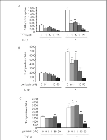
that PP1, at concentrations of 1 µM or higher, blocked the thymidine incorporation induced by IL-1b (Fig. 4A). Involvement of tyrosine kinases in IL-1b-stimulated SF proliferation was confirmed by the inhibition of 3
H-thymidine incorporation by genistein at concentrations of 0.1 µM or higher (Fig. 4B). Finally, genistein also inhibited the increase in 3H-thymidine incorporation induced by
completely inhibits PGE2 production by SFs [19]. Figure 5 indicates that indomethacin, like A77 1726, increased
3H-thymidine incorporation in presence of IL-1b or
TNF-a (Fig. 5). Accordingly, PGE2, one of the prostTNF-aglTNF-andins synthesized by the COX, dose-dependently decreased SF proliferation in absence of IL-1b and TNF-a (Fig. 6). However, in the presence of inflammatory cytokines and
Fig. 4. Inhibition of tyrosine kinases blocks IL-1b- or TNF-a-stimulated
SF proliferation. Human SFs were stimulated or not for 4 days with 1 ng/ ml IL-1b (A and B) or 10 ng/ml TNF-a (C), in combination with various levels of PP1 (A) or genistein (B). Genistein or PP1 were added 2 h prior to stimulation with cytokines. 3H-thimidine incorporation was quantified
as detailed in the materials and methods section. Each bar represents the mean +SEM of triplicate cultures of a representative experiment. *P < 0.05 as compared with control; **P < 0.05 as compared with cells treated with the corresponding cytokine.
Fig. 5. Inhibition of COX mimics A77 1726 effects on SF stimulated
with IL-1b or TNF-a. Human SFs were stimulated or not for 4 days with 1 ng/ml IL-1b (A) or 10 ng/ml TNF-a (B), in absence or pres-ence of 5 µg/ml of indomethacin. Indomethacin was added 2 h prior to stimulation with cytokines. 3H-thimidine incorporation was quantified
as detailed in the materials and methods section. Each bar represents the mean +SEM of triplicate cultures of a representative experiment. *P < 0.05 as compared with control; **P < 0.05 as compared with cells treated with the corresponding cytokine.
Fig. 6. PGE2 dose-dependently decreases SF proliferation. Human SFs
were stimulated or not for 4 days with 10 to 1,000 ng/ml of PGE2. 3
H-thimidine incorporation was quantified as detailed in the materials and methods section. Each bar represents the mean +SEM of triplicate cul-tures of a representative experiment. *P < 0.05 as compared with con-trol; **P < 0.05 as compared with cells treated with 10 ng/ml PGE2.
A77 1726, addition of PGE2 did not reverse the effects of A77 1726 (data not shown).
Discussion
RA is characterized by invasive synovial hyperplasia which plays an important role in the mechanisms leading to progressive joint destruction. SFs manifest an abnormal phenotype illustrated by increased proliferation, resistance to apoptosis, and invasiveness of cartilage and bone. They also produce elevated amounts of pro-inflammatory cytokines [25] and proteases, and induce osteoclast formation and activation [26]. Although a recent study showed that cartilage invasion does not depend directly on synoviocyte proliferation [27], overgrowth of the synovial bulk and lack of synovial cell apoptosis are nevertheless thought to play a very important role in the pathogenesis of RA. Thus, cartilage and bone degradation occurring during the progression of RA could be greatly reduced by the inhibition of SF proliferation [14]. In this study, we sought to determine the effects of A77 1726, the active metabolite of leflunomide, on SF proliferation. A77 1726 has already been shown to display a large array of biological effects on SF behavior: it inhibits the production of IL-1b, TNF-a [28], IL-6 [15], nitric oxide [28], PGE2 [15], matrix metalloproteinase-1 [15] and matrix metalloproteinase-3 [28], whereas it increases that of IL-1 receptor antagonist [19]. It appeared reasonable to postulate that A77 1726 might also modulate SF proliferation through inhibition of pyrimidine synthesis, of tyrosine kinase and/or of COX activities. To investigate A77 1726 effects, we used concentrations known to be present in the serum of patients treated with leflunomide [3]. Similar concentrations are very likely to present in the synovial fluid as well, given the rich vascularization and extremely high permeability of the synovial membrane.
Surprisingly, our results indicate that leflunomide increases SF proliferation in presence of IL-1b and/or TNF-a. This is in contrast with a previous study suggesting that leflunomide had no effect on cell viability in culture of synovial tissue [28]. It is unlikely that these discrepancies are due to differences in cell origin, since we observed similar results when SFs from patients with RA or OA were used. They may not be due to cell passage either, because A77 1726 stimulated SF proliferation independently from passage in our study; and since in addition, the passage did not significantly modify SF responsiveness to several cytokines in our previous investigations [18–20]. Actually, the previous report suggesting that leflunomide had no effect on cell proliferation was performed with an assay based on tetrazolium salt reduction, which measures the activity of a variety of enzymes, located not only in mitochondria, but also in other cellular compartments [29]. In the present study, we observed a discrepancy between results obtained when measuring MTS conversion, as compared to thymidine incorporation and cell counts. The dose-dependent decrease in MTS conversion induced by A77 1726, which we observed both in absence and presence of inflammatory cytokines, appears to be due to inhibition by A77 1726 of one or several enzymes responsible for MTS reduction, with no direct relation to cell proliferation. Therefore, determination of the
effects of leflunomide on cell proliferation using tetrazolium salt assays must be considered with much caution.
Because our results indicating that A77 1726 did not inhibit SF proliferation were unexpected, we checked the effects of A77 1726 on Jurkat T cell proliferation. We confirmed that A77 1726 inhibits proliferation of Jurkat T cells and that this effect is reversed by addition of uridine [21]. Blockage of the de novo pyrimidine synthesis pathway through DHODH inhibition by A77 1726 thus inhibits cell division of the highly proliferating Jurkat T cells [21]. In contrast SFs, like other cell types, may maintain sufficient pyrimidine levels to meet their requirements for cell division through the use of salvage pathways and are thus not affected by DHODH inhibition [24]. However, if the fact that A77 1726 does not inhibit SF proliferation in absence of IL-1b or TNF-a can be explained by the utilization of salvage pathways, the significant increase in proliferation induced by A77 1726 in presence of IL-1b and TNF-a appears surprising.
Inhibition of tyrosine kinases is unlikely to be involved in the stimulation of proliferation by A77 1726 because inhibitors of tyrosine kinases blocked the proliferation induced by IL-1b or by TNF-a. In addition, IL-1b likely stimulates SF proliferation through activation of the src tyrosine kinase pathway [14], likewise we also observed that the specific src tyrosine kinase inhibitor PP1 reduced SF proliferation induced by IL-1b. The mitogenic stimulation of A77 1726 thus likely arises from other mechanisms than inhibition of tyrosine kinase activity. We next explored the known effect of leflunomide on COX inhibition [8], because we recently showed that both 100 µM A77 1726 and 5 µg/ml indomethacin completely inhibited the production of PGE2 induced by IL-1b in synovial fibroblasts [19]. In the present study, we observed that indomethacin increased the mitogenic effects of IL-1b and/or those of TNF-a, suggesting that A77 1726 might indeed display its proliferative effects by inhibiting COX. Accordingly, PGE2 dose-dependently inhibited SF proliferation. However, in presence of IL-1b or TNF-a, we could not observe an inhibition of A77 1726 effects by PGE2. This finding may be due to the fact that other prostaglandins or factors modulated by COX are induced by inflammatory cytokines and alter SF proliferation. Moreover, as previously observed for the induction of IL-1Ra production by A77 1726 in SFs, relatively high doses of A77 1726 (100 µM) were required to efficiently increase cell proliferation, while IL-1b-induced PGE2 production is inhibited already at relatively lower
concentrations (10–50 µM) [19]. The dose dependencies of the two effects thus appeared to be slightly different. Nevertheless, it is conceivable that A77 1726 increases SF proliferation at least in part through inhibition of COX and prostaglandin synthesis. This would be consistent with the fact that A77 1726 displayed significant mitogenic effects in the presence of IL-1b or TNF-a, which both induce high levels of prostaglandins, but not in their absence. On the other hand, we cannot exclude the possibility that leflunomide, as well as indomethacin, exerts proliferative effects by yet unknown distinct mechanisms.
Whether this in vitro mitogenic effect of A77 1726 on SF proliferation translates into significant synovial proliferation in patients is still unknown but deserves consideration.
matory action on lymphocytes and monocytes/macrophages, which in turn may contribute to reduce synovial fibroblast activation and proliferation. Finally, it is also conceivable that like in serum [4, 22], A77 1726 levels in the vicinity of the synovial fibroblast exceed 100 µM, and reach 200 or 300 µM, concentrations that do not increase proliferation. However, evidence is lacking to support this hypothesis and today there is still uncertainty as whether the serum level of A77 1726 is correlated with disease activity [4, 22].
There is no doubt that leflunomide is extremely efficient in RA, as well as in other inflammatory arthropathies including psoriatic arthritis [32, 33]. In RA, leflunomide’s efficacy was shown to be comparable to that of methotrexate with effects reported to be more rapid and slightly better with regards to functional outcome at two years radiological progression [2, 34]. This places leflunomide as one of the best non-biological agents available for the treatment of RA [35, 36].
Our results, though, indicate that the array of beneficial effects exerted by leflunomide could be partly lessened by an inducing effect on SF proliferation. It might be that this latter effect is not significant or that it is annihilated by the other, beneficial, activities of leflunomide, including inhibition of IL-1b and TNF-a synthesis [28], and increase in IL-1Ra production [19] (IL-1b and TNF-a are strong stimulators of SF proliferation). It might also be, however, that in some cases the progressive increase of the synovial bulk could be detrimental on the long term; if substantiated, it would warrant modified treatment strategies, which could include associated use of pro-apoptotic or anti-proliferative agents. Alternatively, efforts directed at developing related pharmacological compounds, devoid of this detrimental effect, would be justified. In any event, we believe that the unexpected observation that leflunomide is able to decrease the proliferation of multiple cell types while increasing that of SFs, is biologically of interest.
In conclusion, results of the present study indicate that the active metabolite of leflunomide, A77 1726, at therapeutically applicable doses, stimulates the proliferation of synovial fibroblasts in the presence of the inflammatory cytokines IL-1b and TNF-a, and that this effect could be related to COX activity. This may lessen the beneficial effect of leflunomide in the treatment of RA, and therefore further elucidation of involved mechanisms may prove useful for the utilization of leflunomide, the development of related compounds or elaboration of new therapeutic strategies.
M, Brouwers JR. A rapid and simple determination of A77 1726 in human serum by high-performance liquid chromatography and its application for optimization of leflunomide therapy. J Pharm Biomed Anal 2004; 36: 17–22.
[4] van Roon EN, Jansen TL, van de Laar MA, Janssen M, Yska JP, Keuper R, et al. Therapeutic drug monitoring of A77 1726, the active metabolite of leflunomide: serum concentrations predict response to treatment in patients with rheumatoid arthritis. Ann Rheum Dis 2005; 64: 569–74.
[5] Breedveld FC, Dayer JM. Leflunomide: mode of action in the treatment of rheumatoid arthritis. Ann Rheum Dis 2000; 59: 841–9.
[6] Fairbanks LD, Bofill M, Ruckemann K, Simmonds HA. Importance of ribonucleotide availability to proliferating T-lymphocytes from healthy humans. Disproportionate expansion of pyrimidine pools and contrasting effects of de novo synthesis inhibitors. J Biol Chem 1995; 270: 29682–9.
[7] Zielinski T, Zeitter D, Muller S, Bartlett RR. Leflunomide, a reversible inhibitor of pyrimidine biosynthesis? Inflamm Res 1995; 44 Suppl 2: S207–8.
[8] Hamilton LC, Vojnovic I, Warner TD. A771726, the active metabolite of leflunomide, directly inhibits the activity of cyclo-oxygenase-2 in vitro and in vivo in a substrate-sensitive manner. Br J Pharmacol 1999; 127: 1589–96.
[9] Xu X, Williams JW, Bremer EG, Finnegan A, Chong AS. Inhibition of protein tyrosine phosphorylation in T cells by a novel immunosuppressive agent, leflunomide. J Biol Chem 1995; 270: 12398–403.
[10] Elder RT, Xu X, Williams JW, Gong H, Finnegan A, Chong AS. The immunosuppressive metabolite of leflunomide, A77 1726, affects murine T cells through two biochemical mechanisms. J Immunol 1997; 159: 22–7.
[11] Goldring SR. Pathogenesis of bone and cartilage destruction in rheumatoid arthritis. Rheumatology (Oxford) 2003; 42 Suppl 2: ii11–6.
[12] Pap T, Muller-Ladner U, Gay RE, Gay S. Fibroblast biology. Role of synovial fibroblasts in the pathogenesis of rheumatoid arthritis. Arthritis Res 2000; 2: 361–7.
[13] Yamanishi Y, Firestein GS. Pathogenesis of rheumatoid arthritis: the role of synoviocytes. Rheum Dis Clin North Am 2001; 27: 355–71.
[14] Takayanagi H, Juji T, Miyazaki T, Iizuka H, Takahashi T, Isshiki M, et al. Suppression of arthritic bone destruction by adenovirus-mediated csk gene transfer to synoviocytes and osteoclasts. J Clin Invest 1999; 104: 137–46.
[15] Burger D, Begue-Pastor N, Benavent S, Gruaz L, Kaufmann MT, Chicheportiche R, et al. The active metabolite of leflunomide, A77 1726, inhibits the production of prostaglandin E(2), matrix metalloproteinase 1 and interleukin 6 in human fibroblast-like synoviocytes. Rheumatology (Oxford) 2003; 42: 89–96.
[16] Kusunoki N, Yamazaki R, Kawai S. Induction of apoptosis in rheumatoid synovial fibroblasts by celecoxib, but not by other selective cyclooxygenase 2 inhibitors. Arthritis Rheum 2002; 46: 3159–67.
[17] Gabay C. Cytokine inhibitors in the treatment of rheumatoid arthritis. Expert Opin Biol Ther 2002; 2: 135–49.
[18] Guicheux J, Palmer G, Relic B, Mezin F, Caverzasio J, Apostolides P, et al. Primary human articular chondrocytes, dedifferentiated chondrocytes, and synoviocytes exhibit differential responsiveness to interleukin-4: correlation with the expression pattern of the common receptor gamma chain. J Cell Physiol 2002; 192: 93– 101.
[19] Palmer G, Burger D, Mezin F, Magne D, Gabay C, Dayer JM, et al. The active metabolite of leflunomide, A77 1726, increases the production of IL-1 receptor antagonist in human synovial fibroblasts and articular chondrocytes. Arthritis Res Ther 2004; 6: R181–9.
[20] Palmer G, Mezin F, Juge-Aubry CE, Plater-Zyberk C, Gabay C, Guerne PA. Interferon beta stimulates interleukin 1 receptor antagonist production in human articular chondrocytes and synovial fibroblasts. Ann Rheum Dis 2004; 63: 43–9.
[21] Cao WW, Kao PN, Chao AC, Gardner P, Ng J, Morris RE. Mechanism of the antiproliferative action of leflunomide. A77 1726, the active metabolite of leflunomide, does not block T-cell receptor-mediated signal transduction but its antiproliferative effects are antagonized by pyrimidine nucleosides. J Heart Lung Transplant 1995; 14: 1016–30.
[22] Chan V, Charles BG, Tett SE. Population pharmacokinetics and association between A77 1726 plasma concentrations and disease activity measures following administration of leflunomide to people with rheumatoid arthritis. Br J Clin Pharmacol 2005; 60: 257–64.
[23] Williams JW, Mital D, Chong A, Kottayil A, Millis M, Longstreth J, et al. Experiences with leflunomide in solid organ transplantation. Transplantation 2002; 73: 358–66.
[24] Fox RI, Herrmann ML, Frangou CG, Wahl GM, Morris RE, Strand V, et al. Mechanism of action for leflunomide in rheumatoid arthritis. Clin Immunol 1999; 93: 198–208.
[25] Isomaki P, Punnonen J. Pro- and anti-inflammatory cytokines in rheumatoid arthritis. Ann Med 1997; 29: 499–507.
[26] Takayanagi H, Oda H, Yamamoto S, Kawaguchi H, Tanaka S, Nishikawa T, et al. A new mechanism of bone destruction in rheumatoid arthritis: synovial fibroblasts induce osteoclastogenesis. Biochem Biophys Res Commun 1997; 240: 279–86.
[27] Seemayer CA, Kuchen S, Kuenzler P, Rihoskova V, Rethage J, Aicher WK, et al. Cartilage destruction mediated by synovial fibroblasts does not depend on proliferation in rheumatoid arthritis. Am J Pathol 2003; 162: 1549–57.
[28] Elkayam O, Yaron I, Shirazi I, Judovitch R, Caspi D, Yaron M. Active leflunomide metabolite inhibits interleukin 1beta, tumour necrosis factor alpha, nitric oxide, and metalloproteinase-3 production in activated human synovial tissue cultures. Ann Rheum Dis 2003; 62: 440–3.
[29] Berridge MV, Tan AS. Characterization of the cellular reduction of 3-(4,5-dimethylthiazol-2-yl)-2,5-diphenyltetrazolium bromide
(MTT): subcellular localization, substrate dependence, and involvement of mitochondrial electron transport in MTT reduction. Arch Biochem Biophys 1993; 303: 474–82.
[30] Reece RJ, Kraan MC, Radjenovic A, Veale DJ, O‘Connor PJ, Ridgway JP, et al. Comparative assessment of leflunomide and methotrexate for the treatment of rheumatoid arthritis, by dynamic enhanced magnetic resonance imaging. Arthritis Rheum 2002; 46: 366–72.
[31] Kraan MC, Reece RJ, Barg EC, Smeets TJ, Farnell J, Rosenburg R, et al. Modulation of inflammation and metalloproteinase expression in synovial tissue by leflunomide and methotrexate in patients with active rheumatoid arthritis. Findings in a prospective, randomized, double-blind, parallel-design clinical trial in thirty-nine patients at two centers. Arthritis Rheum 2000; 43: 1820–30.
[32] Kaltwasser JP, Nash P, Gladman D, Rosen CF, Behrens F, Jones P, et al. Efficacy and safety of leflunomide in the treatment of psoriatic arthritis and psoriasis: a multinational, double-blind, randomized, placebo-controlled clinical trial. Arthritis Rheum 2004; 50: 1939–50.
[33] Reich K, Hummel KM, Beckmann I, Mossner R, Neumann C. Treatment of severe psoriasis and psoriatic arthritis with lefluno-mide. Br J Dermatol 2002; 146: 335–6.
[34] Cohen S, Cannon GW, Schiff M, Weaver A, Fox R, Olsen N, et al. Two-year, blinded, randomized, controlled trial of treatment of active rheumatoid arthritis with leflunomide compared with meth-otrexate. Utilization of Leflunomide in the Treatment of Rheuma-toid Arthritis Trial Investigator Group. Arthritis Rheum 2001; 44: 1984–92.
[35] Aletaha D, Stamm T, Kapral T, Eberl G, Grisar J, Machold KP, et al. Survival and effectiveness of leflunomide compared with methotrexate and sulfasalazine in rheumatoid arthritis: a matched observational study. Ann Rheum Dis 2003; 62: 944–51.
[36] Wolfe F, Michaud K, Stephenson B, Doyle J. Toward a definition and method of assessment of treatment failure and treatment effectiveness: the case of leflunomide versus methotrexate. J Rheumatol 2003; 30: 1725–32.
Abbreviations
COX = cyclooxygenase; DHODH = dihydroorotate dehy-drogenase; DMARD = disease-modifying anti-rheumatic drug; IL-1b = interleukin-1b; OA = osteoarthritis; PGE2 = prostaglandin E2; RA = rheumatoid arthritis; SF = synovial fibroblast; TNF-a = tumor necrosis factor-a.
Competing interests
This work was supported by Aventis Pharma (Frankfurt am Main, Germany).