HAL Id: hal-01054288
https://hal.archives-ouvertes.fr/hal-01054288
Submitted on 6 Aug 2014
HAL is a multi-disciplinary open access
archive for the deposit and dissemination of
sci-entific research documents, whether they are
pub-lished or not. The documents may come from
teaching and research institutions in France or
abroad, or from public or private research centers.
L’archive ouverte pluridisciplinaire HAL, est
destinée au dépôt et à la diffusion de documents
scientifiques de niveau recherche, publiés ou non,
émanant des établissements d’enseignement et de
recherche français ou étrangers, des laboratoires
publics ou privés.
The crystal structure of Fe�S� quinolinate synthase
unravels an enzymatic dehydration mechanism that uses
tyrosine and a hydrolase-type triad.
Mickael V Cherrier, Alice Chan, Claudine Darnault, Debora Reichmann,
Patricia Amara, Sandrine Ollagnier de Choudens, Juan-Carlos
Fontecilla-Camps
To cite this version:
Mickael V Cherrier, Alice Chan, Claudine Darnault, Debora Reichmann, Patricia Amara, et al.. The
crystal structure of Fe�S� quinolinate synthase unravels an enzymatic dehydration mechanism that uses
tyrosine and a hydrolase-type triad.. Journal of the American Chemical Society, American Chemical
Society, 2014, 136 (14), pp.5253-5256. �10.1021/ja501431b�. �hal-01054288�
The crystal structure of Fe
4
S
4
quinolinate synthase unravels
an enzymatic dehydration mechanism that uses tyrosine and
a hydrolase-type triad.
Mickaël V. Cherrier,
,Alice Chan,
§Claudine Darnault, Debora Reichmann,
§Patricia Amara,
Sandrine Ollagnier de Choudens
*,§and Juan C. Fontecilla-Camps
*,Metalloproteins Unit, Institut de Biologie Structurale, Commissariat à l’Energie Atomique–Centre National de la Recherche Scientifique–Université Grenoble-Alpes, 38000 Grenoble, France.
UMR 5086, BMSSI, CNRS - Université Lyon 1, Institut de Biologie et Chimie des Protéines, 7 passage du Vercors, F-69367 Lyon, France.
§DSV/iRTSV/CBM, UMR 5249 CEA-Université Grenoble I-CNRS/Equipe Biocatalyse, CEA-Grenoble, 17 Rue des
Martyrs, 38054 Grenoble Cedex 09, France
Univ. Grenoble Alpes, iRTSV-LCBM, F-38000Grenoble, France CNRS, IRTSV-LCBM, F-38000Grenoble, France
CEA, iRTSV-LCBM, F-38000Grenoble, France
Supporting Information Placeholder
ABSTRACT:Quinolinate synthase (NadA) is a Fe4S4
cluster-containing dehydrating enzyme involved in the synthesis of quinolinic acid (QA), the universal precursor of the essential nicotinamide adenine dinucleotide (NAD) coenzyme. A previously determined apoNadA crystal structure revealed the binding of one substrate analog, providing partial me-chanistic information. Here, we report on the holo X-ray structure of NadA. The presence of the Fe4S4 cluster
gene-rates an internal tunnel and a cavity in which we have docked the last precursor to be dehydrated to form QA. We find that the only suitably placed residue to initiate this process is the conserved Tyr21. Furthermore, Tyr21 is close to a conserved Thr-His-Glu triad reminiscent of those found in proteases and other hydrolases. Our mutagenesis data show that all these residues are essential for activity and strongly suggest that Tyr21deprotonation, to form the reac-tive nucleophilicphenoxide anion, is mediated by the triad. NadA displays a dehydration mechanism significantly differ-ent from the one found in archetypical dehydratases such as aconitase, which use a serine residue deprotonated by an oxyanion hole. The X-ray structure of NadA will help us unveil its catalytic mechanism, the last step in the under-standing of NAD biosynthesis.
Biosynthesis of nicotinamide adenine dinucleotide (NAD), an essential and ubiquitous cofactor in biology, involves in all living organisms the formation of quinolinic acid (QA).1QA is generated in most prokaryotes from L-aspartate
and dihydroxyacetone phosphate (DHAP) through the con-certed action of two enzymes, the L-aspartate oxidase NadB and the quinolinate synthase NadA.2Although the oxidation
of L-aspartate to iminoaspartate (IA) catalyzed by NadB and the reactions from QA to NAD have been well characterized, the condensation ofIA and DHAP to form QA, catalyzed by NadA, is still poorly understood at the molecular level2
(Fig-ure 1). In vitro studies demonstrated that in all systems stu-died so far NadA contains an oxygen sensitive Fe4S4 cluster
essential for activity.3-5NadA is functionally and structurally similarto the citric acid cycle enzyme aconitase, i.e. both catalyze a dehydration process and coordinate a Fe4S4 cluster
with only three cysteine protein ligands, which in NadA are organized in a CX86-112CX86-99C motif.5-7 Consequently, it was
suggested that the unique iron site of the NadA Fe4S4 cluster
should also be directly involved in dehydration steps, acting as a Lewis acid. Partial mechanistic information has been obtained from the two available crystal structures of NadA,8,9
which correspond to the inactive apo form of the enzyme. These structures reveal a common fold with two other Fe4S4
enzymes, IspH10 and Dph211 where the cluster is involved in
redox reactions. To gain insight into the reaction catalyzed by NadA, we have determined the 1.65 Å resolution crystal structure of the active, Fe4S4-bound NadA from
Thermoto-gamaritima (holoTmNadA).
Initially grown holo recombinant wild-type TmNadA crys-tals could not be used to determine its X-ray structure. Sub-sequently, two holoTmNadA proteins, containing purposely engineered K219R and K280R mutations were produced. Only the former mutant, along with a His tag added at the protein N-terminus to facilitate purification, yielded a cluster of polycrystalline material (see Supporting Information for details, Figures S1 and S2). A fragment from this cluster was used to collect data at the European Synchrotron Radiation Facility (ESRF). The structure of TmNadA K219R was
subse-quently solved by molecular replacement and refined at 1.65 Å resolution (Table S1 and Figure S1C). Examination of the crystal packing revealed the central role played by both the K219R mutation and, rather unexpectedly, the His tag (Fig-ure S2). The holoTmNadA K219R mutant exhibits biochemi-cal (Fe and S content)and spectroscopic properties as the wild-type protein and displays both in vivo and in vitroNadA activity (Figure S3).
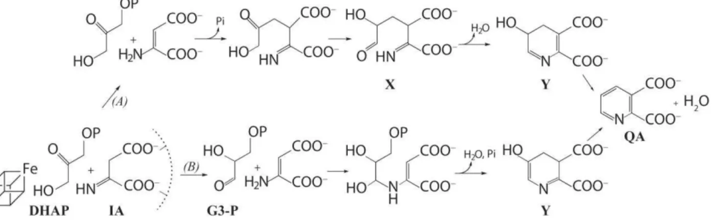
Figure 1.Previously proposed catalytic mechanisms for the synthesis of quinolinate from IA and DHAP by (A)Sakurabaet al.8and
(B) Begley et al.2X = 2-imino, 3-carboxy, 5-hydroxy, 6-oxo hexanoic acid. Y = 5-hydroxy, 4,5-dihydroquinolate. The Fe
4S4 cluster,
which is essential for catalysis, is shown here, before the first step of each mechanism, at interacting distance from DHAP as suggested in reference.12 The malate-based8 postulated interactions of IA with the protein (depicted as a semi-circle in dashed
lines) are also indicated.
The presence of the natural Fe4S4 prosthetic group (Figure
2) in TmNadA K219R re-organizes the interaction of the three protein domains relative to the apo malate-bound
PyrococcushorikoshiiNadA (PhNadA),8 (Figure S4).As
ex-pected, the binding of the Fe4S4 cluster also results in the
ordering of the Cys-bearing inter-domain stretches of
TmNadA K219R. The inter-domain interactions in the holo
structure generate a long tunnel connecting the Fe4S4 cluster
to the molecular surface (Figure 3A). The mobility of domain III relative to domains I and II, as indicated by the PhNadA structure (Figure S4), may finely modulate the size and shape of the tunnel when substrate is bound. Indeed, a comparison between apoPhNadA and our structure shows that binding of malate induces the collapse of the internal tunnel (Figure 3B). Although this conformational change may result from the absence of the Fe4S4 cluster it would make sense for the
active enzyme to shield the unstable IA from the exterior.13 A
comparison of the interaction energies of domains I and II with domain III in our structure and PhNadA indicates that a conformational change of the latter domain should not in-volve a significant relative energy change (Table S2).
Figure 2.(A) Omit (Fo-Fc) electron density map (green mesh) atthe Fe4S4 cluster and its solvent ligand with its
environ-ment shown within its matching (2Fo-Fc) map (blue mesh). Atom color codes: orange: Fe, yellow: S, red: HxO, gold: C.
The dashed line connects the unique Fe to the solvent mole-cule that completes its tetrahedral coordina-tion.(B)anomelectron density map at the Fe4S4 cluster(X-ray
data collected at = 0.979 Å). As expected, peaks are found at the Fe ions positions.
Soriano et al.,9 have modeled the binding cavity of NadA
from a combination of the apoPh and Pyrococcusfuriosus (Pf) X-ray NadA structures. Furthermore, they have placed sub-strates and the last QA precursor in their model. However, the modeled positions of some relevant amino acid side chains appear to be significantly different from their coun-terparts in the holoTm K219R NadA X-ray structure (not shown). Conversely, the apoPhNadA structure reported by Sakuraba et al.8defines the binding site of IA because the
bound malate is analogous to this substrate. However, in the absence of the Fe4S4 cluster, the DHAP binding site,
includ-ing the unique iron cluster site and essential functional ami-no acids,isundefined.
Figure 3. (A) (left) structure of holoTmNadA K219R. The three domains (green, purple and gold) define a tunnel that connects the Fe4S4 cluster to the molecular surface; (right) a
section through the long tunnel of the structure rotated by 90° about the vertical axis. The Fe4S4 cluster is displayed as
balls and sticks. (B) A section of the apoPhNadA structure8
(same orientation as rotatedfigure in B). Note the absence of the tunnel that is found in holoTmNadA. Bound malate is displayed as balls and sticks.
Besides a different orientation of domain III, the compari-son of TmNadA K219R and PhNadA reveals changes at the malate-binding site. These include a 2.5 Å shift of the -helix formed by residues 35-45 of domain I, and a less extensive displacement of the loop containing Ser124 of domain II, both directed towards the carboxylate groups of the IA ana-log.These two localized conformational changes (Figure S5) were built into our holoTmNadA K219R structure using homology modeling.This allowed us to reproduce the bind-ing of the COOH groups of the IA-derived fragment of the last QA precursor 5-hydroxy, 4,5-dihydroquinolinate (5-OH-4,5-DHQA; Y in Figure 1) in the active site structure. Subse-quently, a mechanistic study was initiated using two anchor-ing points to dock the precursor into the catalytic cavity of the above model (see Supporting Information for modeling details): i) the two malate carboxylate groups positions, and ii) the expected direct interaction of the -OH group of the precursor with the unique Fe from the cluster (as determined for the –SH groups of the 4,5-dithiohydroxyphtalic acid (DTHPA) inhibitor12). Subsequently, the position of residues
interacting with the 5-OH-4,5-DHQA models was optimized. Among the possible isomers, we found that the C5-centered
(R)model of the docked 5-OH-4,5-DHQA, with one of the
two possible ring puckerings, satisfied the anchoring re-quirements.Furthermore, it provided a structural basis for catalysis (Figure 4). Indeed, in this conformation a suitable functional group, the –OH from Tyr21, is properly posi-tioned to become the nucleophile that deprotonates C4, a step required in the dehydration of 5-OH-4,5-DHQA to form QA (Figures 1 and 4C). In the alternative orientation for the anchoring carboxylate groups, the potentially catalytic Tyr107 and Ser36 are too far from C4. Furthermore, their geometry is not appropriate for C4 proton abstraction and there is no obvious way of generating a reactive alkoxide nucleophile from the hydroxyl group of either residue (not shown).
Figure 4. (A) View of the active site cavity of holoTmNadA;
(B) The most suitable 5-OH-4,5-DHQA (R) precursor of QA (carbon atoms in blue) docked in the NadA modified active site. Hydrogen bonds between the ligand and the protein are indicated in yellow; the possible attack of the H atom of C4 by Tyr21 is depicted in pink. (C) Schematic view of the active site of TmNadA with the docked QA precursor, a modeled H2O molecule bound to Feu, Tyr21, Asn109, and the
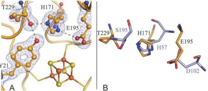
charge-relay system made of Thr229, His171 and. Glu195. Arrows indicate proton subtractions.
Incidentally, we noted that the long tunnel that connects the catalytic unique iron ion of the Fe4S4 cluster to the
mole-cular surface collapses in our TmNadA model in which (R) 5-OH-4,5-DHQA was docked (Figure S6). This suggests that substrate binding controls the opening and closure of the tunnel.
We have subsequently shown that Tyr21 plays a central functional role in NadA catalysis by mutating it to Phe in the
T. maritima enzyme (see Supporting Information). Indeed,
the TmNadA Y21F mutant,which still coordinates a Fe4S4
cluster, displayed no detectable enzymatic activity (Table S3). This result was confirmed with the corresponding Y49F mutant of the more active E. coliNadA(Table S3). In TmNadA K219R the role of Tyr21 in the deprotonation of 5-OH-4,5-DHQA is further substantiated by its interaction with a charge-relay system composed of the strictly conserved resi-dues Thr229, His171 and Glu195. This system, which is similar to the Ser-His-Asp catalytic triad typical of serine proteases and other hydrolases14 should favor the formation
of a nucleophilicphenoxide anion at Tyr21 (which directly interacts with Thr229) (Figures 4C and 5). In order to test this hypothesis, triad mutants were generated in the E. coli-NadA enzyme. They all display a drastically reduced activity showing that the triad residues are also crucial for catalysis (Table S3). The residual activity observed in these mutants is not due to impaired substrate binding because they all bind the DTHPA inhibitor like the wild-type does (Supporting information, Figure S7).
Thr and Glu residues, rather than the classic Ser and Asp, are also used in other triads. For instance, Glu is found in the catalytic triad of acetylcholine esterase from Torpedo
califor-nica and Thr in the triad from asparaginases.14 It should be
noted that His171 also interacts with Cys168, a ligand of the Fe4S4 cluster. However, this interaction is not likely to play a
determinant role in either Fe-S cluster biogenesis or stabili-zation because the corresponding H205F mutant coordinates a Fe4S4cluster. In conclusion, the observed charge-relay triad
is unprecedented and could not have been identified from previous NadA structural data or models derived from them.
Figure 5. (A)Charge-relay triad (Thr229-His171-Glu195) and Tyr21, depicted with their matching (2Fo-Fc) electron densi-ty map, at the NadA active site. (B) Superposition of our proposed Thr-His-Glu triad with the catalytic Ser-His-Asp triad of bovine trypsin (PDB code 4I8G15). The carbon atoms
of TmNadA and trypsin are shown as gold and light blue sticks respectively; blue:N, red:O.
Our results may be compared to those reported for aconi-tase, which is the archetypical enzyme for Fe4S4 cluster-based
dehydration/hydration reactions.16,17 The condensation of IA
with DHAP to generate QA involves two dehydrations by NadA but only the second one is mechanistically equivalent to the synthesis of aconitate from citrate. The first dehydra-tion involves a carbinolamine intermediate, which results from the nucleophilic attack of either –NH2 or =NH2+ to the
>C=O group of glyceraldehyde 3-phosphate (G3-P) resulting from the tautomerization of DHAP, or to the >C=O group of the tautomerized X product (Figure 1). This will depend on which of the previously proposed two mechanisms (Figure 1) is assumed to be the correct one. The reactivity of the >C=O group with the nucleophile will be a function of on its elec-trophilicity. An interaction of the unique Fe ion from the cluster with >C=O would make this group more electrophile and will favor the reaction. The TmNadA K219R structure does not allow us to provide a detailed model for the stereo-chemistry of this reaction but it is certainly compatible with a close (<2.0 Å) terminal >C=O---Feu interaction. The
nuc-leophilic attack has to take place on an aldehydic terminal >C=O to produce the appropriate intermediate in QA syn-thesis (Figure 1). The second dehydration step, that of 5-OH-4,5-DHQA to QA, requires the deprotonation of its C4 fol-lowed by elimination as water of the C5-linked –OH group of the Feu-bound precursor. This reaction is likely to be very
similar to the dehydration of citrate to yield aconitate be-cause it also generates a >C=C< bond, with the additional advantage of resulting in aromatization. The Feu in the
TmNadA K219R structure appears to have a more extensive
coordination than its counterpart in ligand-free aconitase. However, there is a well-defined, most probably H2O/OH-,
ligand that completes the tetrahedral coordination of Feu,
like OH- does in that enzyme16 (Figures 2 and 4A and
Sup-porting Information).
As mentioned above, our structural and functional data indicate that the only plausible candidate for the nucleophile that deprotonates 5-OH-4,5-DHQA in TmNadA is Tyr21. When compared to aconitase there are two major differenc-es: the nature of the alkoxide-bearing residue, which is Ser in that enzyme, and the way the alkoxide is generated. In aconi-tase, the active site Ser642 accepts short H-bonds from an oxyanion hole, formed by the main chain amide and the N atom from Arg644, that lower its pKa enough to facilitate its
deprotonation upon citrate or isocitrate binding.16-18 Con-versely, as discussed above, in NadA the deprotonation of Tyr21 is likely to be unusually carried out by a charge-relay system similar to typical catalytic triads of the hydrolase family (Figures 4C and 5). This process represents a remark-able convergent use of such triad.19
In summary, NadA displays a combination of remarkable features that, besides the dehydration mechanism described above, includes a long tunnel that connects the catalytic unique iron ion of the Fe4S4 cluster with the molecular
sur-face and collapses upon substrate binding. Our efforts will be now concentrated in generating structures of NadA with substrate analogs, inhibitors and NadA variants in order to get further insights into the NadA catalytic mechanism and design potential antibacterial agents. Indeed, NadA consti-tutes a therapeutic target in some pathogens such as
Myco-bacterium leprae and Helicobacter pylori.
ASSOCIATED CONTENT
Supporting Information
Material and methods, additional figures and tables, and tables of crystallographic data collection and refinement statistics.
This material is available free of charge via the Internet at http://pubs.acs.org.
The structure has been deposited in the Protein Data Bank as entry 4P3X.
AUTHOR INFORMATION
Corresponding Authorjuan.fontecilla@ibs.fr or sandrine.ollagnier@cea.fr Notes
The authors declare no competing financial interests.
ACKNOWLEDGMENT
We thank the staff from the BM30A beamline of the ESRF in Grenoble for help with X-ray data collection. We also thank the AgenceNationale pour la Recherche for the NADBIO contract ANR-12-BS07-0018-01.This work was also partially supported by FRISBI (ANR-10-INSB-05-02) within the Gre-noble Partnership for Structural Biology (PSB).
REFERENCES
(1) Frey, P.; Hegeman, A. D. Enzymatic Reaction Mechanisms; Oxford University Press: New York, NY., 2007.
(2) Begley, T. P.; Kinsland, C.; Mehl, R. A.; Osterman, A.; Dorrestein, P. Vitam. Horm.2001, 61, 103.
(3) Ollagnier-de Choudens, S.; Loiseau, L.; Sanakis, Y.; Barras, F.; Fontecave, M. FEBS Lett.2005, 579, 3737.
(4) Cicchillo, R. M.; Tu, L.; Stromberg, J. A.; Hoffart, L. M.; Krebs, C.; Booker, S. J. J. Am. Chem. Soc.2005, 127, 7310.
(5) Marinoni, I.; Nonnis, S.; Monteferrante, C.; Heathcote, P.; Hartig, E.; Bottger, L. H.; Trautwein, A. X.; Negri, A.; Albertini, A. M.; Tedeschi, G. FEBS J.2008, 275, 5090.
(6) Rousset, C.; Fontecave, M.; Ollagnier de Choudens, S. FEBS
Lett.2008, 582, 2937.
(7) Saunders, A. H.; Griffiths, A. E.; Lee, K. H.; Cicchillo, R. M.; Tu, L.; Stromberg, J. A.; Krebs, C.; Booker, S. J. Biochemistry2008, 47, 10999.
(8) Sakuraba, H.; Tsuge, H.; Yoneda, K.; Katunuma, N.; Ohshima, T. J. Biol. Chem.2005, 280, 26645.
(9) Soriano, E. V.; Zhang, Y.; Colabroy, K. L.; Sanders, J. M.; Settembre, E. C.; Dorrestein, P. C.; Begley, T. P.; Ealick, S. E. Acta
crystallogr. Section D, Biol. crystallogr.2013, 69, 1685.
(10) Grawert, T.; Span, I.; Eisenreich, W.; Rohdich, F.; Eppinger, J.; Bacher, A.; Groll, M. Proc. Natl. Acad. Sci. U S A2010,
107, 1077.
(11) Zhang, Y.; Zhu, X.; Torelli, A. T.; Lee, M.; Dzikovski, B.; Koralewski, R. M.; Wang, E.; Freed, J.; Krebs, C.; Ealick, S. E.; Lin, H.
Nature2010, 465, 891.
(12) Chan, A.; Clemancey, M.; Mouesca, J. M.; Amara, P.; Hamelin, O.; Latour, J. M.; Ollagnier de Choudens, S. Angew. Chem.
Int. Ed. Engl.2012, 51, 7711.
(13) Rizzi, M.; Schindelin, H. Curr. Opin. Struct. Biol.2002, 12, 709.
(14) Dodson, G.; Wlodawer, A. Trends Biochem. Sci.1998, 23, 347.
(15) Liebschner, D.; Dauter, M.; Brzuszkiewicz, A.; Dauter, Z.
Acta crystallogr. Section D, Biol. crystallogr.2013, 69, 1447.
(16) Beinert, H.; Kennedy, M. C.; Stout, C. D. Chem. Rev.1996,
96, 2335.
(17) Lloyd, S. J.; Lauble, H.; Prasad, G. S.; Stout, C. D. Protein
Sci.1999, 8, 2655.
(18) Lauble, H.; Kennedy, M. C.; Beinert, H.; Stout, C. D.
Biochemistry1992, 31, 2735.
(19) Gutteridge, A.; Thornton, J. M. Trends Biochem. Sci.2005,