Protein/Protein Interactions (PDZ) in Proximal Tubules
J. Biber, S.M. Gisler, N. Hernando, H. Murer
Institute of Physiology, University of Zurich, 8057 Zu¨rich, Switzerland Received:30 September 2004/Revised:10 February 2005
Introduction
Essentially all cellular functions rely on the correct spatial organization of proteins. In epithelial cells, this is manifested by a separation of the plasma membrane into an apical and a basolateral area, which is required for vectorial transport of solutes, and by the formation of junctional complexes to guarantee barrier function. In this article, we sum-marize the current knowledge of PDZ protein-based multimeric protein complexes in renal proximal tubular (PT) cells (for definition of the PDZ domain, seebelow), which are involved in the organization of the brush border membrane, the basolateral mem-brane and the tight junction of PT cells. In addition, we also consider a fourth subcellular structure, the subapical compartment (SAC). This compartment has been ascribed to the recycling of endocytosed proteins and also to the routing of endocytosed material to the lysosomes.
In renal proximal tubules, the majority of filtered solutes and water are reabsorbed by transepithelial transport processes and by paracellular pathways [68]. In addition, PT-cells exhibit an exceptionally high rate of endocytosis to recover small peptides and hormones. On a morphological basis, three different distinct epithelial cells can be distinguished along the proximal tubules. In the S1 segment, brush borders are formed by long microvilli, whereas the appear-ance of the microvilli in the S2 and S3 segments is less pronounced and varies among different species [37]. Although this longitudinal morphological heteroge-neity of PT cells is also reflected by functional dif-ferences, in this article, we will not distinguish among proximal tubular segments, i.e., the PDZ-based pro-tein complexes discussed below are assumed to be of uniform nature along the entire proximal tubule.
The PDZ module
Numerous protein domains have been described that are implicated in protein-protein interactions (see e.g., www.mshri.on.ca). Based on sequence similarities be-tween the post-synaptic density protein PSD-95, the Drosophilajunctional protein Disc-large and the tight junctional protein ZO1, one such domain was defined as PDZ. PDZ proteins, among other functions, are primarily thought to organize large functional units such as synapses [55] or tight junctions [19]. The characteristics of PDZ domains have been reviewed recently [14, 15, 28, 55] and are summarized as follows: In mammals, over 400 different PDZ proteins have been identified and grouped into three major families according to their domain organization:A first family encompasses proteins, which only contain PDZ domains. The second, MAGUK family (mem-brane-associated guanylate kinases), contains one or more PDZ domains besides a GuK domain (gua-nylate kinase domain) and a SH3 domain. A third family was defined containing proteins of multiple PDZ domains together with a variable number of other protein domains.
The PDZ domain comprises between 80 and 90 amino-acid residues of which the three-dimensional structure has been basically resolved (see http:// smart.enbl-heidelberg.de). The building principle is a sandwich structure of 6 b-strands and two a-helices that form a hydrophobic cleft into which a short pep-tide can be accommodated. As an example, the struc-tural aspect of the CFTR-NHERF interaction has been discussed in detail [38]. In most cases, PDZ domains bind to a C-terminal amino-acid motif. Three different classes of PDZ binding motifs have been recognized. They all include the last four C-terminal amino acids, whose characteristics of interaction can be modulated by more upstream amino acids [55]. Besides the classical canonical determinants, addi-tional PDZ binding motifs have been described and therefore an extended classification has been proposed Correspondence to:J. Biber; email:JuergBiber@access.unizh.ch
DOI:10.1007/s00232-005-0738-7
[29]. Despite the fact that PDZ domains exhibit high sequence similarities, PDZ-mediated interactions are extremely specific. Based on the nature of the residues residing in helix B1 and the bB strand, a classification into 25 different subgroups was proposed [3]. In con-trast to many other protein-protein interactions, PDZ-based interactions are generally regarded as being constitutive and independent of secondary modifica-tions. However, there are no general rules. For exam-ple, the association of the b2-receptor or of the multidrug resistance protein MRP2 with PDZ proteins has been shown to be regulated by phosphorylation reactions [22, 24]. In addition to the binding of PDZ domains to C-termini of proteins, the formation of hetero- and homodimers of PDZ proteins via direct PDZ-PDZ domain interactions has been reported. Furthermore, binding of PDZ domains to lipids, such as phosphatidylinositol-4,5-bisphosphate, was dem-onstrated as well (for review see Ref. 55).
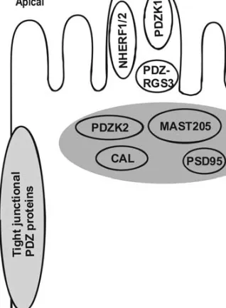
PDZ Proteins Expressedin Renal Proximal Tubules For clarity, we shall assign PDZ proteins of PT-cells to four subcellular regions:the brush border, the subapical compartment, the tight junctional complex and the basolateral membrane (Fig. 1). However, we do not claim that there is no interchange of PDZ proteins between the different regions, e.g., between the brush border and the subapical compartment. BRUSHBORDER
A number of PDZ proteins have been shown to reside within the microvilli at the apical pole of PT-cells. These PDZ proteins may act as scaffolds for a variety of apical transporters and may provide anchoring sites for a correct spatial arrangement of apically localized regulatory elements such as kinases, phos-pholipases and receptors.
NHERF1 (also named EBP50; Ref. 4), originally identified as a regulatory factor of the Na/H-ex-changer NHE3 [76], contains two PDZ domains and one MERM (moesin/ezrin/radixin/merlin) binding domain. Activated ezrin binds to the MERM domain and so provides a link of NHERF1 to the actin net-work [4]. There is good evidence that NHERF1 is localized almost exclusively in the brush borders of all proximal tubular segments [48, 60, 70]. As shown in yeast assays, PDZ domains of NHERF1 also interact with a number of solute transporters, such as the Na/Pi-cotransporter NaPi-lla, the chloride-for-mate exchanger CFEX, the urate/anion exchanger URAT1, the organic cation transporter OCTN1 [2, 17] and the cystic fibrosis transmembrane conduc-tance regulator CFTR [73]. In addition, interaction of NHERF1 with several components of signalling cascades have been reported as well (see below).
NHERF2 (also named E3KARP, Ref. 79), an isoform of NHERF1, contains two PDZ domains in tandem and an ezrin binding site. In heterologous expression systems, such as yeast two-hybrid assays, NHERF2 exhibited a similar interaction pattern with membrane transporters and regulatory proteins as NHERF1 [17]. A difference of the functional roles between the NHERF isoforms may be envisaged because apical expression of NHERF2 appears to be species-dependent:NHERF2 was described in brush borders of mouse renal PTÕs, but has not been ob-served in PT cells of rats [70].
PDZK1 (also named NaPi-Cap1, see Ref. 18), a protein composed of four PDZ domains, was initially identified by a yeast two-hybrid screen performed against MAP17, a 17 kDa protein that is up-regu-lated in kidney carcinomas [33, 34, 35]. Before, a shorter version, diphor-1, which lacks the fourth PDZ domain, had been cloned [12]. In PT cells of rats and mice, PDZK1 is restricted to the brush borders [18, 35, 48].
Results from yeast trap assays suggest that PDZK1 associates with the same transporters as NHERF1 [17]. In addition, MRP2 was reported to interact with PDZK1 [34]. Furthermore, the finding Fig. 1. Putative and established PDZ proteins in the renal proximal tubular cell. PDZ proteins are assigned to the brush borders (api-cal), to the basolateral site, to the subapical compartment (SAC) and to the tight junction.
that PDZK1 associates with D-AKAP2 [16], a dual PKA binding protein [26], indicates that PDZK1 targets PKA activity to the brush border of PT cells. A link of PDZK1 to the cytoskeletal network remains unknown.
PDZ-RGS3 was found to interact with NaPi-lla [18]. PDZ-RGS3 is a member of a protein family that has the property to activate GTPases of heterotri-meric G-proteins via RGS (regulator of G-protein signalling) domain an [31]. Therefore, it could be speculated that PDZ-RGS3 could provide a link be-tween transporters and certain signalling pathways. SUBAPICALCOMPARTMENT(SAC)
Attempts have been made to define this region as an intracellular organelle [23]. The subapical region of PT-cells is free of mitochondria and contains numerous endocytotic and dense, long-shaped vesi-cles; the latter likely deliver endocytosed and newly synthesized material to the apical membrane [9]. Also, the SAC can be conceived of as a decision point of membrane traffic, as demonstrated, for example, in the case of the regulation of the NaPi-lla protein by parathyroid hormone (PTH) [53]. Regarding PDZ proteins residing in the SAC, only sparse information is available.
PDZK2 (earlier referred to as NaPi-Cap2) was identified by a yeast two-hybrid screen against the C-terminus of NaPi-lla and has been localized to the SAC [18]. At the amino-acid level, PDZK2 is 26% identical to PDZK1 and contains four PDZ domains in tandem. Thus far, there is no information about a broader interaction palette of PDZK2. It is of interest that the human ortholog, IKEPP, has been identified in the context of the regulation of guanylyl cyclase C [61], suggesting that PDZK2 may act as a similar scaffolder for regulatory components in the PT cell.
CAL (CFTR-associated ligand), a single-PDZ-domain protein, has been localized in the transgolgi region and is thought to regulate the surface expres-sion of CFTR by retention [8]. As CAL also interacts with NaPi-lla (S. Gisler, unpublished results), it could be hypothesized that CAL may regulate surface expression of NaPi-lla or other apically localized transporters as well. The cellular location of CAL in PT cells, however, is not known.
PSD-95 (Post-Synaptic Density protein) is a member of the MAGUK family. PSD-95 was re-ported to interact with the multiligand endocytic receptor megalin [40]. However, the precise cellular distribution of PSD-95 in PT cells remains to be determined. As megalin is constantly endocytosed and recycled back to the apical membrane [9], this suggests that PSD-95 could orchestrate the trafficking of megalin through the SAC.
MAST205, a microtubule-associated serine/thre-onine kinase, was originally cloned from testis [71].
Besides a kinase domain, MAST205 posseses one PDZ domain at its carboxy end. A link of the dys-trophin/utrophin network to the microtubules via an interaction of the PDZ domain of MAST205 with 2-syntrophin has been described [47]. Recently, an association of MAST205 with NaPi-lla has been re-vealed on the basis of yeast two-hybrid and bio-chemical assays [18]. Although the cellular localization of this protein in PT cells is not known, it could be speculated that MAST205 may act as a linker of the Na/Pi-cotransporter to the microtu-bules. In fact, an involvement of microtubules in the routing of internalized NaPi-lla proteins to lysosomes has been descibed [46].
THETIGHT-JUNCTIONALCOMPLEX
In epithelia, paracellular pathways are controlled by the composition of the tight junctions. To date, 12 PDZ proteins have been associated with this macro-molecular structure (for review, see Ref.19). In addition to the capability to organize the tight-junc-tional complex, tight-junctight-junc-tional PDZ proteins also scaffold diverse proteins that are involved in signal-ling pathways for cell growth and differentiation [59]. Not much is known about the expression and cellular localization of PDZ proteins in tight junctions of PT cells.
The MAGUK protein ZO-1 (zonula occludens 1), has been detected in PT cells [20]. ZO-1 associates through the first PDZ domain with the tetraspan proteins claudins. Out of 18 different claudins, clau-din 2, 10 and 11 have been found in PTÕs [1, 32]. As ZO-2 and ZO-3 can be co-precipitated together with ZO-1, it seems likely that ZO-2/3 are present in PT cells as well. Tight junctional PDZ proteins are not only determinants for the permeability characteristics of tight junctions in PTÕs, but may also be involved in the regulation of transport functions. An example may be the recently described association of the tight-junctional PDZ protein MAGI-3 with frizzled-4, which is part of the Wnt signalling pathway [78]. It is of interest that frizzled proteins have been implicated in the regulation of renal handling of phosphate by the ‘‘phosphatonin’’ frizzled-related protein [56]. THEBASOLATERALMEMBRANE
Most indications of PDZ proteins that reside at the basolateral membrane were obtained from ‘‘non-re-nal’’ experimental systems. Nevertheless, the PDZ proteins mentioned below may be anticipated to be responsible for the proper basolateral localization of certain transporters.
a-SYNTROPHIN contains a single PDZ domain and two PH (plekstrin homology) domains, which confer the ability to bind lipids or phosphorylated serine or threonine residues. This protein is expressed
in several segments of the nephron, including proxi-mal tubules, where it is localized at the basolateral membrane [45]. a-Syntrophin interacts with AQP4, a water channel that has been assigned to the S3 seg-ment of proximal tubules [69]. In astrocytes of a-syntrophin-deficient mice, AQP4 showed a reversed polarization [54]. However, the impact of a-syntro-phin on the polarized distribution of AQP4 in PT-cells has not been analyzed in this model. In HEK cells, it was demonstrated that deletion of the PDZ binding motif (SSV) of AQP4 increased the rate of degradation of the channel and, vice versa, that the expression of a-syntrophin stabilizes the channel in the membrane [54].
CASK (Lin2), another member of the MAGUK family, and VEL1 (Lin7) are PDZ proteins expressed at low levels in the basolateral membrane of PTÕs [65]. A multiprotein complex containing both proteins recruits Kir2 potassium channels (ESE/AI) in brain [43]. Whether or not CASK and VEL1 fulfill similar tasks in the proximal tubule is unknown.
GLUT1CBP (Glut1 C-terminal binding protein) is a single-PDZ-domain protein expressed in kidney but its precise nephron/subcellular distribution has not been determined. GLUT1CBP interacts with the C-terminal DSQV motif of Glut1, a facilitative glu-cose transporter detected in the basolateral mem-brane of proximal tubules, and with a-actinin-1 or other cytoskeletal components [5]. Binding to GLUT1CBP has been suggested to stabilize Glut-1 at the basolateral membrane.
PICK1, a single PDZ domain protein, is able to form homo-oligomers. PICK1 has been proposed to bind (and cluster) several transporters involved in fluid regulation of neurons, such as AQPs and anion exchangers [10]. Although PICK1 was detected in kidney [66], its precise localization and function in renal cells is still an enigma
Functional Impacts of PDZ-mediated Protein-Protein Interactions in PT Cells
With respect to the proximal tubule, direct experi-mentation to elucidate the functional role of diverse PDZ protein interactions has been difficult. Thus far, only a few knock-out mouse models have been gen-erated that allow one to study the impact of PDZ proteins on proximal tubular function. Information has also been obtained from studies performed with cell cultures, notably OK cells, a cell line originally derived from opossum kidney. One should note, however, that this cell line may differ in a number of aspects from the in-vivo situation [51].
Thus far, there is no clear information about possible roles of PDZ proteins in the sorting of newly synthesized proteins to the apical or ba-solateral membrane. However, current data provide
evidence that PDZ proteins retain different trans-porters in the different plasma membrane domains. Furthermore, PDZ proteins may scaffold different transporters in microdomains of membranes that may contribute to the regulation of transport pro-cesses such as by pH changes or alterations of the ionic situation [52]. In addition, PDZ proteins, di-rectly or indidi-rectly, anchor and orchestrate compo-nents of signalling cascades.
SPATIALPOSITIONING
At the apical and basolateral membrane of PT cells, most of the described targets for PDZ proteins are transporters. Elucidation of the postulated functions of PDZ clusters became possible with recently gen-erated PDZ protein knock-out mice and with OK cells.
In proximal tubular brush border membranes of NHERF1-deficient mice, the abundance of the type IIa Na/Pi-cotransporter was reported to be decreased [62]. In these mice, kept either on a normal or a low Pi-diet, NaPi-lla accumulates in subapical, intracel-lular compartments [62, 74]. This suggests that NHERF1 is partially involved in the correct apical localization/positioning of the NaPi-IIa protein. In agreement, impaired apical localization of NaPi-IIa was observed in OK-cells after truncation of its C-terminal PDZ binding motif, TRL, or after over-expression of single PDZ domains [25, 30]. Interest-ingly, NHERF1 deficiency had no effect on the apical content of NHE3, which is explained, at least in mice, by a compensation with NHERF2 [70].
Studies performed with PDZK1-deficient mice indicated that the apical localization of transporters in PT cells is not dramatically affected. Initial studies performed with PDZK1-deficient mice fed a normal chow showed that the absence of PDZK1 does not alter the abundance of NaPi-IIa or the urinary excretion of phosphate [7, 36].
INVOLVEMENT OFPDZ PROTEINS IN THEREGULATION OF
PROXIMALTUBULARTRANSPORT
Na/H-exchange and Na/Pi-cotransport represent paradigms for transport functions in PT cells that are regulated by a variety of hormones and metabolic factors (for review, see Refs. 53 and 75).
A number of proteins implicated in the hormonal control of Na/Pi-cotransport (NaPi-IIa) and Na/H-exchange (NHE-3) have been described to interact with NHERF1/2:The receptor PTHR1 for PTH [49], the adrenergic b2-receptor [6] and phospholipase b1/2 [27, 64]. The observation that NHERF1 can form homodimers and that the formation of NHERF1 homodimers is regulated [41] indicates that NHERF1 participates in a complex way to orchestrate signal-ling cascades required to regulate transport functions.
A clone of OKcells, OKH cells, which expresses low levels of NHERF1, revealed direct evidence that NHERF1 assembles an apical, regulatory complex. Transfection of OKH cells with NHERF1 has been shown to restore the PTH-mediated increase of intracellular calcium, which is compatible with the formation of a complex consisting of NHERF1, PTHR1 and PLC-b [50]. How NHERF1 participates in the regulation of the NaPi-IIa protein when the activation of the apical PTHR1 receptor [67] is im-paired, is currently under investigation using NHERF1-deficient mice.
Besides the signalling components mentioned above, NHERF1 and 2 anchor protein kinase A (PKA) indirectly via ezrin. The role of the NHERF/ ezrin/PKA complex in the regulation of NHE3 has been extensively discussed (for review, see Refs. 63 and 75). Results from primary PT-cell cultures of NHERF1-deficient mice are in agreement with the concept that the NHERF/PKA/ezrin/ complex is necessary for the regulation of NHE3 activity by cyclic AMP [11]. In contrast, results obtained with OK cells suggested that NHERF1 is not required for the regulation of the NaPi-IIa protein in response to cyclic AMP [42]. The precise role of NHERF1 in the cyclic AMP-mediated regulation of NaPi-IIa in kid-ney remains to be determined.
Similar to NHERF1, PDZK1 provides an indi-rect anchor for PKA by sequestering D-AKAP2, which binds both regulatory subunits of PKA [16, 26]. The precise functional role of the PDZK1/D-AKAP2/PKA complex is currently not understood. Surprisingly, in PDZK1-deficient mice, regulation of NaPi-IIa by PTH or by activation of the PKA pathway was normal [7].
More direct control of transporter/channel function by PDZ proteins has been observed for the chloride permeability of CFTR. Interaction of re-combinant NHERF1 with CFTR increases the open probability of CFTR and phosphorylation of NHERF1 influences this channel modulation [57]. Similarly, a potentiation of CFTR channel activity was reported by the interaction of CFTR with PDZK1 [72].
Are NHERF1 or PDZK1 needed for the rapid and/or chronic adaptation of the Na/Pi-cotransporter NaPi-IIa? Dietary content of phosphate robustly regulates the abundance of the NaPi-lla protein in PT-cells, yet, the precise mechanisms are not known. Recent data obtained with NHERF1-and PDZK1-deficient mice indicated that neither NHERF1 nor PDZK1 is important for the regulation of NaPi-IIa by dietary content of Pi, albeit apical expression of NaPi-IIa after a low Pi-diet was slightly impaired in NHERF1-deficient mice [7, 74]. In contrast to the in-vivo findings, up-regulation of NaPi-IIa by a low-Pi medium was abrogated in primary cultures derived from NHERF1-deficient mice [11].
REGULATION OFPDZ INTERACTIONS BY
PHOSPHORYLATION
Until now, only a few examples have been reported that PDZ interactions can be modulated by phos-phorylation reactions either of the PDZ protein itself or of amino acids close or within the PDZ binding motif. It may be envisaged that phosphorylation reactions may allow an on-off mode of PDZ interac-tions. In PT cells, the necessity for an on-off mecha-nism of a PDZ interaction is best illustrated by the down-regulation of the Na/Pi-cotransporter NaPi-IIa in response to PTH. NaPi-IIa, which is localized along the entire length (>1 lm) of the microvilli is only internalized at the base of the microvilli, the inter-microvillar clefts [77]. As upon a stimulation of PTH receptors the localizations of NHERF1 and PDZK1 are not altered (N. Deliot, unpublished results), it is assumed that the affinity of NaPi-IIa to the PDZ do-mains is decreased by PTH in order to enable a higher diffusional mobility of NaPi-IIa. In analogy, increased diffusional mobility has been demonstrated for CFTR after truncation of its PDZ binding motif [21].
NHERF1, but not NHERF2, is constitutively phosphorylated [58]. The role of phosphorylation of NHERF1 has been extensively studied in the context of the PKA-mediated inhibition of NHE3 [75]. Re-cent results obtained in the authorsÕ laboratory by in-vitro experiments using mouse kidney slices showed that PTH induced an increase of NHERF1 phos-phorylation. Augmented phosphorylation of NHERF1 was also observed after individual activa-tion of PKA and PKC, respectively (N. Deliot, unpublished results). Interestingly, cyclic AMP-dependent regulation of NHE3 in OK-cells was not paralleled by an alteration of NHERF1 phosphory-lation [39]. Similarly, in kidney slice experiments, constitutive and regulated phosphorylation of PDZK1 was observed (N. Deliot, unpublished results). Modulations of PDZ interactions by phosphor-ylation reactions have been demonstrated in the fol-lowing cases:i) Phosphorylation of a serine residue within the C-terminal PDZ binding motif SLL of the b2-adrenergic receptor inhibits the interaction of the receptor with NHERF1 [6]; ii) Overlay experiments demonstrated that phosphorylation of the serine residue from the PDZ binding motif STKF of MRP2 has a positive effect on the interaction with NHERF1 [24]; iii) Phosphorylation of the PDZ domain 2 of NHERF1 (on Ser-162) in response to activated PKC resulted in an inhibition of the interaction between NHERF1 and CFTR [57].
IS THEABUNDANCE OFPDZ PROTEINS INPT CELLS
REGULATED?
Sparse information is available about the regulation of the abundance of PDZ proteins in PT-cells.
Transcriptional regulation of NHERF1 by estrogen and up-regulation of PDZK1 in renal carcinomas have been reported [13, 34].
The scaffolding functions of PDZ proteins dis-cussed above may suggest that under conditions that lead to significant up or down-regulation of a specific transport function, the abundance of PDZ proteins may be altered as well. One such potential situation is the intake of a low phosphate diet, which results in up-regulation of the NaPi-lla Na/Pi-cotransporter [44]. However, despite the robust interaction of NaPi-IIa with NHERF1 and PDZK1 as observed in bio-chemical assays, a parallel up-regulation of neither NHERF1 nor PDZK1 was observed in the authorsÕ laboratory [48]. In contrast, up-regulation of the PDZK1 protein by a low Pi-diet has been reported by others [11, 74].
Summary
Using kidney cDNA libraries and single proximal tubular proteins as baits, the yeast two-hybrid tech-nology resulted in the description of numerous po-tential PDZ-based protein-protein interactions. Many of these have been confirmed by biochemical in-vitro assays. In order to assign cellular functions of such proteins, it is first mandatory to define the pre-cise distribution and localization in PT cells of each canditate protein. This prerequisite has been assessed only for a few of the PDZ proteins mentioned in this article. In addition, it remains to be deciphered how the interactions of the identified PDZ proteins are modulated, for example, by phosphorylation reac-tions or by other posttranslational modificareac-tions. As mentioned in this article, PDZ knock-out mouse models may be of help to elucidate the physiological and pathophysiological functions of a particular PDZ interaction. However, current data indicate that, de-spite the robust interactions observed in in-vitro as-says, ablation of a certain PDZ protein does not necessarily result in an expected phenotype, probably due to functional compensation by other PDZ pro-teins. Therefore, it has to be assumed that a large redundancy of known and as yet unidentified PDZ proteins exists in proximal tubular cells.
We acknowledge the financial support by the Swiss National Foundation (Grant No. 31-65397.01 to H.M.) and by the Deutsche Forschungsgemeinschaft (TR-SFB11).
References
1. Alissa, H., Enck, U., Berger, V., Yu, A.S.L. 2001. Claudin-2 is selectively expressed in proximal nephron in mouse kidney. Am. J. Physiol. 281:F966–F974
2. Anzai, N., Miyazaki, H., Noshiro, R., Khamdang, S., Chair-oungdua, A., Shin, H.-J., Enomoto, A., Sakamoto, S., Hirata,
T., Tomita, K., Kanai, Y., Endou, H. 2004. The multivalent PDZ domain-containing protein PDZK1 regulates transport activity of renal urate-anion exchanger URAT1 via its C-ter-minal. J. Biol. Chem. 279:45942–45950
3. Bezprozvanny, I., Maximov, A. 2001. Classification of PDZ domains. FEBS Lett. 509:457–462
4. Bretscher, A., Chambers, D., Nguyen, R., Reczek, D. 2000. ERM-merlin and EBP50 protein families in plasma membrane organization and function. Annu. Rev. Cell. Dev. Biol. 16:113– 143
5. Bunn, R.C., Jensen, M.A., Reed, B.C. 1999. Protein interac-tions with the glucose transporter binding protein GLUT1CBP that provide a link between GLUT1 and the cytoskeleton. Mol. Biol. Cell. 10:819–832
6. Cao, T.T., Deacon, H.W., Reczeck, D., Bretscher, A., Zas-trow, M. 1999. A kinase-regulated PDZ-domain interaction control endocytic sorting of the b2-adrenergic receptor. Nature 401:286–289
7. Capuano, P., Bacic, D., Stange, G., hernando, N., Kaissling, B., Pal, R., Kocher, O., Biber, J., Wagner, C.A., Murer, H. 2005. Expression and regulation of the renal Na/phosphate cotransporter NaPi-IIa in a mouse model deficient for the PDZ protein PDZK1. Pfluegers Arch.-Eur. J. Physiol. 449:392-402 8. Cheng, J., Moyer, B.D., Milewski, M., Loffing, J., Ikeda, M.,
Mickle, J.E., Cutting, G.R., Li, M., Stanton, B.A., Guggino, W.B. 2002. A golgi-associated PDZ domain protein modulates cystic fibrosis transmembrane regulator plasma membrane expression. J. Biol. Chem. 277:3520–3529
9. Christensen, E.I., Birn, H. 2002. Megalin and cubulin:multi-functional endocytic receptors. Nature Rev. Mol. Cell Biol. 3:258–268
10. Cowan, C.A., Yokoyama, N., Bianchi, L.M., Henkemeyer, M., Fritzsch, B. 2000. EphB2 guides axons at the midline and is necessary for normal vestibular function. Neuron 26:417–430 11. Cunninghman, R., Steplock, D., Wang, F., Huang.H., .,
Weinman, E.J. 2004. Defective PTH regulation of NHE3 activity and phosphate adaptation in cultured NHERF1 -/-renal proximal cells. J. Biol. Chem. 279:37815–37821 12. Custer, M., Spindler, B., Verrey, F., Murer, H., Biber, J. 1997.
Identification of a new gene product (Diphor-1) regulated by dietary phosphate. Am. J. Physiol. 273:F801–F806
13. Ediger, T.R., Park, S.E., Katzenellenwogen, B.S. 2002. Estro-gen receptor inducibilitty of the human Na/H exchnager reg-ulatory factor/ezrin-radixin-moesin protein 50 (NHE-RF/ EBP50) gene involving multi half-estrogen respsonse elements. Mol. Endocrin. 16:1828–1839
14. Fanning, A.S., Anderson, J.M. 1999. PDZ domains:funda-mental building blocks in the organization of protein com-plexes at the plasma membrane. J,. Clin. Invest. 103:767–772 15. Garner, C.C., Nash, J., Huganir, R.L. 2000. PDZ domains in
synapse assembly and signalling. Trends Cell Biol. 10:274–280 16. Gisler, S.M., Madjdpour, C., Pribanic, S., Bacic, D., Taylor, S.S., Biber, J., Murer, H. 2003. PDZK1:II. An anchoring site for the PKA-binding protein D-AKAP2 in renal proximal tubular cells. Kidney Int. 64:1746–1754
17. Gisler, S.M., Pribanic, S., Bacic, D., Forrer, P., Sabourin, LA., Tsuji, A., Zhao, Z., Manser, E., Biber, J., Murer, H. 2003. PDZK1:I. A major scaffolder in brush borders of proximal tubular cells. Kidney Int. 64:1733–1745
18. Gisler, S.M., Stagljar, I., Traebert, M., Bacic, D., Biber, J., Murer, H. 2001. Interaction of the type IIa Na/Pi
-cotrans-porter with PDZ proteins. J. Biol. Chem. 276:9206–9213 19. Gonzalez-Mariscal, L., Betanzos, A., Nava, P., Jaramillo, B.E.
2003. Tight junction proteins. Prog. Biophys. Mol. Biol. 81:1–44 20. Gonzalez-Mariscal, L., Namorado, M.C., Martin, D., Luna, J., Valencia, S., Muriel, P., Ponce, L., Reyes, J.L. 2000. Tight
junction proteins ZO-1, ZO-2, and occludin in isolated renal tubules. Kidney Int. 57:2386–2402
21. Haggie, P.M., Stanton, B.A., Verkman, A.S. 2003. Increased diffusinal mobility of CFTR at the plasma membrane after deletion of its C-terminus PDZ binding motif. J. Biol. Chem. 279:5494–5500
22. Hall, RA., Premont, RT., Chow, CW., Blitzer, JT., Pitcher, JA., Claing, A., Stoffel, RH., Barak, LS., Shenolikar, S., Weinman, EJ., Grinstein, S., Lefkowitz, RJ. 1996. The beta2 adrenergic receptor interacts with the Na/H-exchanger regu-latory factor to control Na/H exchange. Nature 392:626–630 23. Hatae, T., Ichimura, T., Ishida, T., Sakurai, T. 1996. Apical
tubular network in the rat kidney proximal tubule cells studied by thick section and scanning electron microscopy. Cell Tissue Res 288:317–325
24. Hegedu¨s, ., Sessler, T., Scott, R., Thelin, W., Bakos, E., Va-radi, A., Szabo, K., Homolya, L., Milgram, S., Sarkadi, B. 2003. C-terminal phosphorylation of MRP2 modulates its interaction with PDZ proteins. Biochem. Biophys. Res. Comm. 302:454–461
25. Hernando, N., Deliot, N., Gisler, S., Lederer, E., Weinman, E.J., Biber Murer, J. H. 2002. PDZ-domain interactions and apical expression of type IIa Na/Pi-cotransporters. Proc. Natl. Acad. Sci. USA 99:11957–11692
26. Huang, L.J., Durick, K., Weiner, J.A., Chun, J., Taylor, S.S. 1997. D-AKAP2, a novel protein kinase A anchoring protein with a putative RGS domain. Proc. Natl. Acad. Sci. USA 94:11184–11191
27. Hwang, J.-I., Heo, K., Shin, K.J., Kim, E., Yun, C.H., Ryu, S.H., Shin, H.S., Suh, P.G. 2000. Regulation of phospholipase C-b3 activity by Na/H exchanger regulatory factor 2. J. Biol. Chem. 275:166632–16637
28. Hung, A.Y., Sheng, M. 2002. PDZ domains:Structural mod-ules for protein complex assembly. J. Biol. Chem. 277:5699– 5702
29. Kang, B.S., Cooper, D.R., Devedjiev, Y., Derewenda, U., Derewenda, Z.S. 2003. Molecular roots of degenerate speci-ficity in synteninÕs PDZ2 domain:Reassessment of the PDZ recognition paradigm. Structure 11:845–853
30. Karim-Jimenez, Z., Hernando, N., Biber Murer, J. H. 2001. Molecular determinants for apical expression of the renal type IIa NaPi -cotransporter, Pfluegers Arch.-Eu. J. Physiol.
442:782–790
31. Kehrl, J.H., Srikumar, D., Harrison, K., Wilson, G.L., Shi, C.-S. 2002. Additional 5Õ exons in the RGS3 locus generate multiple mRNA transcripts, one of which accounts fort he origin of human PDZ-RGS3. Genomics 79:860–868
32. Kiuchi-Saishin, Y., Gotoh, S., Furuse, M., Takasuga, A., Tano, Y., Tsukita, S. 2002. Differential expression patterns of claudins, tight junction membrane proteins, in mouse nephron segments. J. Am. Soc. Nephrol. 13:875–886
33. Kocher, O., Cheresh, P., Lee, S.W. 1996. Identification and partial characterization of a novel membrane associated pro-tein (MAP17) up-regulated in human carcinomas and modu-lating cell replication and tumor growth. Am. J. Pathol. 149:493–500
34. Kocher, O., Comelia, N., Gilchrist, A., Pal, R., Tognazzi, K., Brown, L.F., Knoll, J.H.M. 1999. PDZK1, a novel PDZ do-main-containing protein up-regulated in carcinomas and mapped to chromosome 1q21, interacts with cMOAT (MRP2), the multidrug resistance associated protein. Lab. Invest. 79:1161–1170
35. Kocher, O., Comelia, N., Tognazzi, K., Brown, L. 1998. Identification and partial characterization of PDZK1:a novel protein containing PDZ interaction domains. Lab. Invest. 78:117–125
36. Kocher, O., Pal, R., Roberts, M., Cirovic, C., Gilchrist, A. 2003. Targeted disruption of the PDZK1 gene by homologous recombination. Mol. Cell. Biol. 23:1175–1180
37. Kriz, W., Kaissling, B. 2000. Structural organization of the mammalian kidney. In: The Kidney Physiology & Patho-physiology. Seldin, D.W, Giebisch, G. editors. pp 587–654, Lippincott Williams & Wilkins, Newyork,
38. Ladias, J.A.A. 2003. Structural insights into the CFTR-NHERF interaction. J. Membrane. Biol. 192:79–88
39. Lambrecht, G., Weinman, E.J., Yun, C.H.C. 1998. The role of NHERF and E2KARP in the cAMP-mediated inhibition of NHE3. J. Biol. Chem. 273:29972–29978
40. Larsson, M., Hja¨lm, G., Sakwe, A.M., Engstrom, A., Ho¨gl-und, A., Larsson, E., Robinson, R.C., Sundberg, C., Rask, L. 2003. Selective interaction of megalin with PSD-95-like MAGUK proteins. Biochem. J. 373:381–391
41. Lau, A.G., Hall, R.A. 2001. Oligomerization of NHERF1 and NHERF2 PDZ domains:differential regulation by association with receptor carboxyl-termini and by phosphorylation. Bio-chemistry 40:8572–8580
42. Lederer, E.D., Khundmiri, S.J., Weinman, E.J. 2003. Role of NHERF-1 in regulation of the activity of Na-K ATPase and sodium-phosphate cotransport in epithelial cells. J. Am. Soc. Nephrol. 14:1711–1719
43. Leonoudakis, D., Conti, L.R., Radeke, C.M., McGuire, L.M., Vandenberg, C.A. 2004. A multiprotein trafficking complex composed of SAP97, CASK, Veli, and Mint1 is associated with inward rectifier Kir2 potassium channels. J. Biol. Chem. 279:19051–19063
44. Levi, M., Kempson, S.A., Lo¨tscher, M., Biber, J., Murer, H. 1996. Molecular regulation of renal phosphate transport. J. Membrane Biol. 154:1–9
45. Loh, N.Y., Newey, S.E., Davies, K.E., Blake, D.J. 2000. Assembly of multiple dystrobrevin-containing complexes in the kidney. J. Cell Sci. 113:15–24
46. Lo¨tscher, M., Kaissling, B., Biber, J., Murer, H., Levi, M. 1997. Role of microtubules in the rapid regulation of renal phosphate transporter content. J. Clin. Invest. 99: 1302–1312
47. Lumeng, C., Phelps, S., Crawford, G.E., Walden, P.O., Barald, K., Chamberlain, J.S. 1999. Interactions between b2-syntro-phin and a family of icrotubule-associated serine/threonine kinases. Nature Neuroscience 2:611–617
48. Madjdpour, C., Bacic, D., Kaissling, B., Murer, H., Biber, J. 2004. Segment-specific expression of sodium-phosphate co-transporters NaPi-IIa and -IIc and interacting proteins in mouse renal proximal tubules. Pfluegers Arch.-Europ. J. Physiol. 448:402–410
49. Mahon, M.J., Donowitz, M., Yun, C.C., Segre, G.V. 2002. Na/ H exchanger regulatory factor 2 directs parathyroid hormone 1 receptor signalling. Nature 417:858–860
50. Mahon, M.J., Segre, G.V. 2004. Stimulation by parathyroid hormone of a NHERF1 assembled complex consisiting of the parathyroid receptor I, phospholipase Cbeta and actin in-creases intracellular calcium in opossum kidney cells. J. Biol. Chem. 279:23550–23558
51. McDonough, A.A., Biemesderfer, D. 2003. Does membrane trafficking play a role in regulating the sodium/hydrogen ex-changer isoform 3 in the proximal tubule? Curr. Opin. Nephrol. Hypertens. 12:533–541
52. Moe, O.W. 2003. Scaffolds:Orchestrating proteins to achieve concerted function. Kidney Int. 64:1916–1917
53. Murer, H., Hernando, N., Forster, I., Biber, J. 2000. Proximal tubular phosphate reabsorption. Physiol. Rev. 80:1373–1409 54. Neely, J.D., Amiry-Moghaddam, M., Ottersen, O.P.,
expression and localization of aquaporin-4 water channel protein. Proc. Natl. Acad. Sci. USA 98:14108–14113 55. Nourry C, Grant SGN, and Borg J-P. PDZ domain proteins:
Plug and play. www.stke.org./cgi/content/full/sigtrans;2003/ 179/re7
56. Quarles, L.D. 2003. FGF23, PHEX, and MEPE regulation of phosphate homeostasis and skeletal mineralization. Am. J. Physiol. 285:E1–E9
57. Raghuram, V., Hormuth, H., Foskett, J.K. 2003. A kinase-regulated mechanisms controls CFTR channel gating by dis-rupting bivalent PDZ domain interactions. Proc. Natl. Acad. Sci. USA 100:9620–9625
58. Reczak, D., Berryman, M., Bretscher, A. 1997. Identification of EBP50:a PDZ-containing phosphoprotein that associates with members of the ERM family. J. Cell Biol. 139:169–179 59. Roh, M.H., Margolis, B. 2002. Composition and function of
PDZ protein complexes during cell polarization. Am. J. Physiol. 285:F377–F387
60. Sabolic, I., Herak-Kramberger, C.M., Ljubojevic, M., Bie-mesderfer, D., Brown, D. 2002. NHE3 and NHERF are tar-geted to the basolateral membrane in proximal tubules of colchicine-treated rats. Kidney lnt. 61:1351–64
61. Scott, R.O., Thelin, W.R., Milgram, S.L. 2002. A novel PDZ protein regulates the activity of guanylyl cyclase C, the heat-stable enterotoxin receptor. J. Biol. Chem. 277:22934–22941 62. Shenolikar, S., Voltz, J.W., Minkoff, C.M., Wade, J.B.,
Weinman, E.J. 2002. Targeted disruption of the mouse NHERF1 gene promotes internalization of proximal tubule sodium-phosphate cotransporter type IIa and renal phosphate wasting. Proc. Natl. Acad. Sci. USA 99:11470–11475 63. Shenolikar, S., Weinman, E.J. 2001. NHERF:targeting and
trafficking membrane proteins. Am. J. Physiol. 280:F389–F395 64. Shu, P.-G., Hwang, J.-I., Ryu, S.H., Donowitz, M., Kim, J.H. 2001. The roles of PDZ-containing proteins in PLC-b-medi-ated signaling. Biochim. Biophys. Res. Comm. 288:1–7 65. Straight, S.W., Karnak, D., Borg, J.P., Kamberov, E., Dare,
H., Margolis, B., Wade, J.B. 2000. mLin-7 is localized to the basolateral surface of renal epithelia via its NH(2) terminus. Am. J. Physiol. 278:F464–F475
66. Staudinger, J., Zhou, J., Burgess, R., Elledge, S.J., Olson, E.N. 1995. PICK1:a perinuclear binding protein and substrate for protein kinase C isolated by the yeast two-hybrid system. J. Cell. Biol. 128:263–271
67. Traebert, M., Volkl, H., Biber, J., Murer, Kaissling, H. 2000. Luminal and contraluminal action of 1-34 and 3-34 PTH peptides on renal type IIa Na/Pi-cotransporter. Am. J. Physiol.
278:F792–F798
68. Ullrich, K.J. 1979. Sugar, amino acid, and Na+cotransport in the proximal tubule. Annu. Rev. Physiol. 41:181–195 69. van Hoek, A.N. van , Ma, T., Yang, B., Verkman, A.S.,
Brown, D. 2000. Aquaporin-4 is expressed in basolateral membranes of proximal tubule S3 segments in mouse kidney. Am. J. Physiol. 278:F310–F316
70. Wade, J.B., Liu, J., Coleman, R.A., Cunningham, R., Step-lock, D.A., Lee-Kwon, W., Pallone, T.L., Shenolikar, S., Weinman, E.J. 2003. Localization and interaction of NHERF isoforms in the renal proximal tubule of the mouse. Am. J. Physiol. 285:C1494–C1503
71. Walden, P.D., Cowan, N.J. 1993. A novel 205-kilodalton tes-tis-specific serine/threonine protein kinase associated with microtubules of the spermatide manchette. Mol. Cell. Biol. 13:7625–7635
72. Wang.S., ., Yue.H., ., Derin, R.B. 2000. Acessory protein facilitated CFTR-CFTR interaction, a molecular mechanism to potentiate the chloride channel activity. Cell 103:169–179 73. Wang, S., Raab, R.W., Schatz, P.J., Guggino, W.B., Li, M.
1998. Peptide binding consensus of the NHE-RF-PDZ1 doa-main matches the C-terminal sequence of cystic fibrosis transmembrane conductance regulator (CFTR). FEBS Lett. 427:103–108
74. Weinman, E.J., Boddeti, A., Cunningham, R., Akom, M., Wanf, F., Wang, Y., Liu, J., Steplock, D., Shenolikar, S., Wade, J.B. 2003. NHERF1 is required for renal adaptation to a low phosphate diet. Am. J. Physiol. 285:F1225–F1232 75. Weinman, E.J., Minkoff, C., Shenolikar, S. 2000. Signal
complex regulation of renal transport proteins:NHERF and regulation of NHE3 by PKA. Am. J. Physiol. 279:F393–F399 76. Weinman, E.J., Steplock, D., Wang, Y., Shenolikar, S. 1995. Characterization of a protein cofactor that mediates protein kinase A regulation of the renal brush border membrane Na-H exchanger. J. Clin. Invest. 95:2143–2149
77. Yang, L.E., Maunsbach, A.B., Leong, P.K.K., McDonough, A.A. 2004. Differential trafficking of proximal tubule NHE3 and NaPi2 during acute hypertension or PTH treatment: NHE3 to base of microvilli vs. NaPi to endosomes. Am. J. Physiol. 287:F896–F906
78. Yao, R., Natsume, Y., Noda, T. 2004. MAGI-3 is involved in the regulation of the JNK signaling pathway as a scaffold protein for frizzled and Ltap. Oncogene 23:6023–6030 79. Yun, C.H., Oh, S., Zizak, M., Steplock, D., Tsao, S., Tse,
C.M., Weinman, E.J., Donowitz, M. 1997. cAMP mediated inhibition of the epithelial brush border Na/H-exchanger, NHE3, requires an associated regulatory protein. Proc. Natl. Acad. Sci. USA 94:3010–3015