HAL Id: hal-01420860
https://hal.sorbonne-universite.fr/hal-01420860
Submitted on 21 Dec 2016
HAL is a multi-disciplinary open access
archive for the deposit and dissemination of
sci-entific research documents, whether they are
pub-lished or not. The documents may come from
teaching and research institutions in France or
abroad, or from public or private research centers.
L’archive ouverte pluridisciplinaire HAL, est
destinée au dépôt et à la diffusion de documents
scientifiques de niveau recherche, publiés ou non,
émanant des établissements d’enseignement et de
recherche français ou étrangers, des laboratoires
publics ou privés.
Distributed under a Creative Commons Attribution| 4.0 International License
Serum IL-33, a new marker predicting response to
rituximab in rheumatoid arthritis
Jérémie Sellam, Elodie Rivière, Alice Courties, Paul Rouzaire, Barbara
Tolusso, Edward M. Vital, Paul Emery, Gianfranco Ferraciolli, Martin
Soubrier, Bineta Ly, et al.
To cite this version:
Jérémie Sellam, Elodie Rivière, Alice Courties, Paul Rouzaire, Barbara Tolusso, et al.. Serum IL-33, a
new marker predicting response to rituximab in rheumatoid arthritis. Arthritis Research and Therapy,
BioMed Central, 2016, 18 (1), pp.294. �10.1186/s13075-016-1190-z�. �hal-01420860�
R E S E A R C H A R T I C L E
Open Access
Serum IL-33, a new marker predicting
response to rituximab in rheumatoid
arthritis
Jérémie Sellam
1,10*†, Elodie Rivière
2†, Alice Courties
1, Paul-Olivier Rouzaire
3, Barbara Tolusso
4, Edward M. Vital
5,
Paul Emery
5, Gianfranco Ferraciolli
4, Martin Soubrier
6, Bineta Ly
2, Houria Hendel Chavez
7, Yassine Taoufik
7,
Maxime Dougados
8and Xavier Mariette
2,9*Abstract
Background: Recent works have suggested a possible link between interleukin (IL)-33 and B-cell biology. We aimed to study the possible association between serum IL-33 detection and response to rituximab (RTX) in rheumatoid arthritis (RA) patients in different cohorts with an accurate enzyme-linked immunosorbent assay (ELISA).
Methods: Serum IL-33, rheumatoid factor (RF), anti-cyclic citrullinated peptide (anti-CCP), and high serum immunoglobulin (Ig)G levels were assessed in 111 RA patients receiving a first course of 2 g RTX (cohort 1) in an observational study and in 74 RA patients treated with the same schedule in routine care (cohort 2). Univariate and multivariate analyses identified factors associated with a European League Against Rheumatism (EULAR) response at 24 weeks.
Results: At week 24, 84/111 (76%) and 54/74 (73%) patients reached EULAR response in cohorts 1 and 2, respectively. Serum IL-33 was detectable in only 33.5% of the patients. In the combined cohorts, the presence of RF or anti-CCP (odds ratio (OR) 3.27, 95% confidence interval (CI) 1.13–9.46; p = 0.03), high serum IgG (OR 2.32, 95% CI 1.01–5.33; p = 0.048), and detectable serum IL-33 (OR 2.40, 95% CI 1.01–5.72; p = 0.047) were all associated with RTX response in multivariate analysis. The combination of these three factors increased the likelihood of response to RTX. When serum IL-33 detection was added to seropositivity and serum IgG level, 100% of the patients with the three risk factors (corresponding to 9% of the population) responded to RTX (OR versus patients with none of the three risk factors 29.61, 95% CI 1.30–674.79; p = 0.034).
Conclusion: Detectable serum IL-33 may predict clinical response to RTX independently of, and synergistically with, auto-antibodies and serum IgG level.
Trial registration: NCT01126541; 18 May 2010.
Keywords: Rheumatoid arthritis, Interleukin 33, Rituximab, B-cell, Personalized medicine
* Correspondence:jeremie.sellam@aphp.fr;xavier.mariette@aphp.fr
†Equal contributors
1Université Paris 06, AP-HP St-Antoine hospital, Rheumatology Department,
INSERM UMRS_938, DHU i2B, Paris, France
2
Université Paris-Sud, AP-HP Hôpitaux Universitaires Paris-Sud, Rheumatology Department, Center for Immunology of Viral Infections and Autoimmune Diseases INSERM U1184, Le Kremlin Bicêtre, France
Full list of author information is available at the end of the article
© The Author(s). 2016 Open Access This article is distributed under the terms of the Creative Commons Attribution 4.0 International License (http://creativecommons.org/licenses/by/4.0/), which permits unrestricted use, distribution, and reproduction in any medium, provided you give appropriate credit to the original author(s) and the source, provide a link to the Creative Commons license, and indicate if changes were made. The Creative Commons Public Domain Dedication waiver (http://creativecommons.org/publicdomain/zero/1.0/) applies to the data made available in this article, unless otherwise stated.
Background
Interleukin (IL)-33 is one of the most recently discov-ered members of the IL-1 cytokine family mediating its biological effects via its binding to its receptor suppres-sion of tumorigenicity (ST)2 [1]. IL-33 is upregulated in both resident cells and inflammatory infiltrating cells and is released in case of cell injury, thus acting as an alarmin [2, 3]. IL-33 induces production of Th2 cyto-kines and eosinophilia, and may activate mast cells that can in turn release several pro-inflammatory cytokines [4]. IL-33 investigations have been mainly devoted to asthma and allergy, with the development of a targeted IL-33/ST2 axis therapeutic strategy [5].
IL-33 may also be involved in rheumatoid arthritis (RA) pathogenesis. IL-33 administration exacerbates collagen-induced and K/BxN serum-mediated murine arthritis, and disease severity is reduced in mice treated with sST2-Fc fusion protein or anti-IL-33 monoclonal antibody [6–8]. Extracellular IL-33 is a critical enhancer of tumor necrosis factor (TNF)-induced RA synovial fibroblast activation [9] and could activate osteoclasto-genesis [10, 11]. In patients with RA, biomarker studies have suggested that the serum level of IL-33 could re-flect clinical activity [12] and disease severity, or predict carotid plaque progression [13]. However, the role of IL-33 could be paradoxical since, in K/BxN serum transfer-induced arthritis, ST2 but not IL-33 blockade may improve arthritis [14, 15]. Moreover, IL-33-stimulated mast cells could also suppress monocyte activation [16] and intracellular IL-33 also has anti-osteoclastogenic and anti-inflammatory properties [11].
Some recent works have suggested a possible link between IL-33 and B-cell biology [17]. In mice, IL-33 en-hances immunoglobulin (Ig)M synthesis and markedly induces and activates B1 cells in an ST2-dependent manner [18]. Additionally, IL-33 could also induce regu-latory B cells to produce IL-10, attenuating mucosal inflammation in the gut [19].
Using a transcriptomic approach, we have found that increased IL-33 mRNA expression in the whole blood of patients with RA was predictive of the response to ritux-imab (RTX), a targeted B cell-depleting agent [20]. We aimed to investigate, using an accurate and simple enzyme-linked immunosorbent assay (ELISA), the pos-sible association between a detectable serum level of the IL-33 protein and a response to RTX in RA patients in different cohorts.
Methods
Patients
A total of 224 patients with RA for at least 6 months and fulfilling the American College of Rheumatology (ACR) 1987 criteria were included in the SMART study (NCT01126541). This study is a 2-year, national,
multicenter, randomized open-label study evaluating the efficacy and tolerability of two doses of RTX for re-treatment after one initial course of RTX at a usual dose (1000 mg on days 1 and 15) described previously [21]. All patients had active disease, defined by a Disease Activity Score in 28 joints (DAS28) using C-reactive pro-tein (CRP) (DAS28-CRP) >3.2, with ≥6/66 swollen and ≥6/68 tender joints, or a CRP ≥10 mg/L, or an erythro-cyte sedimentation rate (ESR) ≥28 mm/h. Erosive status was based on the reading of hand and feet X-rays by the investigators in each center. Each patient received a stable dose of methotrexate (MTX) (≥10 mg/week for at least 4 weeks) and had experienced an inadequate response or intolerance to TNF inhibitors, or had con-traindications to TNF inhibitors. All patients in the SMART study received one course of RTX (two 1000 mg infusions, given on days 1 and 15) with usual pre-medication (methylprednisolone, acetaminophen, and antihistamine). In this ancillary study, 111 patients from the 224 were included due to the availability of the serum samples for IL-33 assessment and were desig-nated as cohort 1.
We also studied 32 RA patients from Leeds (UK) and 42 from Clermont-Ferrand (France) treated in real life with a first course of RTX using the same schedule. In order to match the numbers of patients in cohort 1, these two groups of patients from Leeds and Clermont-Ferrand were merged and called cohort 2. There were no overlaps between the two cohorts.
For all patients, treatment efficacy was evaluated 24 weeks after the first RTX infusion according to European League Against Rheumatism (EULAR) response [22]. Patients were then classified as responders (good or moderate) or non-responders after this first course of RTX.
Assessment of serum IL-33, auto-antibodies, and serum IgG
We first used an ELISA IL-33 kit (DuoSet, R&D Sys-tems) for protein assessment, and preliminary presented as an abstract an association between serum IL-33 level and RTX response in a single cohort [23]. However, this kit was not validated for human sera and additional experiments have confirmed the need for caution about the accuracy of this kit for sera measurements [24]. Recommendations from the manufacturer advise us to use another assay for human sera, named Quantikine and also provided by R&D Systems. We aimed to use this more accurate ELISA in order to investigate the possible association between a detectable serum level of IL-33 and a response to RTX in RA patients in the different cohorts.
Using a sample obtained prior to the initiation of RTX in the two cohorts, we measured the serum IL-33 level using the Quantikine kit, which is a solid-phase ELISA
with a 6.25 pg/mL lower limit threshold according to the manufacturer’s instruction [24]. The assessment of rheumatoid factor (RF), anti-cyclic citrullinated peptide (anti-CCP) antibodies, and serum IgG level were also performed. The upper limit of normal (ULN) for serum IgG was 12.7 g/L and high serum IgG level was a value above this cut-off, as described previously [25].
Statistical analysis
Continuous data are described as means and standard deviations (mean ± SD) or as medians and ranges (median (range)).
For all analyses, since serum IL-33 is not systematic-ally detectable, we presented qualitative results as detect-able serum IL-33 or undetectdetect-able serum IL-33. Among patients of the SMART cohort, we compared patients who were assessed for serum IL-33 level and those who were not due to the availability of the serum samples. Student and Wilcoxon tests were used to compare quan-titative values and the Chi-squared test was used for qualitative values.
We analyzed by logistic regression the relationship between EULAR response at 24 weeks and the following four explanatory variables: 1) high disease activity defined as DAS28-CRP >5.1; 2) high serum IgG level; 3)
auto-antibody status (i.e., the presence or absence of RF and/or anti-CCP antibodies), since all of them have been found previously as associated with subsequent RTX response [25, 26]; and 4) detectable serum IL-33. All these explanatory variables were entered in a stepwise multivariate model systematically adjusted on RF or anti-CCP positivy (entry level,p = 0.15; level for staying in the model,p = 0.10). Results are expressed as odds ra-tios (ORs) with 95% confidence intervals (CIs). Ap value <0.05 was considered as significant. Statistical analysis was performed with SAS 9.4 software.
Results
Characteristics of the populations
The characteristics of patients from cohort 1 are presented in Table 1. There was no significant difference between the 111 patients that underwent IL-33 assess-ment and the others that participated in the main SMART trial (n = 113) who did not (Table 1). Available characteristics of patients from cohort 2 are presented in Additional file 1 (Table S1).
Serum IL-33 levels
Using the validated Quantikine assay, most of the patients had undetectable serum IL-33: 84/111 (76%) and
Table 1 Baseline characteristics of RA patients included in the IL-33 ancillary study from the SMART trial and a comparison between the patients in the SMART cohort included in the IL-33 study (cohort 1, n = 111) and the rest of the cohort not assessed for serum IL-33 level (n = 113)
Cohort 1 Patients in the SMART cohort without IL-33 dosage
Whole SMART cohort Inter-group comparison (n = 111) (n = 113) (n = 224)
Age (years) 56 ± 11 56 ± 11 56 ± 11 0.99
Female 95 (86%) 92 (81%) 187 (84%) 0.401
Disease duration (years) 11 (1–42) 10 (0.8–45) 11 (0.8–45) 0.094 DAS28-CRP 5.8 ± 0.8 5.8 ± 0.9 5.8 ± 0.9 0.791
HAQ-DI 1.8 ± 0.6 1.8 ± 0.6 1.8 ± 0.6 0.753
Prednisone treatment 76 (68%) 88 (78%) 165 (74%) 0.11 Prednisone dosage (mg/day) 8 (2–20) 10 (2–15)* 10 (2–20)* 0.243
MTX (mg/week) 14 ± 4 14 ± 4 14 ± 4 0.351
Previous biologic: None/mAb/Eta 8 (7%)/66 (60%)/37(33%) 7 (6%)/70(62%)/36(32%) 15 (7%)/136 (61%)/73(33%) 0.914 Positive RF 76 (69%) 68 (61%) 144 (65%) 0.226 Positive anti-CCP 92 (83%) 78 (70%) 170 (76%) 0.02 Serum IgG level (g/L) 11.8 (4.6–19.2) 12 (5.4–23.3) 12 (4.6–23.3) 0.564 CRP level (mg/L) 12 (0.4–139) 11 (0.3–102) 11 (0.3–139) 0.938 Radiographic erosions 100 (90%) 98 (87%) 198 (88%) 0.432 MTX duration (years) 3.6 (0.1–20.8) 3.2 (0.3–20.5) 3.5 (0.1–20.8) 0.856 Time since last anti-TNF (months) 2.7 (0.6–64.4) 2.8 (1.2–73.6) 2.7 (0.6–73.6) 0.458
Values are shown as means and standard deviations (mean ± SD) or medians (range) for continuous data, and n (%) for qualitative data *Some patients had a deviation protocol concerning the authorized prednisone dosage
anti-CCP anti-cyclic citrullinated peptide antibody, CRP C-reactive protein, DAS28 Disease Activity Score in 28 joints, Eta etanercept, HAQ-DI Health Assessment Questionnaire—Disease Index, IL interleukin, mAb monoclonal antibody, MTX methotrexate, RF rheumatoid factor, TNF tumor necrosis factor
39/74 (53%) in cohorts 1 and 2, respectively. In patients with detectable IL-33, mean serum levels were 63.39 ± 87.31 pg/mL and 45.35 ± 87.19 pg/mL in cohorts 1 and 2, respectively (Fig. 1).
Factors associated with subsequent response to RTX at week 24 in cohort 1
Among the 111 patients, a total of 84 patients (76%) were classified as EULAR responders to RTX at week 24; 27 pa-tients (24%) were non-responders. Detectable serum IL-33 was found in 27 (24.3%) patients. In univariate analysis, the three explanatory variables including DAS28-CRP >5.1, high serum IgG level, and auto-antibody status were associated with RTX response and were included in the multivariate model since they had ap value <0.15 for the association with EULAR response (Table 2).
The multivariate analysis indicated that high disease activity (OR 5.24, 95% CI 1.85–14.85) and auto-antibody status (OR 3.48, 95% CI 1.04–11.60) were independently associated with RTX response. A non-significant associ-ation between EULAR response and high serum IgG level was found (OR 2.82, 95% CI 0.97–8.14) (Table 2). There was no association with serum IL-33 detection in multivariate analysis in cohort 1.
Factors associated with subsequent response to RTX at week 24 in cohort 2
In order to replicate these results in RA patients from routine care, we tested whether the DAS28-CRP >5.1, high serum IgG level, auto-antibody status, and detect-able serum IL-33 were associated with response to RTX
in cohort 2 in which there were 73% EULAR responders among 74 patients.
In univariate analysis, high disease activity and detect-able serum IL-33 were associated with RTX response, but not auto-antibody status or high serum IgG (Table 2). In multivariate analysis, detectable serum IL-33 was the sole factor associated with RTX response (OR 3.73, 95% CI 1.12–12.38), with a non-significant association for auto-antibody status (OR 32.76 (0.7– 999). The presence of RF and anti-CCP auto-antibodies in 97% of the patients in cohort 2 might limit analysis on these markers.
Factors associated with subsequent response to RTX at week 24 in the combination of cohorts 1 and 2
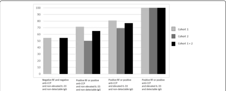
Multivariate analyses indicated that four factors were independently and significantly associated with response to RTX: high disease activity (OR 4.10, 95% CI 1.90– 8.85), auto-antibody status (OR 3.27, 95% CI 1.13–9.46), high IgG level (OR 2.32, 95% CI 1.01–5.33), and detect-able serum IL-33 (OR 2.40, 95% CI 1.01–5.72) (Tdetect-able 3 and Fig. 2). Since the presence of RF and/or anti-CCP antibodies and high serum IgG level have already been identified as predictive markers of subsequent EULAR response, we aimed to investigate whether the addition of detectable serum IL-33 increased the likelihood of response. While 55% of patients without these three factors responded to RTX, the presence of RF and/or anti-CCP antibodies and elevated serum IgG level fur-ther increased the likelihood of this response (77% responders; OR = 2.72, 95% CI 0.67–10.98; p = 0.16 versus patients without any of these three factors). When we
Fig. 1 Serum IL-33 levels in patients from cohort 1, cohort 2, and the combined cohorts. Dosage performed with Quantikine ELISA IL-33 kit (R&D Systems). Each dot represents one patient; means are presented. IL interleukin
added detectable serum IL-33 to these two factors, 100% of the patients with the three risk factors (corresponding to 9% of the combined population) responded to RTX (OR 29.61, 95% CI 1.30–674.79; p = 0.034 versus patients with none of the three risk factors) (Fig. 3).
If we restricted the analysis to seropositive patients from both cohorts (94/111 from cohort 1 and 72/74 from cohort 2), IL-33 detection was still highly predictive of EULAR response (OR 3.03, 95% CI 1.24–7.4; p = 0.015), confirming that this biomarker was independent of seropositivity.
Discussion
In this study, we have identified serum IL-33 detection as a novel biomarker associated with RTX response in RA, in addition to auto-antibody status, in real life patients.
This study was based on our previous observation of the upregulation of IL-33 mRNA expression in the whole blood that was associated with RTX response in a microarray study performed in patients randomly selected from the SMART trial [20]. Since the IL-33 pro-tein may be easily quantified in the serum, we first assessed serum IL-33 levels in the same population using
Table 2 Factors associated with rituximab response in cohorts 1 and 2
Characteristic EULAR non-responders
EULAR responders
Univariate p value Multivariate p value OR (95% CI) OR (95% CI)
Cohort 1 (n = 111) DAS28-CRP 3.2–5.1 (Ref) 12 (46.2%) 14 (53.8%) 4.0 (1.55–10.36) 0.004 5.24 (1.85–14.85) 0.002 >5.1 15 (17.6%) 70 (82.4%)
RF or anti-CCP Negative (Ref) 7 (41.2%) 10 (58.8%) 2.59 (0.88–7.67) 0.08 3.48 (1.04–11.60) 0.04 Positive 20 (21.3%) 74 (78.7%)
IgG > ULN (g/L) No (Ref) 21 (30.9%) 47 (69.1%) 2.76 (1.01–7.52) 0.048 2.82 (0.97–8.14) 0.056 Yes 6 (14.0%) 37 (86.0%)
Detectable IL-33 No (Ref) 23 (27.4%) 61 (72.6%) 2.17 (0.62–6.95) 0.19 – – Yes 4 (14.8%) 23 (85.2%)
Cohort 2 (n = 74) Disease activity DAS28-CRP
3.2–5.1 (Ref) 14 (36.8%) 24 (63.2%) 2.92 (0.97–8.73) 0.056 – – >5.1 6 (16.7%) 30 (83.3%)
RF or anti-CCP Negative (Ref) 2 (100%) 0 (0%) 14.72 (0.34–629.71) 0.16 32.76 (0.7–999) 0.076 Positive 18 (25.0%) 54 (75%)
IgG > ULN (g/L) No (Ref) 15 (37.5%) 25 (62.5%) 2.10 (0.58–7.57) 0.26 – – Yes 4 (22.2%) 14 (77.8%)
Detectable IL-33 No (Ref) 14 (35.9%) 25 (64.1%) 2.71 (0.91–8.10) 0.07 3.73 (1.12–12.38) 0.03 Yes 6 (17.1%) 29 (82.9%)
Values are given as n (%) of responders or non-responders with a given characteristic Rituximab response was evaluated at week 24 according to EULAR response
anti-CCP anti-cyclic citrullinated peptide antibody, CI confidence interval, DAS28-CRP Disease Activity Score in 28 joints by C-reactive peptide, EULAR European League Against Rheumatism, Ig immunoglobulin, OR odds ratio, Ref referent, RF rheumatoid factor, ULN upper limit of normal (i.e., 12.7 g/L)
Table 3 Factors associated with rituximab response in the combination cohort (cohorts 1 and 2)
Characteristics EULAR non-responders (n = 47) EULAR responders (n = 138) Univariate OR (95% CI) p value Multivariate OR (95% CI) p value DAS28-CRP 3.2–5.1 (Ref) 26 (40.6%) 38 (59.4%) 3.26 (1.64–6.47) 0.0007 4.10 (1.90–8.85) 0.0003 >5.1 21 (17.4%) 100 (82.6%)
RF or anti-CCP Negative (Ref) 9 (47.4%) 10 (52.6%) 3.03 (1.15–8.001) 0.025 3.27 (1.13–9.46) 0.03 Positive 38 (22.9%) 128 (77.1%)
IgG > ULN (g/L) No (Ref) 36 (33.3%) 72 (66.7%) 2.55 (1.16–5.60) 0.02 2.32 (1.01–5.33) 0.048 Yes 10 (16.4%) 51 (83.6%)
Detectable IL-33 No (Ref) 37 (30.1%) 86 (69.9%) 2.24 (1.03–4.87) 0.04 2.40(1.01–5.72) 0.047 Yes 10 (16.1%) 52 (83.9%)
Values are given as n (%) of responders or non-responders with a given characteristic Rituximab response was evaluated at week 24 according to EULAR response
anti-CCP anti-cyclic citrullinated peptide antibody, CI confidence interval, DAS28-CRP Disease Activity Score in 28 joints by C-reactive peptide, EULAR European League Against Rheumatism, Ig immunoglobulin, OR odds ratio, Ref referent, RF rheumatoid factor, ULN upper limit of normal (i.e., 12.7 g/L)
the DuoSet ELISA IL-33 kit (R&D System) and prelimin-ary reported in an abstract that the serum IL-33 level was associated with the RTX response [23]. However, additional experiments in patients with Sjögren’s syndrome or RA have raised caution about the accuracy of this kit for sera measurements [24]. Consequently, we discarded these preliminary results and examined serum IL-33 again with an accurate ELISA kit (Quantikine) validated for sera, in two separate and then merged populations.
Here, we have found in univariate and multivariate analysis a significant association between serum IL-33 de-tection and EULAR response in cohort 2. Furthermore, when we combined the two cohorts, the association was significant with an odds ratio in the same range as high serum IgG level (i.e., approximately 2). We thus confirmed at the protein level the results found at the mRNA level, which demonstrates that transcriptomic analysis with non a-priori hypotheses might open the way to new patho-genic pathways.
Fig. 3 Frequency of EULAR response according to the presence of auto-antibodies, the detectability of serum IL-33, and a serum IgG above the upper limit of normal in cohort 1, cohort 2, and the combined cohorts. Results are presented for patients having auto-antibodies and/or elevated serum IL-33 level and/or elevated serum IgG level compared with patients having no auto-antibodies, a non-elevated serum IL-33 and a non-elevated IgG level (referent). anti-CCP anti-cyclic citrullinated peptide antibody, Ig immunoglobulin, IL interleukin, RF rheumatoid factor
Fig. 2 Association between the four explanatory variables and EULAR response at 24 weeks after the first rituximab infusion in the combination of cohorts 1 and 2. Results from the multivariate analysis are presented as odds ratios (OR) (95% confidence intervals (CI)) for each factor. anti-CCP anti-cyclic citrullinated peptide antibody, DAS28-CRP Disease Activity Score in 28 joints by C-reactive peptide, IL interleukin, RF rheumatoid factor Sellam et al. Arthritis Research & Therapy (2016) 18:294 Page 6 of 8
This association was independent of strong predictive factors associated with RTX response, especially the presence of RF or anti-CCP antibodies, which strengthens the possible interest in serum IL-33 assess-ment. We previously reported that patients with a pres-ence of auto-antibodies and high serum IgG level had a better response to RTX in comparison with patients having none of these characteristics [25]. If serum IL-33 detection is added to these two predictive factors, the likelihood of response to RTX reaches 29.6 (95% CI 1.3– 675) in comparison with absence of these three factors. Indeed, 100% of patients displaying these three factors simultaneously were responsive to RTX in our combined cohort. However, these patients represented 9% of the study population.
Auto-antibody status was associated with RTX response in SMART, as previously reported [25], but also in the combination of cohorts. The absence of association in cohort 2 alone may be explained by the presence of these auto-antibodies in almost all the patients (97%) limiting analysis on these markers. Lastly, as in every trial, it is easier to have a response when the starting DAS28 is high and high disease activity was associated with a better response to RTX, as previously observed in SMART [25].
Despite a number of strengths, and especially the use of a replication cohort, this study has several limitations. First, serum IL-33 assessment needs the use of an accur-ate assay such as the Quantikine kit, but the relatively low frequency of patients with detectable IL-33 level (33.5% in the merged population) justifies qualitative IL-33 detection rather than quantitative IL-IL-33 values. Sec-ond, we have no data on the association between serum IL-33 detection and response to other biologic agents in RA, limiting our findings to RTX. Third, the association between serum IL-33 detection and response to RTX is statistically significant, but the usefulness of such a measurement for clinical practice needs to be further in-vestigated. Finally, the presence of RF and anti-CCP auto-antibodies in 97% of the patients in cohort 2 might limit analysis on these markers, as well as the difference in terms of high disease activity (DAS 28-CRP >5.1) frequency between the two cohorts (cohort 1 = 77% versus cohort 2 = 49%).
Conclusions
In conclusion, serum IL-33 assessment with a robust assay may represent a novel, easy biomarker predicting RTX response in synergy with auto-antibody status and high serum IgG level. Such a finding concerning these three biomarkers being easy to monitor in clinical prac-tice represents a further step towards the goal of person-alized medicine to determine the best approach to the therapeutic management of RA [27].
Additional file
Additional file 1: Table S1. Baseline characteristics of RA patients from Clermont and from Leeds. (DOC 33 kb)
Abbreviations
anti-CCP:Anti-cyclic citrullinated peptide; CI: Confidence interval; CRP: C-reactive protein; DAS28: Disease Activity Score in 28 joints; ELISA: Enzyme-linked immunosorbent assay; ESR: Erythrocyte sedimentation rate; EULAR: European League Against Rheumatism; Ig: Immunoglobulin; IL: Interleukin;
MTX: Methotrexate; OR: Odds ratio; RA: Rheumatoid arthritis; RF: Rheumatoid factor; RTX: Rituximab; ST: Suppression of tumorigenicity; TNF: Tumor necrosis factor; ULN: Upper limit of normal
Acknowledgements
We thank the other members of the scientific committee of the SMART study: Pr. J. Sibilia (Strasbourg, France), Pr. J. Tebib (Lyon, France), Pr. B. Combe (Montpellier, France), and Pr. X. Le Loët (Rouen, France). We thank all the SMART investigators: Dr. I. Azais, Poitiers; Dr. J.C. Balblanc, Belfort; Dr. F. Berenbaum, Paris; Dr. P. Bertin, Limoges; Dr. M.-C. Boissier, Bobigny; Dr. P. Bourgeois, Paris; Dr. A. Cantagrel, Toulouse; Dr. P. Carli, Toulon; Dr. P.-Y. Chouc, Marseille; Dr. M. Couret, Valence; Dr. L. Euller-Ziegler, Nice; Dr. P. Fardellone, Amiens; Dr. P. Fauquert, Berck/Mer; Dr. R.-M. Flipo, Lille; Dr. P. Gaudin, Echirolles; Dr. J.-L. Grauer, Aix en Provences; Dr. A. Heraud, Libourne; Dr. P. Hilliquin, Corbeil; Dr. S. Hoang, Vannes; Dr. E. Houvenagel, Lomme; Dr. D. Keita, Paris; Dr. K. Lassoued, Cahors; Dr. L. Le Dantec, Lievin; Dr. J.-M. Le Parc, Boulogne; Dr. L. Lequen, Pau; Dr. F. Lioté, Paris; Dr. C. Marcelli, Caen; Dr. O. Meyer, Paris; Dr. J.-L. Pellegrin, Pessac; Dr. A. Perdriger, Rennes; Dr. G. Rajzbaum, Paris; Dr. S. Redeker, Abbeville; Dr. J.-M. Ristori, Clermont-Ferrand; Dr. A. Saraux, Brest; Dr. G. Tanguy, La Roche sur Yon; Dr. T. Thomas, Saint-Priest-en-Jarez; Dr. L. Zabraniecki, Toulouse, Dr. C. Zarnitski, Montivilliers, France. We thank Dr. Pascale Boisseaux and François Gavini (Roche, France) for supporting this ancillary study.
The authors thank Laura Smales (BioMed Editing, Toronto, Canada) for editing the manuscript and Statitec (Toulouse, France) for independent statistical analysis.
Funding
This work was supported by Roche France, who sponsored the study but were not involved in the interpretation of the data or in the preparation of the manuscript.
Role of the study sponsor: Roche France designed the SMART study but did not participate in the design, data collection, or interpretation of the results of this ancillary study, which was proposed by an independent scientific committee. Roche France supported the measurement of serum biomarkers and the statistical analysis. Roche France was not involved in the writing of the manuscript. Their agreement to submit the manuscript for publication was not required, their approval of the content of the submitted manuscript was not required, and publication of the manuscript was not contingent upon their approval.
Availability of supporting data
Supporting data are available and authors had full access to all of the data in the study.
Authors’ contributions
JS: participated in conception and design of the study, participated in the statistical analysis and interpretation of the results, and wrote the manuscript. ER: performed acquisition of the data, participated in the statistical analysis and interpretation of the results, and wrote the manuscript. AC: participated in statistical analysis and interpretation of the data, and wrote the manuscript. MD: participated in conception and design of the study, acquisition and
interpretation of the data, and manuscript preparation. XM: participated in conception and design of the study, statistical analysis and interpretation of the results, and wrote the manuscript. POR, BT, EV, PE, GF, MS, BL, HHC and YT: participated in acquisition and interpretation of the data, and manuscript preparation. All authors reviewed and approved the final manuscript.
Authors’ information
JS and XM had full access to all of the data in the study and take responsibility for the integrity of the data and the accuracy of the data analysis.
JS and ER made equal contribution to this study. Competing interests
XM and MD belonged to the scientific committee of the SMART study and received honoraria from Roche (less than $10,000 each) for this task. A research grant was provided from Roche for the IL-33 serum biomarkers measurements and for independent statistical analysis.
Consent for publication
We confirm that all authors approved the manuscript for submission. Ethical approval and consent to participate
This study was approved by each local ethic committee: Groupe Hospitalier Pitié-Salpêtrière, Paris, France; Leeds West Research Ethics Committee; and Ethic Committee of Clermont-Ferrand. Consent has been obtained from all patients who participated in this study.
Author details
1Université Paris 06, AP-HP St-Antoine hospital, Rheumatology Department,
INSERM UMRS_938, DHU i2B, Paris, France.2Université Paris-Sud, AP-HP Hôpitaux Universitaires Paris-Sud, Rheumatology Department, Center for Immunology of Viral Infections and Autoimmune Diseases INSERM U1184, Le Kremlin Bicêtre, France.3Biological Immunology Department, ERTICa
Research Group, Clermont-Ferrand University Hospital, Clermont-Ferrand EA4677, France.4Rheumatology Department, Catholic University of the
Sacred Heart, Roma, Italy.5NIHR Leeds Musculoskeletal Biomedical Research Unit, Leeds Teaching Hospitals NHS Trust, Leeds, UK and Leeds Institute of Rheumatic and Musculoskeletal Medicine, University of Leeds, Leeds, UK.
6Rheumatology Department, Clermont-Ferrand University Hospital,
Clermont-Ferrand, France.7AP-HP Bicêtre Hospital, Biological Immunology Department, INSERM U1184, Le Kremlin Bicêtre, France.8Department of
Rheumatology - Hôpital Cochin, Paris Descartes University, Assistance Publique - Hôpitaux de Paris, INSERM (U1153), Clinical Epidemiology and Biostatistics, PRES Sorbonne Paris-Cité, Paris, France.9Service de Rhumatologie, Hôpital de Bicêtre, 78 rue du Général Leclerc, Le Kremlin Bicêtre 94275, France.10Service de Rhumatologie, Hôpital Saint-Antoine, 184 rue du Faubourg Saint-Antoine, Paris 75012, France.
Received: 29 June 2016 Accepted: 21 November 2016
References
1. Liew FY, Pitman NI, McInnes IB. Disease-associated functions of IL-33: the new kid in the IL-1 family. Nat Rev Immunol. 2010;10(2):103–10. 2. Theoharides TC, Petra AI, Taracanova A, Panagiotidou S, Conti P. Targeting
IL-33 in autoimmunity and inflammation. J Pharmacol Exp Ther. 2015;354(1):24–31.
3. Millar NL, Murrell GAC, McInnes IB. Alarmins in tendinopathy: unravelling new mechanisms in a common disease. Rheumatol Oxf Engl. 2013;52(5):769–79.
4. Schmitz J, Owyang A, Oldham E, Song Y, Murphy E, McClanahan TK, et al. IL-33, an interleukin-1-like cytokine that signals via the IL-1 receptor-related protein ST2 and induces T helper type 2-associated cytokines. Immunity. 2005;23(5):479–90.
5. Stolarski B, Kurowska-Stolarska M, Kewin P, Xu D, Liew FY. IL-33 exacerbates eosinophil-mediated airway inflammation. J Immunol Baltim Md 1950. 2010;185(6):3472–80.
6. Xu D, Jiang H-R, Kewin P, Li Y, Mu R, Fraser AR, et al. IL-33 exacerbates antigen-induced arthritis by activating mast cells. Proc Natl Acad Sci U S A. 2008;105(31):10913–8.
7. Xu D, Jiang H-R, Li Y, Pushparaj PN, Kurowska-Stolarska M, Leung BP, et al. IL-33 exacerbates autoantibody-induced arthritis. J Immunol. 2010;184(5): 2620–6.
8. Athari SK, Poirier E, Biton J, Semerano L, Hervé R, Raffaillac A, et al. Collagen-induced arthritis and imiquimod-Collagen-induced psoriasis develop independently of interleukin-33. Arthritis Res Ther. 2016;18(1):143.
9. Kunisch E, Chakilam S, Gandesiri M, Kinne RW. IL-33 regulates TNF-α dependent effects in synovial fibroblasts. Int J Mol Med. 2012;29(4):530–40.
10. Malcolm J, Awang RA, Oliver-Bell J, Butcher JP, Campbell L, Adrados Planell A, et al. IL-33 exacerbates periodontal disease through induction of RANKL. J Dent Res. 2015;94(7):968–75.
11. Lee E-J, So MW, Hong S, Kim Y-G, Yoo B, Lee C-K. Interleukin-33 acts as a transcriptional repressor and extracellular cytokine in fibroblast-like synoviocytes in patients with rheumatoid arthritis. Cytokine. 2016;77:35–43. 12. Xiangyang Z, Lutian Y, Lin Z, Liping X, Hui S, Jing L. Increased levels of
interleukin-33 associated with bone erosion and interstitial lung diseases in patients with rheumatoid arthritis. Cytokine. 2012;58(1):6–9.
13. Shen J, Shang Q, Wong C-K, Li EK, Wang S, Li R-J, et al. IL-33 and soluble ST2 levels as novel predictors for remission and progression of carotid plaque in early rheumatoid arthritis: a prospective study. Semin Arthritis Rheum. 2015;45(1):18–27.
14. Martin P, Talabot-Ayer D, Seemayer CA, Vigne S, Lamacchia C, Rodriguez E, et al. Disease severity in K/BxN serum transfer-induced arthritis is not affected by IL-33 deficiency. Arthritis Res Ther. 2013;15(1):R13. 15. Talabot-Ayer D, Martin P, Seemayer CA, Vigne S, Lamacchia C, Finckh A,
et al. Immune-mediated experimental arthritis in IL-33 deficient mice. Cytokine. 2014;69(1):68–74.
16. Rivellese F, Suurmond J, Habets K, Dorjée AL, Ramamoorthi N, Townsend MJ, et al. Ability of interleukin-33- and immune complex-triggered activation of human mast cells to down-regulate monocyte-mediated immune responses. Arthritis Rheumatol Hoboken NJ. 2015;67(9):2343–53. 17. Komai-Koma M, Gilchrist DS, McKenzie ANJ, Goodyear CS, Xu D, Liew FY.
IL-33 activates B1 cells and exacerbates contact sensitivity. J Immunol Baltim Md 1950. 2011;186(4):2584–91.
18. Ahmed A, Koma MK. Interleukin-33 triggers B1 cell expansion and its release of monocyte/macrophage chemoattractants and growth factors. Scand J Immunol. 2015;82(2):118–24.
19. Sattler S, Ling G-S, Xu D, Hussaarts L, Romaine A, Zhao H, et al. IL-10-producing regulatory B cells induced by IL-33 (Breg(IL-33)) effectively attenuate mucosal inflammatory responses in the gut. J Autoimmun. 2014; 50:107–22.
20. Sellam J, Marion-Thore S, Dumont F, Jacques S, Garchon H-J, Rouanet S, et al. Use of whole-blood transcriptomic profiling to highlight several pathophysiologic pathways associated with response to rituximab in patients with rheumatoid arthritis: data from a randomized, controlled, open-label trial. Arthritis Rheumatol Hoboken NJ. 2014;66(8):2015–25. 21. Mariette X, Rouanet S, Sibilia J, Combe B, Le Loët X, Tebib J, et al. Evaluation
of low-dose rituximab for the retreatment of patients with active rheumatoid arthritis: a non-inferiority randomised controlled trial. Ann Rheum Dis. 2014;73(8):1508–14.
22. van Gestel AM, Anderson JJ, van Riel PL, Boers M, Haagsma CJ, Rich B, et al. ACR and EULAR improvement criteria have comparable validity in rheumatoid arthritis trials. American College of Rheumatology European League of Associations for Rheumatology. J Rheumatol. 1999;26(3):705–11. 23. Sellam J, Hendel-Chavez H, Rouanet S, Vernet N, Ly B, Marion-Thore S, et al.
Serum IL-33 level is increased in rheumatoid arthritis and predicts response to rituximab in combination with high serum IgG level and autoantibody positivity: an open-label, prospective, multicentre biological trial. Arthritis Rheum. 2014;66 Suppl 10:S1279–80.
24. Rivière E, Ly B, Boudaoud S, Chavez H, Nocturne G, Chanson P, et al. Pitfalls for detecting interleukin-33 by ELISA in the serum of patients with primary Sjögren syndrome: comparison of different kits. Ann Rheum Dis. 2016;75(3): 633–5.
25. Sellam J, Hendel-Chavez H, Rouanet S, Abbed K, Combe B, Le Loët X, et al. B cell activation biomarkers as predictive factors for the response to rituximab in rheumatoid arthritis: a six-month, national, multicenter, open-label study. Arthritis Rheum. 2011;63(4):933–8.
26. Couderc M, Mathieu S, Pereira B, Glace B, Soubrier M. Predictive factors of rituximab response in rheumatoid arthritis: results from a French university hospital. Arthritis Care Res. 2013;65(4):648–52.
27. van den Broek M, Visser K, Allaart CF, Huizinga TWJ. Personalized medicine: predicting responses to therapy in patients with RA. Curr Opin Pharmacol. 2013;13(3):463–9.