HAL Id: hal-03041304
https://hal.archives-ouvertes.fr/hal-03041304
Submitted on 5 Dec 2020
HAL is a multi-disciplinary open access
archive for the deposit and dissemination of
sci-entific research documents, whether they are
pub-lished or not. The documents may come from
teaching and research institutions in France or
abroad, or from public or private research centers.
L’archive ouverte pluridisciplinaire HAL, est
destinée au dépôt et à la diffusion de documents
scientifiques de niveau recherche, publiés ou non,
émanant des établissements d’enseignement et de
recherche français ou étrangers, des laboratoires
publics ou privés.
sequentially in response to the actomyosin force
Clémence Vigouroux, Véronique Henriot, Christophe Le Clainche
To cite this version:
Clémence Vigouroux, Véronique Henriot, Christophe Le Clainche. Talin dissociates from RIAM and
associates to vinculin sequentially in response to the actomyosin force. Nature Communications,
Nature Publishing Group, 2020, 11 (1), �10.1038/s41467-020-16922-1�. �hal-03041304�
Talin dissociates from RIAM and associates
to vinculin sequentially in response to the
actomyosin force
Clémence Vigouroux
1
, Véronique Henriot
1
& Christophe Le Clainche
1
✉
Cells reinforce adhesion strength and cytoskeleton anchoring in response to the actomyosin
force. The mechanical stretching of talin, which exposes cryptic vinculin-binding sites,
trig-gers this process. The binding of RIAM to talin could regulate this mechanism. However, the
mechanosensitivity of the talin-RIAM complex has never been tested. It is also not known
whether RIAM controls the mechanosensitivity of the talin-vinculin complex. To address
these issues, we designed an in vitro microscopy assay with puri
fied proteins in which the
actomyosin force controls RIAM and vinculin-binding to talin. We demonstrate that
acto-myosin triggers RIAM dissociation from several talin domains. Actoacto-myosin also provokes the
sequential exchange of RIAM for vinculin on talin. The effect of RIAM on this
force-dependent binding of vinculin to talin varies from one talin domain to another. This
mechanism could allow talin to biochemically code a wide range of forces by selecting
different combinations of partners.
https://doi.org/10.1038/s41467-020-16922-1
OPEN
1Université Paris-Saclay, CEA, CNRS, Institute for Integrative Biology of the Cell (I2BC), 91198 Gif-sur-Yvette, France. ✉email:christophe.leclainche@i2bc. paris-saclay.fr
123456789
M
echanical cues govern a variety of biological processes.
During migration, cells sense and respond to changes in
intracellular and extracellular forces by adapting their
shape, dynamics, and adhesion. Focal adhesions (FAs), that play a
major role in this mechanosensitive process, are composed of
transmembrane integrins that mechanically couple the
extra-cellular matrix (ECM) to the actomyosin cytoskeleton, via
actin-binding proteins (ABPs)
1–6. The mechanosensitivity of FAs
allows cells to adapt adhesion strength to the internal force of the
actomyosin cytoskeleton and to the physical properties of the
ECM
7–13. However, the biochemical mechanisms that govern FA
mechanosensitivity remain largely unexplored.
Several lines of evidence indicate that the force-dependent
conformational change of the actin-binding protein talin might be
the initial switch that triggers the maturation of short-lived
nas-cent adhesions into stable FAs, which withstand higher adhesion
and actomyosin traction force
3,14–16. Although it is thought that
distinct force-dependent conformations of talin select specific
binding partners to trigger appropriate mechanical responses, a
limited number of these mechanisms have been discovered.
The mechanosensitive talin–vinculin interaction has long been
the hallmark of FA maturation
3. Experiments of mechanical
stretching of talin by atomic force microscopy and magnetic
tweezers revealed how force exposes one or more of the 11 cryptic
vinculin-binding sites (VBSs) located in the 13 helical bundles of
the talin rod domain (R1–R13, Fig.
1a)
14,17–20. Using an in vitro
reconstitution strategy, we demonstrated that the actomyosin
force is sufficient to stretch talin, allowing its binding to
vinculin
21,22. A series of cellular and biochemical studies showed
that the force-dependent formation of the talin–vinculin complex
reinforces actin anchoring to FAs
21,23–26. This reinforcement is
further enhanced by the stability of the talin–vinculin complex in
which vinculin locks talin in its stretched conformation
20,21.
Less is known about the specific talin-binding partners in nascent
adhesions. First observations in cells showed that the
Rap1-GTP-interacting adaptor molecule (RIAM) is localized at the leading edge
of migrating cells, where it cooperates with talin and Ena/VASP
proteins to promote the formation of actin-based membrane
protrusions
27,28. During this early phase, RIAM could bind to the
F3 domain of talin head, which disrupts the autoinhibition of talin,
allowing it to activate integrins
29,30. Interestingly, RIAM, which is
initially enriched in nascent adhesions, is replaced by vinculin in
mature FAs, that are known to experience higher traction force
31,32.
Structural studies showed that RIAM also interacts with exposed
residues in the talin helical bundles R2, R3, R8, and R11
33. In R2,
R3, and R8, RIAM-binding sites and VBSs overlap
33–35, suggesting
a complex interplay between RIAM, vinculin, and talin.
Altogether these observations suggest that the actomyosin force
triggers the mechanosensitive transition between nascent
adhe-sions and FAs by controlling the binding of RIAM and vinculin to
talin. However, the mechanosensitivity of the talin–RIAM
com-plex has never been tested. It is also not known whether
RIAM-binding to talin controls the mechanosensitive formation of the
talin–vinculin complex. Furthermore, several domains of talin
that interact with RIAM and vinculin may respond to force
dif-ferently. To determine the mechanosensitivity, sequence and
interdependence of these talin-associated reactions, we designed
an in vitro microscopy assay with purified proteins. In this assay,
the actomyosin force controls the binding of RIAM and vinculin
to a micropatterned surface coated with talin constructs, which
contain variable RIAM- and vinculin-binding sites (Fig.
1a).
Results
In vitro reconstitution of talin
–RIAM complexes. The
stoi-chiometry of the talin–RIAM complex was a major problem to
start this study. RIAM contains two talin-binding site (TBS1 and
TBS2). TBS1 binds to the R2, R3, R8, and R11 helix bundles of the
talin rod and to the subdomain F3 of the head
30,33,35(Fig.
1a).
TBS2 has a very low affinity for talin
35. However, TBS2 could
reinforce TBS1 anchoring by binding to a neighboring site if
available. Hence, RIAM TBS1–TBS2 interacts with talin R2–R3
with a 2:2 stoichiometry and a higher affinity than the TBS1–R3
complex
33. Based on this information, we designed a
fluorescent
mCherry-RIAM 1-306 protein, encompassing TBS1 and TBS2, to
visualize the RIAM–talin interaction in microscopy (Fig.
1a,
Supplementary Fig. 1a, b). We also designed a series of minimal
talin constructs, made of the F2–F3 domains of the head, that
anchor the protein to a micropatterned surface
21, followed by an
exchangeable cassette containing variable RIAM-binding sites,
and the C-terminal actin-binding domain ABD3 (R13) that binds
to the actomyosin cytoskeleton (Fig.
1a, Supplementary Fig. 1a,
b). We selected the three following RIAM-binding regions for
insertion in our minimal talin constructs: R1–R2–R3 that
includes the high affinity R2–R3 part, R7–R8 in which the
RIAM-binding R8 bundle is inserted in R7, and the single helical bundle
R11 (Supplementary Fig. 1a). We found that RIAM binds
spe-cifically and with high affinity to disk-shaped micropatterns
coated with these three talin constructs (Fig.
1b, c). In contrast,
RIAM does not bind to talin F2–F3 alone, whereas it binds to the
same construct fused to R1–R2–R3 (Supplementary Fig. 2). This
result indicates that, in our experimental setup, talin F3 does not
interact with RIAM because it is masked by its interaction with
the surface or because, as reported by others, its affinity is too low
(K
d= 32 µM)
30.
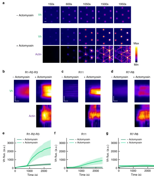
Actomyosin-dependent binding of vinculin to talin bundles.
Before testing the effect of actomyosin on talin–RIAM
interac-tions, we
first established the mechanosensitivity of our minimal
talin constructs using the approach that we developed
previously
21,22. In this method, the force applied to disk-shaped
talin-coated micropatterns is sufficient to expose cryptic
vinculin-binding sites in talin and recruit
fluorescent EGFP-vinculin head
(Vh) (Fig.
1a). Here, the force is produced by the association of
myosin II with slowly polymerizing actin
filaments and applied to
talin through its actin-binding domain (R13). Although
acto-myosin self-organizes transiently, it remains in the disks for at
least 1000 s. In this assay, talin R1–R2–R3 shows a strong binding
to Vh in the presence of actomyosin, compared with the low
constitutive binding measured in the absence of actomyosin
(Fig.
2a, b, e, Supplementary Movie 1). Using a procedure that we
have already validated for full-length talin
21, we confirmed that
the actomyosin-dependent increase in Vh–talin interaction
depends on myosin II and not on the bundling and
poly-merization of the actin network, even for a minimal talin like
R1–R2–R3 (Supplementary Fig. 3a, b). We also observed the
actomyosin-dependent binding of Vh to talin R11 (Fig.
2c, f,
Supplementary Movie 2). R1–R2–R3 recruits more Vh than R11
because it contains
five VBSs, whereas R11 contains only one
(Supplementary Fig. 1a). However, talin R7–R8 does not bind to
Vh in the presence of the same concentration of actin and myosin
used to stimulate R1–R2–R3 and R11 (Fig.
2d, g, Supplementary
Movie 3). The fact that R7–R8 does not bind Vh at all in the
presence of actomyosin also rules out the mechanical exposure of
the single VBS of ABD3 in our series of three constructs (Fig.
2d,
g). Altogether, our data showed that the actomyosin force is
efficiently transmitted to talin through ABD3 to stretch
R1–R2–R3 and R11 but not R7–R8.
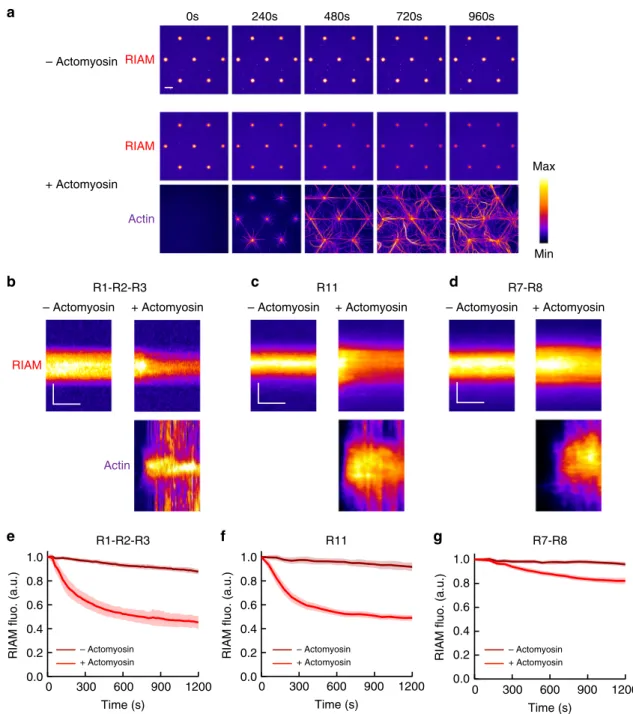
Actomyosin-dependent dissociation of RIAM from talin. To
test the mechanosensitivity of the three talin–RIAM complexes,
we measured the variation in
fluorescence of mCherry-RIAM in
talin-coated disks submitted to the force of the actomyosin
cytoskeleton. Here, the delay of actomyosin accumulation in the
disks makes the onset of force application easy to correlate with
force-dependent events. The time lapses and the kymographs
revealed that RIAM starts to dissociate from talin
R1–R2–R3-coated disks as soon as actomyosin accumulates (Fig.
3a, b, e,
Supplementary Movie 4). At the end of the kinetics, 50% of
RIAM is dissociated from talin (Fig.
3e). As a control, we also
showed that the
fluorescence of RIAM bound to talin
R1–R2–R3 is stable in the absence of actomyosin (Fig.
3a, b, e,
Supplementary Movie 4). Like the actomyosin-dependent
binding of Vh to talin constructs, the actomyosin-dependent
dissociation of RIAM from this minimal talin does not require
the polymerization of actin, nor the formation of actomyosin
bundles, and depends on the presence of myosin II in the assay
(Supplementary Fig. 3c, d). This result demonstrates the
mechanosensitivity of the talin–RIAM interaction. We used the
same method to test the mechanosensitivity of the other two
RIAM-binding sites of talin. Similarly, we observed that the
actomyosin force triggers the efficient dissociation of RIAM
from talin R11 (Fig.
3c, f, Supplementary Movie 5). However,
we found that the actomyosin force only provokes a mild
dis-sociation of RIAM from talin R7–R8 in the presence of the same
concentration of actin and myosin used to stimulate R1–R2–R3
and R11 (Fig.
3d, g, Supplementary Movie 6). This result is in
agreement with the weak mechanosensitivty of talin R7–R8
observed in Fig.
2g.
0 0.5 1 0 0.2 0.4 0.6 0.8 1 2 3 ? R13 ? 10 µmb
c
a
R1-R2-R3 (Kd = 134 nM) R11 (Kd = 36 nM) R7-8 (Kd = 38 nM)RIAM fluo. (a.u.)
RIAM concentration (µM) – talin R1-R2-R3 R11 R7-R8 R7 R1 R2 R3 R4 R5 R6 R8 R9 R10 R11 R12 R13 0 2 3 RIAM Talin mCherry-RIAM Alexa647-Actin Myosin EGFP-Vh R2 R1 R3 or R11 or R7-R8
Talin full length
11 Vinculin binding sites (VBSs) 5 RIAM binding sites (RBSs)
DD
1
Fig. 1 In vitro reconstitution of talin–RIAM complexes. a Top panel: organization of full-length talin featuring RIAM- and vinculin-binding sites. The vinculin-binding sites (VBSs) are the dark green helices. RIAM binds to the R2, R3, R8, and R11 domains of talin. R13 is the C-terminal actin-binding domain (ABD) of talin. Bottom panels: basic principle of the in vitro microscopy assay. Alexa647-labeled actin and myosin II self-assemble to apply force to talin R1–R2–R3, R11, or R7–R8 immobilized in micropatterns, which controls the binding of EGFP-vinculin head (EGFP-Vh) and mCherry-RIAM. Talin is represented as a monomer for convenience but it contains a dimerization domain (DD).b Representative images of thefluorescence of mCherry-RIAM 1-306 (1µM) in non-coated control disks and disks coated with 1 µM talin R1–R2–R3 or R11, or R7–R8. Scale bar = 10 µm. This experiment was repeated three times independently with the same results.c Binding of RIAM to disks coated with talin R1–R2–R3, R11, and R7–R8. Conditions: 0–1 µM mCherry-RIAM 1-306, 1µM of talin during the coating step. Data are mean ± SD. n = 150 disks all points, except (R1–R2–R3 + 0.5 µM RIAM) n = 137 disks. Source data are provided as a Source datafile.
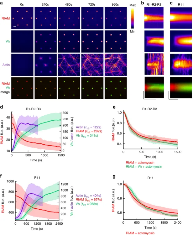
Actomyosin-induced exchange of RIAM for vinculin on talin.
To determine the relationship between the mechanosensitive
talin–RIAM and talin–vinculin interactions observed in Fig.
2
and Fig.
3, we compared the kinetics of RIAM, Vh, and actin in
disks coated with talin R1–R2–R3 and talin R11 in our assay. The
time lapses and the kymographs revealed that actomyosin
accumulation is associated with the concomitant RIAM
dis-sociation and Vh asdis-sociation in disks coated with talin R1–R2–R3
(Fig.
4a, b, d, Supplementary Movie 7) and talin R11 (Fig.
4c, f,
Supplementary Movie 8). The kinetics halftimes indicate a clear
sequence in which actomyosin accumulation is followed by RIAM
dissociation and Vh association (Fig.
4d, f). Our observations are
a
150s 600s 1050s 1500s 1950sb
d
Min Max Time (s)g
Vh fluo. (a.u.)e
Vh fluo. (a.u.)f
c
R1-R2-R3 R11 R7-R8 Vh Vh Actin Vh Actin– Actomyosin + Actomyosin – Actomyosin + Actomyosin – Actomyosin + Actomyosin
R1-R2-R3 R11 R7-R8 – Actomyosin + Actomyosin – Actomyosin + Actomyosin 3000 2000 1000 0 2000 1000 0 3000 2000 1000 0 Time (s) 2000 1000 0 Time (s) 2000 1000 0 Vh fluo. (a.u.) 3000 2000 1000 0 – Actomyosin + Actomyosin – Actomyosin + Actomyosin
Fig. 2 Talin domains bind to vinculin differently in response to the actomyosin force. a Time lapses showing the recruitment of Vh to talin R1 –R2–R3-coated disks in the absence (top) or presence of actomyosin (Vh is shown on the middle and actin on the bottom panel). This experiment was repeated five times independently with the same results. b–d Kymographs of EGFP-Vh (top) and actin (bottom) along a cross-section of a disk coated with talin R1–R2–R3 (b) or R11 (c) or R7–R8 (d) in the absence (left) or presence (right) of actomyosin. Conditions: 100 nM EGFP-Vh, 2.4 µM actin (2% Alexa594-labeled), 50 nM myosin, and 1µM talin during the coating step. The images are color coded using the fire LUT of ImageJ. Scale bar in time lapses = 10 µm. In kymographs, horizontal bar= 500 s, vertical bar = 5 µm. e–g Kinetics of the mean fluorescence of EGFP-Vh corresponding to the conditions described in (b–d). Data are mean ± SD. e n = 80 (−actomyosin), n = 60 disks (+actomyosin). f n = 60 (−actomyosin), n = 75 disks (+actomyosin). g n = 80 (−actomyosin), n = 76 disks (+actomyosin). Source data are provided as a Source data file. See Supplementary Movie 1, Supplementary Movie 2, and Supplementary Movie 3.
consistent with a mechanism in which talin switches from a
RIAM-specific conformation to a vinculin-specific one.
Alter-natively, actomyosin could trigger a competition between RIAM
and Vh for talin. However, Vh does not affect the dissociation
rate of RIAM from talin R1–R2–R3 nor from talin R11 (Fig.
4e,
g). The fact that Vh binding to talin follows RIAM dissociation,
without influencing it, rules out a direct competition mechanism
and demonstrates that talin behaves as a force-dependent
con-formational switch.
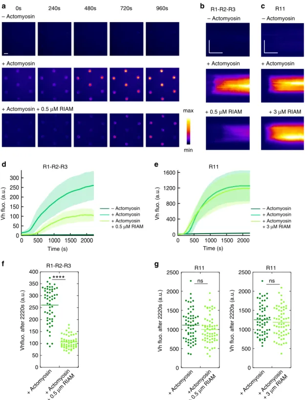
Effect of RIAM on the force-induced vinculin
–talin complex.
Although Vh does not affect RIAM dissociation (Fig.
4e, g),
RIAM could affect talin stretching and its subsequent binding to
a
Time (s)
RIAM fluo. (a.u.)
b d
c
RIAM
Actin
– Actomyosin + Actomyosin – Actomyosin + Actomyosin – Actomyosin + Actomyosin
R1-R2-R3 R11 R7-R8 Min Max RIAM RIAM Actin – Actomyosin + Actomyosin
g
e f
R1-R2-R3 R11 R7-R8 – Actomyosin + Actomyosin 0s 240s 480s 720s 960s 1.0 0.8 0.6 0.4 0.2 0.0 1200 900 600 300 0 Time (s) 1200 900 600 300 0 Time (s) 1200 900 600 300 0RIAM fluo. (a.u.)
1.0 0.8 0.6 0.4 0.2 0.0
RIAM fluo. (a.u.)
1.0 0.8 0.6 0.4 0.2 0.0 – Actomyosin + Actomyosin – Actomyosin + Actomyosin
Fig. 3 The actomyosin force provokes RIAM dissociation from several talin domains. a Time lapses showing the binding of RIAM 1-306 to talin R1–R2–R3-coated disks in the absence (top) or presence of actomyosin (RIAM is shown on the middle and actin on the bottom panel). This experiment was repeated 11 times independently with the same results.b–d Kymographs of mCherry-RIAM 1-306 (top) and actin (bottom) along a cross-section of a disk coated with talin R1–R2–R3 (b) R11 (c) or R7–R8 (d) in the absence (left) or presence (right) of actomyosin. For better comparison, the fluorescence of mCherry-RIAM 1-306 in kymographs was normalized as the maximalfluorescence in the R11- and R7–R8-coated disks. Conditions: 100 nM mCherry-RIAM 1-306, 2.4µM actin (1% Alexa647-labeled for R1–R2–R3, 2% Alexa488 for R11 and R7–R8), 50 nM myosin, 1 µM talin during the coating step. The images are color coded using thefire LUT of ImageJ. Scale bar in time lapses = 10 µm. In kymographs, horizontal bar = 500 s, vertical bar = 5 µm. e–g Kinetics of the meanfluorescence of mCherry-RIAM 1-306 corresponding to the conditions described in (b–d). Data are mean ± SD. e n = 63 (−actomyosin), n = 62 disks (+actomyosin). f n = 50 (−actomyosin), n = 60 disks (+actomyosin). g n = 49 (−actomyosin), n = 60 disks (+actomyosin). Data were first normalized to 1 as the maximal mCherry-RIAM 1-306fluorescence and synchronized using this maximal value as t0before being averaged. Source data are
a b
d
c
RIAM + actomyosin RIAM + Vh + actomyosin RIAM fluo.(a.u.)e
g
f
Actin (t1/2 = 404s) RIAM (t1/2 = 657s) Vh (t1/2 = 958s) Vh / actin fluo. (a.u.) RIAM fluo. (a.u.) Time (s) RIAM + actomyosin RIAM + Vh + actomyosin Time (s) RIAM fluo.(a.u.) Actin (t1/2 = 122s) RIAM (t1/2 = 202s) Vh (t1/2 = 341s) RIAM fluo. (a.u.) Vh / actin fluo. (a.u.) Time (s) R1-R2-R3 R11 R1-R2-R3 R11 R1-R2-R3 Vh RIAM Actin RIAM Vh merge Min Max 0s 240s 480s 720s 960s R11 40 30 20 10 1500 1000 500 0 300 250 200 150 100 50 0 1000 800 600 400 2400 1800 1200 600 0 1200 1000 800 600 400 200 0 1.0 0.8 0.6 0.4 1.0 0.8 0.6 2400 1800 1200 600 0 Time (s) 1500 1000 500 0Fig. 4 The actomyosin force provokes the sequential exchange of RIAM for vinculin on talin. a Time lapses showing the concomitant dissociation of RIAM 1-306, association of Vh, accumulation of actomyosin, and a Vh/RIAM merge in the same disks coated with talin R1–R2–R3. This experiment was repeated 4 times independently with the same results.b, c From top to bottom: kymographs of RIAM, Vh, actin, and Vh/RIAM merge along the cross-section of a disk coated with talin R1–R2–R3 (b) or R11 (c). Conditions: 100 nM mCherry-RIAM, 100 nM EGFP-Vh, 2.4 µM actin (1% Alexa647-labeled), 50 nM myosin, and 1µM talin during the coating step. The images are color coded using the fire LUT of ImageJ. Scale bar in time lapses = 10 µm. In kymographs, horizontal bar= 1000 s, vertical bar = 5 µm. d, f Kinetics of the mean fluorescence of mCherry-RIAM 1-306, EGFP-Vh and Alexa647-labeled actin in disks coated with talin R1–R2–R3 (d) and R11 (f) corresponding to the conditions described in (b) and (c). Actin fluorescence is multiplied by 3. Data are mean ± SD.n = 63 (d), n = 59 disks (f). e Kinetics of mCherry-RIAM 1-306 dissociation from disks coated with talin R1–R2–R3 in the presence of actomyosin with or without 100 nM EGFP-Vh as described in Fig.3b andb respectively.n = 62 (RIAM + actomyosin), n = 63 disks (RIAM + Vh + actomyosin).g Kinetics of mCherry-RIAM 1-306 dissociation from disks coated with talin R11 in the presence of actomyosin with or without 100 nM EGFP-Vh as described in Fig.3c andc respectively.n = 59 disks. e, g Data are mean ± SD. Data were first normalized to 1 as the maximal mCherry-RIAM 1-306 fluorescence and synchronized using this maximal value as t0before being averaged. Source data are provided as a Source datafile. See Supplementary
Vh. We therefore measured the effect of a high concentration of
RIAM on the actomyosin-dependent binding of Vh to talin. A
weak interaction between Vh and RIAM has been reported (K
d=
5 µM) and could affect our interpretations
33. However, this weak
interaction cannot compete with the high affinity binding of Vh
for a construct of talin corresponding to R1–R2–R3 in which one
VBS (helix 12) is exposed (K
d= 140 nM)
36. Similarly, this weak
interaction between Vh and RIAM is unlikely to affect the high
affinity binding of RIAM 1-306 to our talin R1–R2–R3 and talin
R11 (K
d= 36 nM and 134 nM respectively, Fig.
1c). In addition,
we showed that Vh does not affect the constitutive binding of
RIAM to talin R1–R2–R3 in the absence of actomyosin
(Sup-plementary Fig. 4), demonstrating that RIAM and Vh do not
sequester each other through direct binding in our experimental
conditions. After this clarification, we showed that the
actomyosin-dependent binding of Vh to talin R1–R2–R3 is
severely impaired by a high concentration of RIAM, compared
with the control without RIAM (Fig.
5a, b, Supplementary
Movie 9). Kinetic analysis and steady-state measurement
con-firmed that the final level of Vh bound to stretched talin is lower
in the presence of RIAM (Fig.
5d, f), suggesting that the
talin–RIAM complex requires a higher force for unfolding than
talin alone. Our observations demonstrate that RIAM protects
talin R2–R3 bundles from mechanical unfolding, which implies
that the force-dependent dissociation of RIAM is a prerequisite
for the exposure of VBSs in R2–R3. Surprisingly, RIAM does not
affect the actomyosin-dependent binding of Vh to talin R11, at
the concentration tested for talin R1–R2–R3 (0.5 µM) and at a
higher concentration (3 µM) (Fig.
5c, e, g, Supplementary
Movie 10). Therefore, unlike talin R1–R2–R3, the dissociation of
RIAM from talin R11 is not a prerequisite for the
force-dependent binding of vinculin.
Discussion
Our in vitro reconstitution demonstrates that talin dissociates
from RIAM and associates to vinculin sequentially in response to
the actomyosin force. The force-dependent dissociation of the
talin–RIAM interaction is one of the rare elementary
mechan-osensitive reactions of adhesion complexes which has been
dis-covered. This process could control the mechanosensitive
maturation of FAs (Fig.
6a).
We found that RIAM protects R2–R3 against stretching and its
dissociation is a prerequisite for vinculin binding. Such a bistable
mechanism is the desirable behavior for a mechanosensitive
switch involved in a cellular decision-making process. This
behavior likely results from the overlap between the
RIAM-binding sites and VBSs in R2 and R3
33. R1–R2–R3 is a key
mechanosensitive part of talin, since a construct, almost identical
to our talin R1–R2–R3, can rescue spreading, polarization and
migration of talin null cells
37. In this region of talin, R3 has a
critical role, since mutations that destabilize this bundle affect
ECM sensing
38. The stretching of single molecules clearly
revealed that R3 is the weakest bundle of the R1–R2–R3 region
and should unfold
first in response to force
19,20. The inhibition of
the mechanical exposure of VBSs in talin R1–R2–R3 by RIAM
could therefore result from the stabilization of R3.
Our study reveals that talin R11 binds to vinculin in response
to force. In contrast with talin R1–R2–R3, the mechanosensitive
binding of R11 to vinculin is not affected by RIAM. Although the
structure of the R11–RIAM complex is not known, our data
suggest that the binding sites for RIAM and vinculin do not
overlap in R11. Talin R11 can therefore dissociate from RIAM
and associate to vinculin independently. Interestingly, simulations
predicted that the
first and last helices of the
mechanically-stretched R11 should unfold
first, leaving a 3-helix intermediate
bundle containing a cryptic VBS
39. Whether RIAM dissociates
after the detachment of these
first and last helices of R11 remains
to be determined.
The different effects of RIAM on talin R1–R2–R3 and R11
imply that RIAM increases the threshold force for the exposure of
the VBSs located in R2–R3 but does not affect the exposure of the
single VBS of R11. However, the sequential exchange of RIAM for
vinculin on talin R11 implies that the force required to dissociate
RIAM is lower than the one required to expose the single VBS of
R11 (Fig.
6b).
R7–R8 is difficult to stretch in conditions that provoke both
RIAM dissociation and vinculin association for R1–R2–R3 and
R11 (Fig.
6b). In response to force, Vh does not bind to the two
VBSs located in R7 and R8 for several possible reasons. The single
VBS of R8 is probably not exposed because R8 is protected from
unfolding by its insertion in R7. The single VBS of R7 could
remain stably attached to R7 in response to force or, if exposed,
its affinity for Vh is low as previously reported
40. The weak
dissociation of RIAM and the total absence of vinculin association
suggest that the RIAM-binding site is disrupted at a level of force
that is not sufficient to expose the single VBS of R8. Altogether,
our observations are in agreement with a previous report showing
that unfolding of isolated R8 occurs at a force of 5 pN, whereas
unfolding of R7–R8 occurs at 15 pN, demonstrating that R8 is
mechanically protected by its insertion in R7
18. Mutations that
prevent the mechanical stretching of R7 favor signaling cascades
downstream of R8, revealing the importance of this
mechan-osensitive reaction controlled by a high threshold force
41.
The mechanosensitivity of the domains of talin is influenced by
their position relative to the ABDs in the full-length protein. By
placing FRET sensors at different positions along talin, Ringer
et al. revealed an intramolecular tension gradient characterized by
high forces between the head and ABD2 and lower forces between
ABD2 and ABD3
42. However, we showed that a construct
com-posed of R1–R2–R3 fused to ABD2 is not stretched compared
with the same construct where ABD2 is replaced by ABD3
(Supplementary Fig. 5). This result can be easily explained by the
fact that ABD2 is masked by R3 in talin
23. The traction force
applied to ABD3 would unfold R3, leading to the exposure of
ABD2. The fact that R8 is the C-terminal part of ABD2 allows
alternative interpretations of the weak mechanosensitivity of
R7–R8
23. Indeed, we cannot exclude that the binding of actin
filaments stabilizes R8 and prevents RIAM dissociation and
vinculin association. Alternatively, if actomyosin generates a
pulling force on R13 (ABD3) and R7–R8 (ABD2) simultaneously,
the apparent tension between R13 and R7–R8 could be reduced,
as suggested by FRET measurement in cells
42, leading to weak
dissociation of RIAM and association of vinculin.
The structure of talin reveals auto-inhibitory contacts between
F3 and R9, and also F2 and R12
43,44. RIAM binding to the talin
rod does not require the disruption of the F3–R9
autoinhibi-tion
35. In contrast, the release of the F3-R9 intramolecular contact
appears critical to initiate talin–vinculin interaction
indepen-dently of force application in nascent adhesion
45. However, this
talin conformation can be further stretched to recruit more
vin-culin in FAs. Whether the disruption of the talin auto-inhibitory
contacts facilitates the stretching of individual bundles to
dis-sociate RIAM and bind vinculin remains to be determined. It
would also be interesting to determine whether the recently
reported autoinhibition of RIAM controls its interaction with
talin
46.
Vinculin autoinhibition influences the mechanosensitivity of
the talin–vinculin complex. Several biochemical, structural, and
cellular studies compared the recruitment of the constitutively
active Vh and the autoinhibited full-length vinculin (VFL) in FAs,
leading to apparent discrepancies. In cells, Vh remains associated
– Actomyosin
b
a
0s 240s 480s 720s 960sc
+ Actomyosin + Actomyosin + 0.5 µM RIAM min maxg
Vh fluo. (a.u.) Time (s) R11 Vh fluo. (a.u.) Time (s) R1-R2-R3 – Actomyosin – Actomyosin + Actomyosin + 0.5 µM RIAM + 3 µM RIAMf
e
+ Actomyosin R11 R1-R2-R3 R11Vhfluo. after 2220s (a.u.) Vh fluo. after 2220s (a.u.) Vh fluo. after 2220s (a.u.)
ns ns R11 R1-R2-R3 – Actomyosin + Actomyosin + Actomyosin + 0.5 µM RIAM
d
0 500 1000 1500 2000 2500 0 500 1000 1500 2000 2500 0 + Actomyosin+ Actomyosin + 0.5 µm RIAM 50 100 150 200 250 300 350 400 300 250 200 150 100 50 0 2000 1500 1000 500 0 1600 1200 800 400 0 2000 1500 1000 500 0 + Actomyosin+Actomyosin + 0.5 µm RIAM + Actomyosin+ Actomyosin + 3 µm RIAM – Actomyosin + Actomyosin + Actomyosin + 3 µM RIAMFig. 5 RIAM inhibits the actomyosin-dependent binding of vinculin to talin R1–R2–R3 but not to R11. a Time lapse showing the recruitment of Vh in disks coated with talin R1–R2–R3 in the absence of actomyosin (top), presence of actomyosin (middle), and presence of actomyosin and RIAM (bottom). This experiment was repeated twice independently with the same results.b, c Kymographs of EGFP-Vh along a cross-section of a disk coated with talin R1–R2–R3 (b) and R11 (c). Conditions: 100 nM EGFP-Vh, 2.4 µM actin, 50 nM myosin, 500 nM (b) or 3 µM (c) mCherry-RIAM, 1 µM talin during the coating step. The images are color coded using thefire LUT of ImageJ. Scale bar in time lapses = 10 µm. In kymographs, horizontal bar = 1000 s, vertical bar= 5 µm. d, e Kinetics of the mean fluorescence of EGFP-Vh corresponding to the conditions described in (b, c). Data are mean ± SD. d n = 54 (−actomyosin) and (+actomyosin), n = 51 disks (+actomyosin + 0.5 µM RIAM). e n = 59 disks. f, g Steady-state binding of Vh (2220 s after sealing the chamber) in disks coated with talin R1–R2–R3 (f) or R11 (g) in the absence and presence of RIAM. f, g Same conditions as in (b, c). Each data point represents the meanfluorescence of Vh in one disk. The bar shows the mean. f n = 54 (+actomyosin), n = 51 disks (+actomyosin + 0.5 µM RIAM). A significant difference was found using a two-tailed t test (P = 3.95 × 10⁻²²).g Left panel:n = 60. No significant difference was found using a two-tailed t test (P = 0.1126). Right panel: n = 59 disks. No significant difference was found using a two-tailed t test (P = 0.3575). ****P < 0.0001 using a two-tailed t test; ns nonsignificant. Source data are provided as a Source data file. See Supplementary Movie 9 and Supplementary Movie 10.
to talin in FAs after myosin inhibition by blebbistatin, whereas
VFL dissociates
24,47,48. The recruitment of VFL to FAs is restored
by cell stretching, demonstrating the force-dependence of the
talin–vinculin interaction, whereas Vh binding is not increased
24.
The slow dissociation of Vh from talin after force release,
observed in vitro
20,21, could explain the slow dissociation of Vh in
cells after blebbistatin treatment, whereas the fast dissociation of
VFL could result from the reassociation of the tail to the head of
vinculin in the absence of actomyosin. Indeed, actomyosin force
acts on vinculin to maintain the active open conformation of
vinculin
48,49. The saturation of talin by Vh would explain why
talin does not rectruit more Vh after cell stretching. Thus,
in vitro, and probably in cells, the mechanosensitivity of the
talin–Vh interaction depends on Vh concentration. Because Vh is
not autoinhibited like VFL, it binds to partially exposed VBSs in
the least stable helical bundles of non-stretched talin, provided
that Vh concentration is high enough
21. At the low concentration
of Vh used in our past and present in vitro studies (22–100 nM),
Vh mimics VFL by displaying a very weak constitutive binding,
which is greatly enhanced by the application of actomyosin force
to talin (Fig.
2e).
Talin bundles have several binding partners other than RIAM
and vinculin. Interestingly, talin R7 interacts with KANK and R8
interacts with both DLC and paxillin
33,41,50,51. The catch-to-slip
bond switching behavior of the R7–KANK complex is thought to
control KANK recruitment at FAs
52. The mechanical unfolding
of R8 should provoke the dissociation of DLC from talin, leading
to upregulation of actomyosin contractility, and acceleration of
cell migration
41. Interestingly, R11 interacts with the
β-subunit of
integrins. However, it is not known whether R11, like the
F3 subdomain of the head, promotes the inside–out activation of
integrins and whether this activity is influenced by the
force-dependent dissociation of RIAM and association of vinculin. This
high number of combinations of talin partners, associated with
specific mechanically-stretched talin conformations, provides the
cell with a precise means of informing itself about variations in
intracellular and extracellular forces. Conversely, the binding of
talin partners, as exemplified by the present study on RIAM, can
modify the mechanosentivity of talin bundles differently, leading
to a change in the hierarchy of their response to force.
Methods
cDNA constructs. All talin constructs are derived from a cDNA encoding for human talin-1 containing a C-terminal His6tag. Talin R1–R2–R3, corresponding
to F2–F3–R1–R2–R3–R13, was cloned into a pETM plasmid with an N-terminal StrepTagII and a C-terminal His6tag. This construct was made in three steps. First
the R2–R3 fragment was PCR amplified using primers 1 and 2 and cloned into the KpnI/BamHI sites of pETM, leading to the intermediate plasmid pETM–R2–R3. R13 was PCR amplified using primers 3 and 4 and cloned into the BamHI/EcoRI sites of pETM–R2–R3, leading to the intermediate plasmid pETM–R2–R3–R13. Finally, F2–F3–R1–R2–R3 was PCR amplified using primers 5 and 6 and cloned into the KpnI/NcoI sites of the pETM–R2–R3–R13, leading to pETM–F2–F3– R1–R2–R3–R13. Talin R11, corresponding to F2–F3–R11–R13 and talin R7–R8, corresponding to talin F2–F3–R7–R8–R13 were cloned into a pET-29a(+) plasmid with an N-terminal StrepTagII and a C-terminal His6tag. Talin R7–R8 and R11
have been synthesized by Genscript. The cDNAs encoding for talin F2–F3 (talin 196–405), talin F2–F3–R1–R2–R3 (talin 196–911), and talin R1–R8 (talin 196–1659) were PCR amplified using the primer pairs 7–8, 7–9, 7–10 and cloned into the BamH/XhoI, BamHI/EcoRI, and BamHI/EcoRI sites, respectively, of a
a
Actomyosin force Talin / vinculin in focal adhesions Leading edge Talin / RIAM in nascent adhesionsb
TBS1 TBS2 Vh RIAM Vinculin Force 1 2 3 7 8 11 1 2 3 7 8 11 1 7 8 3 2 11 1 7-8 3 2 11 TalinFig. 6 Model for the actomyosin-dependent binding of RIAM and vinculin to talin. a Scheme illustrating that RIAM, which is initially enriched in nascent adhesions, is replaced by vinculin in mature FAs in response to the force exerted by the actomyosin stressfibers. b Model describing how talin dissociates from RIAM and associates to vinculin sequentially in response to the actomyosin force.
pGEX6P1 plasmid (GE Healthcare) with an N-terminal GST tag and a C-terminal His6tag. Our constructs do not include the F0 and F1 subdomains of the head
(talin 1–195) because this part of talin reduces the expression quality in our hands and is not involved in the binding of RIAM and vinculin. A preexisting cDNA encoding for human vinculin 1–851, corresponding to Vh, was cloned into the SalI/ NotI sites of a homemade pGEX-6P1-EGFP plasmid. The cDNA encoding for mouse RIAM 1-306 was PCR amplified using primers 11 and 12, and cloned into the BamHI/XhoI sites of a homemade pGEX-6P2-mCherry plasmid with a C-terminal His6tag. Primers used for cloning the DNA constructs in this study are
listed in Supplementary Table 1.
Protein purification. All the recombinant proteins were expressed in Escherichia coli (BL21 DE3, Invitrogen). After transformation, bacteria were grown in 4–12 l of LB medium containing 0.1 mg ml−1of ampicillin or kanamycin at 37 °C until absorbance reached 0.8 at 600 nm. The recombinant proteins were expressed upon addition of 1 mM isopropyl-β-D-thiogalactoside for 16 h at 16 °C. After cen-trifugation, the bacterial pellet was submitted to specific purification steps22.
Talin R1–R2–R3, R11, and R7–R8 were bound to Ni-NTA (Ni2+-nitrilotriacetic
acid)-Agarose (Macherey-Nalgene), washed with 50 mM Tris pH 7.8, 500 mM NaCl, 20 mM imidazole, 1 mMβ-mercaptoethanol (BME), eluted with 50 mM Tris pH 7.8, 500 mM NaCl, 250 mM imidazole, 1 mM BME, dialyzed in 20 mM Tris pH 7.8, 100 mM KCl, 1 mM DTT, frozen in liquid nitrogen, and stored at−80 °C.
mCherry-RIAM 1-306, talin F2–F3, F2–F3–R1–R2–R3, R1–R8, and EGFP-Vh containing a N-terminal GST (Glutathione-S-transferase) tag, were bound to glutathione-Sepharose (GE Healthcare), washed with 50 mM Tris pH 7.8, 500 mM NaCl, and eluted with 50 mM Tris pH 7.8, 500 mM NaCl and 50 mM reduced L-Glutathione (Sigma-Aldrich). For mCherry-RIAM 1-306 and EGFP-Vh, GST was cleaved by PreScission protease (GE Healthcare) in 50 mM Tris pH 7.8 and 500 mM NaCl and GST was eliminated by Glutathione-Sepharose
chromatography. mCherry-RIAM 1-306 and talin F2–F3, F2–F3–R1–R2–R3, and R1–R8 were then bound to Ni-NTA-Agarose, washed with 50 mM Tris pH 7.8, 500 mM NaCl, 20 mM imidazole, eluted with 50 mM Tris pH 7.8, 500 mM NaCl, 250 mM imidazole, dialyzed in 20 mM Tris pH 7.8, 100 mM KCl, 1 mM DTT, frozen in liquid nitrogen and stored at−80 °C. EGFP-Vh was further centrifuged at 300,000 × g for 30 min, purified by gel filtration (Superdex 200, 16/60, GE Healthcare) in 20 mM Tris pH 7.5, dialyzed in 20 mM Tris pH 7.8, 100 mM KCl, frozen in liquid nitrogen, and stored at−80 °C.
Actin was purified from rabbit skeletal muscle acetone powder. After cycles of polymerization and depolymerization, actin was gelfiltered on a Superdex G-200 column (GE Healthcare) in 5 mM Tris pH 7.8, 0.2 mM ATP, 0.1 mM CaCl2, 1 mM
DTT. Actin was labelled with Alexa Fluor 488, 594, and 647 Succinimidyl Ester (Invitrogen)22. Myosin II was extracted from rabbit skeletal muscles in a buffer
containing 500 mM KCl, 100 mM K2HPO4. After grinding and centrifugation, the
actin-containing pellet is discarded. The supernatant is submitted to cycles of precipitation in low-salt buffer, centrifugation, and resuspension in high-salt buffer. Finally, the protein was dialyzed in 20 mM KH2PO4/K2HPO4pH 7.5, 500 mM KCl,
1 mM EDTA, and stored at−20 °C after addition of 50% glycerol.
Sample preparation for the in vitro assay. Micropatterning was performed by modifying an existing method as follows53,54. Glass coverslips (22 mm × 32 mm,
Thermo Scientific/Menzel-Glaser) were first washed with milliQ water and ethanol, sonicated and irradiated for 1 min under a deep UV lamp (Ossila). The coverslips were incubated for 2 h in 0.1 mg ml−1PLL-g-PEG (SuSoS) dissolved in 10 mM HEPES pH 7.8 and washed with milliQ water. The chrome–quartz photomask (Toppan, France), designed with disks of 5 µm in diameter, regularly spaced by 30 µm (Fig.1a), was cleaned by deep UV irradiation for 1 min, placed on the PLL-g-PEG-coated coverslip, and exposed to deep UV for 3 min. The chamber was made of a micropatterned coverslip attached to a glass slide (Super Frost, Thermo Scientific) with double-sided adhesive tape. The volume of a typical chamber was 50 µl. The chamber wasfirst incubated with talin (1 µM) for 5 min at room tem-perature. Unbound talin was washed out with 200 µl of F-buffer (10 mM Tris pH 7.8, 25 mM KCl, 1 mM MgCl2, 0.2 mM CaCl2, 1 mM DTT). The surface of the
disks was passivated with 100 µl of F-buffer containing 10% BSA for 5 min at room temperature and washed with 200 µl F-buffer. Finally, 100 µl of the reaction was added and the chamber was sealed with VALAP (1:1:1 mixture of vaseline, lanolin, and paraffin). A typical reaction contained: 2.4 µM actin (containing 1% Alexa647-labeled or 2% Alexa488-Alexa647-labeled or 2% Alexa561-Alexa647-labeled actin), 50 nM myosin II, 1% BSA, a salt mix (2 mM MgCl2, 0.2 mM EGTA, and 25 mM KCl), and an ATP
regenerating mix (2 mM ATP, 2 mM MgCl2, 10 mM creatine phosphate, 3.5 U/ml
creatine kinase) in G-fluo buffer (10 mM Tris pH 7.8, 0.2 mM CaCl2, 0.4%
methylcellulose, 5 mM DABCO and 20 mM DTT). Additional proteins such as EGFP-Vh and mCherry-RIAM 1-306 were also added. The gelsolin-capped actin filaments used in Supplementary Fig. 3 have been prepared by mixing the barbed-end capping protein gelsolin with actinfilaments at a 1:600 gelsolin/actin molar ratio22.
Microscopy observations. Images were acquired with a Nikon Ti Eclipse E microscope equipped with a 60X oil immersion objective (Apochromat, 1.49 NA) and coupled to a sCMOS camera (Photometrics, Prime 95B or Hamamatsu, Orca
Flash04), using the spinning disk mode (Yokogawa CSU-X1-A1) or the TIRF mode. EGFP-Vh (or Alexa488-Actin), mCherry-RIAM 1-306 (or Alexa594-actin) and Alexa647-actin were excited with 488, 561, and 642 nm lasers, respectively. Data analysis. Images were acquired with MetaMorph and analyzed with ImageJ. For steady-state data, each point of the dot plots represents the meanfluorescence of a single disk (background subtracted). The bar indicates the mean. Kinetics were obtained by averaging kinetics in a large number of single disks, after background subtraction, normalization, and synchronization on the maximal value of mCherry-RIAM for each disk (Figs.3e–g, 4e, g), only synchronization (Fig.4d, f), or only background subtraction (Figs.2e–g, 5d, e, and Supplementary Fig. 5b, c). The affinity of RIAM for talin constructs was obtained by plotting the average fluorescence of RIAM in a high number of talin-coated disks as a function of the total concentration of RIAM (Fig.1c). Since we assumed that the amount of talin in the disks is negligible compared to the total concentration of RIAM in solution, we estimated the value of the Kdas the concentration of RIAM at half saturation. Note
that this assumption can only underestimate the affinity.
The graphs were assembled using Igor Pro or Kaleidagraph. Statistical analysis was performed using Student t test in Microsoft Excel. Experiments were reproduced 2–11 times with the same conclusions.
Reporting summary. Further information on research design is available in the Nature Research Reporting Summary linked to this article.
Data availability
Data supporting this paper are available from the corresponding author upon reasonable request. A reporting summary for this article is available as a Supplementary Information file. The source data underlying Figs.1c,2e–g,3e–g,4d–g,5d–g, and Supplementary
Figs. 1b, 2, 3b, d, 4, 5b, c are provided as a Source datafile. Source data are provided with this paper.
Received: 26 February 2020; Accepted: 26 May 2020;
References
1. Le Clainche, C. & Carlier, M.-F. Regulation of actin assembly associated with protrusion and adhesion in cell migration. Physiol. Rev. 88, 489–513 (2008).
2. Wehrle-Haller, B. Structure and function of focal adhesions. Curr. Opin. Cell Biol. 24, 116–124 (2012).
3. Ciobanasu, C., Faivre, B. & Le Clainche, C. Integrating actin dynamics, mechanotransduction and integrin activation: the multiple functions of actin binding proteins in focal adhesions. Eur. J. Cell Biol. 92, 339–348 (2013). 4. Schwarz, U. S. & Gardel, M. L. United we stand– integrating the actin
cytoskeleton and cell–matrix adhesions in cellular mechanotransduction. J. Cell Sci. 125, 3051–3060 (2012).
5. Bachmann, M., Kukkurainen, S., Hytönen, V. P. & Wehrle-Haller, B. Cell adhesion by integrins. Physiol. Rev. 99, 1655–1699 (2019).
6. Romero, S., Le Clainche, C. & Gautreau, A. M. Actin polymerization downstream of integrins: signaling pathways and mechanotransduction. Biochem. J. 477, 1–21 (2020).
7. Gardel, M. L., Schneider, I. C., Aratyn-Schaus, Y. & Waterman, C. M. Mechanical integration of actin and adhesion dynamics in cell migration. Annu. Rev. Cell Dev. Biol. 26, 315–333 (2010).
8. Wolfenson, H., Yang, B. & Sheetz, M. P. Steps in mechanotransduction pathways that control cell morphology. Annu. Rev. Physiol. 81, 585–605 (2019).
9. Humphrey, J. D., Dufresne, E. R. & Schwartz, M. A. Mechanotransduction and extracellular matrix homeostasis. Nat. Rev. Mol. Cell Biol. 15, 802–812 (2014). 10. Wolfenson, H., Lavelin, I. & Geiger, B. Dynamic regulation of the structure
and functions of integrin adhesions. Dev. Cell 24, 447–458 (2013). 11. Sun, Z., Guo, S. S. & Fässler, R. Integrin-mediated mechanotransduction. J.
Cell Biol. 215, 445–456 (2016).
12. Geiger, B., Spatz, J. P. & Bershadsky, A. D. Environmental sensing through focal adhesions. Nat. Rev. Mol. Cell Biol. 10, 21–33 (2009).
13. Jansen, K. A., Atherton, P. & Ballestrem, C. Mechanotransduction at the cell-matrix interface. Semin. Cell Dev. Biol. 71, 75–83 (2017).
14. Haining, A. W. M., Lieberthal, T. J., Hernández, A. & del, R. Talin: a mechanosensitive molecule in health and disease. FASEB J. 30, 2073–2085 (2016).
15. Gough, R. E. & Goult, B. T. The tale of two talins—two isoforms to fine-tune integrin signalling. FEBS Lett. 592, 2108–2125 (2018).
16. Goult, B. T., Yan, J. & Schwartz, M. A. Talin as a mechanosensitive signaling hub. J. Cell Biol. 217, 3776–3784 (2018).
17. del Rio, A. et al. Stretching single talin rod molecules activates vinculin binding. Science 323, 638–641 (2009).
18. Yao, M. et al. The mechanical response of talin. Nat. Commun. 7, 11966 (2016).
19. Haining, A. W. M., von Essen, M., Attwood, S. J., Hytönen, V. P. & del Río Hernández, A. All subdomains of the talin rod are mechanically vulnerable and may contribute to cellular mechanosensing. ACS Nano 10, 6648–6658 (2016).
20. Yao, M. et al. Mechanical activation of vinculin binding to talin locks talin in an unfolded conformation. Sci. Rep. 4, 4610 (2015).
21. Ciobanasu, C., Faivre, B. & Le Clainche, C. Actomyosin-dependent formation of the mechanosensitive talin–vinculin complex reinforces actin anchoring. Nat. Commun. 5, 3095 (2014).
22. Ciobanasu, C., Faivre, B. & Le Clainche, C. Reconstituting actomyosin-dependent mechanosensitive protein complexes in vitro. Nat. Protoc. 10, 75–89 (2015).
23. Atherton, P. et al. Vinculin controls talin engagement with the actomyosin machinery. Nat. Commun. 6, 10038 (2015).
24. Hirata, H., Tatsumi, H., Lim, C. T. & Sokabe, M. Force-dependent vinculin binding to talin in live cells: a crucial step in anchoring the actin cytoskeleton to focal adhesions. Am. J. Physiol. Cell Physiol. 306, C607–C620 (2014). 25. Austen, K. et al. Extracellular rigidity sensing by talin isoform-specific
mechanical linkages. Nat. Cell Biol. 17, 1597–1606 (2015).
26. Thievessen, I. et al. Vinculin–actin interaction couples actin retrograde flow to focal adhesions, but is dispensable for focal adhesion growth. J. Cell Biol. 202, 163–177 (2013).
27. Lafuente, E. M. et al. RIAM, an Ena/VASP and profilin ligand, interacts with Rap1-GTP and mediates Rap1-induced adhesion. Dev. Cell 7, 585–595 (2004).
28. Lagarrigue, F. et al. A RIAM/lamellipodin–talin–integrin complex forms the tip of stickyfingers that guide cell migration. Nat. Commun. 6, 8492 (2015).
29. Han, J. et al. Reconstructing and deconstructing agonist-induced activation of integrinαIIbβ3. Curr. Biol. 16, 1796–1806 (2006).
30. Yang, J. et al. Conformational activation of talin by RIAM triggers integrin-mediated cell adhesion. Nat. Commun. 5, 5880 (2014).
31. Lagarrigue, F., Kim, C. & Ginsberg, M. H. The Rap1-RIAM-talin axis of integrin activation and blood cell function. Blood 128, 479–487 (2016). 32. Lee, H.-S., Anekal, P., Lim, C. J., Liu, C.-C. & Ginsberg, M. H. Two modes of
integrin activation form a binary molecular switch in adhesion maturation. Mol. Biol. Cell 24, 1354–1362 (2013).
33. Goult, B. T. et al. RIAM and vinculin binding to talin are mutually exclusive and regulate adhesion assembly and turnover. J. Biol. Chem. 288, 8238–8249 (2013).
34. Baxter, N. J., Zacharchenko, T., Barsukov, I. L. & Williamson, M. P. Pressure-dependent chemical shifts in the r3 domain of talin show that it is thermodynamically poised for binding to either vinculin or RIAM. Structure 25, 1856–1866.e2 (2017).
35. Chang, Y.-C. et al. Structural and mechanistic insights into the recruitment of talin by RIAM in integrin signaling. Structure 22, 1810–1820 (2014). 36. Patel, B. et al. The activity of the vinculin binding sites in talin is influenced by
the stability of the helical bundles that make up the talin rod. J. Biol. Chem. 281, 7458–7467 (2006).
37. Rahikainen, R., Öhman, T., Turkki, P., Varjosalo, M. & Hytönen, V. P. Talin-mediated force transmission and talin rod domain unfolding independently regulate adhesion signaling. J. Cell Sci. 132, jcs226514 (2019).
38. Rahikainen, R. et al. Mechanical stability of talin rod controls cell migration and substrate sensing. Sci. Rep. 7, 3571 (2017).
39. Mykuliak, V. V., Haining, A. W. M., von Essen, M., del Río Hernández, A. & Hytönen, V. P. Mechanical unfolding reveals stable 3-helix intermediates in talin andα-catenin. PLoS Comput. Biol. 14, e1006126 (2018).
40. Gingras, A. R. et al. Mapping and consensus sequence identification for multiple vinculin binding sites within the talin rod. J. Biol. Chem. 280, 37217–37224 (2005).
41. Haining, A. W. M. et al. Mechanotransduction in talin through the interaction of the R8 domain with DLC1. PLoS Biol. 16, e2005599 (2018).
42. Ringer, P. et al. Multiplexing molecular tension sensors reveals piconewton force gradient across talin-1. Nat. Methods 14, 1090–1096 (2017). 43. Dedden, D. et al. The architecture of talin1 reveals an autoinhibition
mechanism. Cell 179, 120–131 (2019). e13.
44. Goult, B. T. et al. The structure of an interdomain complex that regulates talin activity. J. Biol. Chem. 284, 15097–15106 (2009).
45. Atherton, P. et al. Relief of talin autoinhibition triggers a force-independent association with vinculin. J. Cell Biol. 219.https://doi.org/10.1083/
jcb.201903134(2020).
46. Chang, Y.-C. et al. Molecular basis for autoinhibition of RIAM regulated by FAK in integrin activation. Proc. Natl Acad. Sci. 116, 3524–3529 (2019). 47. Humphries, J. D. et al. Vinculin controls focal adhesion formation by direct
interactions with talin and actin. J. Cell Biol. 179, 1043–1057 (2007). 48. Carisey, A. et al. Vinculin regulates the recruitment and release of core focal
adhesion proteins in a force-dependent manner. Curr. Biol. 23, 271–281 (2013).
49. Grashoff, C. et al. Measuring mechanical tension across vinculin reveals regulation of focal adhesion dynamics. Nature 466, 263–266 (2010). 50. Bouchet, B. P. et al. Talin-KANK1 interaction controls the recruitment of
cortical microtubule stabilizing complexes to focal adhesions. Elife 5.https://
doi.org/10.7554/eLife.18124(2016).
51. Zacharchenko, T. et al. LD motif recognition by talin: structure of the talin-DLC1 complex. Structure 24, 1130–1141 (2016).
52. Yu, M. et al. Force-dependent regulation of talin–KANK1 complex at focal adhesions. Nano Lett. 19, 5982–5990 (2019).
53. Reymann, A.-C. et al. Nucleation geometry governs ordered actin networks structures. Nat. Mater. 9, 827–832 (2010).
54. Azioune, A., Storch, M., Bornens, M., Théry, M. & Piel, M. Simple and rapid process for single cell micro-patterning. Lab Chip 9, 1640–1642 (2009).
Acknowledgements
This work was supported by Agence Nationale pour la Recherche Grants ANR-16-CE13-0007-02 PHAGOMECANO and ANR-18-CE13-0026-01 RECAMECA (to C.L.C.). The present work has benefited from the Light Microscopy facility of Imagerie‐Gif, (http:// www.i2bc.paris-saclay.fr), member of IBiSA (http://www.ibisa.net), supported by “France‐BioImaging” (ANR‐10‐INBS‐04‐01), and the Labex “Saclay Plant Sciences” (ANR-10-LABX-0040-SPS). We thank Annabelle Fente for initial observations of mCherry-RIAM binding to talin, Jun Qin (Cleveland Clinic, USA) for the gift of the mouse RIAM cDNA. We thank Florence Niedergang and the members of the “Cytos-keleton Dynamics and Motility” team for helpful discussions.
Author contributions
C.V. performed the microscopy experiments, analyzed the data, and prepared thefigures. V.H. cloned cDNAs. C.V., V.H., and C.L.C. purified and characterized the proteins used in this study. C.L.C. designed the experiments, supervised the project, and wrote the paper.
Competing interests
The authors declare no competing interests.
Additional information
Supplementary informationis available for this paper at https://doi.org/10.1038/s41467-020-16922-1.
Correspondenceand requests for materials should be addressed to C.L.C. Peer review informationNature Communications thanks Masahiro Sokabe and the other, anonymous, reviewer(s) for their contribution to the peer review of this work. Peer reviewer reports are available.
Reprints and permission informationis available athttp://www.nature.com/reprints
Publisher’s note Springer Nature remains neutral with regard to jurisdictional claims in published maps and institutional affiliations.
Open Access This article is licensed under a Creative Commons Attribution 4.0 International License, which permits use, sharing, adaptation, distribution and reproduction in any medium or format, as long as you give appropriate credit to the original author(s) and the source, provide a link to the Creative Commons license, and indicate if changes were made. The images or other third party material in this article are included in the article’s Creative Commons license, unless indicated otherwise in a credit line to the material. If material is not included in the article’s Creative Commons license and your intended use is not permitted by statutory regulation or exceeds the permitted use, you will need to obtain permission directly from the copyright holder. To view a copy of this license, visithttp://creativecommons.org/ licenses/by/4.0/.