http://jfm.sagepub.com/
Journal of Feline Medicine and Surgery
http://jfm.sagepub.com/content/15/7/611
The online version of this article can be found at:
DOI: 10.1177/1098612X13489224
2013 15: 611
Journal of Feline Medicine and Surgery
D Radford, Etienne Thiry, Uwe Truyen and Marian C Horzinek
Herman Egberink, Tadeusz Frymus, Tim Gruffydd-Jones, Margaret J Hosie, Hans Lutz, Fulvio Marsilio, Karin Möstl, Alan
Maria Grazia Pennisi, Katrin Hartmann, Albert Lloret, Lluis Ferrer, Diane Addie, Sándor Belák, Corine Boucraut-Baralon,
Cryptococcosis in cats: ABCD guidelines on prevention and management
technique does not amount to an endorsement of its value or quality, or the claims made by its manufacturer. those of the authors and the inclusion in this publication of material relating to a particular product, method or of animals and interpretation of published materials lies with the veterinary practitioner. The opinions expressed are from actions or decisions based on information contained in this publication; ultimate responsibility for the treatment
arising country. The authors, editors, owners and publishers do not accept any responsibility for any loss or damage advertising material, it is the responsibility of the reader to check that the product is authorised for use in their own bear this in mind and be aware of the prescribing laws pertaining to their own country. Likewise, in relation to Furthermore, drugs may be mentioned that are licensed for human use, and not for veterinary use. Readers need to formulations that are not available or licensed in the individual reader's own country.
The Journal of Feline Medicine and Surgery is an international journal and authors may discuss products and Disclaimer
Published by:
International Society of Feline Medicine
American Association of Feline Practitioners
and
http://www.sagepublications.com
can be found at: Journal of Feline Medicine and Surgery
Additional services and information for
http://jfm.sagepub.com/cgi/alerts Email Alerts: http://jfm.sagepub.com/subscriptions Subscriptions: http://www.sagepub.com/journalsReprints.nav Reprints: http://www.sagepub.com/journalsPermissions.nav Permissions:
What is This?
- Jun 27, 2013
Version of Record
>>
at Universite de Liege on September 3, 2013 jfm.sagepub.com
C L I N I C A L
R E V I E W
European Advisory Board on Cat Diseases www.abcd-vets.org
Corresponding author: Maria Grazia Pennisi Email: [email protected]
CRYPTOCOCCOSIS IN CATS
ABCD guidelines on prevention
and management
Maria Grazia Pennisi, Katrin Hartmann, Albert Lloret, Lluis Ferrer*, Diane Addie, Sándor Belák, Corine Boucraut-Baralon, Herman Egberink, Tadeusz Frymus, Tim Gruffydd-Jones, Margaret J Hosie, Hans Lutz, Fulvio Marsilio, Karin Möstl, Alan D Radford, Etienne Thiry, Uwe Truyen and Marian C Horzinek
Overview: Cryptococcosis is worldwide the most common systemic fungal disease in cats; it is caused by the Cryptococcus neoformans–
Cryptococcus gattii species complex, which
includes eight genotypes and some subtypes (strains) with varying geographical distribution, pathogenicity and antimicrobial susceptibility. Cats acquire the infection from a contaminated environment. The prognosis is favourable in most cases, provided a diagnosis is obtained sufficiently early and prolonged treatment is maintained. Infection: Basidiospores are the infectious propagules of Cryptococcus species as they penetrate the respiratory system and induce primary infection. Asymptomatic colonisation of the respiratory tract is more common than clinical disease. Avian guanos, particularly pigeon droppings, offer favourable conditions for the reproduction of C neoformans. Both Cryptococcus species are associated with decaying vegetation. Disease signs: Cryptococcosis caused by
C neoformans or C gattii is indistinguishable clinically.
The disease can present in nasal, central nervous system (which can derive from the nasal form or occur independently), cutaneous and systemic forms. Diagnosis: An easy and reliable test for
cryptococcosis diagnosis is antigen detection in body fluids. Only isolation and polymerase chain reaction allow identification of the species genotype. Disease management: Amphotericin B, ketoconazole, fluconazole and itraconazole have all been used to treat cats. Surgical excision of any nodules in the skin, nasal or oral mucosa assists recovery. Continued treatment is recommended until the antigen test is negative.
Prevention: Efficient preventive measures have not been demonstrated. Vaccines are not available.
Agent properties
Feline cryptococcosis, discovered over a century ago, is a non-contagious systemic fungal disease acquired from a contaminated environment. For this reason it is not considered a zoonotic disease; animals may serve as sentinel hosts.
Feline cryptococcosis is caused by basidiomycetous yeasts of the genus Cryptococcus belonging to the C neoformans–C gattii complex. A previous classification distinguished five serotypes (A, B, C, D, AD) according to antigenic characteristics of the capsular polysaccharide.1 The updated nomenclature based also on genotyping differentiates two main species affecting cats: C neoformans – including the varieties
C n var grubii (former serotype A) and C n var neoformans (former
serotype D) – and C gattii (former sero -types B and C). According to molecular char-acterisation, iso-lates from the C
neoformans–C gattii
complex include eight genotypes and some subtypes (strains) with varying geographical distribution, pathogenicity and antimicrobial susceptibility.2
Small-size infectious propagules such as basidio spores (<2 μm) and desiccated yeast cells (<3 μm) are easily dispersed by air flow and can penetrate the respiratory system where the primary infection takes place. The fungus can differentiate into several morphological forms including yeast, chlamydospores, pseudohyphae and hyphae under certain conditions, but it is typically present in the yeast form in mam-malian hosts, reproducing by mitosis in animal tissues.3,4
Other species that have been rarely reported are C albidus, which may affect immunocompromised cats, and C magnus isolated in cats affected by otitis.5,6
European Advisory Board on Cat Diseases
The European Advisory Board on Cat Diseases (ABCD) is a body of experts in immunology, vaccinology and clinical feline medicine that issues guidelines on prevention and management of feline infectious diseases in Europe, for the benefit of the health and welfare of cats. The guidelines are based on current scientific knowledge of the diseases and available vaccines concerned.
The latest version of the crytococcosis in cats guidelines is available at www.abcd-vets.org
*The ABCD is grateful to Professor Lluís Ferrer, of the Foster Hospital for Small Animals, Cummings School of Veterinary Medicine, Tufts University, USA, who, though not a member of the Board, contributed to this article.
612
JFMSCLINICAL PRACTICEEpidemiology
Cryptococcosis affects humans, cats, dogs, ferrets, horses, goats, sheep, cattle, dolphins, birds, koalas and other marsupials.1It has a worldwide distribution and is observed more commonly in cats than in dogs.7
Unfortunately, Cryptococcus is not usually identified to the species and molecular level with routine diagnostic sampling, and data regarding the feline disease in Europe are from single case reports or small case series, since the disease usually occurs sporadically.8 Larger retrospective studies are available from Canada, Australia and California.7,9–13
The disease is usually rare or sporadic. However, in 1999, a large-scale outbreak of cryptococcosis caused by C gattii for the first time involved humans, terrestrial (dogs, cats, ferrets, llamas, horses, birds) and marine (porpoises Phocoenoides dalli) animals; it occurred on southern Vancouver Island, British Columbia, Canada in a region charac-terised by wet, mild winters and dry, warm summers. It is now well known that C gattii has a worldwide distribution with a high preva-lence along the Pacific coast of North America. In Europe, it has been reported from Austria, Denmark, France, Germany, Greece, Italy, the Netherlands, Portugal, Spain, Sweden and the United Kingdom.2 C n var grubii also has a worldwide distribution and is commonly isolated from affected individuals in various animal species. C neoformans is considered a cosmopolitan opportunistic pathogen in human urban populations, whereas C gattii is a true pathogen, more prevalent in rural areas.1
Environmental exposure and asymptomatic colonisation of the respiratory tract are more common than the clinical disease.14,15 Asymptomatic carriage of C gattii has been recognised in 4.3% of cats, 1.1% of dogs and in 2% of wild animals (squirrels) trapped in British Columbia.10,16
C neoformans ecology is usually related to the
presence of avian guanos, particularly pigeon droppings, which offer favourable conditions for the mitotic amplification and reproduction of the fungus, but both Cryptococcus species have been associated with decaying vegetation such as eucalyptus leaves.17 Pigeons serve as
C neoformans carriers, which likely contributes
to the worldwide distribution, as they carry
Cryptococcus species on their beaks, feathers
and legs.18 Animals, plants, soil and water-ways are the sources from where the potential pathogen may be contracted.
Cats are five to six times more likely to be affected by the disease than dogs, and three times more than horses.7Retrospective studies of feline cases tended to show a preponder-ance in males, although this finding was not
confirmed in other studies.7,13,19–23 Pedigree breeds such as Ragdoll, Birman, Siamese and Himalayan were considered more often affect-ed than non-paffect-edigree domestic cats but, again, this finding has not been confirmed in more recent studies.7,12,13,20,24 In contrast with other animal species, where usually young adults contract the infection, cats of all ages may be affected.7,20No seasonal trend in the diagnosis of infection has been observed.7Also lifestyle does not seem to be a risk factor – the disease has been reported in indoor cats, too.
Pathogenesis
Cryptococcus is primarily an airborne
pathogen, and the nasal cavity is usually the primary site of infection in cats and dogs. In most cases there is only a subclinical colonisa-tion without the invasion of the epithelium.10 When invasion of mucosal tissues occurs, disease develops locally and/or systemically. In both people and cats, the infection may fol-low ingestion of desiccated yeast cells or, more rarely, cutaneous inoculation of fungal forms. The incubation period varies from months to years, and the source of infection often remains unknown. The virulence (genotype) and burden of the inhaled organisms influence the outcome of infection. From the upper res-piratory tract the infection may spread locally to the central nervous system (CNS) through the ethmoid bone, and rarely also to the lower respiratory tract or systemically.25
There are temperature-sensitive strains which are unable to grow at temperatures >37.0°C and may cause infections only at body sites where the temperature is lower (skin, nose, scrotum).26,27
Immunity
Antibodies produced against capsular antigens are not protective. Persistent infections can occur because the capsule of cryptococcal yeast forms inhibits phagocytosis, and other virulence factors such as melanin production protect the yeast cells from oxidative damage. The organ-ism is, therefore, able to survive inside phago-cytic cells such as macrophages and neutrophils and can be disseminated with these cells.2,24,28
Some studies have suggested that crypto-coccosis has a higher prevalence or a less favourable outcome in feline leukaemia virus-or feline immunodeficiency virus-infected cats,19,21 but this conclusion has not been shared by others.12,13,20,29,30 The disease has been reported in cats undergoing chemother-apy or with a concurrent opportunistic infec-tion; hence, a role for immunocompetence cannot be excluded in the pathogenesis of feline cryptococcosis.24,31
Environmental
exposure and
asymptomatic
colonisation of
the respiratory
tract are
more common
than clinical
disease.
R E V I E W/ABCD guidelines on cryptococcosis
at Universite de Liege on September 3, 2013 jfm.sagepub.com
Clinical signs
Cryptococcosis caused by C
neoformans or C gattii is
clini-cally indistinguishable. This disease can present in several different clinical forms, including the nasal form, CNS form (which can derive from the nasal form or occur inde-pendently), the cutaneous form and the systemic form. Geographical differences in the prevalence of some clinical
presentations are postulated as being a conse-quence of the distribution of genotypes with different virulence. Abnormalities in blood tests are non-specific, if
pres-ent, showing an inflammatory process.
Nasal form
The nasal form is the most common in cats, presenting as a chronic sinonasal disease, either alone or together with local spread to the skin, sub-cutis, bones and regional (submandibular) lymph nodes.7,12,20 It induces naso-facial swelling followed by deep non-healing ulceration
draining gelatinous exudate, chronic nasal discharge (monolateral or bilateral) with serous, mucopurulent or bloody aspect, stertor and inspiratory dyspnoea, sneezing and snuffling and submandibular lymphadenopa-thy (Figures 1–3). Anorexia and subsequent weight loss may also be a result of anosmia affecting cats with chronic nasal disease.
Cryptococcus is an important differential in cats
with chronic nasal discharge, regardless of whether or not facial swelling and/or skin ulceration is present.
In some cases, a fleshy mass may protrude from one or both nostrils. Nasopharyngeal granulomas (resembling polyps or cancer) presenting with stertor, inspiratory dyspnoea and open-mouth breathing have also been described.32 Proliferative or ulcerated lesions in the oral cavity or pharynx may additionally develop. Otitis media/interna with vestibular signs may occur.33,34 Lower respiratory tract disease may follow and may manifest radio-logically as only pulmonary or mediastinal nodules.
CNS form
CNS involvement most likely arises following local dissemination through the cribriform plate; in such cases, sudden blindness due to optical neuritis appears, together with seizure or behavioural changes. In other cases dissem-ination probably occurs haematogenously and induces granulomatous encephalo -myelitis with solitary or multiple lesions.13,35 Many cats show head or spinal pain; other signs of meningeal involvement (hyperaesthe-sia, nuchal rigidity) are not common.13
Cutaneous form
Cutaneous forms are characterised by solitary or multiple dermal to subcutaneous nodules in the skin: the former are suggestive of direct inoculation, the latter of haematogenous spread from the primary site of infection.1The nodules are usually non-pruritic and not painful, and commonly accompanied by regional lymphadenopathy.
Systemic form
Systemic forms may occur through haematogenous dissemination and manifest with signs of meningoencephalomyelitis (see CNS form), uveitis, chorioretinitis, osteomyelitis and polyarthritis, systemic lym-phadenitis or multi-organ involvement, including the kidneys (Figures 4 and 5). Apathy and cachexia appear in cats with severe dissemination during the prolonged chronic course of the disease. The systemic form arising from dis-semination may or may not follow classi-cal nasal disease.25,36
Figure 1Nasal cryptococcosis: chronic monolateral nasal discharge and mild nasal deformity.
Courtesy of Maria Grazia Pennisi
Figure 2 Cryptococcal disease: severe nasofacial swelling and deformity. Courtesy of Maria Grazia Pennisi
Figure 3Cryptococcal disease: ulcerated skin nodules on the face.
Courtesy of Maria Grazia Pennisi
Cryptococcosis caused by C neoformans
or C gattii is clinically indistinguishable.
Figure 4 Cryptococcal disease: keratouveitis and cryptococcoma in the anterior chamber. Courtesy of
R E V I E W/ABCD guidelines on cryptococcosis
614
JFMSCLINICAL PRACTICEAntigen detection
Antigen detection in blood is the test of choice, if available, because it is fast, reliable and min-imally invasive. Cryptococcal capsular antigen may be detected by latex cryptococcal antigen agglutination test (LCAT) on serum, CSF or urine. The sensitivity and specificity of the test is improved by pre-treating samples with heat and a proteinase (pronase, often included in commercial diagnostic kits) and is considered good in cats [EBM grade III].1In some cases, false-negative results may occur [EBM grade IV].35If the antigen test is negative, and cryp-tococcosis is still a possibility, tissue samples should be submitted for cytology, histology and culture. In case of titres <200 a confirmato-ry cytology, culture or PCR is suggested.
LCAT titre is also an efficient way of moni-toring the efficacy of therapy. Treatment is usually continued until a negative LCAT is obtained, but it has been reported that the titre continues to decrease after stopping therapy in cats with clinical resolution but still remains positive [EBM grade III].29
Cytology
Cytology can be an easy tool to diagnose cryp-tococcosis because the appearance of the organisms is characteristic and the number of yeasts in the lesions is usually high; however, a negative result does not exclude the diagno-sis. Appropriate cytological samples can be obtained using impression smears from ulcerated skin lesions, fine needle aspirates of nodules, impression smears of biopsy samples or bronchoalveolar lavage fluid or CSF taps. In the case of renal involvement yeast may be seen in the urinary sediment.37
Smears stained with Romanowsky-type stain (Wright, Diff Quick, Giemsa) may show pink to violet, round or budding extracellular yeasts that vary in size (4–15 μm) and shape and are typically surrounded by a more or less clear, thick halo corresponding to the unstained capsule (Figures 6 and 7). If Gram stain is used, the organism appears
Gram-Diagnosis
An easy and reliable test for cryptococcosis diagnosis is antigen detection in body fluids. Alternatively, samples can be collected from lesions and submitted for cytology, culture, histopathology and polymerase chain reaction (PCR). These include: pleural or peritoneal effusions; cerebrospinal fluid (CSF); speci-mens collected from bronchoalveolar lavage; fine needle aspirates from nodules or enlarged lymph nodes; or biopsies taken from any affected tissues.
An increased risk of cerebellar herniation after CSF collection is suspected and this inva-sive procedure should be considered only when a CNS disease compatible with feline cryptococcosis is not confirmed using other suitable biological samples [EBM grade III].13
Isolation and PCR give the opportunity to identify the species and the genotype (PCR only) involved.
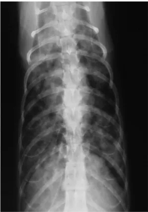
Figure 5 Thoracic radiography, ventrodorsal view: diffuse, multiple, poorly defined nodules with blurred margins in the lungs of a cat with systemic cryptococcosis. Courtesy of
Maria Grazia Pennisi
Cytology
can readily
diagnose
cryptococcosis
because the
appearance
of the
organisms is
characteristic
and the
number of
yeasts in the
lesions is
usually high.
Figure 6 Diff Quick stained smear of nasal exudate from a cat with C neoformans infection. Note the prominent capsule (clear halo) and narrow-necked budding (arrow). Courtesy of
Richard Malik, University of Sydney Veterinary School, Australia
Figure 7 Diff Quick stained smear of fine needle aspirate of a cryptococcal lesion. Note the enormous capsule surrounding the yeast cells. Courtesy of Mark Krockenberger
at Universite de Liege on September 3, 2013 jfm.sagepub.com
positive with a Gram-negative (pink) capsule. A pyogranulomatous inflammatory pattern is usually seen. Although filamentous forms are not commonly observed in tissues, these atyp-ical morphologatyp-ical forms of C neoformans may be present in cats.26,27
Histology
Biopsy samples of nasal mucosa, lymph nodes or skin nodules may be obtained for histology, but they may also provide impression smears for cytology and material for culture and PCR. Haematoxylin-eosin stained sections show eosinophilic bodies surrounded by a clear halo and a pyogranulomatous reaction (Figure 8). Mayer’s mucicarmine method specifically stains the capsule of Cryptococcus. Immunohistochemistry on tissue sections is used for species differentiation, using mono-clonal antibodies (Figure 9).38
Culture
Culture should be performed if the antigen test is negative, when titres are low or absent. Only samples from nasal biopsies should be submitted for culture, because the presence of
Cryptococcus in nasal discharge cultures is not
considered evidence of disease. Positive culture of biopsy samples and histological changes consistent with infection are consid-ered diagnostic and may be used to test the sensitivity towards antifungal drugs.
Culture of biopsy samples is more sensitive
than cytology in confirming infection [EBM grade III]. Cryptococcus is easily isolated in Sabouraud dextrose agar after incubation at 25°C and 37°C for 10 days but also on bacteri-al standard media. It is now possible to differ-entiate C neoformans from C gattii by a specific agar test.2
When samples are contaminated by bac -teria, as occurs in nasal secretions, media containing antibiotics are useful.1
PCR
PCR has been developed for genetic identifi-cation in CSF, urine, serum and biopsy sam-ples, but is not used routinely in practice.39–41
Antibody detection
Antibody detection is not a diagnostic tool because it cannot distinguish subclinical infec-tion from disease.
Diagnostic imaging
Advanced diagnostic imaging techniques (CT and MRI) are frequently used in the eval -uation of chronic nasal and CNS signs. Abnormal findings in feline cryptococcosis are the presence of chronic rhinitis, frontal sinusitis and/or intranasal or intracranial focal solitary or multifocal masses or fluid-filled lesions [EBM grade III].13Confirmation of diagnosis is not possible by imaging alone, but resolution of a mass lesion can be followed up by MRI in cats under medical therapy.42,43 MRI findings may also include meningeal enhancement, and optic nerve and cribriform plate involvement.13
Prognosis
The prognosis is favourable in most cases, provided the diagnosis is obtained sufficiently early (before dissemination or before the devel-opment of irreversible lesions) and patients and owners comply with a long course of treat-ment (months) and follow-up (years).
Although information on outcomes is quite limited, it seems that cats have a more favourable prognosis than dogs or horses, which more frequently develop lower respira-tory, disseminated and neurological disease with associated higher mortality [EBM grade III].7,11,13,29
In one retrospective study, disease severity did not influence outcome, although the pres-ence of CNS involvement had a significantly adverse impact on the outcome of therapy [EBM grade III].29 By contrast, alteration of the mental status was the only negative prognos-tic factor in a retrospective study on cats with the CNS form of cryptococcosis, and complete recovery was also documented in cats with a CNS form [EBM grade III].13,43
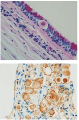
Figure 8Early invasion of
C gattii into the respiratory
epithelium of a koala. Note the eosinophilic body surrounded by a clear halo.
Courtesy of Mark Krockenberger Figure 9Use of immunohistology to demonstrate C gattii in histological sections. It is possible to conclusively identify Cryptococcus species in paraffin-embedded formalin-fixed tissue sections using monoclonal antibodies directed against different capsular epitopes. These show up as brown precipitates, highlighting both the yeast cell body and its capsule. Note also the narrow neck budding.
Courtesy of Mark Krockenberger
EBM grades The ranking system for grading the level of evidence of various statements within this article is described on page 533 of this Special Issue.
616
JFMSCLINICAL PRACTICER E V I E W/ABCD guidelines on cryptococcosis
Treatment
No prospective controlled studies exist on the treatment of feline cryptococcosis and all data are based on retrospective studies and case reports. Treatment guidelines have not been established and the choice of appropriate anti-fungal drug depends on many factors. Owner compliance is crucial, because of the high costs in terms of both money and time com-mitment required for treatment.
Some retrospective studies on treatment outcomes in feline cryptococcosis have been reported, using a variety of criteria for evalu-ating the success of therapy.21,44,45 In the largest retrospective study performed on 59 cats, 68% had a successful outcome [EBM grade III].29Most of them needed one single course of therapy of several months (1–24) duration and few cats received a second course of therapy because of clinical recur-rence or raised LCAT titre. According to a more recent retrospective study, the clinical outcome may be favourable in approximately two-thirds of treated cats [EBM grade III].7 Most recovered cats were presented with sinonasal or single skin, subcutis or intestinal lesions, and the ones that did not recover had CNS or disseminated disease.
Amphotericin B, ketoconazole, fluconazole and itraconazole have all been used to treat cats. With regard to the effect of different ther-apeutic protocols, there was no significant dif-ference in outcome between cats treated with amphotericin B-containing protocols and those treated with azole monotherapy using fluconazole or itraconazole [EBM grade III].29
The median cumulative dose of ampho-tericin B for cats cured at the first attempt was 16 mg/kg (range 7–23 mg/kg). This was high-er than the previously recommended cumula-tive dose of 4–8 mg/kg. The median duration of treatment for fluconazole-treated cats was significantly shorter (4 months; range 1–8 months) than the median for the itraconazole group (9 months; range 3–24 months). Liposomal formulations of amphotericin B may be better tolerated but are very expensive and not easily available. Recommendations for treatment based on case studies are that fluconazole or itraconazole are good choices. In CNS or systemic cases, amphotericin B alone or in combination with flucytosine may be the first choice, followed by prolonged treatment with fluconazole or itraconazole.29 Cats with pre-existing renal disease should be treated with itraconazole or fluconazole only. Fluconazole seems to be more effective than itraconazole for infections involving the CNS, eye and urinary tract, and is also better toler-ated [EBM grade III].1,24,43 Resistance to flu-conazole was reported with some isolates that
nevertheless were susceptible to other azoles.2 The clinical condition of cats with cerebral cryptococcosis may worsen soon after starting amphotericin B therapy, presumably due to an inflammatory response and increased intra cranial pressure. Shortacting cortico -steroid (dexamethasone or prednisolone sodi-um succinate) therapy is reported to be of immediate benefit in such cases and associat-ed with improvassociat-ed chance of survival in the short term.13,29
Surgical excision of any nodules located in the skin, nasal or oral mucosa must be consid-ered as a valuable aid in cats under medical therapy [EBM grade III].46
In general, treatment is recommended until the antigen test is negative. If the antigen test is negative at the time of diagnosis and the disease was confirmed by other methods, or if the antigen test is not available, treatment should be continued until at least 2–4 months after resolution of clinical signs. See Table 1 for treatment options.
Prevention
Free-roaming cats in rural areas are potential-ly more exposed to Cryptococcus, even though urban cats can be contaminated through pigeon guano. The presence of avian guanos, particularly pigeon droppings, and some decaying vegetation substrates such as euca-lyptus leaves, may be considered a risk fac-tor.17A knowledge of local fungal habitats that carry the largest risks of exposure and about seasonal variations in the production of infec-tious propagules would be useful to develop preventive measures for both human and animal infection.
Drug/therapy Dose and duration Comments
Itraconazole 50–100 mg/cat
q24h
Good absorption without food. Oral solution better than capsules. Hepatotoxicity possible; monitor liver enzymes periodically/monthly
Amphotericin B 0.25 mg/kg q48h IV
to a total dose of 4–16 mg/kg
Treatment of choice for CNS infection and/or systemic disease. Significant nephrotoxicity; monitor renal function frequently/weekly
Flucytosine 25–50 mg/kg PO
q6h
Synergistic with amphotericin B; do not use as single treatment
Fluconazole 50 mg/cat q12h Suggested treatment of choice, especially
for CNS infection. Good absorption without food. Monitor liver enzymes
Terbinafine 10 mg/kg q24h Use if resistance to azoles
Surgical excision
Skin, oropharyngeal and nostril granulomas
iv = intravenous, PO = oral, CNS = central nervous system Treatment of cryptococcosis Table 1
Owner
compliance
is crucial,
because of the
high costs
in terms of
both money
and time
commitment
required for
treatment.
at Universite de Liege on September 3, 2013 jfm.sagepub.com
Funding
The authors received no specific grant from any funding agency in the public, commercial or not-for-profit sectors for the preparation of this article. The ABCD is supported by Merial, but is a scientifically independent body.
Conflict of interest
The authors do not have any potential conflicts of interest to declare.
References
1 Sykes JE and Malik R. Cryptococcosis. In: Greene CE (ed). Infectious diseases of the dog and cat. 4th ed. St Louis: Saunders, Elsevier, 2012, pp 621–634.
2 Lester SJ, Malik R, Bartlett KH and Duncan CG.
Cryptococcosis: update and emergence of Cryptococcus
gattii. Vet Clin Pathol 2011; 40: 4–17.
3 Alspaugh JA, Davidson RC and Heitman J. Morphogenesis of Cryptococcus neoformans. Contrib Microbiol 2000; 5: 217–238. 4 Lin X and Heitman J. The biology of the Cryptococcus
neofor-mans species complex. Annu Rev Microbiol 2006; 60: 69–105. 5 Kano R, Kitagawat M, Oota S, Oosumit T, Murakami Y,
Tokuriki M, et al. First case of feline systemic Cryptococcus albidus infection. Med Mycol 2008; 46: 75–77.
6 Kano R, Hosaka S and Hasegawa A. First isolation of Cryptococcus magnus from a cat. Mycopathologia 2004; 157: 263–264.
7 McGill S, Malik R, Saul N, Beetson S, Secombe C, Robertson I, et al. Cryptococcosis in domestic animals in Western
Australia: a retrospective study from 1995–2006. Med Mycol
2009; 47: 625–639.
8 Castella G, Abarca L and Cabanes FJ. Criptococosis y
ani-males de Compañía. Rev Iberoam Micol 2008; 25: S19–S24.
9 Craig S, Lester S, Black W, Fyfe M and Raverty S. Multispecies
outbreak of cryptococcosis on southern Vancouver Island, British Columbia. Can Vet J 2002; 43: 792–794.
10 Duncan C, Stephen C, Lester S and Bartlett KH. Follow-up
study of dogs and cats with asymptomatic Cryptococcus
gattii infection or nasal colonization. Med Mycol 2005; 43: 663–666.
11 Duncan C, Stephen C and Campbell J. Clinical characteristics
and predictors of mortality for Cryptococcus gattii infection in dogs and cats of southwestern British Columbia. Can Vet J
2006; 47: 993–998.
12 O’Brien CR, Krockenberger MB, Wigney DI, Martin P and Malik R. Retrospective study of feline and canine crypto
-coccosis in Australia from 1981 to 2001: 195 cases. Med Mycol
2004; 42: 449–460.
13 Sykes JE, Sturges BK, Cannon MS, Gericota B, Higgins RJ, Trivedi SR, et al. Clinical signs, imaging features, neuro
-pathology, and outcome in cats and dogs with central nerv-ous system cryptococcosis from California. J Vet Intern Med
2010; 24: 1427–1438.
14 Malik R, Wigney DI, Muir DB and Love DN. Asymptomatic
carriage of Cryptococcus neoformans in the nasal cavity of dogs and cats. J Med Vet Mycol 1997; 35: 27–31.
15 Connolly JH, Krockenberger MB, Malik R, Canfield PJ, Wigney DI and Muir DB. Asymptomatic carriage of Cryptococcus neoformans in the nasal cavity of the koala (Phascolarctos cinereus). Med Mycol 1999; 37: 331–338.
16 Bartlett KH, Fyfe MW and MacDougall LA. Environmental
< Cryptococcosis is worldwide the most common systemic fungal disease in cats caused by the C neoformans–C gattii species complex.
< Cryptococcosis is a non-contagious rare or sporadic disease acquired by cats, usually by inhalation of organisms from a contaminated environment.
< Cryptococcosis is not considered a zoonosis; animals may serve as sentinels for exposure of human beings.
< The disease can present in several clinical forms, including the nasal form, CNS form (which can either derive from the nasal form or occur independently), cutaneous form and systemic form.
< Diagnosis can be confirmed using a rapid agglutination antigen test on serum or body fluids.
< The prognosis is favourable, if the diagnosis is obtained early and owners cooperate with a long course of treatment (months) and follow-up (years).
< Surgical excision of nodules located in the skin, nasal or oral mucosa is valuable adjunctive treatment for cats undergoing antibiotic therapy.
< Antifungal treatment choices include amphotericin B, ketoconazole, fluconazole or itraconazole, based on individual assessment.
< Avian guano, particularly pigeon droppings, and decaying vegetation substrates such as eucalyptus leaves are risk factors.
618
JFMSCLINICAL PRACTICER E V I E W/ABCD guidelines on cryptococcosis
Cryptococcus neoformans var gattii in British Columbia,
Canada. Am J Respir Crit Care Med 2003; 167: A499.
17 Fortes ST, Lazéra MS, Nishikawa MM, Macedo RC and Wanke B. First isolation of Cryptococcus neoformans var gattii from
a native jungle tree in the Brazilian Amazon rainforest.
Mycoses 2001; 44: 137–140.
18 Pal M. Cryptococcus neoformans var neoformans and munia
birds. Mycoses 1989; 32: 250–252.
19 Gerds-Grogan S and Dayrell-Hart B. Feline cryptococcosis: a
retrospective study. J Am Anim Hosp Assoc 1997; 33: 118–122.
20 Malik R, Wigney DI, Muir DB, Gregory DJ and Love DN.
Cryptococcosis in cats: clinical and mycological assessment of 29 cases and evaluation of treatment using orally admin-istered fluconazole. J Med Vet Mycol 1992; 30: 133–144.
21 Jacobs GJ, Medleau L, Calvert C and Brown J. Cryptococcal
infection in cats: factors influencing treatment outcome, and results of sequential serum antigen titers in 35 cats. J Vet
Intern Med 1997; 11: 1–4.
22 Flatland B, Greene RT and Lappin MR. Clinical and serologic
evaluation of cats with cryptococcosis. J Am Vet Med Assoc
1996; 209: 1110–1113.
23 Lester SJ, Kowalewich NJ, Bartlett KH, Krockenberger MB, Fairfax TM and Malik R. Clinicopathologic features of an
unusual outbreak of cryptococcosis in dogs, cats, ferrets, and a bird: 38 cases (January to July 2003). J Am Vet Med Assoc
2004; 225: 1716–1722.
24 Trivedi SR, Sykes JE, Cannon MS, Wisner ER, Meyer W, Sturgess BK, et al. Clinical features and epidemiology of
cryptococcosis in cats and dogs in California: 93 cases (1988–2010). J Am Vet Med Assoc 2011; 239: 357–369.
25 Martins DB, Zanette RA, França RT, Howes F, Azevedo MI, Botton SA, et al. Massive cryptococcal disseminated infection
in a immunocompetent cat. Vet Dermatol 2011; 22: 232–234.
26 Bemis DA, Krahwinkel DJ, Bowman LA, Mondon P and Kwon-Chung KJ. Temperature-sensitive strain of Cryptococcus neoformans producing hyphal elements in a
feline nasal granuloma. J Clin Microbiol 2000; 38: 926–928.
27 Lin X. Cryptococcus neoformans: morphogenesis, infection,
and evolution. Infect Genet Evol 2009; 9: 401–416.
28 Urban CF, Lourido S and Zychlinsky A. How do microbes
evade neutrophil killing? Cell Microbiol 2006; 8: 1687–1696.
29 O’Brien CR, Krockenberger MB, Martin P, Wigney DI and Malik R. Long-term outcome of therapy for 59 cats and 11
dogs with cryptococcosis. Aust Vet J 2006; 84: 384–392.
30 Norris JM, Bell ET, Hales L, Toribio JA, White JD, Wigney DI, et al. Prevalence of feline immunodeficiency virus infection
in domesticated and feral cats in eastern Australia. J Feline
Med Surg 2007; 9: 300–308.
31 Graham KJ, Brain PH, Spielman D, Martin PA, Allan GS and Malik R. Concurrent infection with Cryptococcus neoformans/ gattii species complex and Mycobactcerium avium affecting
the subcutis and bone of a pelvic limb in a cat. J Feline Med
Surg 2011; 13: 776–780.
32 Malik R, Martin P, Wigney DI, Church DB, Bradley W, Bellenger CR, et al. Nasopharyngeal cryptococcosis. Aust Vet J 1997; 75: 483–488.
33 Beatty JA, Barrs VR, Swinney GR, Martin PA and Malik R.
Peripheral vestibular disease associated with cryptococcosis in three cats. J Feline Med Surg 2000; 2: 29–34.
34 Paulin J, Morshed M and Armién AG. Otitis interna induced
by Cryptococcus neoformans var grubii in a cat. Vet Pathol
2013; 50: 260–263.
35 Belluco S, Thibaud JL, Guillot J, Krockenberger MB, Wyers M, Blot S and Colle MA. Spinal cryptococcoma in an
immuno-competent cat. J Comp Pathol 2008; 139: 246–251.
36 Tisdall PL, Martin P and Malik R. Cryptic disease in a cat with
painful and swollen hocks: an exercise in diagnostic reason-ing and clinical decision-makreason-ing. J Feline Med Surg 2007; 9:
418–423.
37 Brandt LE and Blauvelt MM. What is your diagnosis? Urine
sediment from a southern California cat with weight loss.
Vet Clin Pathol 2010; 39: 517–518.
38 Krockenberger MB, Canfield PJ, Kozel TR, Shinoda T, Ikeda R, Wigney DI, et al. An immunohistochemical method that
dif-ferentiates Cryptococcus neoformans varieties and serotypes in formalin-fixed paraffin-embedded tissues. Med Mycol
2001; 39: 523–533.
39 Kano R, Fujino Y, Takamoto N, Tsujimoto H and Hasegawa A.
PCR detection of the Cryptococcus neoformans CAP59 gene from a biopsy specimen from a case of feline cryptococcosis.
J Vet Diagn Invest 2001; 13: 439–442.
40 Okabayashi K, Kano R, Watanabe T and Hasegawa A.
Serotypes and mating types of clinical isolates from feline cryptococcosis in Japan. J Vet Med Sci 2006; 68: 91–94.
41 Meyer W, Castañeda A, Jackson S, Huynh M and Castañeda E.
Molecular typing of IberoAmerican Cryptococcus
neofor-mans isolates. Emerg Infect Dis 2003; 9: 189–195.
42 Karnik K, Reichle JK, Fischetti AJ and Goggin JM. Computed
tomographic findings of fungal rhinitis and sinusitis in cats.
Vet Radiol Ultrasound 2009; 50: 65–68.
43 Hammond JJ, Glass EN, Bishop TM, Kent M and De Lahunta A. Imaging diagnosis – intracranial cryptococcal mass in a
cat. Vet Radiol Ultrasound 2011; 52: 306–308.
44 Medleau L, Jacobs GJ and Marks MA. Itraconazole for the
treatment of cryptococcosis in cats. J Vet Intern Med 1995; 9:
39–42.
45 Davies C and Troy GC. Deep mycotic infections in cats. J Am Anim Hosp Assoc 1996; 32: 380–391.
46 Hunt GB, Perkins M, Foster SF, Barrs VR, Swinney GR and Malik R. Nasopharyngeal disorders of dogs and cats: a
review and retrospective study. Compend Contin Educ Pract
Vet 2002; 24: 184–199.
Available online at jfms.com
Reprints and permission: sagepub.co.uk/journalsPermissions.nav
at Universite de Liege on September 3, 2013 jfm.sagepub.com