PRODUCTION OF CHITOSANASE BY
RECOMBINANT STREPTOMYCES LIVIDANS
AND ENZYMATIC PREPARATION
OF CHITOSAN OLIGOMERS
par
Li, Tong
Memoire presente au departement de biologie
en vue de 1'obtention du grade de maitre es sciences (M.Sc.)
DEPARTEMENT DE BIOLOGIE
FACULTE DES SCmNCES
UNIVERSITE DE SHERBROOKE
Sherbrooke, Quebec, Canada, juin 19931*1
National Libraryof Canada du CanadaBibliotheque nationale Acquisitions and
Bibliographic Services Branch 395 Wellington Street
Ottawa, Ontario
K1AON4
Direction des acquisitions et des services bibliographiques
395, rue Wellington
Ottawa (Ontario) K1AON4
Your file Votre ee'fQrwce Our file Notre reference
The author has granted an
irrevocable non-exclusive licenceallowing the National Library of
Canada to reproduce, loan,distribute or sell copies of
his/her thesis by any means and
in any form or format, makingthis thesis available to interested
persons.
L'auteur a accorde une licence irrevocable et non exclusive
permettant a la Bibliotheque
nationale du Canada de
reproduire, prefer, distribuer ou vendre des copies de sa these de quelque maniere et sousquelque forme que ce soit pour
mettre des exemplaires de cettethese a la disposition des
personnes interessees.
The author retains ownership of
the copyright in his/her thesis.
Neither the thesis nor substantial
extracts from it may be printed or
otherwise reproduced without
his/her permission.
Uauteur conserve la propriete du
droit cTauteur qui protege sa
these. Ni la these ni des extraits substantiels de celle-ci nedoivent etre imprimes ou
autrement reproduits sans son autorisation.ISBN 0-315-93130-2
ABSTRACT
Elicitors are molecules which can trigger phytoalexin and pathogenesis-related proteins biosynthesis in plants. Chitosan as an elicitor has important roles in the interaction between pathogenic fungi and plants. Chitosan can inhibit fungal DNA transcription into mRNA, and it can also trigger the specific defensive genes which encode for at least 20 kinds of proteins related to resistant mechanism in
plants.
Chitosanase is an endoglycosidase that can hydrolyze chitosan into oligosaccharide fragments. Chitosan heptamer-more fractions showed maximal activity in both antifungal properties and induction of plant resistance. In this work, recombinant strains of Streptomyces lividans were used for production of chitosanase, then we used this enzyme to prepare the active chitosan oligomers and to test their antifungal properties and their ability to induce defensive responses in
plants.
In order to produce large amounts of chitosanase, the chitosanase gene from Streptomyces N174 was cloned in the high copy-number vector pFD666 and transformed into protoplasts of S. lividans TK24 and 10-164. These recombinant
Ill
strains could produce chitosanase efficiently. The chitosanase activity could reach
up to 95 units per millilitre of fermentation liquid on natural substrate (mycelium
oiMucor rouxii). When DNA sequences were deleted in upstream or downstream of the chitosanase gene, chitosanase production became lower. Maybe, these sequences have some functions for the chitosanase gene expression and stability. In the chitosan medium, the chitosanase activity of the recombinant strains S.
lividans TK24 decreased quickly after 3 days. We have shown that it related to the
production of a specific proteolytic enzyme for chitosanase. On the contrary, the chitosanase activity could maintain high level after 3 days in M. rouxii mycelium as fermentation substrate. If S. lividans 10-164 was used as host for carrying the chitosanase gene, chitosanase activity reached high level after 2 days and the same level was maintained for a few days in D-glucosamine medium. The enzyme was recovered by polyacrylic acid precipitation. The enzyme prepared with this method has stable activity for long time.
Using chitosanase hydrolysis, the heptamer-more fractions were prepared. These chitosan oligomers could inhibit fungal growth and could induce the production of pathogenesis-related proteins such as B-l,3-glucanase, chitinase and chitosanase by plants.
ACKNOWLEDGEMENT
I sincerely thank my supervisors, Dr. Ryszard Brzezinski and Dr. Carole Beaulieu, for their guidance, genuine interest and constant encouragement during this study.
I also desire to extend my appreciation to Dr. Claude V. Dery and some graduate students in our laboratory for their helps to my laboratory works.
Canadian International Development Agency has provided a student fellowship for my master study in Canada.
Finally, special thanks are due to my parents for their continued support and encouragement throughout my study.
TABLE OF CONTENTS
ABSTRACT ... ii
ACKNOWLEDGEMENT ... iv
TABLE OF CONTENTS ... v
LIST OF ABBREVIATIONS ... ix
LIST OF TABLES ... xii
LIST OF FIGURES ... xiv
CHAPTER 1 - INTRODUCTION ... 1
CHAPTER 2 - MATERIALS AND METHODS ... 12
2.1 Plants, microorganisms and plasmids ... 12
2.2 Media and culture conditions ... 12
2.2.1 Streptomyces lividans ... 12
2.2.2 Fungus ... 14
2.2.3 Plants ... 15
2.3 Enzymes assays ... 15
2.3.1 Standard substrates ... .15
2.3.2 Enzymatic reactions and measurements ... 16
VI
2.4 Total protems determination ... 19
2.5 SDS-polyacrylamide gel electrophoresis ... 19
2.6 Preparation of the active chitosan oligomers by chitosanase hydrolysis ... 20
2.6.1 Preparation of the oligomers ... 20
2.6.2 Testing the chitosan oligomers by thin-layer chromatography ... 21
2.7 Purification of chitosanase with polyacrylic acids ... 21
2.8 Transformation of plasmids into •S'^^o/nyc^ protoplasts ... 23
2.9 Extraction of the cmde enzymes from plants ... 23
2.9.1 Plant seedling extracts ... 23
2.9.2 Plant leaves extracts ... 24
CHAPTER 3 - RESULTS ... 26
3.1 Optimization of the conditions for chitosanase production ... 26
3.1.1 The optimal fermentation conditions of chitosanase production by recombinant S.lividans TK24(pRL226) . 29 Step 1. Effect of carbon source on chitosanase production ... 29
vii
enrichment ... 32
Step 3. Effect of nitrogen source on chitosanase
production ... 34
Step 4. Optimization of inoculum ... 34
Step 5. Chitosanase production on natural substrates . . 38
Conclusion ... 41
3.1.2 Comparison between different clones carrying the
chitosanase gene ... 41
3.1.3 Study on the protease in Streptomyces lividans TK24 . . 48 3.1.4 Study on Streptomyces lividans 10-164 carrying the
chitosanase gene ... 52
3.2 Purification of chitosanase with polyacrylic acid ... 57
3.3 Preparation of the active chitosan oligomers by chitosanase
hydrolysis ... 57
3.4 Antifungal properties of the chitosan oligomers and induction of
defensive responses in plants ... 62
3.4.1 Inhibition of fungal growth ... 62
3.4.2 Induction ofpathogenesis-related proteins during tomato
Vlll
3.4.3 Induction of pathogenesis-related proteins in mature
plants ... 70
CHAPTER 4 - DISCUSSION ... 73
4.1 Streptomyces: a good host for heterologous gene expression ... 73
4.2 Pre-culture to help Streptomyces spore germination ... 75
4.3 Investigation on possible induction of chitosanase gene
expression by chitosan ... 76
4.4 Function of other DNA sequences in the cloned chitosanase
gene ... 77
4.5 Extracellular protease in Streptomyces lividans TK24 ... 79 4,6 Chitosan oligomers antifungal properties and induction of
defensive responses in plants ... 82
4.6.1 Solution of chitosan oligomers ... 82
4.6.2 Interaction of chitosan oligomers, fungus and plants ... 83
CONCLUSION ... 85 REFRENCES ... 87
LIST OF ABBREVIATIONS
act.:BSA:
C.A.:CHS:
chs:DMSO:
EDTA: ferm.: g: GlcN:(GlcN)^:
(GlcN)3:
(GlcN)4:
(GlcN)5:
(GlcN),:
(GlcN)^:
GlcNAc: activityBovine Semm Albumin casamino acid
chitosanase
chitosanase-encoding gene
dimethylsulfoxide
ethylenediaminetetra-acetic acid (disodium salt) fermentation gram glucosamine chitobiose chitotriose chitotetraose chitopentaose chitohexaose chitoheptaose N-acetyl- glucosamine
x HAc:
HC1:
hr: Kb: kDa: L: M: M.E.: mg: mm: ml: mM: mol:MOPS:
mpv: MS: mU: nm: NaAc: acetic acid chlorhydric acid hour kilobase kilodalton liter molar malt extract milligram minute milliliter millimolar mole 4-Morpholineprc milliliters ofpa( minimal salts milliunit nanometer sodium acetateXl neo: O.D.: PA:
PAGE:
PDA:PMSF:
P.-R.: pro.: rpm:SDS:
TSA:
TSB: u:w'
^1: [im: pmol:aminoglycoside resistance gene
optical density polyacrylic acid
polyacrylamide gel electrophoresis potato dextrose agar
phenylmethyl sulfonyl fluoride
pathogenesis-related protein
rotations per minute
sodium dodecyl sulfate
Tryptic Soy Agar Tryptic Soy Broth
unit
microgram
microliter
micrometer micromole
LIST OF TABLES
Table 1: Minimum inhibitory concentration of chitosan and chitosan
hydrolysate for fungi ... 9
Table 2: The plants, organisms and plasmids used in this study ... 13 Table 3: Preliminary experiments for chitosanase production
by recombinant S. lividans TK24 (pRL226) ... 28 Table 4: Effect of carbon source on chitosanase production ... 30
Table 5: Optimization of the ratio between chitosan and enrichment . . 33
Table 6: Effect of nitrogen source on chitosanase production ... 38
Table 7: Optunization of inoculum ... 39
Table 8: Chitosanase production on natural substrates ... 41
Table 9: Comparison between the different clones carrying the
chitosanase gene ... 44
Table 10: Purification of chitosanase from S. lividans TK24 (pRL226)
with polyacrylic acid ... 59
Table 11: Effect of chitosan oligomers on inhibition of radial growth of
Fusarium oxysporum f.sp. radicis-lycopersici ... 64 Table 12: Effect of chitosan oligomers on tomato seeds germination.. ... .67
Xlll
Table 13: Enzymatic activities in tobacco leaves treated with
chitosan oligomers ... 72
Table 14: Enzymatic activities in tomato leaves treated with
LIST OF FIGURES
Fig. Fig. Fig. Fig. Fig. Fig. Fig. Fig. 1: 2: 3: 4: 5: 6: 7: 8:Structure of chitin and chitosan ... 2
Model of chitosan in plant-fungal pathogen interactions .... 4 The cloned chitosanase gene and its position in respect to the neo
gene of the vector ... 11
Special tube setting for the preparation of leaves extract .... 25
Effect of carbon source on chitosanase production ... 31
Optimization of inoculum ... 37
Chitosanase production on natural substrates ... 40
Comparison of chitosanase production between the different
clones carrying the chitosanase gene ... 44
Fig. 9 A: SDS-PAGE analysis of cmde chitosanase production of different
clones in starch and chitosan-starch fermentation media .... 45
Fig. 9 B: SDS-PAGE analysis of cmde chitosanase production of different
clones in starch and chitosan-starch fermentation media .... 46
Fig .10: Comparison of chitosanase production by S. lividans TK24 (pRL226)
and (pRL270) with different inocula. ... 47
Fig. 11: Time-course of chitosanase production in different fermentation
XV
Fig. 12: Time-course of protease activity in different fermentation media of
S. lividans TK24 (pRL226) ... 50
Fig. 13: Time-course of chitosanase production (A) and protease activity (B) in chitosan-starch fermentation media of two S. lividans TK24
clones ... 51
Fig. 14: SDS-PAGE analysis of proteins produced by S. lividans TK24
(pRL226) in different fermentation media . ... 53
Fig. 15: The time course of chitosanase production in fermentation liquid of
recombinant strains 5'. lividans 10-164. ... 55
Fig. 16: The time course of chitosanase activity (A) and protease activity (B)
in fermentation liquid of recombinant strains 5'. lividans 10-164
(pRL226). ... 56
Fig. 17: Thin layer chromatography showing the different degree of chitosan
hydrolysis by cmde chitosanase. ... 60
Fig. 18: Thin layer chromatography showing the optimal degree of chitosan
hydrolysis by cmde chitosanase ... 61
Fig. 19: Effect of chitosan oligomers on inhibition of radial growth of
XVI
Fig. 20: Induction of chitinase activity by the chitosan oligomers in tomato
seedlings. ... 67
Fig .21: Induction of chitosanase activity by the chitosan oligomers in tomato
seedlings. ... 68
Fig. 22: Induction of B-l,3-glucanase activity by the chitosan oligomers in
CHAPTER 1
INTRODUCTION
Elicitors are molecules of microbial origin which trigger the production of
phytoalexins and pathogenesis-related proteins. Elicitors act as a signal inducing plant resistant responses and regulating biochemical and physiological processes. Elicitors include: glucans, chitosan, glucoproteins, polysaccharides and fatty acids.
Chitosan is a polymer ofB-l,4-glucosamine. In fact, it is deacetylated chitin (Fig. 1). As a practical approach, the chitosan polymer is defined as containing glucosamine moieties partially N-acetylated, with the degree (percentage) of
acetylation cited (Fenton and Eveleigh 1981). Chitosan was first identified as a
minor component of cell walls of Phycomyces blakesleeanus (Kreger, 1954) and further investigation established its presence throughout the cell walls of Zygomycete fungi, and in the cell walls of other fungi such as Agaricus, Puccinia, Fusarium and Saccharomyces (Ouakfaoui and Asselin 1992). Chitosan is a component of the cell walls of many plants pathogenic fungi, but it is not a normal compound of plant tissues. However, most commercially available chitosan is
H
H
HO
(b) CHITOSAN
Fig. 1 Structure of chitin and chitosan
3
Chitosan as an elicitor has important roles in the interaction between plant and pathogenic fungi. For example, in the interaction between pea endocarp-tissue and F. solani, naturally released chitosan can accumulate in both the fungal cell and the adjacent host plant cells within 15-30 min, following inoculation of the
fungi (Hadwiger and Beckman, 1980, 1981). Chitosan can inhibit germination of
F. solani spores and growth of fungi at a concentration lower than 10 jng/ml. When chitosan was applied to pea tissue before the inoculation with F. solani f. sp. pisi., chitosan induced defensive responses which developed a complete
immunity of the pea to this pathogen. Chitosan can inhibit the fungal growth and
induce the defensive responses of plant, because it has a strong affinity for DNA
due to multiple positive charges (Hadwiger and Beckman, 1980) (Fig. 2). First, chitosan can inhibit fungal DNA transcription mto mRNA, directly inhibiting the
fungal growth. Second, chitosan can activate plant defense genes (Daniels et al.,
1986) and induce the biosynthesis of at least 20 kinds of pathogenesis-related
proteins, such as B-l,3-glucanase, chitinase and chitosanase. These enzymes
enhance plant potential to degrade the cell wall of fungi which results in the
release of additional chitosan that can amplify the elicitor signalling process by feedback. In addition, chitosan induces the plant synthesis of phenylalanine ammonia lyase (PAL, a key enzyme in the phenylpropanoid pathway in plants).
P- glucan- ^hTO-^l-.^g-g-S-S-?
hltTn—A-^FUNGUS
nucleus DNA-^RNA'—-> Enzymes —Chitosan^inhibits^Y
V-V-V~VW-V^V-^7h-V~?~VL~V--Vh-V~V-•-RNA
j3-.o-.o-o- o-o-o-o-o-o o-o-o o-o
PLANT CELL WALI
-y-y-^-v, v-t-Yi VT^L ^1 • • • BI:-B—A-B;-»-R-<?; 'o-o-Q-^-o-5^-0-^-^ B-GLUCANASE ICHITINASE"! PHENYLPROPANOID PATHWAY ENZYMES
CHITOSAN actjvates genesh
RNA
DNA
PLANT NUCLEUS.
5
Phytoalexins are produced by this pathway (Ryan, 1987; Loschke et al., 1983;Kendra and Hadwiger, 1984). Application of chitosan solution at a concentration of 100 fJig/ml provides protection against F. solani f. sp. pisi. in pea pod tissue for periods of at least 5 days, while a concentration of 10 /zg/ml can maintain resistance for up to 3 days (Kendra et al., 1989). There are also homolog of the resistance in potatoes (Matton and Brisson, 1989), soybean (Leguay et al., 1988), parsley (Somssich et al., 1988), birch, alder (Breiteneder et al., 1989) and bean (Walter etal., 1990).
The mechanism for triggering gene activation by chitosan is still not
understood. Kendra and Hadwiger (1986) suggested that chitosan can directly
interact with plant DNA to activate specific defense genes (Daniels et al., 1986). After an external application of chitosan, as well as cell wall components purified from the fungi F. solani, chitosan can be detected in the cytoplasm of pea cells
within 20 min of application (Hadwiger and Loschke, 1981). This means that
chitosan can penetrate the target cell plasma membrane very fast. This observation raises the possibility of an internal receptor different from the plasma membrane transduction system. Kendra and Hadwiger (1987) showed that there was no correlation between the chitosan or F. solani induction of disease resistant
6
responses in pea pod tissue and fluctuation in [ Ca++ ], inhibition of calmodulin, blockage of Ca++ channels or host membrane leakage. These results further indicated that cell wall or plasma membrane-bound Ca++ does not play a direct role in the chitosan activation of the pea resistance response mechanism. In contrast to these results, Young et al. (1982, 1983) have found that a linear correlation was found between calcium release from chitosan-treated whole cells or isolated cell wall and the amount of bound chitosan. The effect of chitosan on membrane permeability is due to its polycationic properties. Kohle et al. (1985) speculated that the Ca++ release after chitosan application may act as a secondary messenger to trigger host gene expression. However, most of the workers agree with Young et al. (1982). They showed that hyphal wall component can cause rapid depolarization of the transmembrane potential and may inhibit the electrogenic ion pump in the plasmalemme. Pelissier and Esquerre-Tugaye (1984)found that H+ extmsion from plant cells is inhibited by fungal elicitors in plant and
this effect is reversible. Recent data have shown that oligosacccharides cause the phosphorylation of a small protein in plasma membranes isolated from tomato and potato leaves (Ryan, 1988), and that a protein kinase seems to play an important role in the elicitation of the defense reaction in pea plant (Shiraishi et al., 1990). Verapamil, a Ca++ channel blacker, and K-252a, a strong inhibitor of protein
7
kinase, inhibit pisatin accumulation in pea epicotyl which had been treated with
elicitor isolated from M. pinodes. Potential roles of Ca++, cy die AMP and phosphatidylinositide system in the induction resistance process need further
investigations (Ryan, 1988; Shiraishi et al., 1990; Oku, 1992).
Chitosanase, a new class of enzyme, is an endoglycosidase that can hydrolyse chitosan into oligosaccharide fragments. The distinction between chitinase and chitosanase is not strict, as both enzymes have the ability to degrade
a variety of chitosans with different degrees of acetylation (Ohtakara, 1988). The
term chitosanase is usually given to enzymes having higher activity against highly deacetylated chitosan than chitin (Monaghan et al., 1973; Pelletier et al., 1990; Sakai et al., 1991). Chitosanases are produced by many micro-organisms, including fungi, bacteria and actinomycetes. In 1980, Hadwiger and Beckman reported that extracts of pea endocarp containing chitosanase could degrade F. solani f. sp. pisi and f. sp. phaseoli cell walls to produce carbohydrates fragments (chitosan and its oligomers) that have antifungal effect on F. solani and that act as
powerful elicitors of the induction of pisatin (an isoflavinoid phytoalexin in the
pods). Kendra and Hadwiger (1984) showed that monomer and dimer units have no antifungal activity but induce little pisatin production. Trimer through pentamer
8
units showed antifungal activity at high concentrations and have a moderate ability to induce pisatin formation. A sharp increase in antifungal activity and pisatm formation was noted for the hexamer units, while the heptamer and more fractionshowed maximal activity in both inhibition of fungal growth and pisatin induction.
These results mdicated that the high-molecular-weight chitosan fragments are more active in both antifungal and pisatin formation activities than the intermediate and low-molecular-weight fragments. In practice, chitosan oligomers prepared by chitosanase hydrolysis are a mixture of oligomer fractions with different degree of polymerization. For economic reasons, a mixture of oligomers is used. Chitosan
hydrolysate, which was prepared from chitosan by about 5% hydrolysis with
chitosanase, has shown to be maximal in both antifungal and antibacterial activities
(Table 1) (Uchida et al., 1989). This suggested that we should find the optimal
degree of hydrolysis of chitosan in order to obtain a maximal effect both on antifungal activity and the induction of defensive responses in plants.
In order to produce chitosanase in large amounts, which could be used for the large scale preparation of the active chitosan oligomers, several chitosananolytic actinomycetes were isolated from soil by Brzezinski's research group (Dupuy, 1991). One of these strains, Streptomyces N174, was isolated from
Table 1 Minimum inhibitory concentration of chitosan and chitosan hydrolysate for fungi.
Fungi Fusarium solani F. oxysporum F. oxysporum cepae Minimum Chitosan 0.07 0.09 0.08
inhibitory concentration (MIC), % Chitosan hydrolyzate* 0.035 0.050 0.035 Chitosan oligomer I
NE
NE NE* 50 mg total reducing sugar (TRS)/g of chitosan NE = No effect at 1.0 %
10
a slightly acidic soil in a sugar maple grove near Sherbrooke (Pink et al., 1991;
Boucher et al., 1992). This strain was highly effective in degradation of chitosan. The chitosanase gene from Streptomyces N174 was cloned in the high copy-number vector pFD666 (Pink et al., 1991) and several subclones were constructed (Masson et al., 1993; see also Fig. 3) and transformed into protoplasts of S. lividans TK24 and 10-164. Recombinant strains could produce chitosanase efficiently. This chitosanase was analyzed by SDS-PAGE. The molecular weight of the enzyme is 29.5 kDa; the isoelectric point is 7.5. The maximal activity of chitosan degradation was observed at 65°C, when the pH was maintained at 5.5. The K^ of the enzyme was 0.088 mg/ml and the V^ax was 96.5 U/mg. The enzyme degraded chitosan with a range of acetylation degrees from 1 to 60% but not chitin or CM-cellulose. The enzyme showed an endo-splitting type activity and the end-product of chitosan degradation contained a mixture of dimers and trimers of D-glucosamine (Boucher et al., 1992). Here, I describe further studies on the production of chitosanase by recombinant S. lividans carrying the chitosanase gene from Streptomyces N174, and application of chitosanase to prepare the active chitosan oligomers as an elicitor.
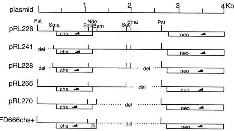
plasmid
pRL226
pRL241 del-4^
--pFD666chs+
J3S. deljaftQ-Fig. 3 The cloned chitosanase gene and its position in respect to the neo gene of the vector.
CHAPTER 2
MATERIALS AND METHODS
2.1 Plants, microorganisms and plasmids
Plants, microorganisms and plasmids used in this study are listed in Table 2.
2.2 Media and culture conditions
2.2.1 Streptomyces lividans
The spomlation medium was SLM-3 with 50 ^tg/ml of Kanamycin (DeWitt,
1985). Spares were prepared from SLM-3 plates heavily inoculated with mycelial cultures and incubated for 2 weeks at 30°C.
Pre-culture medium was TSB containing 10 /Ag/ml of Kanamycin. The spares were directly taken from SLM-3 plates and were used to inoculate the TSB liquid medium. Culture was incubated 20-24 hr at 30°C on rotary shaker at 300 rpm, then the pre-cultare was centrifuged and the volume of the pellet was
13
Table 2 The plants, organisms and plasmids used in this study.
Name Streptomyces Uvidans TK24 Streptomyces lividans 10-164 Mucor rouxii Aspergillus niger Saccharomyces cerevisiae Rhizopus oryzae Fusarium oxysporum f.sp. radicis-lycopersici pRL241 pRL226
pRL228
pRL266
pRL270
pFD666 chs+ Tomato seeds Tobacco seeds Source D. A. HopwoodJohn Innes Institute Norwich, England
Dieter Kluepfel
Institut Armand-Frappier
ville de Laval, Quebec, Canada
ATCC1 #24905
ATCC #336
ATCC #2360
NRRL2#395
Pierre Mathieu Charest
Dept. de phytologie, F.S.A.A.
University de Laval Quebec, Canada Jean-Yves Masson Isabelle Boucher Dept. de biologie University de Sherbrooke
Sherbrooke, Quebec, Canada F. Denis Institut de Recherches Cliniques de Montreal HYB.PERRON2000 W.H.Perron
XANTHI
W.H.Perron) American Type Culture Collection
14
measured (expressed as mpv). The pellet was washed with physiological salt solution water (NaCl 0.85%), and used for the inoculation of fermentationmedium.
Fermentation medium contained the appropriate carbon source (at a
concentration of 2% (w/v)) in minimal salts (MS) solution (Neugebauer et al,
1991). In some experiments, the nitrogen source was omitted from MS solution.
Then Img/ml riboflavin and 1M MOPS (pH 7.0) were added to the medium after sterilization (riboflavin and MOPS final concentration was 1^1/ml and 0.1M respectively) (Boucher, 1992). The fermentation liquid media were inoculated at
different densities with mycelium taken from the pre-culture of S. lividans and the cultures were incubated at 30°C on a rotary shaker at 300 rpm.
2.2.2 Fungus
Liquid medium contained malt extract (30g/L), proteose peptone (3g/L) and
agar (15g/L). Fungi were incubated on rotary shaker at 120 rpm at 25°C.
15
chitosan oligomers were tested on PDA medium containing various concentrations of the chitosan oligomers. Fungus inoculum was set down in the centre of PDA Petri plate and incubated at 28°C.2.2.3 Plants
Seeds were sterilized in 10% (v/V) bleach for 10 min and washed twice with
sterile distilled water. Then, seeds were put on 0.6% agar Petri dishes mixed with various concentrations of chitosan oligomers for germination (Hirano et al., 1988). They were incubated at room temperature for 6 days.
Plants were grown in 50% vermiculite and 50% planta-mix soil, with a 16 hours photo-period in a growth chamber (23-25°C).
2.3 Enzymes assays
2.3.1 Standard substrates
16
dissolved in 25 ml of 1 % HAc, then pH was adjusted at 5.5 with 50 mM sodium
acetate and completed with distilled water in 100 ml. The final concentration of chitosan solution was 0.25%.
Chitinase: chitin (practical grade C-7170, Sigma) was suspended in 0.05 M
Mcllvaine buffer, pH 6.8. The concentration of chitin was 2%.
B-l,3-glucanase: laminarin (L-9634, Sigma) was dissolved in 0.05 M Mcllvaine buffer, pH 6.8. The concentration of laminarin was 2%.
Protease: azocasein (Sigma) was dissolved in 0.1 M Tris-Cl buffer, pH 8.0. The concentration of azocasein was 6 mg/ml.
2.3.2. Enzymatic reactions and measurements
Chitosanase: the chitosanase activity standard assay contained 950 ^1 of 0.25 % chitosan substrate, 1-20 mU of enzyme and distilled water to a final volume of 1.0 ml. The reaction mixture was incubated in a water bath at 37°C for 10 min.
17
the reaction tube in ice for 10 min, the reaction tube was centrifuged in order to eliminate the chitosan precipitate. Soluble reducing sugars were measured by Nelson-Somogyi assay (Spiro, 1966) as follows: 0.5 ml of supernatant was put into a tube containing 0.5 ml of alkaline copper reagent. After boiling 15 min in water bath, the reaction tube was chilled under tap water, then 0.5 ml of arsenomolybdate reagent and 4.5 ml of distilled water were added before centrifagation. Reducing sugars were measured spectrophotometrically at 520 mn.Chitinase and B-l,3-glucanase: the procedure was the same as for chitosanase, except that the reaction incubation time was 30 min.
Protease: the overall proteolytic activity standard assay contained 0.5 ml of substrate and 0.5 ml of the respective enzyme cmde extract. After 30 min
incubation at 50°C, undigested substrate was precipitated by adding 0.5 ml 10%
trichloroacetic acid (TCA). The absorbency of the supernatant was determined at 366nm after centrifugation.
18
2.3.3 Definition of enzyme activity unitOne unit (U) of chitosanase activity was defined as the amount of enzyme that liberated 1 /xmol equivalent D-glucosamine in 1 min under the above conditions.
One unit (U) of chitinase activity was defined as the amount of enzyme that liberated 1 /zmol equivalent N-acetyl-glucosamine in 1 min under the above conditions.
One unit (U) of B-l,3-glucanase activity was defined as the amount of enzyme that liberated 1 ^mol equivalent glucose in 1 min under the above
conditions.
One unit of protease activity was defined as the amount of enzyme required
19
2.4 Total proteins determination
The protein determination was done according to Bradford's method
modified by Stoscheck (Bradford, 1976; Stoscheck, 1990). Ten mg of Serva blue G (C. 1.42655, Serva) was dissolved in 10 ml of 85% phosphoric acid and 5 ml of 95% ethanol, the solution was then diluted to 100 ml with distilled water, filtrated
on two layer of S&S filter paper #410, and stored at 4°C. Seventy-five jul of 1M NaOH was added to 1 ml of this stock reagent before mixing with the protein. The reactive solutions were gently mixed and incubated 5 min at room temperature. Optical density at 590 mn was measured. The results were compared with the BSA standard curves in low absorbance level (O.D. and jug protein in linear relation), and high absorbance level (log O.D. and log /Ag protein in linear relation)
(Stoscheck, 1990).
2.5 SDS-polyacrylamide gel electrophoresis
Samples were analyzed by SDS-PAGE (12% acrylamide). The
electrophoresis conditions were described by Laemmli (1970). The gel was stained with Coomassie Blue (Sambrook et al., 1989).
20
2.6 Preparation of the active chitosan oligomers by chitosanase
hydrolysis
2.6.1 Preparation of the chitosan oligomers
The procedure of preparation of the chitosan oligomers by chitosanase hydrolysis was done as follows: 2 grams of chitosan were dissolved in 1% acetic acid and pH was adjusted to 5.5 with ammonium hydroxide. The chitosan solution
was divided into eight tubes (each tube containing 250 mg chitosan). Various
amounts of chitosanase were added in each tube: 0.5U, l.OU, 1.5U, 2.0U, 3.0U, 4.0U, 5.0U and 6.0U (for optunising the units ofchitosanase). After mixing, tubes were incubated at 37°C in a water bath for 10 min. The reaction was terminated by boiling 20 min. Tubes were cooled down at room temperature. Then two volume of 95% ethanol were added dropwise, tubes were mixed gently and put on table until flocculation occurred. After 30 min centrifugation, the pellet was taken
and washed twice with 95% ethanol to eliminate HAc (ethanol should be added 2
or 3 times volume of pellet). The pellet was washed with ether twice again and dried at room temperature. The dry weight was determined.
21
2.6.2 Testing the chitosan oligomers by thin-layer chromatographyThe chitosan oligomers were analyzed by thin-layer chromatography (silica
gel Whatman AL SIL G/UV). The developing solvent was a solution containing
n-propanol, distilled water and ammonium hydroxide (70:30:1). The staining solution contained p-anisaldehyde, 95% ethanol, concentrated sulfuric acid and
glacial acetic acid (1:18:1:0.2). Sugars were visualized by spraying staining solution and incubating at 95°C for about 5 min (Chaplin, 1986). The standard used were 14 mM GlcN, 14 mM GlcNAc, 10 mM (GlcNAc)2, 30 mM (GLcN)3 and 30 mM (GLcN)4, (putting 5-10 pl of sample per lane).
2.7 Purification of chitosanase with polyacrylic acids
The procedure of precipitation of chitosanase from fermentation liquid with PA (Sternberg, 1976; Boucher et al., 1992) was done as follows: the chitosanase activity, the total proteins and the volume of the fermentation liquid supernatant were measured. The fermentation liquid supernatant was cooled down to 4°C (this temperature was maintained throughout the procedure) and the pH was adjusted down to 4.5 with 5 M HAc. The fermentation liquid was kept in ice bath and
22
mixed with a magnetic stirrer. A 4% (w/v) solution of PA was added drop by drop. The quantity of this solution which should be added was determmed empirically (Boucher et al., 1992) as follows:
Volume (ml) of 4% PA solution =
Volume (ml) of ferm. liquid X concentration of proteins (ptg/ml) x 10~4
After mixing with a magnetic stirrer another 30 min, the precipitate was collected by centrifugation at 5000 rpm for 30 min, and the pellet was resuspended
in 1/5 of the original volume of distilled water. NaOH (1 M) was added until the
pH reached 8.5. To remove residual PA, calcium acetate solution (1 M) was added dropwise (the final concentration reached up to 35 mM) and mixed with a magnetic stirrer for 30 min again until calcium polyacrylate precipitated. The precipitate was removed by centrifugation. The supernatant (containing chitosanase) was acidified down to pH 5.0 with 1 M HAc. The chitosanase activity, the total proteins and the volume of the cmde enzyme were measured again. This cmde enzyme extract could be used directly or stored at -20°C after adding 1 volume of sterile glycerol.
23
2.8 Transformation of plasmids into Streptomyces protoplasts
The plasmids pRL226 and pRL270 were transformed into S. lividans 10-164
protoplasts following the procedure ofHopwood et al. (1985). Transformants were recovered on R2YE regeneration medium. After incubating at 30°C for 14-20 hrs,
the plates were overlaid with soft agar containing Kanamycin (5 mg per plate) to
select the resistant colonies.
2.9 Extraction of the crude enzymes from plants
2.9.1 Plant seedling extracts
Plant enzymes from seedlings were recovered according to the procedure of Hirano et al. (1988) modified as follows: whole seedlings were collected from agar plates and put m a mortar kept in ice bath. Ten volumes of 0.05 M potassium
phosphate buffer solution (pH 6.8) were added to seedlings. The seedlings were
homogenized with quartz sand. The plant seedlings debris were eliminated by centrifagation (5000 rpm, 20 min at 4°C). The supernatant was either immediately tested for enzymatic activities or frozen at -20°C until further analysis.
24
2.9.2 Plant leaves extractsIntercellular fluids of plant leaves were extracted according to Parent and
Asselin (1984) with the following modifications: freshly collected leaves were cut
with scissor into pieces of 4 to 5 cm2. Pieces were infiltrated in vacuo, with gentle agitation in a large excess of a cold (4°C) mixture: 0.05 M potassium phosphate
buffer, pH 6.8, containing 0.5 mM PMSF (PMSF is dissolved in isopropanol) for
at least three periods of 30 seconds. Leaves pieces were gently blotted until dry, rolled up and placed in a special tube setting shown on Fig. 4. The leaves
intercellular fluid was collected by centrifuging at 1000 rpm for 15 min at 4°C in
the small tube. It was either used immediately for enzymatic analysis or frozen at -20°C until further analysis.
25
Centrifuge tube
Container of leaves pieces
Intercellular fluids of leaves
^
CHAPTER 3
RESULTS
3.1 Optimization of the conditions for chitosanase production
Before producing large amounts of chitosanase, the optimal fermentation conditions for the chitosanase production by recombinant S. lividans had to be determined.
In preliminary experiments, the recombinant S. lividans TK24 (pRL226)
was inoculated in different media, and the chitosanase activities were measured.
First, S. lividans TK24 (pRL226) spares (taken from SLM-3 plate) were
inoculated directly into different fermentation media. The detected chitosanase activity was very low, less than 0.5 units per ml of fermentation liquid (Table 3), and the culture growth was not abundant. Probably, in fermentation media, spore germinadon is not easy. Consequently, a rich complex medium was used to help the spares germination as in experunent II.
27
(containing 10 /xg of Kanamycin per ml) for pre-culture before inoculating into the fermentation media. By this way, the chitosanase activities increased almost up to
20 times compared with the first experiment (Table 3).
From these preliminary experiments, we found that: first, it is very important to search the optimal fermentation condition of chitosanase gene expression and chitosanase production in recombinant S. lividans, because when we used different fermentation conditions we got very different activity level. Second, in order to approach the optimal fermentation conditions, it is necessary to pre-culture the spores in TSB medium for help spore germination (see section
4.2).
These results also led us to set up a new series of experiments to define the optimal fermentation conditions for maximal chitosanase gene expression.
28
Table 3 Preliminary experunents for chitosanase production
by recombinant S. lividans TK24 (pRL226). I. Spores inoculated directly into ferm. liquid
Medium Chitosan 1.0% Casamino acid 1.0% Chitosan 0.5% + Casamino acid 0.5% Starch 1.0% Ferm. time* (days) 1 1
6
CHS act.(U/ml)
0
0.25 0.28 0.49 Specific act. (U/mg-pro.) 6.25 4.31 13.07II. Spores were pre-cultured one-day in TSB medium
before inoculation into ferm. liquid
Chitosan Casamino acid 1.0% Chitosan 0.5% + Casamino acid 0.5% Starch 1.0%
1
1
9
0
0.51 4.20 7.90 12.75 42.00 77.07*: The time taken for the chitosanase activity to reach the maximum. **: The ferm. media use 0.05% asparagine as nitrogen source.
29
3.1.1 The optimal fermentation conditions of chitosanase production by recombinant 5. lividans TK24 (pRL226)
Step 1. Effect of carbon source on chitosanase production
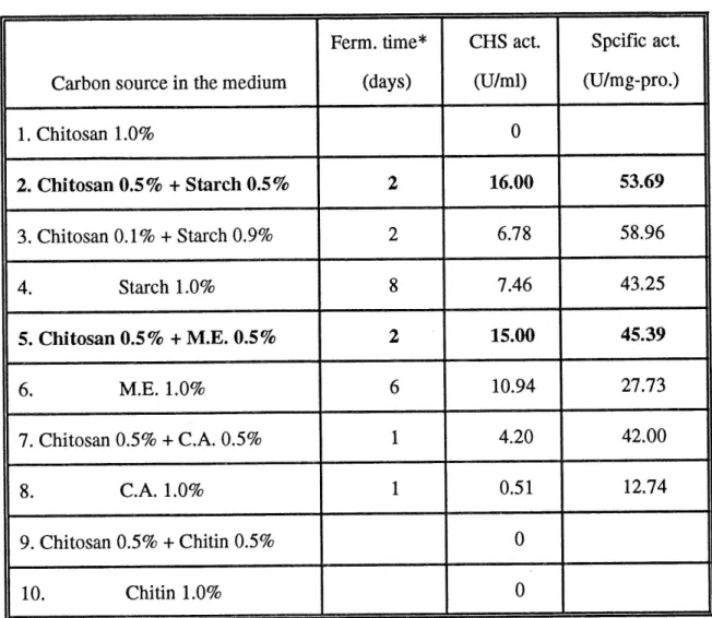
In order to find appropriate carbon source, chitosanase production was
compared by S. lividans TK24 (pRL226) in fermentation media containing
different carbon sources (or different combinations of carbon sources). The chitosanase activity and total proteins were measured in culture supernatant every
day (Table 4 and Fig. 5).
From this experiment, it was found that chitosanase production was higher in the presence than that in the absence of chitosan in the fermentation media. It is possible that chitosan could act as an inducer of chitosanase gene expression (see
section 4.3). However, when chitosan was used as the sole carbon source in the
medium, the chitosanase gene could not be expressed. Probably, S. lividans growth is not easy in medium with chitosan as sole carbon source because S. lividans needs synthesis of other kinds of proteins before expression of the
30
Table 4 Effect of carbon source on chitosanase productionCarbon source in the medium 1. Chitosan 1.0% 2. Chitosan 0.5% + Starch 0.5% 3. Chitosan 0.1% + Starch 0.9% 4. Starch 1.0% 5. Chitosan 0.5% + M.E. 0.5% 6. M.E. 1.0% 7. Chitosan 0.5% + C.A. 0.5% 8. C.A. 1.0% 9. Chitosan 0.5% + Chitin 0.5% 10. Chitin 1.0% Ferm. time* (days)
2
2
8
26
1
1
CHS act.(U/ml)
0
16.00 6.78 7.46 15.00 10.94 4.20 0.510
0
Spcific act (U/mg-pro.) 53.69 58.96 43.25 45.39 27.73 42.00 12.74*: The time taken for the chitosanase activity to reach the maximum.
**: The strain used was SMvidans TK24 (pRL226).
31
23456 Time (Days) Starch0.5%+Chitosan0.5% OStarchl.O% M.E.0.5%+Chitosan0.5% D M.E. 1.0% ^ C.A.0.5%+Chitosan0.5%32
chitosanase gene.
After comparison, chitosan plus starch or malt extract media were chosen as the appropriate carbon sources for chitosanase production. Table 4 and Fig. 5 showed that more activity was produced in the chitosan-starch medium; but the activity was more stable in chitosan-malt extract medium (after reaching a maximum, the activity decreased slower).
Step 2. Optimization of the ratio between chitosan and enrichment
Table 4 showed that the chitosanase production was dependent on the ratio between chitosan and starch. Consequently, some additional experiments were carried out to determine the optimal ratio between chitosan and enrichment in fermentation medium. According to our results, 1.5% of chitosan plus 0.5% of enrichment as carbon source was the optimal ratio for getting maxunal activity and
higher specific activity (Table 5). Along with the decrease of chitosan ratio, the
chitosanase production became lower. In 1.5% of chitosan plus 0.5% of malt extract fermentation medium, the chitosanase activity could reach up to 41.9 units per ml of fermentation liquid and the specific activity could reach up to 65.47 units
33
per mg of proteins (this value was very close to the purified chitosanase which hada specific activity of 66 units per mg of proteins) (Table 5).
Table 5 Optimization of the ratio between chitosan and enrichment.
No 1. 2. 3. 4. 5. Medium Chitosan% + M.E.% 1.5 0.5 1.0 1.0 0.5 1.5 0.2 1.8 0.0 2.0 Ferm. time* (days)
3
2
5
4
6
CHS act.(U/ml)
41.90 23.25 29.75 15.00 12.00 Specific act. (U/mg-pro.) 65.47 45.81 44.07 33.44 33.48* : The time taken for the chitosanase activity to reach the maximum.
** : The strain used was S.Uvidans TK24 (pRL226).
34
Step 3. Effect of nitrogen source on chitosanase productionIn order to find a cheap and efficient nitrogen source, we tested chitosanase activity in media with different nitrogen sources. We found that 0.2% of anunonium sulphate was the best one (Table 6). It was cheaper and more efficient (four times) than asparagine which used before. When urea was used as nitrogen source, the chitosanase production could not be detected, even if 5'. lividans could grow in this medium. Using 0.2% of ammonium sulphate as nitrogen source in the fermentation medium, the chitosanase activity was 39.7 units per ml of fermentation liquid with a specific activity of 39.08 units per mg of proteins.
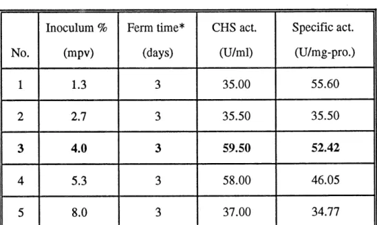
Step 4. Optimization of inoculum
We also analysed the effect of the quantitative inoculum on chitosanase production. To our surprise, the chitosanase activity greatly varied with the amount of inoculum (Table 7, Fig. 6). This difference was more than 20 units per ml of fermentation liquid. It indicated the importance of optimization of inoculum for chitosanase production. The results showed that addition of 4% mpv ofpre-culture to the fermentation medium was the optimal quantitative inoculum for chitosanase
35
Table 6 Effect of nitrogen source on chitosanase production.
Nitrogen
1. Asparagine 0.05%
2. Ammonium sulphate 0.2% 3. Urea 0.2%
4. Corn steep liquor 1.0%
Ferm. time* (days)
2
4
2
CHS act.(U/ml)
9.52 39.700
12.10 Specific act. (U/mg-pro.) 56.00 39.08 16.93*: The time taken for the chitosanase activity to reach the maximum.
**: The strain used was S. lividans TK24 (pRL226).
Table 7 Optimization of inoculum.
36
No.1
23
4
5
Inoculum % (mpv) 1.3 2.7 4.0 5.3 8.0 Ferm time* (days)3
3
3
3
3
CHS act.(U/ml)
35.00 35.50 59.50 58.00 37.00 Specific act. (U/mg-pro.) 55.60 35.50 52.42 46.05 34.77*: The time taken for the chitosanase activity to reach the maximum.
**: The strain used was S. lividans Tk24 (pRL226).
***: The ferm. media were 1.5% chitosan + 0.5% M. E., as well as 0.2% ammonium sulphate as nitrogen source.
37
I
:a
.^ >••I
<L> V rt 20
60 50 40 30 20 10I.-ll/
'7
"I !
U' !
F/ /
t.i'
1s/
?/
'!/ • /J_L
»<\ /^-\ ''•••// ^ '•:
/
/
-L^ .-.
\^ y' \
v<
^\
\ \
\ \
\ \
'.» '.1 '.\ . \ '. N ^J_L
\
\
\
\
\
\ \
\ \
\
Y\
^s:'Y ''<">. ss '•»...I 0 1 2345 Time (days) Inoculumare 1.3, 2.7, 4.0, 5.3, 8.0, % mpv38
production (Table 7, Fig. 6). With this amount of inoculum, chitosanase activity
could reach up to 59.5 units per ml of fermentation liquid with a specific activity
of 52.42 units per mg of proteins.
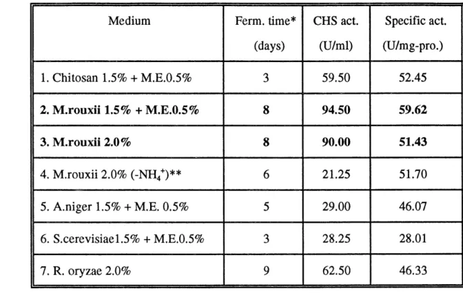
Step 5. Chitosanase production on natural substrates
As previously mentioned, chitosan is a natural component of fungal cell walls. Fungal cell walls should be an economical fermentation substrate for chitosanase production. We used four kinds of sterile microorganism cells and mycelia instead of chitosan as substrate for fermenting recombinant S. lividans. We found that all of them could act as substrates for chitosanase production. Specially, it was observed that the chitosanase activity could be maintained at high level for a few days when M. rouxii mycelium was used as substrate. The activity reached up to 94.5 units per ml of fermentation liquid and the specific activity was 59.62 units per mg of proteins after 8 days. The same phenomenon was observed with R. oryzae mycelium as substrate. The activity could reach up to 62.5 units per ml of fermentation liquid after 9 days. But in chitosan substrate, the chitosanase activity reached only 59.5 units per ml of fermentation liquid after 3 days, then decreased quickly (Fig. 7, Table 8). These showed that fungal
39
mycelium could be a convenient substrate for large scale production ofchitosanase.
Table 8 Chitosanase production on natural substrates.
Medium 1. Chitosan 1.5% + M.E.0.5% 2. M.rouxii 1.5% + M.E.0.5% 3. M.rourii 2.0% 4. M.rouxii 2.0% (-NH4+)** 5. A.niger 1.5% + M.E. 0.5% 6. S.cerevisiael.5% + M.E.0.5% 7. R. oryzae 2.0% Ferm. time*
(days)
3
8
8
6
5
3
9
CHS act.(U/ml)
59.50 94.50 90.00 21.25 29.00 28.25 62.50 Specific act. (U/mg-pro.) 52.45 59.62 51.43 51.70 46.07 28.01 46.33*: The time taken for the chitosanase activity to reach the maximum. **: There was not nitrogen source in MS solution.
40
1 234567 8 c Time (days) -X- Chitosanl.5%+M.E.0.5% • M.rouxiil.5%+M.E.0.5% 0 A.nigerl.5%+M.E.0.5% D R.oryzae 2.0% A S.cerevisiael .5%+M.E.0.5% 1041
Conclusion
We can conclude that the chitosanase production was very efficient in chitosan medium enriched with starch or malt extract, but the highest chitosanase activity level was obtained in medium containing M.rouxii mycelium instead of chitosan. The chitosanase activity could reach up to 94.5 units per ml of
fermentation liquid with the specific activity of 59.62 units per mg of proteins.
3.1.2 Comparison between different clones carrying the chitosanase gene
The chitosanase gene from Streptomyces N174 was cloned into the vector
pFD666 at Pstl site, as plasmid pRL226. By deleting some DNA sequences in
upstream or downstream of the chitosanase gene, several other derivatives were
constructed: pRL241, pRL228, pRL266 and pRL270 (Masson et al., 1993; see
also Fig. 3). Furthermore, to test the chitosanase gene expression driven by the synthetic consensus promoter (Denis and Brzezinski, 1991), we used a clone in which the native chitosanase gene promoter was replaced with the synthetic consensus promoter: the clone pFD666 chs+ (obtained from Dr. F. Denis, Institut
42
de Recherches Cliniques de Montreal). The restriction maps of these plasmids are shown in Fig. 3. The plasmids were transformed into S.lividans TK24. When we inoculated these recombinant strains in the optimal fermentation conditions
established for S. lividans TK24 (pRL226), the chitosanase production was lower than that in S. lividans TK24 (pRL226). Production was particularly low in S.
lividans TK24 (pFD666 chs-^-); The chitosanase activity was only 0.58 units per
ml of fermentation liquid (Table 9, Fig. 8).
We used the SDS-PAGE to analyze the extracelluar proteins produced by the different clones carrying the chitosanase gene. The results showed that the molecular weight of chitosanase was the same (29.5 kDa) for all clones (Fig. 9).
In fact, when the quantitative inoculum less than 1% (mpv) was used, without changing other fermentation conditions, the chitosanase gene in pRL270 could be expressed very well (Fig. 10). Using 0.67% and 4% inocula, chitosanase
activity of S. lividans TK24 (pRL226) was very similar (about 40 U/ml) , except
that the time course of enzyme production was different. But, using 0.67% and
4% ofinocula, the chitosanase activity of S. lividans TK24 (pRL270) showed very large difference (47 U/ml and 18 U/ml respectively).
Table 9 Comparison of chitosanase production between the different clones carrying the chitosanase gene
43
Recombinant Strain: S. Uvidans TK24 1. pRL226 2. pRL241 3. pRL228 4. pRL266 5. pRL270 6. pFD666c/i.y+ Ferm. time* (days)4
4
5
3
4
2
CHS act.(U/ml)
74.00 8.19 9.94 19.75 9.81 0.58 Specific act. (U/mg-pro.) 66.73 25.36 23.20 61.15 28.89 5.27* : The time taken for the chitosanase activity to reach the maximum. ** : The ferm. media contained 1.5% Chitosan + 0.5% Starch, as well as
44
123456
Time (days)
pRL226 pRL241 pRL228 pRL266 pRL270 pRL666 chs+
Fig. 8 Comparison of chitosanase production between the different clones carrying the chitosanase gene.
45
M.W. (kDa)
-CHS
1 2 3 4 56 7 89 Fig. 9 A SDS-PAGE analysis of cmde chitosanase production of
different clones in starch and chitosan-starch fermentation media. 1) Standard proteins.
Medium: 2-5) In starch ferm. medium;
6-9) In chitosan-starch fenn. medium.
Strain: 2,9) S. Uvidans TK24 (pRL270); 3,8) S. lividans TK24 (pRL266);
4,7) S. lividans TK24 (pRL241); 5,6) S. lividans TK24 (pRL266).
*) The samples were taken for the chitosanase activity to reach the maximum days.
46
M.W. (kDa)
CHS-Fig. 9 B SDS-PAGE analysis of crude chitosanase production of different clones in starch and chitosan-starch fermentation media.
1) Standard protems.
Medium: 2,4) In starch ferm. medium;
3,5) In chitosan-starch ferm. medium.
Stram: 2,3) S. lividans TK24 (pRL226); 4,5) S. lividans TK24 (pRL228),
*) The samples were taken for the chitosanase activity to reach
47
0 234
Time (days)
Fig. 10 Comparison of chitosanase production by S. lividans TK24 (pRL226) and (pRL270) with different inocula.
0 pRL226 and ® pRL270, inoculated at 0.67 % mpv; D pRL226 and H pRL270, inoculated at 4.0 % mpv.
48
3.1.3 Study on the protease in Streptomyces lividans TK24
When we studied the chitosanase activity, we found that the time course of chitosanase production was different on various substrates (Fig. 7). For example, the time courses of chitosanase production in starch, chitosan-starch and M. rouxii mycelium media, are shown on Fig. 11. In M. rouxii mycelium and starch substrates, the chitosanase activity showed slightly decreased after 4 days and returned to the original level after 5 days, remaining stable for a few days. But in chitosan-starch substrates, after 4 days, the activity decreased very quickly and could not be recovered. This probably indicated that concomitant with chitosanase production, a specific proteolytic enzyme for chitosanase was produced in fermentation medium (see section 4.5). To test this hypothesis, we also measured
the overall protease activity in fermentation liquid (Fig. 12). We found that there
are some relations between the tune courses of chitosanase activity and protease
activity.
We got similar results in the other experiments. Fig. 13 indicated that in each culture of pRL226 and pRL270, the increase of protease activity precisely corresponded to the moment of a serious decrease of chitosanase activity.
49
50
0
1
2345
Time (days)
0 Mjouxii 2.0 % AStarch 2.0 % D Chitosan 1.5 % + Starch 0.5 %
Fig. 11 Tune-course of chitosanase production in different
50
90
I-0 123456
Time (days)
0 M. rouxii 2.0 % A Starch 2.0 % D Chitosan 1.5 % + Starch 0.5 %
Fig. 12 Tune-course of protease activity in different
A) 50 40 h ^ .a p. >-> -1—> >
i
cd u^
30 h 20 h cos
2 0 10 It I, 'IL I ft j I / aJ_L
f I/° /
Jf\fl:f\
r/ » f I I \ \t I I I \ \ \ I 13 \ ^ J. s '^ \ 1 k ^ \ \ \ -Ih. t \ \ ^ \ \ \b
2345 Time (days) 2345 Time (days)Fig. 13 Time-course of chitosanase production (A) and protease activity (B) in chitosan-starch fermentation media of two S. lividans TK24 clones, each inoculated at two different densities.
0 pRL226 and ® pRL270, inoculated at 0.67% mpv; pRL226 and • pRL270, inoculated at 4.0 % mpv.
52
The N174 chitosanase is thus sensitive to degradation by one or several proteases produced by S. lividans TK24. This was also illustrated by the progressive decrease of intensity of the chitosanase band observed in CoomasieBlue stained gels (Fig. 14).
The proteolytic degradation of chitosanase activity was not inhibited by PMSF, but it was partially inhibited by 2.5 mM EDTA (data not shown).
For above reasons, we wanted to study chitosanase production in a host strain which is protease deficient or has very low protease activity. A possible candidate for such strain, S. lividans 10-164, which has been said to have low protease activity, was offered by Dr. Dieter KluepfeL
3.1.4. Study on Streptomyces lividans 10-164 carrying the chitosanase gene
S. lividans 10-164 is a B-l,4-glucan-glucanohydrolase (endocellulase) and xylanase-negative double mutant (Mondou et al., 1986). The plasmids pRL226 and pRL270 were transformed into the protoplasts of 5'. lividans 10-164 and two new
53
-CHS
8
10
Fig. 14 SDS-PAGE analysis of proteins produced by S. Uvidans TK24 (pRL226) in different fennentation media.
1, 2 and 3: samples were taken from the cultures of 2, 4 and 6 days m 1.5% chitosan + 0.5% M.E. medium;
4, 5, 6 and 7: samples were taken from the cultures of 2, 4, 6 and 8 days in 1.5% M. rouxii mycelium + 0.5% M.E. medium;
8, 9 and 10: samples were taken from the cultures of 2, 4 and 6 days
54
recombinant strains were named S. lividans 164 (pRL226) and S. lividans 10-164 (pRL270). The chitosanase gene in these new recombinant strains was expressed very well in glucosamine medium, but expressed at a lower level in chitosan-glucosamine and M. rouxii mycelium media (Fig. 15). From theseexperiments, we found that: (1) the S. lividans 10-164 (pRL226) and (pRL270)
could use the glucosamine as the carbon and nitrogen source for chitosanase
production; (2) the chitosanase activity is stable (after 2 days it reached a high level and this level could be kept for a few days) (Fig. 15); (3) the protease activity of S. lividans 10-164 was lower than that in S. lividans TK24 (Fig. 16).
and (4) no correlation was found between chitosanase and protease activities (Fig.
16).
These suggested that S. lividans 10-164 is really different from S. lividans
TK24 as the host for chitosanase gene expression and that further studies should be necessary to fully explore the potential of this new host.
12345678 Time (days)
® 10-164-226 in Glucosamine2% D in Chitosanl.5%+Glucosamine0.5% • 10-164-270 in Glucosamine2% 0 in Chitosanl.5%+Glucosamine0.5%
^< 10-164-226 in Mucor rouxii2%
Fig. 15 The time course of chitosanase production m fermentation liquid of recombinant strains S. lividans 10-164. Inoculum was 1.3 mpv/lOOml (MS solution without nitrogen source, the carbon sources are indicated on the figure).
^
345
Time (days)
345
Time (days) Fig. 16 The time course of chitosanase activity (A) and protease activity (B) in fermentation
liquid ofrecombinant strains S. lividans 10-164 (pRL226). Inoculum was
1.3 mpv/100ml (MS solution without nitrogen source, the carbon source is indicated on the figure).
0 Glucosamine 1.5% + Starch 0.5% D Glucosamine 1.5% + M.E. 0.5% A Glucosamine 1.5% + Glycerol 0.5%
LTt
57
3.2 Purification of chitosanase with polyacrylic acid
In order to concentrate and stabilize the chitosanase from the fermentation liquid in an economical way, we precipitated the chitosanase protein with PA at pH 4 (Table 10). This cmde enzyme extract could be stored for one year at -20°C
in 50% glycerol, retaining 50% of its activity. After precipitation, the overall proteolytic activity decreased to 5% of the initial level. This suggested that most of the proteolytic activity produced by S. lividans TK24 have acidic pl's.
Furthermore, the activity that degrades the chitosanase protein became undetectable
when the PA precipitate was dissolved in a buffer containing 2.5 mM EDTA (data
not shown).
3.3 Preparation of the active chitosan oligomers by chitosanase
hydrolysis
In order to find the optimal degree of hydrolysis of chitosan, chitosan solutions were incubated with different concentrations ofchitosanase. The products of hydrolysis of chitosan by the chitosanase were analyzed by thin layer
58
Table 10 Purification of chitosanase with polyacrylic acid.
Volume (ml)
Proteins (mg/ml)
Activity (U/ml)
Total protein (mg) Total act. (units) Specific act. (U/mg)
Yield (%)
Purification factor Ferm. liquid160
1.8880
301
12800
42
100
1
Precipitation of enzyme45
3.56160
160
7200
45
57
1.159
chromatography. The results, shown in Fig. 17, mdicated that the chitosanase first hydrolysed chitosan to produce longer chitooligosaccharides and then these oligosaccharides were hydrolysed to shorter chitooligosaccharides as the final products. This mechanism is similar to that described for the chitosanase fromNocardia orientalis (Sakai et al., 1991)
The results clearly showed that the enzyme hydrolysed chitosan to produce long chitooligosaccharides, such as chitopentaose, chitohexaose, chitoheptaose and longer oligomers, when 1 to 2 units of chitosanase were added to chitosanase
solution (Fig. 18). According to Kendra and Hadwiger (1984), we should select
one unit chitosanase per 250 mg of chitosan as the optimal concentration for preparation of the active chitosan oligomers, which should display maxunal activities in both fangal growth inhibition and induction of defense responses in
60
1
7 8 9 10 11 12
Fig. 17 Thm layer chromatography showmg the different degrees
of chitosan hydrolysis by cmde chitosanase.
1) Standard of GlcN; 2) Standard of GlcNAc; 3,4) Oligomers of GlcN;
5-12) 250 mg of chitosan was hydrolysed by different
units of chitosanase (0.5U, 1U, 1.5U, 2U, 3U, 4U, 5U and 6U). (see section 2.6.1).
61
GlcN
(GlcN)2
(GlcN)3
(GlcN)4
(GlcN)5
(GlcN)«
(GlcN)^
1
Fig. 18 Thin layer chromatography showing the optimal degree
of chitosan hydrolysis by cmde chitosanase.
1) Standard of GlcN;
2,3) Oligomers of GlcN;4) 250 mg of chitosan hydrolysed with 1U of CHS. 5) 250 mg of chitosan hydrolysed with 1.5U of CHS. 6) 250 mg of chitosan hydrolysed with 2U of CHS.
62
3.4 Antifungal properties of the chitosan oligomers and
induction of defensive responses in plants
After getting the high-molecular-weight chitosan oligomers, we pursued the possibility of their commercial use. We analysed their antifimgal properties and their ability to induce defense reactions m plants.
3.4.1 Inhibition of fungal growth
Fusarium oxysporum f. sp. radicis-lycopersici is a widespread soil borne pathogen that is responsible for severe tomato yield losses in green house soil and hydroponic systems (Jar vis, 1989; Jenkins and Averre, 1983). As with other vascular wilt pathogens (Beckman, 1987), F. oxysporum f. sp. radicis-lycopersici gains entry into roots through wounds and colonizes in the direction of xylem vessels, where it abundantly multiplies (Charest et al., 1984).
Our results showed that the chitosan oligomers prepared by our procedure significantly reduced the radial growth of F. oxysporum f.sp. radicis-lycopersici (Table 11). The marked effect was observed at a concentration of 2 mg/ml, which
63
indicated a growth inhibition of 60%. The fungal hyphae growth on plates
containing chitosan oligomers was dense and short, whereas the fungal growth on
control plates developed normally and actively (Fig. 19).
Table 11 Effect of chitosan oligomers on inhibition of radial
growth of Fusarium oxysporum f.sp. radicis-lycopersici.
PDA containing different
concentration of chitosan oligomers 0.00 mg/ml 0.25 mg/ml 0.50 mg/ml l.OOmg/ml 2.00 mg/ml
The diameter of radial
growth of 5-day-old F.oxysporum (mm) 72(d:l.l)b 67(±2.6)
60(±LO)
51(±1.2) 32(±1.5) Inhibition of radial growth(%)
0
7.6 18.2 31.8 60.6a. Fungi were inoculated with a 6-mm-diameter mycelial plug taken from the margin a 5-day-old Fusarium oxysporumf.sp.radiacis-lycopersici culture.
64
Fig. 19 Effect of chitosan oligomers on inhibition of radial
growth of Fusarium oxysporum f.sp. radicis-lycopersici.
(The PDA medium contains chitosan oligomers with different concentrations shown in figure).
65
3.4.2. Induction of pathogenesis-related proteins during tomato seedsgermination process
Our attention was focused on the biological function of chitosan oligomers present durmg seeds germination process for increasmg the plant resistance. Our experunent results showed that P.-R. protems were induced by our chitosan oligomers durmg tomato seeds germination process.
Chitmase, B-l,3,-glucanase and chitosanase were mduced durmg tomato
seeds germmation process by the chitosan oligomers, and the enzymatic activities were measured in 6-day-old tomato seedlmgs. The activities mcreased along with the mcrease of concentration of chitosan oligomers (Fig. 20-22), but a toxic effect for tomato seedlmgs was observed when the chitosan oligomers were used at a concentration higher than 500 /zg/ml. This phenomenon was also described with bean plants (Pospieszny and Atabekov, 1989). The optimal concentration of the chitosan oligomers for induction of P.-R. proteins synthesis was 200 jug/ml. In addition. Table 12, which shows the effect of chitosan oligomers on tomato seeds germmation efficiency, also mdicated that our chitosan oligomers were of benefit to tomato seeds germmation at a proper concentration (lower than 200 /zg/ml).
66
Higher concentrations (such as 500 /zg/ml) were toxic and inhibited germmation.Table 12 Effect of chitosan oligomers on tomato seeds germination.
Concentration of chitosan oligomers in 0.6% agar plates water 50{ig/ml lOO^ig/ml
200ng/ml
500ng/ml 1000»ig/ml Germination (%)(After incubation days)
r
0
0
0
0
0
0
2
56b52
47
41
0
0
3
84
85
90
80
68
47
4
85
88
90
88
78
68
5
85
89
90
90
80
77
6
85C89
90
90
80
80
a. The number is the day of incubation.
. Each seed germination experiment was done with 100 seeds. °. The producer also expected 85% germination of those seeds.
water 50 100 200 500
Concentration of the chitosan oligomers (ug/ml)
1000
Fig. 20 Induction of chitmase activity by the chitosan oligomers in tomato seedlings.
*: The datum has significant difference with control.
**: Each of the data is expressed per 100 seedlings and is an average of three experiments.
<?»
60 50 gMO
I
>> •r so •s u cd uI
§20
Q
10 water 50 100 200 500Concentration of the chitosan oligomers (ug/ml)
1000
Fig. 21 Induction of chitosanase activity by the chitosan oligomers in tomato seedlings. *: The datum has significant difference with control.
**: Each of the data is expressed per 100 seedlings and is an average of three experiments.
0-\ 00
m
water 50 100 200 500
Concentration of the chitosan oligomers (ug/ml)
1000
Fig. 22 Induction of B-l,3-glucanase activity by the chitosan oligomers m tomato seedlings. *: The datum has significant difference with control.
**: Each of the data is expressed per 100 seedlings and is an average of three experiments.
0^ <b
70
3.4.3 Induction of pathogenesis-related proteins in mature plants
In order to test the effect of our chitosan oligomers on the induction of P.-R. protems synthesis m mature plants, we sprayed the chitosan oligomers on a mature plant leaf. After waiting for the induction of proteins synthesis and systemic movement from treated to untreated leaves (about 5 days), three plant leaves were collected from each plant (one from plant upper part, one from middle part, another from lower part). The intercellular fluid of leaves was collected and the enzymatic activities of P.-R. protems were measured (Table 13-14).
The experiment showed that the P.-R. protems were mduced by the chitosan oligomers m tobacco leaves. The enzymatic activities of chitosanase, chitmase and B-l,3-glucanase of plant fluids obtamed from tobacco leaves treated with the chitosan oligomers at the concentration of 10 mg/ml were significantly different from the control plants (Table 13). We repeated this experunent three tunes and got similar results. These indicated that the chitosan oligomers were efficient to