ii
Université de Sherbrooke
Radiolysis of water induced by the recoil ions of the 10B(n,)7Li nuclear reaction: Calculation of the yields of primary species up to 350 °C and in situ generation of
ultrafast transient “acid spikes” along the radiation tracks Par
Muhammad Mainul ISLAM
Département de médecine nucléaire et radiobiologie
Mémoire présenté à la Faculté de médecine et des sciences de la santé en vue de l’obtention du diplôme de maître ès sciences (M.Sc.) en "sciences des radiations et imagerie biomédicale"
Sherbrooke, Québec, Canada
April 2018
Jury
Pr Éric Rousseau Examinateur, Département d'obstétrique-gynécologie, Faculté de médecine et des sciences de la santé
Pr Johannes Van Lier Examinateur, Département de médecine nucléaire et radiobiologie, Faculté de médecine et des sciences de la santé
Pr Jean-Paul Jay-Gerin Directeur de recherche, Département de médecine nucléaire et radiobiologie, Faculté de médecine et des sciences de la santé
iii RÉSUMÉ
Radiolyse de l'eau induite par les ions de recul de la réaction nucléaire 10B(n,α)7Li:
calcul des rendements des espèces primaires jusqu'à 350 °C et génération in situ de pics d’acidité ultra-rapides transitoires le long des trajectoires de rayonnement
Muhammad Mainul ISLAM
Département de médecine nucléaire et radiobiologie
Mémoire présenté à la Faculté de médecine et des sciences de la santé en vue de l’obtention du diplôme de maître ès sciences (M.Sc.) en "sciences des radiations et imagerie biomédicale", Faculté
de médecine et des sciences de la santé, Université de Sherbrooke, Sherbrooke, Québec, Canada J1H 5N4
La réaction nucléaire 10B(n,α)7Li de capture de neutrons par le bore est très importante pour
l'industrie nucléaire ainsi que pour la radiobiologie, car la thérapie par capture neutronique par le bore est utilisée dans des radiothérapies biochimiques ciblées pour plusieurs traitements anticancéreux. L'acide borique enrichi en 10B est utilisé dans les réacteurs nucléaires pour contrôler
le flux de neutrons et la réactivité dans le cœur. Cependant, les noyaux de recul (particules α de 1,47 MeV et ions 7Li3+ de 0,84 MeV) résultant de la réaction 10B(n,α)7Li agissent comme sources
de rayonnement à transfert d'énergie linéaire élevé (TEL) compliquant ainsi le processus radiolytique à l'intérieur du réacteur. La simulation Monte-Carlo est utilisée dans ce travail pour prédire les rendements (valeurs G) des radicaux et des produits moléculaires dus à la radiolyse de l'eau par la réaction 10B(n,α)7Li en fonction de la température de 25 à 350 °C. Nos calculs montrent
des rendements plus bas en radicaux libres et plus élevés en produits moléculaires en comparaison avec un rayonnement de faible TEL (e.g., rayons γ de 60Co). Les résultats de nos simulations
concordent bien avec les estimations expérimentales existantes à 20 et 289 °C. Cependant, l'inflexion prédite par certains auteurs dans les rendements moléculaires H2 et H2O2 au-dessus de
~150 °C ne peut être confirmée dans la mesure où l’on adopte la constante de vitesse de la réaction (e−
aq + e−aq) en solution neutre ou légèrement acide. De plus, dans cette étude, nous avons
également calculé la concentration de H3O+ formé in situ le long des trajectoires du rayonnement en
considérant un « modèle de trajectoire cylindrique » pour un rayonnement à fort TEL. Pour ces ions, le pH le long des trajectoires est proche de 0 jusqu'à ~100 ps, puis revient progressivement à un pH neutre (7) à ~0,1 ms. Cependant, dans l'eau cellulaire, le "pic d'acidité" demeure plus longtemps à cause de la faible mobilité du proton dans ce milieu, ce qui soulève plusieurs questions en relation avec la boroneutrothérapie et, plus généralement, l’hadronthérapie.
Mots-clés: Radiolyse de l'eau, réaction de capture de neutrons par le bore, ions hélium et lithium de recul, transfert d'énergie linéaire (TEL), température, simulations Monte-Carlo, rendements radicalaires et moléculaires, pH, effet de pic acide dans les trajectoires en fonction du temps, réacteurs nucléaires, boroneutrothérapie, hadronthérapie.
iv ABSTRACT
Radiolysis of water induced by the recoil ions of the 10B(n,)7Li nuclear reaction: Calculation of the yields of primary species up to 350 °C and in situ generation of
ultrafast transient “acid spikes” along the radiation tracks Muhammad Mainul ISLAM
Département de médecine nucléaire et radiobiologie
Thesis presented at the Faculty of Medicine and Health Sciences in order to obtain the Master of Sciences (M.Sc.) degree in “Radiation Sciences and Biomedical Imaging”, Faculty of Medicine and
Health Sciences, Université de Sherbrooke, Sherbrooke, Québec, Canada J1H 5N4
The 10B(n,α)7Li nuclear reaction is very important for the nuclear industry as well as in
radiobiology, as boron neutron capture therapy (BNCT) is used in biochemically targeted radiotherapies for several malignant cancer treatments. Boric acid enriched with 10B is used
in nuclear reactors to control the neutron flux and the reactivity in the core. However, recoil nuclei (1.47 MeV -particles and 0.84 MeV 7Li3+ ions) resulting from this reaction
act as sources of high linear energy transfer (LET) radiation, thereby complicating the radiolytic processes inside the reactor. Monte Carlo simulations are used to predict the yields (G-values) of the radicals and molecular products due to the radiolysis of water by the 10B(n,)7Li recoil ions as a function of temperature from 25 to 350 °C. Our computed
yields show lower yields of free radicals and higher yields of molecular products in comparison with low-LET radiation (60Co γ-rays). Our simulation results agree well with
existing experimental estimates at 20 and 289 °C. However, the non-monotonic downward inflection of the yields of molecular H2 and H2O2 above ~150 °C can be confirmed if we
could get the rate constant of the (e−
aq + e−aq) reaction measured under neutral or slightly
acidic conditions. Moreover, in this study, we also calculated the in situ concentrations of H3O+ and the corresponding pH values along the radiation tracks using a “cylindrical track
model” characteristic of high-LET radiation. For both considered recoil ions, the pH along the tracks is near zero up to ~100 ps, after which it progressively returns to neutrality at ~0.1 ms. However, in cellular water, this “acid spike” retains for a longer period of time due to the slower diffusion of free protons in this medium, a result that may have several implications in BNCT and, more generally, in the overall field of hadrontherapy.
Keywords: Water radiolysis, boron neutron capture reaction, He and Li recoil ions, linear energy transfer (LET), temperature, Monte Carlo simulations, radical and molecular yields, pH, “acid spike” effect along the radiation tracks as a function of time, nuclear reactors, BNCT, hadrontherapy.
v TABLE OF CONTENTS Résumé...iii Abstract...iv Table of contents...v List of figures...vii List of table………..xiv List of abbreviation...xv 1. Introduction………...………..1
1.1 Energy deposition events and interaction of ionizing radiation…...………7
1.1.1 Linear energy transfer (LET) and interaction ………...…………9
cross section for heavy charged particles 1.2 Radiolysis of water………..……..11
1.2.1 Track structure in radiation chemistry of water………...……12
1.2.1.1 Low LET radiation and track structure………...12
1.2.1.2 High LET radiation and track structure………..….15
1.2.2 Time scale of events and formation of primary products………17
in neutral water radiolysis 1.3 Boron Neutron Capture nuclear reaction...24
1.3.1 Boron Neutron Capture nuclear reaction ………....24
in nuclear industry 1.3.2 Boron Neutron Capture nuclear reaction………..25
in cancer treatment. 1.4 Effect of temperature on water radiolysis due to the ………....26
10B(n,α)7Li nuclear reaction. 1.5 Formation of H3O+ in spurs and tracks...28
1.6 Research objective...31
2. Monte Carlo simulations...32
2.1 The IONLYS code...34
vi
3. Stopping and range of ions in matter (SRIM) ………...42 4. Article no. 1...44
Muhammad Mainul Islam, Phantira Lertnaisat, Jintana Meesungnoen,
Sunuchakan Sanguanmith,Jean-Paul Jay-Gerin, Yosuke Katsumura, Satoru Mukai, Ryuji Umehara, Yuichi Shimizu and Masashi Suzuki
“Monte Carlo track chemistry simulations of the radiolysis of water induced by the recoil ions of the 10B(n,α)7Li nuclear reaction. 1. Calculation of the yields of
primary species up to 350 °C”
Royal Society of Chemistry Advances, 2017, vol. 7, pp. 10782-10790.
5. Article no. 2………...66
Muhammad Mainul Islam, Vanaja Kanike, Jintana Meesungnoen, Phantira
Lertnaisat, Yosuke Katsumura, Jean-Paul Jay-Gerin
“In situ generation of ultrafast transient “acid spikes” in the 10B(n,α)7Li radiolysis
of water”
Chemical Physics Letters, 2018, vol. 693, pp. 210-215.
6. Discussion……….93 6.1 Yield of H2 and importance of the bimolecular self-reaction of eaq…………..93
6.2 Yield of H2O2……….……….97
6.3 Yields of radicals………….…….………...98 6.4 Local acidity inside of the regions encased by radiation tracks……..…..…….99 7. Conclusion………...………...106 8. References………...109 9. Appendices……….129
vii LIST OF FIGURES
Chapter 1: Introduction
Figure 1.1 Simplest representation of the mode of action ………...4 (direct and indirect) of radiation on a cell. Absorption of ionizing radiation may damage the cell directly attacking the DNA or producing the radiolysis products of water or perturbing the function of the mitochondria. From AZZAM et al. (2012).
Figure 1.2 Scheme of the nuclear reaction resulting from the ……….……5 low-energy (< 0.5 eV) thermal neutron capture by a 10B atom. After
absorption, 94% of the reactions leave the 7Li ion in its first excited state
(7Li*) which rapidly de-excites to the ground state by releasing a 478-keV
γ-ray. For the remaining 6% of the reactions, the 7Li ion is left directly in its
ground state resulting in the emission of a 1.78 MeV α-particle and a 1.02 MeV 7Li ion. Note that the 4He and 7Li recoil ions are in opposite directions
(i.e., at a 180° angle), away from the site of the compound nucleus, and hence they form one straight track.
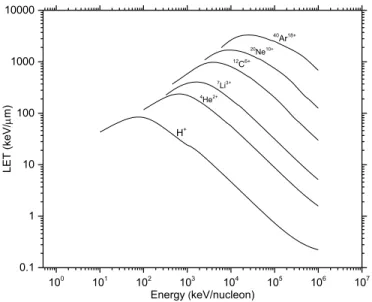
Figure 1.3 LET of some heavy ions as a function of energy………..11 in liquid water using SRIM program (WATT, 1996).
Figure 1.4 Track structure entities classified as spurs ………..14 (spherical entities, up to 100 eV), blobs (spherical or ellipsoidal, 100-500 eV) and short tracks (cylindrical, 500 eV- 5 keV) for a primary high energy electron (not to scale) (BURTON, 1969).
Figure 1.5 Monte Carlo track simulation of 300 MeV protons………..15 (a) and 150 keV protons (b) (LET~ 0.3 and 70 keV/µm) incident on liquid water at 25 °C (KANIKA, 2015a).
Figure 1.6 Primary energy-loss events in high-LET radiation………..15 tracks (FERRADINI, 1979)
viii
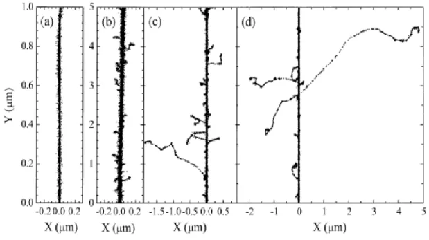
Figure 1.7 Projections over the XY-plane of track segments calculated ………16 (at ~10-13 s) for (a) H+ (0.15 MeV), (b) 4He2+ (1.75 MeV/nucleon), (c) 12C6+
(25.5 MeV/nucleon), and (d) 20Ne10+ (97.5 MeV/nucleon) impacting ions.
Ions are generated at the origin and along the Y axis in liquid water under identical LET conditions (~70 keV/μm). Dots represent the energy deposited at points where an interaction occurred. From MUROYA et al. (2006), with permission.
Figure 1.8 Time scale of events that occur in the low-LET ………18 radiolysis of neutral, deaerated water (MEESUNGNOEN, 2007; MEESUNGNOEN and JAY-GERIN, 2010). As a guide to the eyes, we use different colors in the figure in order to contrast the individual processes occurring during the radiolysis of water.
Figure 1.9 Graphical representation of the Boron Neutron Capture Therapy…………26
Chapter 2: Monte Carlo simulations
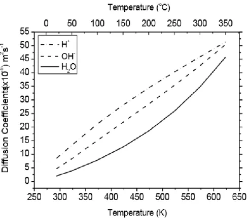
Figure 2.1 Diffusion coefficients (D) for the various track species ………..40 involved in our simulations (ELLIOT and BARTELS, 2009).
Chapter 4 – Article No. 1
Figure 1 Scheme of the nuclear reaction resulting from ………...………..49 the low-energy (<0.5eV) thermal neutron capture by a 10B atom. After
absorption, 94% of the reactions leave the 7Li ion in its first excited state
(7Li*) which rapidly de-excites to the ground state by releasing a 478 keV
γ-ray.For the remaining 6% of the reactions, the 7Li ion is left directly in its
ground state resulting in the emission of a 1.78 MeV α-particle and a 1.02 MeV 7Li ion. Note that the 4He and 7Li recoil ions are in opposite directions
(i.e., at a 180o angle), away from the site of the compound nucleus, and
hence they form one straight track.
ix
helium and lithium ions of the 10B(n,α)7Li reaction into liquid water at room
temperature: (a) simulated ion trajectories; (b) and (c) variations of the energy and LET of the two ions as a function of penetration depth, respectively (the points selected in this study are indicated by arrows). Total ions calculated: 1000.
Figure 3 G-values (in molecule per 100 eV) for the ………...55 10B(n,α)7Li radiolysis of pure, deaerated liquid water as a function of
temperature in the range of 25–350 oC: (a) G(e−
aq); (b) G(•OH); (c) G(•H);
(d) G(H2O2); and (e) G(H2). Our simulated results, obtained at 10-7 and 10-6
s, are shown as solid, blue and red lines, respectively. Symbols are the water decomposition yields induced by the 10B(n,α)7Li reaction estimated by
Cohen (ref. 15) at 20 oC (based on the approximate relationship between
LET and G-values given in Fig. 5.3 of Allen (ref. 43), using an average initial LET of 240 eV nm-1)(B) and by Christensen (ref. 19) at 289 oC (C).
The primary (or “escape”) yields for the low-LET (~0.3 eV nm-1) radiolysis
of water (ref. 29) obtained using our previously calculated spur lifetimes between 25–350 oC (ref. 44) are also given (black dashed lines) for
comparison purposes. Note that all yield curves shown in this figure were obtained under exactly the same conditions as in ref. 29 as far as the temperature dependences of the different parameters intervening in the early physicochemical stage (e.g., the electron thermalization distance called rth in
ref. 29) and in the subsequent chemical stage [e.g., the (e−
aq + e−aq) reaction
rate constant, represented by the non-Arrhenius black dashed line k = ka in
Fig. 4(a)] of the radiolysis are concerned.
Figure 4 (a) Rate constant for the self-reaction of two hydrated……….57 electrons as a function of temperature (ref. 49). The black dashed line (denoted ka) shows the (e−aq + e−aq) reaction rate constant measured under
alkaline conditions (ref. 20). The symbols (■) are experimental data. The red solid line (denoted kb) shows the (e−aq + e−aq) reaction rate constant obtained
x
(b) and (c) The red solid lines show our Monte Carlo simulation results for
G(H2) and G(H2O2) (in molecule per 100 eV), at 10-6 s, as a function of
temperature, when kb was used. A comparison is made with the
corresponding yields of H2 and H2O2 obtained when ka was used
[represented here by the black dashed lines, which are the same as the red solid lines in Fig. 3(e) and (d)]. The symbols (○) (ref. 15) and (●) (ref. 19) are the same as in Fig. 3(e) and (d).
Figure 5 G-values (in molecule per 100 eV) for the 10B(n,α)7Li ………...….59
radiolysis of deaerated 0.4 M H2SO4 aqueous solutions (pH 0.46 at 25 oC) as
a function of temperature in the range of 25–350 oC. Note that, at this high
concentration of H2SO4, the H+ ions very rapidly (<10-9 s) scavenge most, if
not all, of the e−
aq radicals in the tracks to form •H atoms (ref. 37). Note also
that, in our simulations, the direct action of ionizing radiation on the sulfuric acid anions (mainly HSO4−) has been neglected. The solid curves represent
the results of our Monte Carlo simulations for (a) G(•H), (b) G(•OH), (c) G(H2), and (d) G(H2O2) at 10-6 s after the initial energy deposition. The
yields of primary species induced by the 10B(n,α)7Li reaction measured by
Barr and Schuler (ref. 23) in acidic solutions at 25 oC are given by (●). The
primary (or “escape”) yields for the low-LET (~0.3 eV nm-1) radiolysis of
0.4 M H2SO4 aqueous solutions (ref. 53) obtained from our previously
calculated spur lifetimes between 25–350 oC (ref. 44) are also shown
(dashed lines) for the sake of comparison. Finally, in all calculations, the reaction of the •H atom with water: •H +H
2O H2 + •OH was assumed to
follow an Arrhenius temperature dependence over the 25–350 °C range studied, with a rate constant of 4.6 × 10⁻⁵ M-1s-1 at 25 oC and 104 M-1 s-1 at
300 oC, in agreement with recent muon spin spectroscopy experiments using
muon as an analogue of a hydrogen atom (ref. 54).
Figure 6 Yields of H2 (panel a) and H2O2 (panel b) ………...60
(in molecule per 100 eV) formed during the 10B(n,α)7Li radiolysis of
xi
the range of 25–350 oC. The black dashed lines show our Monte Carlo
simulation results for G(H2) and G(H2O2) at 10-6 s when the (e−aq + e−aq)
reaction rate constant k2 = ka [see Fig. 4(a)] was used (note that these curves
are the same as the lines in Fig. 5(c) and (d). A comparison is made with the corresponding yields of H2 and H2O2 obtained when k2 = kb [see Fig. 4(a)]
was used (represented by the red solid lines).
Chapter 5 – Article No. 2
Figure 1 Schematic of the nuclear reaction resulting from ……...………...86 the low-energy (< 0.5 eV) neutron capture by a 10B atom. After absorption, an
excited 11B is formed that almost immediately (~10-12 s) undergoes a fission
reaction producing, in 94% of the cases, two high-LET heavy ions, 4He2+
(-particle) and 7Li3+, and a low-LET -ray (see [10]). Note that the 4He and 7Li
recoil ions are emitted in opposite directions (i.e., at a 180° angle), away from the site of the compound nucleus, and hence they form one straight track.
Figure 2 SRIM simulation of the penetration of 1000 recoil ………..87 1.47-MeV helium and 0.84-MeV lithium nuclei of the 10B(n,)7Li reaction into
liquid water at 25 °C. A major contribution to the observed straggling comes from the changes in the charge state of the respective ions as they go into and through the water.
Figure 3 Time evolution of G(H3O+) (in molecule/100 eV) for ……….88
the radiolysis of pure, deaerated liquid water by the recoil of -particles and lithium ions (“dose average” LET of ~196 and 216 eV/nm, respectively) from the 10B(n,)7Li nuclear reaction at 25 °C from ~1 ps to 1ms. The red solid line
shows the hydrogen ion yield values obtained from our Monte Carlo simulations (see text). Our computed yields of e
-aq, •OH, OH- and H• are shown
as blue, grey, red, and green dashed lines, respectively.
Figure 4 Time dependence of the extents G(H3O+) (in molecule per 100 eV) ……..89
xii
from our Monte Carlo simulations of the 10B(n,)7Li radiolysis of pure,
deaerated water at 25 °C, in the interval of ~1 ps to 1 ms.
Figure 5 Simulated track history (at ~10-13 s, projected into ………..90 the XY plane of figure) of a 0.3 MeV helium ion (LET ~ 196 eV/nm) (A) and of a 0.4 MeV lithium ion (LET ~ 216 eV/nm) (B) traversing through liquid water at 25 °C. The irradiating ion is generated at the origin and starts traveling along the Y-axis. Dots represent the energy deposited at points where an interaction occurred. The tracks can be described as two coaxial cylindrical volumes centered on the path of the ions. The inner cylindrical volume (i.e., the region adjacent to the trajectory) is the track “core” with radius rc. Surrounding the
core is a much larger region called the “penumbra” where all of the energy is deposited by energetic secondary electrons (γ-rays) created in knock-on collisions by the ion.
Figure 6 Variation of pH with time calculated for pure, deaerated ……….91 liquid water at 25 °C and in the interval of ~1 ps to 1 ms, for irradiating 0.3 MeV helium (Zeff = 1.6, LET ~ 196 eV/nm) and 0.4 MeV lithium (Zeff = 1.7,
LET ~ 216 eV/nm) using the axially homogeneous cylindrical track model (characteristic of high-LET radiation) with rc = 2 nm for both ions (see text).
The pH values reported here are simply the average of the pH obtained for the two ions. For the sake of comparison, the dashed line shows the time evolution of pH in an isolated (spherical) “spur” (characteristic of low-LET radiation) [20] as calculated previously for 300 MeV incident protons (which mimic 60Co
/fast electron irradiation; LET ~ 0.3 eV/nm) using an initial spur radius of 11.7 nm (see Fig. 4 of [20]).
Figure 7 Variation of pH with time calculated for deaerated ……….92 bulk cellular water at 25 °C and in the interval of ~1 ps to 1 µs, under the same irradiation conditions as in Fig. 6, using the axially homogeneous cylindrical track model with rc = 2 nm for both He and Li ions (solid line). Simulations
were performed using an intracellular proton mobility 100 times lower than that in free liquid water (note that, in the calculations, the diffusion coefficients of
xiii
all other species were also lowered by a factor of 100 relative to their liquid water values). Comparison is made with the pH values calculated for irradiated free liquid water (as shown in Fig. 6) (dashed line).
Chapter 6: Discussion
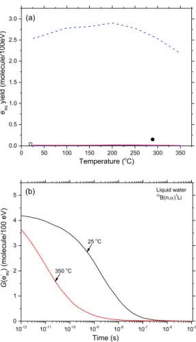
Figure 6.1 (a) Yields of e−aq due to the 10B(n,α)7Li radiolysis of pure, ……..………94 deaerated water at 10-6 and 10-7 s, shown as the red and blue solid lines,
respectively. The dashed blue line shows the “escape” yield of e−aq for low-LET irradiation (γ-rays or fast electrons). (b) Comparison of the decay of e−aq at 25 °C (black line) and 350 °C (red line) as a function of time.
Figure 6.2 Yields of the different chemical species ………98 (in molecule/100 eV) for the radiolysis of pure, deaerated liquid water by the recoil of α-particles and lithium ions from the 10B(n,α)7Li nuclear reaction at
25 °C from ~1 ps to 1ms. Our computed yields of e
-aq, •OH, and H• are
shown as blue, grey, and green dashed lines, respectively.
Figure 6.3 Qualitative representation of the lateral distribution ………...105 of the radiolytic products formed after a heavy -particle has traversed water (right side of the figure). The separation of the positive (core of the track) and negative (at some distance from the track) charges is clearly visualized (LEA, 1946; MORRISON, 1950)
xiv LIST OF TABLES
Table 1 Main spur/track reactions and rate constants (k) for the………...41 radiolysis of pure liquid water at 25 ºC (from MEESUNGNOEN, 2007). Some values of k have been updated by using the most recently available data of ELLIOT and BARTELS (2009).
xv
LIST OF ABBREVIATION
10B(n,α)7Li Boron Neutron Capture Nuclear (BNCT) reaction
D Diffusion coefficient
DEA Dissociative electron attachment
DNA Deoxyribonucleic acid
e
aq Hydrated electron
eV Electron-volt
GX or g(X) Primary yield of the radiolytic species X G(X) Experimental yield of the final product X Gy/s Gray/second (dose rate)
IRT Independent reaction times
k Reaction rate constant keV Kilo-electron-Volts LET Linear energy transfer
MC Monte Carlo
MeV Mega-electron-Volts
µm Micrometer
µs Microsecond
ps Picosecond
SOD Superoxide dismutase
1 1. INTRODUCTION
Radiation chemistry is a comprehensive field of study including many areas of research. The absorption of ionizing radiation in matter causes chemical changes in the matter, which is known as radiation chemistry. Radiation chemistry has been originated with the discovery of X-ray in 1895. In the following year, Becquerel discovered the radioactivity of uranium. Pierre Curie and Marie Curie conducted their research on different mineral salts to investigate the ‘radioactivity’ of the materials. However, the term ‘radioactivity’ had been used by them first. They discovered polonium and radium, which are also highly radioactive materials (see, for example: FERRADINI and BENSASSON, 1989; SPINKS and WOODS, 1990; ZIMBRICK, 2002). In 1901, Curie and Debierne observed the evolution of hydrogen and oxygen from water and from the solution of radium salts (CURIE and DEBIERNE, 1901; DEBIERNE, 1914). This study was the first trigger on the radiolysis of water by ionizing radiation. Sir William Ramsay and Frederick Soddy did a quantitative study of some simple radon-induced reactions (RAMSAY and SODDY, 1903). BRAGG (1907) analyzed the data obtained from Ramsay and Soddy and reported that the number of the water molecules decomposed was nearly equal to the number of ions produced in air by the radon employed. In 1912, Samuel Lind modeled several studies to determine the relation between the number of ion-pair formed by α-rays in a gas and the number of chemicals subjected to chemical changes (LIND, 1912, 1921).
By the 1930’s, the advent of powerful X-ray equipment for medical and industrial purposes instigated the research in this field. X-ray is more penetrating and better suited to the irradiation of bulk liquid and solid samples. The development of X-ray equipment and use of radiation in the medical field also increased the interest of research in the effect of X-rays and other types of radiation on aqueous solution including biological materials. Hugo Fricke, another true pioneer in the field of radiation chemistry, published several systematic studies on the effect of radiation on aqueous solutions. A solution of ferrous ions in 0.4 M sulfuric acid at this time has now become a routine chemical means to measure the energy absorption in irradiated systems, constituting the so-called “Fricke dosimeter” (SPINKS and WOODS, 1990; FRICKE and HART, 1966).
2
A great impetus on research in radiochemistry occurred in 1942 with the advent of nuclear energy. The research asserts on the two important practical problems, namely, the study and prevention of undesired radiation effects or radiation damage, and the utilization of the tremendous amounts of radiation energy for beneficial purposes. The former includes the study of the damage of materials due to the radiation used in reactor construction and in the processing of radioactive fuels as well as the development of radiation resistant materials. Another extremely important area of research is the effect of radiation on living systems and utilization of radiation for medical treatment. To use the radiation energy for humans, it is very important to understand physical and chemical processes of the ionizing radiation in a material. Though the fundamental ionization processes are similar in all system, the properties of the medium such as phase, polarity, and composition can greatly affect the chemistry. In all systems, it is very important to understand what chemical species are formed, what the internal energies are, how they are dispersed spatially, and what reactions can occur (JONAH, 1995). The understanding of the simpler chemical systems is necessary for unraveling the complexity of events incurred by irradiation of living systems.
A large fraction of the radiation chemistry studies has been concerned with the studies of water and aqueous solutions because of the unique importance of water in biological systems. Moreover, it also has a large number of practical applications, for instance, radiotherapy and diagnostic radiology, environmental management of radioactive waste materials, nuclear power generation and radiation effect in space (LAVERNE, 2004;
MEESUNGNOEN and JAY-GERIN, 2011).
All biological systems are very sensitive to ionizing radiation. Since living cells and tissue consist mainly of water (~70-80% by weight), the knowledge of the radiation chemistry of aqueous solutions is very critical to our understanding of the early stages in the complicated chain of radiobiological events that follows the passage of radiation. Therefore, we have to understand and clearly model the interaction of ionizing radiation with water and aqueous solutions and analyze the subsequent effect of the species produced due to ionizing radiation. Not only for biological aspect analysis but also to understand the
3
corrosion of materials and safety of the nuclear power reactors, such kind of studies are very relevant.
The absorption of ionizing radiation by living cells can cause damage of different molecular structures of biological relevance, can disrupt the biochemical processes in the cell and can produce new reactive chemical species that may damage nucleic acids, proteins and lipids. The biological cell damage by ionizing radiation can be done by ‘direct’ or ‘indirect’ effect. In case of the direct effect, the biological species absorb the energy from the ionizing radiation directly, which disrupts their initial constituents and functions. In the indirect effect, however, the water molecules in the cell absorb the ionizing radiation and produce several radiolytic products such e−
aq, H•, •OH, HO2•/O2•−,
H2, H2O2, O2, etc., which subsequently might act as triggers of signalling or other
damaging effects (AZZAM et al., 2012; MUROYA et al., 2006; NATHAN, 2003; FORMAN et al., 2004; VEAL et al., 2007).
Deoxyribonucleic acid (DNA) is a large molecule with a double helix structure which carries the genetic instructions and biological information of the living cells. DNA is a highly charged poly-anion that is hydrated with water molecules. Exposure to ionizing radiation causes a plethora of DNA damage, which is responsible for genomic instability, potential tumorigenesis, mutagenicity and finally, cell death. Different types of DNA damages and repair mechanisms have been reported and studied over the years. The main types of DNA damages are base damage, apyrimidinic/apurinic (AP) site, single-strand break (SSB), double-strand break (DSB), tandem lesions and various clustered lesions (von SONNTAG, 2006). It is considered that the complex lesions or multiply damaged sites (MDS) composed of more than one lesion (base damage, base loss or strand break) within one or two helical turns of DNA, have significant biological effects, including mutation and cytotoxicity (CHAPMAN, 1980; NIKJOO et al., 1997; GOODHEAD, 1994; WARD, 1994). The complexity and nature of DNA damages induced by ionizing radiation largely depend on the cellular phenotype, type of ionizing radiation, radiation quality or “linear energy transfer” (LET) (i.e., stopping power, -dE/dx), dose, and dose rate. For example, exposure to high-LET radiations (e.g., -particles, highly charged and high energy particles), the yield of locally multiply damaged (LMDS) sites in DNA is greatly increased
4
(KRYSTON, 2011; WARD, 1985; GEORGAKILAS, 2011). Moreover, radiation-induced generation of reactive oxygen (ROS) and nitrogen species (RNS) may spread from targeted cells to non-targeted bystander cells through intercellular communication mechanism. More importantly, mitochondria are the richest source of ROS. The premature leakage of electrons reduces O2 to produce superoxide radicals (O2•). Radiation-induced ROS/RNS
production by mitochondria plays multiple roles in signaling cascades, mediates apoptosis, mutation of mitochondrial DNA, autophagy and propagation of non-targeted responses (PETKAU, 1987; HEI et al., 2011, PRISE, 2009; WERNER and WERB, 2002; MALAKHOVA et al., 2005; AZZAM et al., 2012). The research in radiobiology is very important to improve treatment and imaging procedures. The research in this domain also opens a new horizon to early detect cancer cells and specifically treat them.
Fig. 1.1: Simplest representation of the mode of action (direct and indirect) of radiation on a cell. Absorption of ionizing radiation may damage the cell directly attacking the DNA or producing the radiolysis products of water or perturbing the function of the mitochondria.FromAZZAM et al. (2012).
Boron-10 is one of the stable isotopes of boron with a natural abundance of ~20%. It is known to exhibit a high propensity to absorb thermal neutrons with a neutron-capture cross-section of 3835 barns (1 barn = 10-28 m2), which is about six times greater than that
of uranium-235 and three orders of magnitude greater than that of the nuclei of living tissues. When boron-10 (10B) captures slow neutrons, a fission reaction takes place which
5
MeV, respectively. The excited lithium ion returns to the ground stage with the emission of a low energy gamma (γ)-ray. Though α-particles and excited lithium ions produce high-LET tracks, the path length of those ions is limited (e.g., 5-8 µm) (ISLAM et al., 2017). A schematic boron neutron capture nuclear reaction is shown in Fig. 1.2.
478 keV 10
B
1n thermal ˂ 0.5 eV 7Li* 0.84 MeV 5 µm 8 µm γ-rays 1.47 MeV α-rays (4He)⟿
Fig. 1.2: Scheme of the nuclear reaction resulting from the low-energy (< 0.5 eV) thermal neutron capture by a 10B atom. After absorption, 94% of the reactions leave the 7Li ion in its first excited state (7Li*) which rapidly de-excites to the ground state by releasing a 478-keV γ-ray. For the remaining 6% of the reactions, the 7Li ion is left directly in its ground state resulting in the emission of a 1.78 MeV α-particle and a 1.02 MeV 7Li ion. Note that the 4He and 7Li recoil ions are in opposite directions (i.e., at a 180° angle), away from the site of the compound nucleus, and hence they form one straight track.
The Boron Neutron Capture nuclear reaction 10B(n,α)7Li has been used in clinical
studies of biochemically targeted radiotherapies for cancer treatment known as “boron neutron capture therapy” (BNCT) (SAUERWEIN, 2012; HOSMANE, 2012). The
10B(n,)7Li nuclear reaction is also a very important reaction in the nuclear industry. In the
nuclear industry, boron carbide (B4C) rods are used to control the reaction by absorbing
neutrons inside the reactor in the Boiling Water Reactor (BWR). Moreover, boric acid (H3BO3) is generally added as a water-soluble neutron poison in the primary coolant of
pressurized water reactors (PWRs) to control the neutron flux and the reactivity in the core (PUCHEAULT, 1952; KOIKE et al., 1969; COHEN, 1980). The 10B(n,)7Li nuclear
reaction inside the nuclear reactor produces several oxidizing species that contribute to the corrosion of the reactor and piping materials. Understanding the radiation chemistry inside
6
the reactor is very important to maintain the proper chemical environment and minimize the corrosion of the materials.
Monte Carlo computer simulations are now a standard tool in scientific fields such as condensed matter physics, including surface-physics and applied physics problems, chemical physics, including studies of solutions, chemical reactions, polymer statistics. Monte Carlo simulations, as well as other simulation methods, are used to investigate and answer subtle theoretical questions that arise in a complex system. Monte Carlo simulation methods are well suited to take into account the stochastic nature of the complex sequence of events that are generated in aqueous systems following the absorption of ionizing radiation. Simulations allow the reconstruction of the intricate action of radiation. The relationship between initial radiation track structure, the ensuing chemical processes, and the stable products formed in the radiolysis of both pure water and different solutions have been studied using this simulation tool. Stochastic simulation codes employing Monte Carlo procedures have been used with success by a number of investigators to model the entire water radiolysis process as a function of time, LET of the radiation, pH, presence or absence of oxygen, temperature, etc. (for reviews, see, for example: BALLARINI et al., 2000; UEHARA and NIKJOO, 2006; KREIPL et al., 2009; MEESUNGNOEN and JAY-GERIN, 2011). Those theoretical modeling and calculations for water radiolysis process provide a realistic description of the early physical aspect of the radiation track structure and spatio-temporal development of the track. It also depicts the diffusion of different water radiolysis products and the reactions with one another or the milieu (MUROYA et al., 2006; MEESUNGNOEN and JAY-GERIN, 2011).
In such perspective, we used the Monte Carlo track chemistry simulations to predict the yields (G-values) of all primary radical and molecular species produced in the radiolysis of pure, neutral water and 0.4 M sulfuric acid aqueous solutions by the recoil ions of the 10B(n,α)7Li nuclear reaction as a function of temperature from 25 to 350 °C. The
calculations were performed individually for 1.47- MeV α-particles and 0.84 MeV lithium nuclei with “dose-average” linear energy transfer (LET) values of ~196 and 225 eV/nm at 25 °C, respectively (ISLAM et al., 2017). We also analyzed the change of pH along the track structure region produced due to the passage of ionizing radiation through pure,
de-7
aerated water during and shortly after the irradiation. The concentrations of hydronium ions (H3O+) generated in situ in water induced by the recoil ions (α-particles and lithium nuclei)
released from the 10B(n,α)7Li nuclear reaction were obtained from our calculated yields (or G-values) of H3O+ as a function of time (in the interval of ~1 ps to 1 μs). We observed that
for these two high linear energy transfer (LET) irradiating ions, the pH remains near 0 on a time scale of ~100 ps after which the system gradually returns to neutral pH at ~1 ms. In bulk cell water, these initial conditions of high acidity persist over a much longer period of time due to the much lower value of the intracellular diffusion coefficient of the free proton. Apparently, this ultrafast transient “acid spike” effect has never been explored in water or in a cellular environment exposed to high-LET (densely ionizing) radiations. In this regard, the present work raises the question as to the implications of this effect in “Boron Neutron Capture Therapy” (BNCT), a therapeutic modality that is used for treating locally malignant tumors, or other high-LET radiations therapeutic modality such as ‘Carbon therapy’. Moreover, this present study also prompts a number of important questions about the effect of the change of acidity due to radiation inside the nuclear reactor in terms of the material corrosion and damage.
1.1 Energy deposition events and interaction of ionizing radiation
Ionizing radiations are defined as those types of energetic particles and electromagnetic radiations that, either directly or indirectly, cause ionization of a medium,
i.e., the removal of a bound orbital electron from an atom or a molecule and thereby, the
production of a residual positive ion radical. Some molecules, instead of being ionized, may also be excited to upper electronic states (e.g., see: EVANS, 1955; ANDERSON, 1984; IAEA, 1995; MOZUMDER, 1999; TOBUREN, 2004). Directly ionizing radiations are fast moving charged particles (e.g., electrons, protons, α-particles, stripped nuclei, or fission fragments) that produce ionizations through direct Coulomb interactions. In this case, note that particle-particle contact is not necessary since the Coulomb force between the incoming particle and the molecular electrons acts at a distance. Indirectly ionizing radiations are energetic electromagnetic radiations (like X- or γ-ray photons) or neutrons that can also liberate bound orbital electrons, but secondarily to a preliminary interaction. For photons, this interaction is predominantly via production of Compton electrons and
8
photoelectrons (and, if the incident photon energy is greater than 1.02 MeV, then there is the production of electron-positron pairs). The final common result in all modes of absorption of ionizing radiation depends on the formation of tracks of physical energy-loss events in the form of ionization and excitation processes and in a geometrical pattern that depends on the type of radiation involved.
Generally, the electrons ejected in the ionization events may themselves have sufficient energy to ionize one or more other molecules of the medium. In this way, the primary high-energy electron can produce a large number (~ 4 × 10⁴ for a 1 MeV particle)
of secondary or higher-order generation electrons (it is customary to refer to all electrons that are not primary as “secondary”) along its track as it gradually slows down (ICRU REPORT 31, 1979). From atomic physics, it is known that most energy-loss events by fast electrons involve small transfers of energy. In fact, the probability of a given energy transfer, Q, varies inversely with the square of that energy loss (EVANS, 1955). “Distant” or “soft” collisions, in which the energy loss is small, are therefore strongly favored over “close” or “hard” collisions, in which the energy loss is large (MOZUMDER, 1999). The vast majority of these secondary electrons have low initial kinetic energies with a distribution that lies essentially below 100 eV and a most probable energy below 10 eV
(LAVERNE and PIMBLOTT, 1995; SANCHE, 2002; AUTSAVAPROMPORN, 2006). In
most cases, they lose all their excess energy by multiple quasi-elastic (i.e., elastic plus vibrational excitations) and inelastic interactions with their environment, including ionizations and/or excitations of electronic, intramolecular vibrational or rotational modes of the target molecules (MICHAUD et al., 2003) and quickly reach thermal equilibrium (i.e., they are “thermalized”). Determining exactly which of these competing interaction types will take place is a complex function of the target medium and the energy range of the incident electron. By definition, a measure of the probability that any particular one of these interactions will occur is called the “cross section” (expressed in units of area) for that particular interaction type (see, for example, JOACHAIN, 1975). The total interaction cross section σ, summed over all considered individual processes i, is used to determine the distance to the next interaction, and the relative contributions σi to σ are used to determine
the type of interaction. Actually, the mean distance between two consecutive interactions or “mean free path” λ is defined by
9
λ = 1 𝑁σ⁄ (1.1)
where N is the number of atoms or molecules per unit volume, and
σ = ∑ σ𝑖 i (1.2)
In a dilute aqueous environment, thermalized electrons undergo trapping and hydration in quick succession (within ~10 ps) as a result of the water electric dipoles rotating under the influence of the negative charge (BERNAS et al., 1996). Some electrons that have kinetic energies lower than the first electronic excitation threshold of the medium, the so-called “subexcitation” electrons (PLATZMAN, 1955), may also undergo, prior to thermalization, prompt geminate ion recombination (FREEMAN, 1987) or induce the production of energetic (~1-5 eV) anion fragments via formation of dissociative negative ion states (resonances) (i.e., dissociative electron attachment, or DEA) (CHRISTOPHOROU et al., 1984; BASS and SANCHE, 2003). As a consequence of the energy gained by the medium, a sequence of very fast reactions and molecular rearrangements lead to the formation of new, highly non-homogeneously distributed 5 chemical species in the system, such as charged and/or neutral molecular fragments, reactive free radicals, and other excited chemical intermediates. The trail of the initial physical events, along with the chemical species, is generally referred to as the track of a charged particle and its overall detailed spatial distribution, including contributions from secondary electrons, is commonly known as “track structure” (see, for example PARETZKE, 1987; MAGEE and CHATTERJEE, 1987; KRAFT and KRÄMER, 1993; PARETZKE et al., 1995; MOZUMDER, 1999; LAVERNE, 2000, 2004).
1.1.1 Linear energy transfer (LET) and interaction cross sections for heavy charged particles
Different experiments have been done to determine the radiation-chemical effects in liquid water, where we observed that the yields of different radiolytic products depended on the types of the radiation. The total energy deposition and the spatial distribution of this energy are the determining elements for the quantitative yields of the species. The “Linear Energy Transfer”, or LET, is the measure of the energy deposition along and within the
10
track of a penetrating charged particle. This value is also termed as “stopping power” in radiation physics. The quantity of the energy deposition or LET value of a particular type of radiation is very important to evaluate the overall chemical effect. It is defined as
𝐿𝐸𝑇 = −𝑑𝐸
𝑑𝑥 (1.3)
where dE is the average energy locally (i.e., in the vicinity of the particle track) imparted to the medium by the particle in traversing a distance dx (ICRU REPORT 16, 1970). Usually, LET values are in the units of keV per micron (keV/μm) (the conversion into SI unit is: 1 keV/μm ≈ 1.602 × 10¹⁹ J/nm).
The Bethe theory of stopping power describes the average energy loss due to the electromagnetic interactions between fast charged particles and the electrons in absorber atoms (see, for example: FANO, 1963). For kinetic energies of ions that are small compared to their rest-mass energy, the non-relativistic stopping power formula of Bethe (BETHE, 1930; BETHE and ASKIN, 1953) is given by (in SI units):
−𝑑𝐸 𝑑𝑥 = ( 1 4𝜇𝜀) 2 4𝜋𝑍2 𝑒4 𝑚0𝑉2 N 𝑙𝑛 ( 2𝑚0𝑉2 𝐼 ) (1.4)
where Ze is the charge of on the incident ion, V is the ion velocity, mo is the mass of
electron, N is the number of electron per cubic meter of the absorbing medium, and I is the mean of all the ionization and excitation potentials of the bound electrons in the absorber. For liquid water, I = 79.7 ± 0.5 eV (BICHSEL and HIRAOKA, 1992).
We can observe that the LET value of an incident particle depends on the velocity of the particle. The velocity term in the numerator of the logarithm term and in the dominator of the pre-logarithmic term gives rise to the familiar Bragg peak, i.e., with decreasing velocity of the incident particle the LET increases to a maximum and then decreases to lower velocities (LAVERNE, 2000, 2004) (Fig. 1.3).
We also can see from the equation (1) that the LET value is also proportional to the square of the projectile charge number Z2. This Z2 is very important for providing the cross
section for the intrinsic scattering by fully ionized or stripped (in another word “bare”) ion projectiles. On average the net positive (or effective) charge on an incident ion decreases
11
when the speed decreases (IAEA, 1995; ICRU REPORT 55, 1996; LAVERNE, 2004;
MEESUNGNOEN, 2007).
Fig. 1.3: LET of some heavy ions as a function of energy in liquid water using SRIM program (WATT, 1996).
1.2 Radiolysis of water
The term radiolysis refers to any chemical changes induced by ionizing radiation, and includes synthesis as well as degradation. Water radiolysis of water is defined as the chemical decomposition of the water molecules due to the action of the ionizing radiation. A large fraction of radiation chemistry studies has been concerned with the studies of water and aqueous solutions because of the unique importance of the water in biological system and in different industrial purpose. Moreover, water or aqueous solutions are used in different industries where radiation is involved, for instance, nuclear power industry uses water to cool down the reactor and control the reactions in the reactor and the environmental management of the radioactive materials or waste. Since a large portion of living cells and tissue is water (~70-85% by weight), the knowledge of water radiolysis is very important in case of radiotherapy and diagnostic radiology (LAVERNE, 2004; GARRETT et al., 2005; MEDIN, 2006). The radiolysis of water depends on the absorbed dose and the quality of the radiation. Due to the influence of ionizing radiation, radiolysis
100 101 102 103 104 105 106 107 0.1 1 10 100 1000 10000 20Ne10+ 40Ar18+ 7Li3+ 4He2+ LE T (keV / m) Energy (keV/nucleon) H+ 12C6+
12
of water takes place and produces different radicals such as e−
aq, H•, •OH, and HO2•/O2•−,
and also the molecular products such as H2, H2O2, and O2. 1.2.1 Track structure in radiation chemistry of water
The distribution of the track structure is mainly defined by the distribution of the physical energy deposition events and their geometrical dispositions. Interestingly, the track structure is known as “LET effects” as most of the early studies used this parameter to characterize the different radiation chemical yields for various radiation-induced ions in liquid water. Furthermore, the radiation track structure is an important concept in identifying the precise spatial location of the radiolytic species and free-radical intermediates generated in the tracks, and their subsequent radiobiological action at the molecular and cellular levels. However, the tracks are not static. As a function of time, the tracks are constantly expanding due to the diffusion of different reactive species. It can be noted that the diffusion co-efficient of different species in the liquid water medium is different (FRONGILLO et al., 1998). Overall, the scientific community agrees that different qualities of radiation must be analyzed in terms of track structure (CHATTERJEE and HOLLEY, 1993; MUROYA et al., 2006).
1.2.1.1 Low-LET radiation and track structure
In case of low-LET radiation, the deposition of energy along the track is comparatively lower. The energy loss of a fast-moving charged particle in a medium takes place due to the electromagnetic interactions and collisions between the fast-moving charged particles and the absorber atom. The LET value for high energy ionizing radiation such as fast electrons generated from X- or γ-ray beams is very low. For example, the average LET of a 1-MeV Compton electron in water is ~0.3 keV/μm. The track-averaged mean energy loss per collision event by such an electron is in the region ~47-57 eV
(COBUT, 1993; LAVERNE and PIMBLOTT, 1995; COBUT et al., 1998;
AUTSAVAPROMPORN, 2006; KOHAN et al., 2013). This means that the energy-loss events are, on the average, separated by distances of ~200 nm. This non-homogeneous distribution of energy deposition events in space gives rise to the “spur” theory for low-LET track structure (ALLEN, 1948; MAGEE, 1953; MOZUMDER and MAGEE,
13
1966a,b), according to which the entire track is to be viewed as a random succession of (more or less spherical) spurs, or spatially localized energy-loss events. The few tens of electron-volts deposited in a spur cause a secondary electron to be ejected from a molecule. As the ejected electron moves away, it undergoes collisions with surrounding water molecules, loses its excess energy, and becomes thermalized (~0.025 eV at 25 °C) within ~8-12 nm of its geminate positive ion (GOULET et al., 1990, 1996; PIMBLOTT and MOZUMDER, 2004; MEESUNGNOEN and JAY-GERIN, 2005a; UEHARA and NIKJOO, 2006). This average “electron thermalization distance” or “penetration range” (rth) can be viewed as an estimate of the spur’s initial radius, prior to spur expansion. Thus,
the individual spurs produced by low-LET radiation are so far apart along the track that they are not initially overlapping (but they will overlap somewhat later as they develop in time).
To model the radiation-chemical consequences of different energy-loss processes, MOZUMDER and MAGEE (1966a,b) considered, somewhat arbitrarily, a low-LET track as composed of a random sequence of three types of essentially non-overlapping entities: “spurs, blobs, and short tracks” (Fig. 1.4). The spur category contains all track entities created by the energy losses between the lowest excitation energy of water and 100 eV; in most cases, there are one to three ion pairs in such isolated spatial areas and about the same number of excited molecules (PIMBLOTT and MOZUMDER, 1991). Blobs were defined as track entities with energy transfers between 100-500 eV, and short tracks as those with energy transfers between 500 eV and 5 keV. Secondary electrons produced in energy transfers above 5 keV were considered as “branch tracks”. Short and branch tracks are, collectively, described as δ-rays. This old concept of track entities proved to be very helpful in greatly facilitating the visualization of track processes and in modeling radiation-chemical kinetics. It is still a useful approach for the classification of track structures, since it takes into account the spatial arrangements of initial species, which affect their subsequent reactions.
14 Fig. 1.4: Track structure entities classified as spurs (spherical entities, up to 100 eV), blobs (spherical or ellipsoidal, 100-500 eV) and short tracks (cylindrical, 500 eV-5 keV) for a primary high energy electron (not to scale) (BURTON, 1969).
The partition between the three entities strongly depends on the incident particle energy (PIMBLOTT et al., 1990). In case of low LET radiation, the tracks are formed initially by well-separated “spurs” (spherical in shape). With the increase of LET, the distance between the spurs decreases and the isolated spur structure changes to a situation in which the spurs overlap and form a dense continuous column. For instance, when α- particles (4He2+) with 0.84 MeV energy passes through liquid water, it produces a
cylindrical track with high concentration of water radiolytic products (ISLAM et al., 2017). However, we observe several distinct “spur” in water when it is irradiated by 300 MeV protons (LET ~ 0.3 keV/µm) (KANIKE et al., 2015a).
15 Fig. 1.5: Monte Carlo track simulation of 300 MeV protons (a) and 150 keV protons (b) (LET~ 0.3 and 70 keV/µm) incident on liquid water at 25 °C(KANIKA, 2015a).
1.2.1.2 High-LET radiation and track structure
High-LET tracks produced by the heavy particles consist initially of a cylindrical “core” and a surrounding region traversed by the emergent, comparatively low-LET secondary electrons, called the “penumbra” (MOZUMDER et al., 1968; CHATTERJEE and SCHAEFER, 1976; FERRADINI, 1979; MAGEE and CHATTERJEE, 1980, 1987;
MOZUMDER, 1999; LAVERNE, 2000, 2004).
Fig. 1.6: Primary energy-loss events in high-LET radiation tracks (FERRADINI, 1979).
16 Fig. 1.7: Projections over the XY-plane of track segments calculated (at ~10-13 s) for (a) H+ (0.15 MeV), (b) 4He2+ (1.75 MeV/nucleon), (c) 12C6+ (25.5 MeV/nucleon), and (d) 20Ne10+ (97.5 MeV/nucleon) impacting ions. Ions are generated at the origin and along the Y axis in liquid water under identical LET conditions (~70 keV/μm). Dots represent the energy deposited at points where an interaction occurred. From MUROYA et al. (2006), with permission.
Figure 1.7 illustrates typical two-dimensional representations of short (1-5 μm) track segments of H+, 4He2+, 12C6+, and 20Ne10+ ions. The Monte Carlo simulation code
IONLYS developed in our laboratory was used to calculate the track segment under the same LET conditions (~70 keV/μm). We can observe that these tracks can be considered as straight lines with the ejected high-energy secondary electrons traveling to a greater average distance away from the track core as the velocity of the incident ion increases, from protons to neon ions. In other words, even though all those particles are depositing the same amount of energy per unit path length, that energy is lost in a volume that increases in the order H+ < 4He2+ < 12C6+ < 20Ne10+, indicating that the higher-Z particle (where Z is the
ion charge number) has the lower mean density of reactive species (MUROYA et al., 2006; MEESUNGNOEN and JAY-GERIN, 2011). The fact that tracks of different ions with the same LET have different radial distributions of energy deposited by δ-rays is in accord with Bethe’s theory of stopping power (BETHE, 1930; BETHE and ASHKIN, 1953) and
17
indicates that LET is not a unique descriptor of the radiation chemical effects within heavy-charged particle tracks (SCHULER and ALLEN, 1957; SAUER et al., 1977; LAVERNE and SCHULER, 1987; KAPLAN and MITEREV, 1987; FERRADINI, 1990; FERRADINI and JAY-GERIN, 1999; LAVERNE, 2000, 2004).
1.2.2 Time scale of events and formation of primary products (free radicals and molecules) in neutral water radiolysis
The bombardment of the water molecule with high-energy radiation commences the overall chemical change of the water. Depending on the quality of the radiation, the radiation may cause direct ionization or indirect ionization of water. Electromagnetic radiations (X- and - rays) and neutral particles (neutrons) are indirectly ionizing; on the other hand, heavy charged particles such -rays or 7Li3+ or 12C6+ cause direct ionization of
the water. Indirectly ionizing radiations are always more penetrating than directly ionizing particulate radiations. The overall chemical change of water terminates with re-establishing the chemical equilibrium.
The complex events that accompany the absorption of high energy photons or the passage of fast charged particles in liquid water can be divided in three consecutive, temporal stages: physical, physico-chemical, chemical stages (PLATZMAN, 1958; KUPPERMANN, 1959). These stages correspond with the initial dissipation of energy in the system, the establishment of thermal equilibrium, and the establishment of chemical equilibrium, respectively (Fig. 1.8) (MEESUNGNOEN and JAY-GERIN, 2011). However, in a physiologic system, there follows a biological stage in which the products produced in the physical, physico-chemical, chemical stages interact with the bio-molecules present in the cells (AZZAM et al., 2012).
(i) The physical stage
(ii) The physico-chemical stage (iii) The chemical stage
18 (i) The “physical” stage
The duration of the physical stage is approximately 10-16 s, when the transfer of the
energy from the incident high-energy radiation to the water takes place. Such absorption of the energy by the water molecules, along with the path of the radiation, produces a large amount of ionized and electronically excited water molecules, which are denoted as H2O•+
and H2O*elec, respectively.
Fig. 1.8: Time scale of events that occur in the low-LET radiolysis of neutral, deaerated water (MEESUNGNOEN, 2007; MEESUNGNOEN and JAY-GERIN, 2010). As a guide to the eyes, we use different colors in the figure in order to contrast the individual processes occurring during the radiolysis of water.
19
H2O H2O•+ + e (ionization) (1.5)
H2O H2O*elec (excitation) (1.6)
Note that H2O*elec represents here many excited states, including the so-called
“superexcited” states (PLATZMAN, 1962a) and the excitations of collective electronic oscillations of the “plasmon” type (HELLER et al., 1974; KAPLAN and MITEREV, 1987; LAVERNE and MOZUMDER, 1993; WILSON et al., 2001).
Generally, the electron ejected (which is called a “secondary” electron) in the ionization event has sufficient energy either to ionize or excite one or more other water molecules in the vicinity, and this leads to the formation of “spurs” or “cylindrical track” that contain the products of the events.
(ii) The “physicochemical” stage
The physicochemical stage consists of the processes which lead to the establishment of the thermal equilibrium in the system. The duration of this stage is about 10-12 s. During
this stage, the ions and excited water dissipate their excess energy by bond rupture, luminescence, energy transfer to neighboring molecules, etc.
The secondary (“dry”) electron produced from ionized water molecules undergoes scattering as it moves away from its parent ion. The secondary electrons transfer energy due to collision with other water molecules and eventually, it reaches at thermal equilibrium with the water. However, PLATZMAN (1955) stated that the secondary electrons form subexcitation electrons (esub) before it thermalized (eth). Once it is
thermalized (eth) (after ~10-40 fs at 25 °C; see (GOULET et al., 1990, 1996;
MEESUNGNOEN et al., 2002a), it can get localized or “trapped” (etr) in a pre-existing
potential energy well of appropriate depth in the liquid (then forming the so-called “wet” electron whose exact physicochemical nature is still the subject of investigation) before reaching a fully relaxed, hydrated state (eaq) as the dipoles of the surrounding molecules
orient in response to the negative charge of the electron. In liquid water at room temperature, thermalization, trapping, and hydration can then follow in quick succession (on the time scale of ~240 fs-1 ps, as revealed from time-resolved femtosecond laser
20
spectroscopic studies) (MOZUMDER, 1999; JAY-GERIN et al., 2008; MEESUNGNOEN and JAY-GERIN, 2011):
e → eth→ etr → eaq (1.7)
The ejected electron that escapes process (1.7), after reaching the final stage of the energy degradation, can also be temporarily captured by a water molecule to produce a transient water molecule anion. This anion is unstable and undergoes dissociation mainly into H
and •OH:
e + H2O → H2O• → H + •OH (1.8)
The hydride ion produced in this reaction reacts with another water molecule through a fast proton transfer:
H + H2O → H2 + OH (1.9)
Reactions (1.7)-(1.9) correspond to the so-called “dissociative electron attachment” or DEA process, which has been observed in amorphous solid water at ~20 K for electron energies between about 5 and 12 eV (ROWNTREE et al., 1991). It has been suggested that DEA to water was responsible, at least in part, for the yield of “nonscavengeable” molecular hydrogen observed experimentally in the radiolysis of liquid water at early times (PLATZMAN, 1962b; FARAGGI and DÉSALOS, 1969; GOULET and JAY-GERIN, 1989; KIMMEL et al., 1994; COBUT et al., 1996; MEESUNGNOEN et al., 2015). Experimental works have sustained this proposed mechanism, by showing that the previously accepted “nonscavengeable” yield of H2 is due to precursors of eaq and it can
be lowered with appropriate dry electron scavengers at high concentrations (PASTINA et al., 1999).
In the course of their thermalization, “dry” electrons can be recaptured by their parent ions due to the Coulomb attraction of the latter which tends to draw them back together to undergo electron-cation “geminate” recombination:
H2O•+ + e → H2O*vib (1.10)
As the electron is recaptured, the parent ion is transformed into a (vibrationally) excited neutral molecule.