Université de Montréal
Étude du rôle de la membrane basale spécialisée dans la
maturation de l’émail
par Luiz Claudio Viegas Costa
Département de stomatologie Faculté de médecine dentaire
et
Vice-Décanat des sciences fondamentales et études supérieures Faculté de médecine
Université de Montréal
Mémoire présenté
en vue de l’obtention du grade de Maîtrise en Sciences (M.Sc.) en Sciences biomédicales – Option Médecine expérimentale
Mai 2016
Université de Montréal Faculté des études supérieures
Ce mémoire intitulé :
Étude du rôle de la membrane basale spécialisée dans la
maturation de l’émail
Présenté par : Luiz Claudio Viegas Costa
A été évalué par un jury composé des personnes suivantes :
__________________________________ Dr Louis Gaboury, président-rapporteur __________________________________ Dr Antonio Nanci, directeur de recherche
__________________________________
Dre Florina Moldovan, membre du jury
iii
Résumé
Les cellules épithéliales qui produisent l’émail, les améloblastes, sont séparées de l'émail au niveau de la zone de maturation par une membrane basale spécialisée (MBS) enrichie en laminine 332 (LM-332). Cette protéine hétérotrimérique (composée des chaînes α3, ß3 et γ2) assure l'intégrité structurelle des membranes basales (MB) et influence divers processus cellulaires épithéliaux tels que l'adhésion et la différenciation cellulaire.
Des modèles de souris « knockout » (KO), où les gènes codant pour LM-332 ont été supprimés, meurent peu après la naissance. Néanmoins, ce phénotype létal peut être contourné en substituant chez la souris le gène produisant la chaîne γ2 de la laminine (LAMC2) par sa forme humaine, sous le contrôle de l’expression du promoteur-rtTA, de la cytokératine 14, inductible par la prise de doxycycline (Dox) - (Tet-on).
Le but de ce projet est d’examiner si l’utilisation de cette protéine humaine chez la souris a un effet sur la structuration de la MBS ainsi que sur la maturation de l'émail.
La phase de maturation de l’organe de l’émail chez la souris transgénique a été sévèrement altérée par rapport à une souris normale (WT). La MBS n’est plus visible, une matrice dystrophique s’est formée dans la couche d'émail dans la phase de maturation, et la présence d’une matrice résiduelle de l'émail est observée durant la phase tardive de maturation. Des micro-analyses tomographiques ont révélé une usure excessive des surfaces occlusales des molaires, un écroulement de l'émail sur les pointes des incisives et une hypominéralisation de l'émail.
Cependant, aucune altération structurale due à cette recombinaison transgénique n’a été observée dans d'autres sites épithéliaux, tels que la peau, le palais et la langue.
Ces résultats indiquent que, bien que ce modèle de souris humanisée soit capable de rétablir ses fonctions dans divers tissus épithéliaux, il est incapable de soutenir la structuration d'une MBS à l'interface entre les améloblastes et l’émail en maturation. Cet échec peut être lié à la composition spécifique de la MBS dans la phase de maturation et supporte l’hypothèse que la MBS est essentielle pour la maturation adéquate de l'émail.
iv
Mots-clés : Amélogénèse, Membrane basale spécialisée, Maturation de l’émail, Souris
v
Abstract
The epithelial ameloblasts are separated from the maturing enamel by an atypical basement membrane (BM) that is enriched in laminin 332 (LM-332). This heterotrimeric protein (α3, ß3 and γ2 chains) provides structural integrity to BMs and influences various epithelial cell processes including cell adhesion and differentiation.
Mouse models that lack expression of individual LM-332 chains die shortly after birth. The lethal phenotype of laminin γ2 knockout mice can be rescued by human laminin γ2 (LAMC2) expressed using a doxycycline-inducible (Tet-on) cytokeratin 14 promoter-rtTA. These otherwise normal-looking rescued mice exhibit white spot lesions on incisors.
We therefore investigated the effect of rescue with human LAMC2 on enamel maturation and structuring of the atypical BM. The maturation stage enamel organ in transgenic mice was severely altered as compared to wild type controls, a structured BM was no longer discernible, dystrophic matrix appeared in the maturing enamel layer, and there was residual enamel matrix late into the maturation stage. Microtomographic scans revealed excessive wear of occlusal surfaces on molars, chipping of enamel on incisor tips, and hypomineralization of the enamel layer. No structural alterations were observed at other epithelial sites, such as skin, palate and tongue. These results indicate that while this humanized mouse model is capable of rescue in various epithelial tissues, it is unable to sustain structuring of a proper BM at the interface between ameloblasts and maturing enamel. This failure may be related to the atypical composition of the BM in the maturation stage and reaffirms that the atypical BM is essential for enamel maturation.
Keywords : Amelogenesis, Basement membrane, Enamel maturation, Humanized mouse
vi
Table des matières
Résumé ... iii
Abstract ... v
Table des matières ... vi
Liste des figures ... viii
Liste des figures complémentaires ... ix
Liste des abréviations ... x
Remerciements ... xiii
Chapitre 1 - Introduction ... 14
Chapitre 2 – État des connaissances et mise en contexte ... 18
2.1 - Tissus épithéliaux et la membrane basale ... 18
2.1.1 – Amélogénèse et la membrane basale spécialisée ... 18
2.1.2 - Laminine-332 : …….. ... 19
2.2 – Modèles expérimentaux transgéniques – un aperçu ... 23
2.2.1 – Modèles humanisés : avantages et limites ... 23
Chapitre 3 – Matériel et méthodes ... 25
3.1 – Génération et génotypage des souris LAMC2-dox ... 25
3.2 – Préparation des tissus pour la microscopie optique, microscopie électronique et micro-tomographie ... 26
3.3 – Immunohistochimie ... 27
Chapitre 4 – Article ... 29
4.1 – Résultats publiés et contribution à la recherche ... 29
4.2 – Transfert de connaissance et vulgarisation de la recherche ... 30
vii
Conclusion ... 40
Bibliographie ... 41
Annexe 1 - Demande d’autorisation d’utiliser des animaux – Transgéniques ... 50
Annexe 2 - Demande d’autorisation d’utiliser des animaux – Sauvages (WT) ... 51
viii
Liste des figures
Fig. 1 - Image histologique de la formation de la dent et ses structures ...…... Page 16
Fig. 2 – Image schématique de la structure de la LM-332…....………...………... Page 20
ix
Liste des figures complémentaires
Fig. 4 - Vue clinique des incisives chez les souris WT et LAMC2-Dox ...…... Page 53
Fig. 5 - Microscopie électronique à balayage (MEB) et micro-tomographie (micro-CT scan) des molaires chez des souris WT et LAMC2-Dox ……….…… Page 54
Fig. 6 - Epithélium de jonction de l’incisive des souris LAMC2-Off-Dox – HE…….. Page 55
Fig. 7 - Incisive inférieure des souris LAMC2-Off-Dox, avec les insertions des zones de sécrétion et de maturation de l’émail (Bleu de toluidine) …………...…………...….. Page 56
x
Liste des abréviations
ADN : Acide désoxyribonucléique AI : Amelogenesis Imperfecta AMTN : Amelotine
Ca2+: Ion calcium
CT-Scan : Tomodensitométrie – tomographie Dox : Doxycycline
EDTA : Éthylènediamine disodium tétra-acétique JEB : Épidermolyse jonctionnelle bulleuse
KO : « Knockout » - (anglais) invalidation génique
LAMA3 : Gènes codants de la chaîne α3, de la Laminine 332 humaine
LAMB3 : Gènes codants de la chaîne ß3, de la Laminine 332 humaine
LAMC2 : Gènes codants de la chaîne γ2, de la Laminine 332 humaine LAMC2 : Laminine γ2 (protéine)
Lamc2 : Laminine γ2 murine (gène)
LAMC2-Dox: Souris transgénique exprimant la chaîne γ2 de la Laminine humaine lors de la
consommation de doxycycline
Lamc2-KO : Génotype knockout de la chaîne γ2, de la Laminine 332
LAMC2-Off-Dox: Souris transgénique pour la chaîne γ2 de la Laminine humaine, qui ne reçoit
pas de doxycycline, donc sans l’expression de cette chaîne. LM-332 : Laminine 332
MB : Membrane basale
MBS : Membrane basale spécialisée
xi MET : Microscope électronique à transmission ODAM : Odontogenic Ameloblast-associated PCR : Réaction en chaîne par polymérase PO4-: Ion phosphate
rtTA : Transactivateur inverse (reverse) du promoteur régulable à la tétracycline SCPPPQ1 : Secretory calcium-binding phosphoprotein-proline-glutamine-rich 1 Tet-on : Activation transcriptionnelle contrôlée par la tétracycline
xii
Às pessoas que representam tudo na minha vida: meu Pai, minha Mãe e meu filho, Thales. Amo vocês de coração e alma! Ao meu Mestre e Senhor, Luz que guia meus passos nesta jornada chamada Vida!
xiii
Remerciements
J’aimerais tout d’abord remercier Dr Antonio Nanci, mon directeur de recherche, qui m’a accueilli dans son laboratoire lorsque j’étais dans un moment délicat de mon parcours aux cycles supérieurs à l’UdeM. Vous avez su m’aider et me soutenir, ainsi que m’encourager à mieux connaitre le monde de la recherche scientifique. Grâce à vos enseignements et conseils, j’ai pu repousser mes limites et découvrir de nouveaux horizons. Votre souci du détail, de l’intégrité, de l’esprit analytique et de la cohérence m’ont grandement influencé pendant tout ce temps. Ma vision de ce qu’on appelle « Science » a changé après cette remarquable expérience de vie.
Je voudrais remercier les membres de mon jury, Dr Louis Gaboury et Dre Florina Moldovan, pour avoir gentiment accepté d’évaluer mon mémoire.
Je pourrais écrire un chapitre pour remercier mes collègues de travail. Vous m’avez toujours appuyé et motivé à poursuivre mon chemin pendant mes études. L’ambiance du laboratoire n’aurait pas été aussi agréable et joyeuse sans vous tous. Aurélien, tu as toujours été là, dès le début, pour aider gentiment et ta bonne humeur est incroyablement contagieuse. Alejandra et Dainelys, vous avez fortement contribué à la chaleur et l’amitié pendant chaque instant de notre quotidien. Mon cher Renan, j’apprécie ton esprit taquin et ton sens de l’humour, ainsi que nos bonnes et sérieuses conversations. Ta présence au labo m’a beaucoup soulagé, merci !
Ma chérie amie Clarice, tu m’as donné les bons mots aux moments les plus difficiles. Tu es une personne merveilleuse ! Merci pour tes bons conseils et pour ton amitié !
À toi aussi, Rima, merci beaucoup ! Poursuivre le projet que tu avais entamé a été un honneur et une grande responsabilité. Tes enseignements et ton soutien m’ont énormément aidé.
xiv
Katia, il n’existe pas de façon juste et suffisante pour te remercier. Tu es une personne gentille, solidaire, sensible et généreuse ! Tu es une professionnelle sérieuse, méticuleuse et responsable, un exemple pour nous tous ! Pour ton soutien et l’aide apportée, merci infiniment !
En remerciant Svetlana, Faheem, André, Amal et Daiane, je remercie tous mes collègues du cinquième étage. Vous m’avez aidé avec les bonnes conversations et l’amitié.
En remerciant Mr Mourad Benmiloud, je remercie tous les professionnels qui font de l’Université de Montréal une référence de l’apprentissage et de la recherche mondiale !
Je remercie Dr Emami d’avoir ouvert les portes de l’UdeM pour moi. Certainement sans votre confiance je n’aurais pas pu suivre mes études dans cette excellente université !
Je remercie de tout mon cœur ma famille. Vous avez été là pour moi et pour Thales dans tous les moments. Vous me soutenez depuis toujours. Merci pour tout !
Finalement, en remercient Dr Roy et Dr Kauzman, je remercie tous ceux qui, directement ou indirectement m’ont influencé positivement pendant tout le chemin parcouru !
Chapitre 1 - Introduction
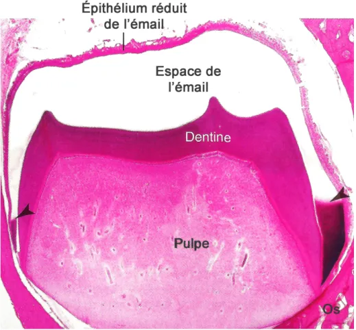
Les améloblastes sont les cellules d’origine épithéliale responsables de l’amélogénèse ; ce processus permet la formation de l’émail en trois étapes fonctionnelles majeures qui sont la pré-sécrétion, la sécrétion et la maturation. Au cours de la pré-pré-sécrétion, une membrane basale (MB) sépare les cellules épithéliales de la matrice de pré-dentine récemment formée et associée aux odontoblastes (1). À ce moment, les améloblastes en voie de différenciation acquièrent leur phénotype, changent de polarité, développent un vaste appareil de synthèse protéique et se préparent à sécréter la matrice organique de l'émail. Juste avant la phase de sécrétion, cette MB disparait. Pendant la phase de sécrétion, les améloblastes élaborent et organisent l'émail pour qu’elles atteignent son épaisseur définitive. Plus tard, au début de l'étape de maturation, lorsque la matrice organique disparait et qu’il y a une accumulation importante de minéraux et la croissance des cristaux, les améloblastes reforment une MB à l'interface entre les cellules et l'émail (1, 2). Une figure montrant les différentes structures de la dent montre la formation de l’émail, une partie déjà maturée, avec l’espace de émail qui a été déminéralisé, et une partie en maturation, où la matrice organique est encore présente (FIG 1) (1). La MB à l’interface améloblastes-émail est dite spécialisée (MBS) de part sa composition atypique puisqu’elle ne contient pas de collagènes de types IV et VII (3-5) mais est enrichie en LM-332 (6, 7) et d’autres part car elles possèdent des fonctions spécifiques, en comparaison aux MB typiques.
16
Fig. 1. Image histologique de la dent et ses structures en formation. L’émail
présente deux phases de formation : on observe l’espace de l’émail formé et déminéralisé pour des fins histologiques et la partie en maturation, où la matrice organique peut être visualisée (flèches) (1). Coloration : HE
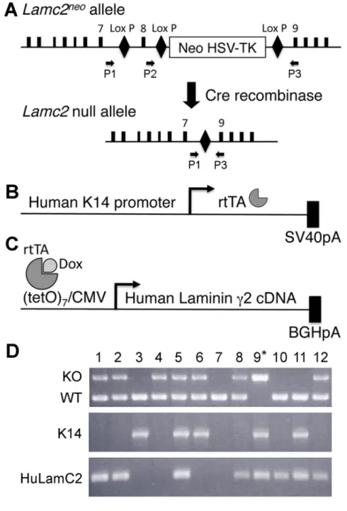
La grande famille des Laminines est formée par des glycoprotéines composées des chaînes α, ß et γ qui jouent chacune un rôle essentiel dans l'intégrité structurale de la MB (8, 9). La LM-332 est un hétérotrimère en forme de croix, composé des chaînes α3, ß3 et γ2 (8, 9), qui sont codées, respectivement par les gènes LAMA3, LAMB3 et LAMC2. (9). La perte d'une seule de ces chaînes mène à de graves défauts du développement et entraîne la létalité précoce des souris, ce qui gêne l’étude des fonctions de chacune de ses chaînes par l’utilisation de modèle « KO » (10, 11). Pour empêcher cette létalité prématurée, un modèle transgénique utilisant la laminine γ2 humaine (LAMC2) dans un modèle de souris Lamc2-KO, a été mis au point (12). Ces animaux produisent la chaîne γ2 humaine sous le contrôle de la doxycycline (Dox) en utilisant le
17
promoteur de la CK-14, sous le contrôle d’un transactivateur inverse (reverse) du promoteur régulable à la tétracycline (rtTA) - LAMC2-Dox (12). Dans ce modèle, les animaux qui reçoivent la Doxycycline expriment la LAMC2 humaine au lieu de celle de souris. L'isoforme humaine a été choisie car on peut suivre son expression, sa localisation et son renouvellement sans réactivité croisée avec la LAMC2 endogène de la souris. Des études antérieures ont montré que les animaux qui reçoivent la Dox ne montrent pas d’atteinte des tissus mous épithéliaux; cependant, l’interruption de l'antibiotique mène à des altérations tissulaires graves en environ trois semaines et aux décès ultérieures des animaux (12, 13). Pourtant, l'effet sur les tissus durs, tels que les dents, n'a pas encore été examiné chez ce modèle transgénique. Ces souris représentent ainsi un bon modèle pour l’étude du rôle de LM-332 dans la formation de la dent, en particulier durant la phase de maturation de l’émail.
Il est connu que la LM-332 est impliquée dans la différenciation terminale des améloblastes au cours de la phase de pré-sécrétion (14), mais on ne connaît pas son rôle lors de la maturation de l’émail.
Curieusement, alors que les animaux LAMC2-Dox ne montrent pas d’altération macroscopique ou histologique des tissus mous (12), l'émail de la portion de l’incisive qui a fait éruption semble atteint. La présente étude vise à explorer ce modèle transgénique conditionnel de la LAMC2 afin d’élucider le rôle de LM-332 dans la structuration et le fonctionnement de la MB spécialisée, qui se forme durant la phase de maturation de l’émail.
L’objectif de cette recherche est donc de caractériser et d’élucider le phénotype dentaire dans la zone de maturation chez les souris transgéniques, qui expriment la LAMC2 humaine.
Chapitre 2 – État de connaissances et Mise en contexte
2.1 - Tissus épithéliaux et la membrane basale
Les épithéliums sont séparés du tissu conjonctif adjacent par une matrice extracellulaire spécialisée appelée membrane basale (MB). Les membranes basales qui délimitent les tissus ont une épaisseur variant entre 40-120 nm. Elles servent à stabiliser la structure cellulaire, à définir la polarité et à établir des barrières physiques. (15, 16). Leur composition contrôle des processus cellulaires tels que la propagation, la migration et l'adhérence. Généralement les MBs sont composées de collagène de types IV, VII et XVIII, des protéoglycans et glycoprotéines, telles que le nidogène et les laminines (3-5, 16).
Les laminines sont considérées comme des constituants majeurs de la MB où elles jouent un rôle très important (8, 16). Ce sont des glycoprotéines de grande taille composées de trois chaînes (α, ß et γ) qui participent à plusieurs fonctions de la MB. Elles laminines sont impliquées tant dans le développement physiologique, que dans les maladies (3, 5, 12, 17-19).
2.1.1 – Amélogénèse et la membrane basale spécialisée
Tout au long de l’amélogénèse, les améloblastes subissent des changements morphologiques et produisent d'importantes molécules (20, 21). Les multiples activités observées durant le cycle de vie des améloblastes peuvent s’autoréguler en fonction du stade de développement de l’email (1). Toutefois, il importe de bien comprendre la régulation du développement de l’émail en raison des défauts observés lors de troubles génétiques spécifiques. Plusieurs facteurs contribuent à une malformation de l’émail : altérations de voies de signalisation ou des facteurs transcriptionnels, atteinte des récepteurs ou de leurs ligands, autant de facteurs qualitatifs ou quantitatifs capables de perturber la formation de l’émail (22, 23).
Les stades observés au cours de l’amélogénèse illustrent différents aspects de l’interface améloblaste-dent. Tout d'abord, la phase de pré-sécrétion est associée à une MB typique (20). Puis, durant la phase de sécrétion cette MB disparait et les améloblastes produisent des
19
protéines qui participent de la minéralisation partielle de l’émail, qui aura son épaisseur définitive au bout de cette phase. Lors de la transition vers la phase de maturation, les améloblastes subissent des changements majeurs au niveau de leur taille et de leur architecture pour entamer la phase de maturation (23). Enfin, une structure analogue à la MB, nommée MB spécialisée (MBS), appelée aussi MB atypique, est formée pendant la phase de maturation, permettant de fixer les améloblastes à la surface minéralisée de l'émail (20, 24-26). Pendant cette étape, les améloblastes vont retirer la matrice organique et promouvoir la croissance des cristaux (23).
Récemment, des approches génomiques ont permis d’identifier les protéines produites par les améloblastes et de mieux caractériser la composition de la MBS présente à l’interface organe de l’émail-dent. Ces études ont identifié des composants spécifiques, tels que les protéines « Odontogenic Ameloblast-associated » (ODAM), « Amelotin » (AMTN) et « Secretory
calcium-binding phosphoprotein-proline-glutamine-rich 1 » (SCPPPQ1) (27, 28).
Ces protéines sont produites aussi par les cellules de l’épithélium de jonction (EJ). Ce dernier consiste en une couche de cellules incomplètement différenciées, qui est formée lors de la fusion entre l’épithélium de la muqueuse buccale et l’organe de l’email au cours de l’éruption de la dent. (29). De ce fait, la MBS est ainsi la matrice extracellulaire associée à l’EJ permettant de fixer les tissus gingivaux à l’émail dentaire. Cette jonction forme la première ligne de défense contre les maladies parodontales (27, 28, 30).
Les études ont révélé que la MBS se distingue en composition des MBs typiques ; elle est enrichie en LM-332, mais ne contiennent ni de laminines contenant la chaîne γ1, ni de collagène de types IV et VII (8, 31-34). Des mutations dans les gènes LM-332 sont impliquées dans la pathogenèse de l'hypoplasie de l'émail (1, 6).
2.1.2 - Laminine-332 :
Parmi les divers types de Laminines, la Laminine-332 (LM-332) est l’isoforme constituée par les chaînes α3, ß3 et γ2, codées respectivement par les gènes LAMA3, LAMB3 et LAMC2. Cet hétérotrimère en forme de croix (voir Figure 2), sécrété par les cellules épithéliales est indispensable pour l'intégrité structurale et la fonction de la MB (9). LM-332 est un composant
20
adhésif majeur de la MB de l'épiderme et possède une structure distincte des autres laminines puisque ses bras sont courts, et que certains domaines sont absents de ses chaînes. Elle présente aussi des fonctions uniques, telles que la migration des cellules de l’épithélium de jonction durant son renouvellement, et aussi l’attachement de ces cellules à la dent via les hémidesmosomes (32, 35).
Fig. 2. Structure de la LM-332. Plusieurs sites sont ciblés pour les mutations menant
à une fonction anormale de la LM-332 et causant des maladies, comme la JEB (19).
L’absence ou les anomalies d’expression de LM-332 chez la souris engendrent des défauts graves du développement, similaires à ceux observés dans l'Épidermolyse Bulleuse
21
Jonctionnelle humaine (JEB) (17). La JEB est définie comme un groupe de maladies, cliniquement et génétiquement hétérogènes, qui se caractérise par le détachement de l'épiderme, des blessures et bulles ou vésicules chroniques de la peau et ses annexes, ainsi que des muqueuses, induites mécaniquement à l’occasion d’un traumatisme mineur; on observe aussi une malformation dentaire chez les personnes atteintes de la JEB. Ce groupe de maladies est causé par des défauts dans la structure et l'expression des protéines de la MB, ainsi que de certaines protéines intra-épidermiques (19, 36).
Chez la souris, ces défauts provoquent une létalité précoce (17, 19, 36). Une suppression ciblée d’une des chaînes de la LM-332 entrave l’étude à long terme de ses fonctions, puisqu’elle entraine la létalité de l’animal (10, 11).
Récemment, un modèle de souris transgénique qui exprime la LM-332 contenant une chaîne γ2 humaine a été développé (FIG 2). Il permet d’éviter la létalité des souris Lamc2-KO et restaure les fonctions de la MB épidermique (12). Ces animaux ne présentent pas de lésions au niveau de la peau ou des muqueuses, ni dans d'autres organes épithéliaux, comme c’est le cas chez les souris déficientes en LM-332 (12, 17). Malgré l’apparente normalité et la survie de ces animaux, le retrait de l'antibiotique engendre de graves lésions tissulaires après les trois premières semaines et le décès à environ 3 mois d'âge (12, 13). Cependant, l'effet de la LM-332 humanisée sur les tissus durs, y compris les dents, n'a pas encore été examiné chez ces souris.
Les animaux LAMC2-Dox constituent un modèle de choix pour l’étude du rôle de la LM-332 dans la zone de maturation de l’émail dentaire où l’on retrouve une MB enrichie de LAMC2.
22
Fig. 3. Schéma de la construction du transgène de la souris LAMC2-dox. Après la
création d’un allèle nul pour le Lamc2 et l’insertion du promoteur CK-14 et du cDNA de la chaîne γ2 humaine, la liaison de la Dox au rtTA permet l’expression de la LAMC2 humaine. L’analyse par PCR confirme que chez ce transgène conditionnel, il est possible de réprimer ou entraîner son expression sous l'effet de la Dox (12).
23
2.2 – Modèles expérimentaux transgéniques – un aperçu
L'étude des maladies humaines est souvent limitée par l'absence de modèles animaux robustes, efficaces et capables de reproduire leurs caractéristiques. Pour bien adresser cette problématique, un animal doit avoir une progéniture abondante, une taille réduite, un développement rapide et un entretien facile (37, 38). La souris (Mus musculus) présente de telles caractéristiques et représente un modèle de choix pour reproduire les changements génétiques à l’origine des maladies humaines. Les études chez la souris ont fourni de précieuses informations scientifiques au cours des dernières décennies (37, 39, 40).
La souris est devenue l'organisme modèle le plus fréquemment utilisé pour l'étude de la physiologie et de la pathologie humaines. La génétique moléculaire de la souris a été raffinée pour permettre la production d'une gamme de mutants utiles en recherche. Ceux-ci comprennent des additifs transgéniques, des souris « knock-in » et « knock-out » (avec des mutations conditionnelles ou inductibles), ces réarrangements ou suppressions génétiques permettant des conditions d’étude analogues aux conditions chez l’humain (39).
En effet, les modèles de souris knockout (celles qui n’expriment pas une protéine donnée), les modèles transgéniques (qui sur-expriment une certaine protéine de type sauvage ou mutée) et des mutants (exprimant une protéine modifiée) ont apporté des informations inestimables pour comprendre le métabolisme de l’améloblaste et ses effets sur l’amélogénèse (1). Des études chez la souris ont apporté d’importantes avancées liées à la pathogénèse de différents types de malformations de l’émail, par exemple lors du développement de l’Amélogénèse Imparfaite (AI) humaine (41). Une autre étude avec des souris KO d’ODAM a montré son rôle dans le maintient de l’intégrité de l’attache épithéliale autour de la dent (30).
2.2.1 – Modèles humanisés : avantages et limites
Plusieurs lignées de souris transgéniques humanisées bien caractérisées ont été établies. Comme les protéines ayant une séquence humaine d'acides-aminés peuvent présenter les caractéristiques biochimiques de leurs homologues de la souris, des animaux transgéniques sont construits avec des séquences codantes de gènes humains, introduites dans le génome la souris. Par contre,
24
l’humanisation comporte toujours des risques, puisque même avec une approche précise du profil d'expression du transgène humain, son fonctionnement dans le contexte de la souris ne peut pas toujours être assuré (38, 39).
Développer de nouvelles approches pour l’humanisation génomique est un défi mais de nouvelles technologies semblent prometteuses. Une question clé sur les souris humanisées concerne l’efficacité de la machinerie transcriptionnelle de la souris pour la lecture de la séquence d'ADN humain. Quand un chromosome humain est intégré au génome de la souris, la séquence d'ADN humain est associée à des modifications qui peuvent influencer les fonctions physiologiques de la protéine produite (39).
Par conséquent, des expériences effectuées uniquement chez la souris ne peuvent pas toujours reproduire avec précision les résultats obtenus chez l'homme. Bien que les modèles de souris génétiquement humanisées aient un grand potentiel, il faut apprécier les limites et les contraintes du modèle. Il est important de se rappeler qu’un modèle de souris génétiquement humanisée concerne l’étude d’une protéine chez une souris et non chez un être humain (38, 40). Quoi qu’il en soit et malgré les limites décrites ci-dessus, les modèles de souris génétiquement humanisés engendrent un intérêt évident et un énorme potentiel pour approfondir notre compréhension des maladies humaines et développer de nouvelles approches pour la prévention, le dépistage et le traitement de ces maladies (38).
Chapitre 3 – Matériel et méthodes
Toutes les procédures concernant les manipulations des souris ont été approuvées par le Comité de déontologie de l'expérimentation sur les animaux de l'Université de Montréal. (ANNEXES 1 et 2)
3.1 – Génération et génotypage des souris LAMC2-Dox
Un modèle de souris déficiente en LAMC2 a été développé par nos collaborateurs (12) afin d’étudier le rôle de cette protéine dans la MB. Les animaux Lamc2-KO ont été secourus par l’ajout d’un transgène LAMC2 humain et son expression, inductible par l’absorption de doxycycline (LAMC2-Dox) sous le contrôle du promoteur CK-14, a permis d’utiliser ce modèle (12).
Les souris LAMC2-Dox ont reçu de façon continue la doxycycline (Dox) lors de leur abreuvage (1 mg / ml avec 5% de saccharose, Sigma-Aldrich Canada Ltd, Oakville, ON, Canada) ou lors de leur alimentation (3,5-4,4 mg par jour, Harlan Laboratories, Indianapolis, IN) afin d’induire l'expression de la protéine LAMC2 humaine. Deux groupes contrôles ont été établis avec les souris C57BL/6 (Charles Rivers Canada ; St-Constant, QC, Canada) utilisées pour le développement d’animaux transgéniques et congéniques. Le premier groupe était composé de deux portées de deux souris sauvages (WT) - femelles en gestation (jour 14) qui ont reçu le même régime Dox. Au moment du sacrifice, ce groupe consistait de douze souris traitées à la Dox durant une période de 8 semaines. Le second groupe contrôle comprenaient également des souris C57BL/6, ayant reçu une alimentation régulière, sans ajout de Dox. Les souris ont été logées dans une installation stérile adaptée, dans des cages ventilées où l'eau, la nourriture et la literie ont toujours été autoclavées.
Le génotypage a été effectué par analyse PCR de l'ADN génomique extrait de la queue de la souris, en utilisant les amorces suivantes :
- allèle Lamc2 de souris (5'AGCTAATACGGGTTCAGCC3 '(sens) et 5' TGTAACCAGAAGCACATTCC3 '(anti-sens)) ;
26
- allèle LAMC2 humaine (5 'AGGCTGTCCAACGAAATGGG3' (sens) et 5'GGAGCTGTGATCCGTACACCA3 '(anti-sens)) ;
- et allèle cytokératine 14 - gène rtTA (5 'GTCCGATGGGAAAGTGTAGCCTG3' (sens) et 5'TTTCTTCTTTAGCGACTTGATGC3 '(anti-sens)).
3.2 – Préparation des tissus pour la microscopie optique,
microscopie électronique et micro-tomographie
Des souris de 8 à 16 semaines d’âge ont été anesthésiées avec une solution 20% de hydrate de chloral (0,4 mg/g de poids corporel ; Fisher Scientific, Whitby, ON, Canada). Elles ont été sacrifiées par perfusion à travers le ventricule gauche avec du lactate de Ringer (Abbott Laboratories, Montréal, QC, Canada) pendant 30 secondes, puis par une solution de fixation constituée de 4% de paraformaldehyde (BDH, Toronto, ON, Canada) et 0,1% de glutaraldéhyde (Electron Microscopy Sciences, Washington, PA) dans un tampon phosphate 0,1M (PB, pH 7,2) pendant 10 min. Les mandibules et maxillaires des animaux sauvages et LAMC2-Dox ont été disséqués puis immergés dans la même solution de fixation pendant 3h. Les échantillons ont été rincés dans le PBS pendant une nuit à 4°C. Les tissus mous des animaux WT et LAMC2-Dox, tels que la langue, le palais et la peau, ont été soumis à la même préparation que les mâchoires pour l’histologie et microscopie optique.
Une partie des échantillons des tissus durs a été décalcifiée avec 4,13% EDTA (éthylènediamine disodium tétra-acétique, Fisher Scientific), solution aqueuse, tel que précédemment décrit par Wazen et al, 2015 (42).
Certains spécimens décalcifiés ont été traités pour l’enrobage dans la paraffine. Des coupes de 5 micromètres (μm) d'épaisseur ont été préparés avec un microtome Leica RM 2155 (Leica Microsystems Canada, Richmond Hill, ON, Canada) et montés sur les lames SuperFrost® / Plus (Fisher Scientific). Ces coupes ont été colorées à l'hématoxyline (Ricca Chemical Company) et l'éosine (Sigma Chemical Company) pour les analyses morphologiques, ou préparées pour la coloration et incubation avec l’immuno-peroxydase (Dako Corporation, Carpinteria, CA) et pour l’immunofluorescence.
27
D'autres échantillons ont été traités pour l’enrobage dans la résine LR-White (London Life, Compagnie résine ; Berkshire, Royaume-Uni), ainsi que dans la résine Époxy (Electron Microscopy Sciences) tel que précédemment décrit par Wazen et al, 2015 (42). Ces échantillons ont été coupés en coupes semi-minces d’1μm, à l’aide d’un ultra-microtome (Reichert Jung Ultracut-E) avant d’être colorées avec le bleu de toluidine. Des coupes ultraminces, de 80-100 nanomètres (nm) ont été transférées sur des grilles de nickel de 200 mailles. Ces grilles ont été précédemment revêtues d’un film de Formvar® (vinyle formate) puis colorées avec une solution 4% uranyl acétate pendant 8 min et avec une solution de citrate de plomb pendant 2 min. Les grilles ont été examinées au microscope électronique à transmission (MET) FEI Tecnai 12 (FEI, Eindhoven, Pays-Bas), à 80 kV.
3.3 – Immunohistochimie
L'immunohistochimie a été réalisée sur les coupes de 5μm à température pièce. Brièvement, les coupes déparaffinées ont été bloquées 20 min avec une solution constituée de tampon phosphate 0,01 M (PBS, pH 7,2) et 5% de lait en poudre (Carnation, Nestlé, Don Mills, ON, Canada), suivi d'un traitement avec la trousse peroxydase Dako EnVision + ™, (Dako Corporation, Carpinteria, CA) selon les recommandations du fabricant. Les coupes ont subi une incubation de 3h avec l’anticorps primaire : - (ODAM lapin anti-rat (1: 10000) (Moffatt et al., 2008); - AMTN lapin anti-rat (1: 2000) (Moffatt et al., 2006b); - SCPPPQ1 lapin anti-rat (1: 2000) (Moffatt et al., 2014); - Albumine lapin anti-rat (1: 2000, ICN Pharmaceutical, Aurora, OH); ou LM-332 lapin anti-humain (1: 100, Abcam, Toronto, ON, Canada). Les anticorps primaires ont été rincés avec une solution constituée de 0,01 M de PBS, pH 7,2, contenant 0,05% de Tween 20 (Fisher Scientific) (0,01 M PBS - Tween 20) pendant 20 minutes (2X 5 minutes et 1X 10 minutes). L’incubation avec les anticorps secondaires a été faite avec la solution 2 de la trousse Dako EnVision + ™, (Dako Corporation, Carpinteria, CA) selon les recommandations du fabricant. Les anticorps secondaires ont été lavés avec une solution constituée de 0,01 M de PBS, pH 7,2, contenant 0,05% de Tween 20 (Fisher Scientific) (0,01 M PBS - Tween 20) pendant 20 minutes (2X 5 minutes et 1X 10 minutes). La réaction a été révélée avec la 3,3'-diaminobenzidine et
28
contre-colorées avec le vert de méthyle (Dako Corporation, Carpinteria, CA). Les coupes ont été examinées au microscope AxioImager M2 (Carl Zeiss, Oberkochen, Allemagne).
D’autres coupes ont été traitées pour l’immunofluorescence. Les coupes ont été bloquées pendant 1h dans du PBS contenant 5% de lait en poudre suivi d’une incubation de 3h avec les anticorps primaires (anti-AMTN, ODAM et/ou SCPPPQ1), et par la suite, de l’incubation dans le noir des anticorps secondaires chèvre-anti-lapin Alexa Fluor 488 et/ou 594 (1:500, 1h, Life Technologies ™, Mississauga, ON, Canada). Entre chaque étape, les lames ont été lavées trois fois pendant 10 minutes avec du PBS-Tween (0,05% (v/v)). Les coupes ont finalement été montées avec une solution de montage contenant du DAPI (ProLong Gold antifade reagent - Life Technologies ™) pour colorer les noyaux. Les contrôles négatifs comprenaient une omission de l'anticorps primaire et l'incubation avec le sérum pré-immunisé. Les coupes ont été examinées au microscope AxioImager M2 (Carl Zeiss, Oberkochen, Allemagne) en fluorescence, ainsi qu’au microscope Elyra PS1 avec l’option « Structured Illumination
Chapitre 4 – Article
4.1 – Résultats publiés et contribution à la recherche
Le modèle animal LAMC2-Dox a été établi à l'Université de Montréal par Dre. Rima Wazen, assistante de recherche du Dr Antonio Nanci, qui a exécuté les études pilotes nécessaires à mon projet.
J’ai par la suite participé aux différentes étapes de l’étude menant à la publication de l’article, comprenant :
1 - l’entretien dans l’animalerie et l’euthanasie des animaux, incluant la perfusion intracardiaque, la dissection et la préparation des tissus à utiliser ;
2 - la préparation des solutions et d’échantillons, pour l’enrobage en paraffine, en résine LR-White et en résine Époxy ;
3 - les coupes en paraffine et ouverture des blocs pour les coupes semifines ;
4 - les colorations en HE, ainsi que l’immunohistochimie utilisée pour les expériences ; 5 - l’analyse et prise d’images en microscopie optique, en fluorescence et électronique ; 6 - la préparation de figures et la rédaction de la première version de l’article.
Pour ma formation et l'exécution de mon travail j'ai bénéficié de la supervision directe de Dre Rima Wazen et du Dr Antonio Nanci.
Le projet a aussi compté sur l’aide et la collaboration d’autres membres du laboratoire, ainsi que d’autres chercheurs de différentes institutions, au Canada et aux États-Unis.
Par rapport à l’article, ma contribution a été considérée égale à celle de deux autres premiers auteurs.
Wazen RM, Viegas-Costa LC, Fouillen A, Moffatt P, Adair-Kirk TL, Senior RM, Nanci, A. Laminin gamma2 knockout mice rescued with the human protein exhibit enamel
30
maturation defects. Matrix biology : journal of the International Society for Matrix Biology. 2016.
Matrix Biol. 2016 Mar 5. pii: S0945-053X(16)30025-7. doi: 10.1016/j.matbio.2016.03.002.
4.2 – Transfert de connaissance et vulgarisation de la recherche
Présentation par affiche
Viegas-Costa LC, Wazen RM, Fouillen A, Moffatt P, Adair-Kirk TL, Senior RM, Nanci, A. Laminin gamma2 knockout mice rescued with the human protein exhibit enamel maturation defects.
J’ai présenté mes résultats de recherche pendant la Journée scientifique 2016 du RSBO, le 11 mars à l'Hôpital Shriners pour enfants, à Montréal. Le travail a été présenté aussi dans les « Journées dentaires internationales du Québec » qui ont eu lieu au Palais des congrès de Montréal les 30 et 31 mai 2016.
Laminin
γ2 knockout mice rescued with the
human protein exhibit enamel maturation
defects
Rima M. Wazena, 1, Luiz C. Viegas-Costaa, 1, Aurélien Fouillena, b, 1, Pierre Moffattc, Tracy L. Adair-Kirkd, Robert M. Seniord and Antonio Nancia, b
a - Laboratory for the Study of Calcified Tissues and Biomaterials, Department of Stomatology, Faculty of Dentistry, Université de Montréal, Montréal, Québec, Canada
b - Department of Biochemistry and Molecular Medicine, Université de Montréal, Montréal, Québec, Canada c - Shriners Hospital for Children, Montréal, Montréal, Québec, Canada
d - Department of Medicine, Washington University School of Medicine, St. Louis, MO
Correspondence toAntonio Nanci: at: Laboratory for the Study of Calcified Tissues and Biomaterials, Department of Stomatology, Faculty of Dentistry, Université de Montréal, Montréal, Québec, Canada.
http://dx.doi.org/10.1016/j.matbio.2016.03.002
Abstract
The epithelial ameloblasts are separated from the maturing enamel by an atypical basement membrane (BM) that is enriched in laminin 332 (LM-332). This heterotrimeric protein (α3, ß3 and γ2 chains) provides structural integrity to BMs and influences various epithelial cell processes including cell adhesion and differentiation. Mouse models that lack expression of individual LM-332 chains die shortly after birth. The lethal phenotype of laminin γ2 knockout mice can be rescued by human laminin γ2 (LAMC2) expressed using a doxycycline-inducible (Tet-on) cytokeratin 14 promoter-rtTA. These otherwise normal-looking rescued mice exhibit white spot lesions on incisors. We therefore investigated the effect of rescue with human LAMC2 on enamel maturation and structuring of the atypical BM. The maturation stage enamel organ in transgenic mice was severely altered as compared to wild type controls, a structured BM was no longer discernible, dystrophic matrix appeared in the maturing enamel layer, and there was residual enamel matrix late into the maturation stage. Microtomographic scans revealed excessive wear of occlusal surfaces on molars, chipping of enamel on incisor tips, and hypomineralization of the enamel layer. No structural alterations were observed at other epithelial sites, such as skin, palate and tongue. These results indicate that while this humanized mouse model is capable of rescue in various epithelial tissues, it is unable to sustain structuring of a proper BM at the interface between ameloblasts and maturing enamel. This failure may be related to the atypical composition of the BM in the maturation stage and reaffirms that the atypical BM is essential for enamel maturation.
© 2016 Published by Elsevier B.V.
Introduction
Formation of enamel proceeds as three major functional steps that include the pre-secretory, secretory and maturation stages [1,2]. During the pre-secretory stage, a typical basement membrane (BM) separates the differentiating epithelial amelo-blasts from the forming mantle dentin matrix and associated odontoblasts [1]. Just prior to the start of secretion, this BM is removed. Later, at the onset of the maturation stage, ameloblasts reform a BM at the interface between the cells and enamel.
However this BM is atypical; it does not contain collagen types IV and VII [3–5] but is enriched in laminin-332 (LM-332)[6,7], which consists ofα3, ß3 andγ2 chains[8,9].
The use of knockout mouse models to study the function of laminins is complicated by the fact that targeted deletion of the chains results in severe developmental defects that lead to early lethality
[10,11]. To circumvent this premature lethality phenotype for LM-332, a double transgenic doxycy-cline (Dox)-inducible human LAMC2 transgene (LAMC2-Dox), under the regulation of the cytokeratin
MATBIO-1245; No. of pages: 12; 4C:
0022-2836/© 2016 Published by Elsevier B.V. Matrix Biol. (2016) xx, xxx–xxx
Article
Please cite this article as: R. M. Wazen, et al., Lamininγ2 knockout mice rescued with the human protein exhibit enamel maturation defects, Matrix Biol (2016),http://dx.doi.org/10.1016/j.matbio.2016.03.002
14 promoter-tetracycline transactivator gene (rtTA) transgene was created [12]. In this model, animals receiving Dox are rescued by expressing the human LAMC2 instead of the mouse one. Progeny of the cross of the human LAMC2 transgenic with LAMC2 knockout mice shows no defects in soft epithelial tissues and appear generally normal. However, turning off human LAMC2 conditional expression (knockout condition) leads to severe tissue alterations as of the third week after stopping administration of Dox, and animals die around 3 months of age[12,13]. It has been shown that LM-332 is implicated in the terminal differentiation of ameloblasts during the pre-secretory stage[14]but little is known on its role during maturation. Our ultimate objective was to exploit this conditional LAMC2 knockout mouse model to elucidate the role of LM-332 in the structuring and functioning of the atypical BM created during the maturation stage. While establishing the colony for these studies it became apparent that incisors of the LAMC2-Dox mice exhibited chalky enamel patches, a feature suggestive of a problem with enamel matura-tion. This study therefore focused on elucidating the origin of this tooth phenotype in the transgenic mice rescued with human LAMC2. While structural orga-nization in the pre-secretory stage appeared nor-mal, in the maturation stage the atypical BM and enamel organ was severely altered, and the removal of the organic matrix was delayed resulting in hypomineralized enamel. Contrary to what occurs at sites where typical BMs are found, these data indicate that LAMC2-Dox mice expressing the human protein are unable to sustain proper
struc-turing of the atypical BM during the maturation stage, and that this interfacial structure is essential for enamel maturation.
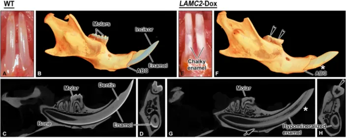
Results
Visual and micro-computed tomographic analyses As compared to age-matched WT mice (Fig. 1A, B), the erupted portion of incisors from LAMC2--Dox animals exhibited chalky patches on the enamel surface, chipping of the enamel layer and blunt tips (Fig. 1E, F). Comparative sagittal and coronal tomographic views revealed that the enamel is also severely abraded indicating that the enamel was softer (Fig. 1C vs 1G, 1D vs 1H). There was an increase in periodontal space between the labial aspect of the incisor and alveolar bone (Fig. 1G, H).
To eliminate the possible effect of Dox on normal enamel formation and maturation, gestating female WT mice and newborn mice were maintained on the same Dox diet as LAMC2-Dox mice. This did not create any significant alterations to amelogenesis (data not shown).
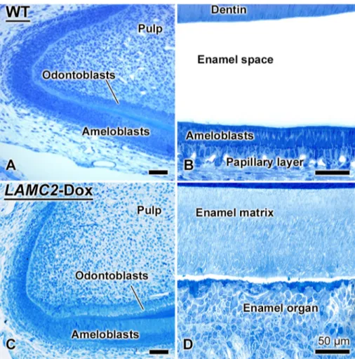
H i s to l o g i c a l a n d s t r u c t u r a l c h a n g e s i n amelogenesis in LAMC2-Dox mice
Major defects in amelogenesis were observed in both maxillary and mandibular incisors of
LAMC2-Fig. 1. Comparative micro-CT views of hemimandibles from wild type (WT, A-D) and human LAMC2-rescued transgenic mice (LAMC2-Dox) (E-H) mice. B, F tomographic reconstructions; C, G, sagittal sections; D, H, cross-sectional views taken at matching physical locations. Visual inspection (A, E) in live mice reveals the presence of chalky enamel patches on the erupted portion of incisors in LAMC2-Dox mice (E). Molars from these mice are excessively abraded and cuspal surfaces (arrowheads) are flattened (F, H). Enamel in LAMC2-Dox mice is hypomineralized (G, H) and frequently flakes off (*) at incisor tips (G). Note the augmented periodontal space (arrows) on the labial aspect of the incisor. ABC, Alveolar bone crest.
2 Lamininγ2 knockout mice rescued with human protein exhibit defects
Please cite this article as: R. M. Wazen, et al., Lamininγ2 knockout mice rescued with the human protein exhibit enamel maturation defects, Matrix Biol (2016),http://dx.doi.org/10.1016/j.matbio.2016.03.002
Dox mice (Fig. 2C, D) as compared to normal WT amelogenesis (Fig. 2A, B). In LAMC2-Dox mice, there were no obvious histological changes in amelogenesis during the pre-secretory stage and mantle dentin formation (Fig. 2A vs 2C). However, organization of the maturation stage ameloblasts and of the other associated enamel organ cells was severely altered (Figs. 2D, 3) as compared to WT mice (Fig. 2A-B). The disruption in cellular organi-zation and ameloblasts polarity (Figs. 2D,3A-C), the presence of dystrophic matrix among cells of the enamel organ and in the enamel layer (Fig. 3C), and the persistence of enamel matrix up to the gingival margin (Fig. 3D) make it difficult to reliably subdivide the maturation stage into early, mid and late portions or to apply reference points such as where enamel matrix is EDTA-soluble[15]. Consis-tent with the hypomineralization observed in micro--computed tomographic scans (Fig. 1), residual enamel matrix extended beyond the alveolar bony
crest, and in some cases up to the gingival margin (Fig. 3D).
At the ultrastructural level (Fig. 4), the typical BM present between differentiating ameloblasts and the forming mantle dentin matrix (pre-secretory stage) (Fig. 4A), and between the papillary layer and blood vessels during the maturation (Fig. 4B) appeared normal in LAMC2-Dox mice. However, the interface between the disrupted enamel organ and maturing enamel was very irregular (Fig. 4C, D). Frequently, there was dystrophic matrix interposed between the enamel organ and enamel, and no distinct atypical BM could be recognized (Fig. 4C, D). Occasionally, a BM-like accumulation was observed at the apical cell surface (Fig. 4F).
Immunohistochemistry
Incubations intended to map the localization of LAMC2 in the maturation stage of amelogenesis in
Fig. 2. During amelogenesis, there is a typical basement membrane along the apical surface of differentiating ameloblasts while during the maturation stage an atypical one is recreated at the interface between of ameloblasts and enamel. There is no difference in the pre-secretory histology between wild type (WT) and human LAMC2-rescued transgenic mice (LAMC2-Dox) (compare A with C). However, in the maturation stage, the LAMC2-dox mouse exhibits dramatic differences (compare B with D); the organization of the enamel organ is disrupted and there is residual enamel matrix through maturation. Toluidine blue stained sections.
3 Lamininγ2 knockout mice rescued with human protein exhibit defects
Please cite this article as: R. M. Wazen, et al., Lamininγ2 knockout mice rescued with the human protein exhibit enamel maturation defects, Matrix Biol (2016),http://dx.doi.org/10.1016/j.matbio.2016.03.002
WT mice showed that ameloblasts express the protein and it accumulated at the interface between ameloblasts and maturing enamel where the atypical BM is found (Fig. 5A). However, in LAMC2-Dox mice, even though there were groups of enamel organ cells immunoreactive for LAMC2, there generally was no such accumulation (Fig. 5B). Some staining, above background, was also seen over enamel matrix (Fig. 5B). In soft epithelial tissues, the typical BM region separating epithelial
cells and the underlying connective tissue was conspicuously labeled (Fig. 5C).
The atypical BM associated with maturation stage ameloblasts provides a permeability barrier that prevents the inflow of albumin into maturing enamel
[16]. To confirm the absence of a functional atypical BM in our mouse model, we have compared the distribution of albumin in WT (Fig. 6A, B) and LAMC2-Dox mice (Fig. 6C, D). Distinctively from WT mice (Fig. 6A, B), albumin was found in maturing Fig. 3. Photomicrographs from hematoxylin and eosin stained histological sections of LAMC2-Dox mice incisors. Entering the maturation stage, ameloblasts gradually lose their columnar appearance, the enamel organ becomes disorganized and its individual cell layers are eventually no longer discernable (A). Cystic spaces containing loose organic material (*, B) and/or patches dystrophic matrix (arrowheads, C) with variable appearance can be found between the disrupted enamel organ and the enamel surface. Some organic matrix also accumulates among cells of the enamel organ (arrows, C). Organic matrix persists well beyond the gingival margin (D).
4 Lamininγ2 knockout mice rescued with human protein exhibit defects
Please cite this article as: R. M. Wazen, et al., Lamininγ2 knockout mice rescued with the human protein exhibit enamel maturation defects, Matrix Biol (2016),http://dx.doi.org/10.1016/j.matbio.2016.03.002
enamel matrix of LAMC2-Dox mice (Fig. 6C, D), indicating a change of BM permeability.
Consistent with previous reports[17–20]in WT mice, AMTN, ODAM and SCPPPQ1 accumulated at the interface between the ameloblasts and maturing enamel matrix (data not shown). In the LAMC2-Dox mice (Fig. 7), the staining patterns for AMTN (Fig. 7A, B), ODAM (Fig. 7C, D) and SCPPPQ1 (Fig. 7E, F) were complex, correlating with the amount of dystrophic matrix and the degree of disruption of enamel organ. When altered ameloblasts were interposed with dystrophic matrix, the immunolabeling for the three proteins at apical surface of ameloblasts was irregular and more diffuse. All three proteins also accumulated over patches of organic matrix among the enamel organ cells.
Discussion
We are exploiting a Dox-inducible human LAMC2 transgene mouse model regulated by the cytokeratin
14 promoter-rtTA construct, to study the role of LM-332 on structuring of the atypical BM found at the interface between ameloblasts and maturing enamel. Such conditional expression allows bypass-ing the early lethality associated with targeted inactivation of LM-332[12]. The atypical BM present during enamel maturation does not contain collagen IV and VII, is enriched in LM-332, and has been shown to comprise AMTN, ODAM, SCPPPQ1, three novel BM components that belong to the SCPP gene cluster [21–23]. This cluster regroups a number of genes coding for proteins that stabilize Ca2 +and PO4−
ions in body fluids and/or guide CaPO4 deposition
into receptive extracellular matrices [21–23]. Our results here demonstrate that expression of the human LAMC2 chain in mice does not affect events associated with the pre-secretory stage of amelogen-esis, consistent with what has been reported in other soft tissues where typical BMs are found [12,13]. However, it cannot compensate for the lack of mouse Fig. 4. Transmission electron micrographs of LAMC2-Dox mice incisors. In these human LAMC2 rescued mice, the typical basal membrane (BM) separating differentiating ameloblasts from the forming mantle dentin during the pre-secretory stage (A, inset), and that associated with blood vessels and the papillary layer (B) during the maturation stage appears normal. On the other hand, the interface between ameloblasts and maturing enamel is generally disrupted. Dystrophic organic matrix frequently accumulates along the apical surface of ameloblasts and sometimes extends between the cells (*, C-E). In these decalcified preparations, disorganized‘crystal ghost’ profiles can be found (E, inset). Occasionally, when there is no intervening, dystrophic matrix, a thin‘fuzzy’ layer or material (F) is present along the cell surface. In general, no well-defined atypical BM (aBM) structure can be identified during the maturation stage in these LAMC2-Dox mice.
5 Lamininγ2 knockout mice rescued with human protein exhibit defects
Please cite this article as: R. M. Wazen, et al., Lamininγ2 knockout mice rescued with the human protein exhibit enamel maturation defects, Matrix Biol (2016),http://dx.doi.org/10.1016/j.matbio.2016.03.002
LAMC2 during enamel maturation leading to major disruption. The organization of the enamel organ is severely altered and dystrophic organic matrix appears in the enamel layer and among cells of the enamel organ. Some of this dystrophic matrix shows similarities to the amyloid-like material found in calcifying epithelial odontogenic tumors, which
cal-cifies in a concentric pattern[24–27]. Removal of the organic matrix is hampered, resulting in hypominer-alized enamel that is soft, and abrades and chips readily. Patients with junctional epidermolysis bullosa due to LM-332 mutations also have similar tooth problems [28]. In the Dox-inducible human LAMC2 transgene mouse model, structuring and Fig. 5. Immunolocalization of laminin-332. In wild type mice (WT), a discrete immunolabeling is present along the apical surface of ameloblasts where an atypical basement membrane (aBM) is found (A). In human LAMC2-rescued transgenic (LAMC2-Dox) mice, the organization of the enamel organ is severely disrupted; while groups of cells are immunoreactive (arrowheads) confirming expression of the transgene, in general, there is little or no staining at the interface between the cells and enamel (B). However, in soft epithelial tissues, the BM region between epithelial cells and the underlying connective tissue conspicuously labeled (C, arrows). Altogether, these data suggest that there is a problem with aBM assembly during the maturation stage in LAMC2-Dox mice. Counterstain is methyl green.
6 Lamininγ2 knockout mice rescued with human protein exhibit defects
Please cite this article as: R. M. Wazen, et al., Lamininγ2 knockout mice rescued with the human protein exhibit enamel maturation defects, Matrix Biol (2016),http://dx.doi.org/10.1016/j.matbio.2016.03.002
functioning of the atypical BM is also affected, resulting in loss of its permeability barrier capacity and inflow into enamel of exogenous molecules such as albumin. The distribution AMTN, ODAM and SCPPPQ1 is altered, and while some molecules may accumulate along the apical surface of the altered ameloblasts, others appear to diffuse away from the surface. There is also ectopic accumulation of these proteins between cells of the enamel organ. Altogether these results indicate that this transgenic model in which mouse LAMC2 is substituted by the human form is capable of sustaining BM formation and rescue in various epithelial tissues but is unable to do so during the maturation stage of amelogenesis. While the generated phenotype is complex, the data suggest that improper structuring of the atypical BM at the interface between ameloblasts and maturing enamel is implicated in alterations that disrupt the maturation process and ultimately result in a significantly hypomineralized enamel layer.
It could be argued that these dramatic effects may at least in part be related to the mice receiving Dox for prolonged periods. To eliminate this possibility we have included a control WT group that was similarly exposed to Dox during gestation until sacrifice. The dentition of these mice showed none of the alterations seen in LAMC2-Dox mice. In addition, dental alterations resulting from thera-peutic intake of Dox are rare and usually negligible
[29–31].
The findings reported here are surprising because expression of the human LAMC2 fully rescues the early lethality observed in absence of mouse LAMC2, and there are in general no epithelial complications. Yet, enamel maturation is perturbed in these transgen-ic mtransgen-ice. Several possibilities could account for the lack of compensatory function by the human LAMC2. Although the human and rodent LAMC2 have quite high homology (~ 85%)[32,33], it cannot be excluded that the human form can have different biochemical characteristics than the mouse [34]. Indeed, the Fig. 6. Immunolocalization of albumin in wild type (WT, A-B) and human LAMC2-rescued transgenic (LAMC2-Dox) mice (C-D). In both cases, albumin was found among the cells of the enamel organ and in cells of the papillary layer. No significant staining for albumin is normally seen in maturing enamel of WT mice (A, B) but it is detected in the enamel matrix of LAMC2-Dox mice (C, D). Counterstain is methyl green.
7 Lamininγ2 knockout mice rescued with human protein exhibit defects
Please cite this article as: R. M. Wazen, et al., Lamininγ2 knockout mice rescued with the human protein exhibit enamel maturation defects, Matrix Biol (2016),http://dx.doi.org/10.1016/j.matbio.2016.03.002
relatively small difference in their amino acid compo-sition may be sufficient to affect its capacity to interact with the other BM molecules constituting the atypical BM, molecules that are not found in typical BMs. In fact, LM-332, ODAM, AMTN and SCPPPQ1 have all been immunodetected during maturation in the LAMC2-Dox mice but their presence and distributions are patchy, irregular, and not restricted to the cell-enamel interface. Alternatively, the cytokeratin 14 promoter used may not sustain high enough levels of LAMC2 synthesis during maturation needed to achieve proper stoichiometric
LM-332 assembly. Definite proof will require further studies looking at quantitative LAMC2 expression levels as compared to WT. As a consequence of these two scenarios, constituents may drift apart and assume altered distribution. Absence of a proper BM along the apical surface of ameloblasts may also affect their polarity, directionality of secretion and produce ectopic accumulations.
Finally, there may be a problem with processing of human LAMC2 in the rescued transgenic LAMC2-Dox mice. Indeed, extracellular processing Fig. 7. Immunohistochemical preparations for amelotin (AMTN, A-B), odontogenic ameloblast-associated (ODAM, C-D) and secretory calcium-binding phosphoprotein-proline-glutamine-rich 1 (SCPPPQ1, E-F) during maturation in LAMC2-Dox mice incisors. All three proteins are present at the interface between the enamel organ and maturing enamel but the labeling is irregular and somewhat diffuse. They are also conspicuously found as patches among the cells of the enamel organ (arrowheads). A, C, E, immunoperoxidase; B, D, F, immunofluorescence preparations observed by structured illumination microscopy (SIM). Counterstain in A, C, E is methyl green. Nuclei are stained in blue in B, D, F. 8 Lamininγ2 knockout mice rescued with human protein exhibit defects
Please cite this article as: R. M. Wazen, et al., Lamininγ2 knockout mice rescued with the human protein exhibit enamel maturation defects, Matrix Biol (2016),http://dx.doi.org/10.1016/j.matbio.2016.03.002
of theα3 and γ2 chains appears to be essential for the correct integration and function of LM-332 in typical BM [35]. However, this appears unlikely since there are no apparent defects in epithelial tissues soft tissues elsewhere in the body[12,13]. In addition, BMs in the pre-secretory stage and those associated with the papillary layer and blood vessels during maturation appear normally struc-tured. Such a problem could only therefore occur if there was a distinct, processing mechanism for the atypical BM.
While albumin is conspicuously found in most tissues, it is generally accepted that it is either absent or found in low abundance in enamel
[16,36]. Its abnormal presence within the maturing enamel layer of LAMC2-Dox mice indicates that the selective permeability barrier normally as-sumed by the enamel organ and/or the atypical BM during enamel maturation is no longer func-tional [37–39]. Continued influx of exogenous organic materials would likely perturb the complex process of enamel maturation and result in the observed hypomineralized enamel. Albumin may also find its way in by incorporation of the intercellular matrix patches into the enamel layer. It has been reported that in some pathological conditions [40–42], there could be abnormal retention of albumin during the maturation stage which may inhibit crystal growth [36]. It therefore cannot be excluded that the excessive albumin detected in the LAMC2-Dox mice may in part derive from such a mechanism.
In conclusion, our mouse model expressing human LAMC2 appears to represent a restricted functional knockout of LM-332 that has provided important information on the role of the atypical BM for proper enamel maturation. While the primary complication is at the level of the BM, the observed maturation phenotype is complex and secondary changes in ameloblast function may also be impli-cated. In addition, it has been reported that LAMC2 is also expressed during the secretory stage of amelogenesis [6]. Although no readily apparent changes were noticed in this stage, the possibility that secretion of a human protein in larger quantities than normal in forming enamel may cause some alterations will need further attention. The LAMC2-Dox mouse model may be further exploited by removing Dox to completely inhibit the expression of LAMC2 while avoiding early lethality of mice. This, however, is likely to result in a more complex phenotype since all stages of amelogenesis would likely be affected. Novel tools using maturation stage-specific gene promoters, such as that for AMTN and/or ODAM, will need to be created to elucidate with more precise control the mecha-nisms at play for atypical BM assembly. All these models will provide a unique exciting opportunity to investigate the role the proper adhesion of the
junctional epithelium on the onset of periodontal disease since an atypical BM is also implicated at this site[43]. Finally, this study brings attention to the fact that outcomes from humanized mouse models must be interpreted with caution [44,45], particularly that there is concern on the extent to which human DNA sequences can be adequately processed by the mouse transcriptional machinery
[34].
Materials and methods
All procedures for mice handling were approved by the Comité de déontologie de l'expérimentation sur les animaux of Université de Montréal.
Generation and genotyping of LAMC2-Dox mice A viable mouse model of LM-332 deficiency has been developed which expresses a Dox-inducible human LAMC2 transgene (LAMC2-Dox) under the cytokeratin 14 promoter on the LAMC2 knockout background [12]. LAMC2-Dox mice continuously received Dox in water (1 mg/ml with 5% sucrose, Sigma-Aldrich Canada Ltd., Oakville, ON, Canada) or in rodent diet (3.5–4.4 mg daily dose, Harlan Laboratories, Indianapolis, IN) to induce the expres-sion of human LAMC2. Gestating (day 14) female C57BL/6 mice received the same Dox rodent diet and newborn mice were maintained on the same diet for 8 weeks. Control mice also included C57BL/6 mice on regular diet. All mice were housed in a sterile, specific pathogen-free animal facility in which ventilated cages, water, food and bedding were autoclaved.
Genotyping was done by PCR analysis of genomic DNA extracted from mouse tail clips, using the following primers: mouse LAMC2 allele (5'AGCTAA-TACGGGTTCAGCC3’ (forward) and 5′ TGTAACCA-GAAGCACATTCC3’ (reverse)); human LAMC2 allele (5′ AGGCTGTCCAACGAAATGGG3’ (forward) and 5'GGAGCTGTGATCCGTACACCA3’ (reverse)); and cytokeratin 14- rtTA gene (5′ GTCCGATGG-G A A A GTCCGATGG-G T GTCCGATGG-G T A GTCCGATGG-G C C T GTCCGATGG-G 3’ ( f o r w a r d ) a n d 5'TTTCTTCTTTAGCGACTTGATGC3’ (reverse)). Tissue preparation for light and electron microscopy and micro-computed tomography
Eight and 16-weeks old mice were anesthetized with 20% chloral hydrate solution (0.4 mg/g body weight; Fisher Scientific, Whitby, ON, Canada) and sacrificed by perfusion through the left ventricle with Ringer's lactate (Abbott Laboratories; Montreal, QC, Canada) for 30 s, followed by a fixative solution consisting of 4% paraformaldehyde (BDH; Toronto, ON, Canada) and 0.1% glutaraldehyde (Electron Microscopy Sciences; Washington, PA) in 0.1 M 9 Lamininγ2 knockout mice rescued with human protein exhibit defects
Please cite this article as: R. M. Wazen, et al., Lamininγ2 knockout mice rescued with the human protein exhibit enamel maturation defects, Matrix Biol (2016),http://dx.doi.org/10.1016/j.matbio.2016.03.002
phosphate buffer (PB, pH 7.2) for 10 min. Mandibles and maxillae from WT and LAMC2-Dox were dissected and the specimens were immersed in the same fixative solution for 3 h. The samples were rinsed in 0.1 M PB (pH 7.2) overnight at 4 °C.
Samples were decalcified with 4.13% disodium ethylenediamine tetra-acetic acid (Fisher Scientific) for 21–28 days. The decalcifying solution was chan-ged each two days and then samples were washed for 24 h in 0.1 M PB buffer, pH 7.2. Some decalcified specimens were processed for paraffin embedding. Sections of 5-μm in thickness were prepared with a Leica RM2155 microtome (Leica Microsystems Canada, Richmond Hill, ON, Canada) and mounted on Superfrost®/Plus slides (Fisher Scientific) and stained with hematoxylin and eosin for morphological analyses or processed for immunoperoxidase stain-ing. Other specimens were processed for embedding in LR White resin (London Resin Company; Berkshire, UK). One micrometer thick semi-thin sections were cut with glass knives on a Reichert Jung Ultracut E ultramicrotome and stained with toluidine blue. Ultrathin 80–100 nm sections were cut with a dia-mond knife, transferred onto Formvar®-coated (poly-vinyl formate) 200-mesh nickel grids, stained with 4% aqueous uranyl acetate for 8 min and with lead citrate for 2 min, and examined at 80 kV with a FEI Tecnai 12 (FEI, Eindhoven, The Netherlands) transmission electron microscope.
Some hemimandibles were left calcified and were scanned with a desktop Micro-CT scanning system (SkyScan 1272, Bruker SkyScan, Aartselaar, Bel-gium). The scanner was operated at 70 kV/142μA with a resolution of 4.8μm/pixel and an aluminum filter of 0.5 mm. The sample rotated at 0.15°/step for 360° with an exposure time of 2 s/step during the scan period. The set of x-ray shadow images obtained from scanning was then reconstructed using the NRecon software (version 1.6.1.3, Bruker SkyScan). The CT-Analyzer software (version 1.10.0.1, Bruker Sky-Scan) was used for the creation of 3D models. Immunohistochemistry
Immunohistochemistry was performed on 5-μm thick sections mounted on Superfrost®/Plus slides. All steps were done at room temperature. Briefly, deparaffinized sections were blocked for 20 min, with a solution consisting of 0.01 M phosphate buffered saline (PBS, pH 7.2), and 5% skim milk (Carnation; Nestle, Don Mills, ON, Canada). They were incubated for 3 h with either rabbit anti-rat ODAM (1:10,000) (Moffatt et al., 2008), anti-rat AMTN (1:2000) (Moffatt et al., 2006b), anti-rat SCPPPQ1 (1:2000) (Moffatt et al., 2014), anti-rat albumin (1:2000, ICN Pharmaceutical, Aurora, OH) or anti-human LM-332 (1:100, Abcam, Toronto, ON, Canada). Primary antibodies were washed with a solution consisting of 0.01 M PBS, pH 7.2,
contain-ing 0.05% Tween 20 (Fisher Scientific) (0.01 M PBS – Tween 20) for 30 min followed by treatment with the Dako EnVision™ + System, peroxidase kit (Dako Corporation, Carpinteria, CA) as recommend-ed by the manufacturer. Visualization was performrecommend-ed with 3,3′-diaminobenzidine and sections were coun-terstained with methyl green (Dako Corporation). Sections were examined under an AxioImager M2 microscope (Carl Zeiss, Oberkochen, Germany).
Some sections were processed for immunoflu-orescence. Sections were blocked in PBS con-taining 5% skim milk for 1 h. They were then incubated with the AMTN, ODAM and SCPPPQ1 primary antibodies followed by a secondary goat-anti-rabbit-AlexaFluor 488 or 594 antibody (1:500, 1 h, Life Technologies™, Mississauga, ON, Canada). Following each steps, the slides were washed three times for 10 min each with PBS-Tween (0.05% (v/v)). Sections were then mounted in ProLong Gold containing DAPI (Life Technologies™) to stain nuclei. Negative controls included omission of primary antibody and incuba-tion with pre-immune serum. Secincuba-tions were exam-ined under structured illumination microscopy the Elyra PS1 microscope (Carl Zeiss).
Acknowledgments
We extend our thanks to Mrs. Katia Julissa Ponce (UdeM) for technical assistance and Dr. Martin Pellicelli (Shriners) for acquisition of the micro-CT scans and imaging. This study was supported in part by the Canadian Institutes of Health Research (grant no. MOP110972), the Network for Oral and Bone Health Research. PM is supported by the Shriners of North America. AF is supported by the Cellular D y n a m i c s o f M a c r o m o l e c u l a r C o m p l e x e s NSERC-CREATE program. RMS was supported by National Heart, Lung, and Blood Institute/NIH P50 HL-29594 and the Alan A. and Edith L. Wolff Charitable Trust/Barnes-Jewish Hospital.
Received 5 January 2016; Received in revised form 3 March 2016; Accepted 3 March 2016 Available online xxxx Keywords: Amelogenesis; Basement membrane; Enamel maturation; Humanized mouse model; Lamininγ2 These authors contributed equally to the work. 10 Lamininγ2 knockout mice rescued with human protein exhibit defects
Please cite this article as: R. M. Wazen, et al., Lamininγ2 knockout mice rescued with the human protein exhibit enamel maturation defects, Matrix Biol (2016),http://dx.doi.org/10.1016/j.matbio.2016.03.002