Université de Montréal
Development of a retroviral strategy that efficiently creates
nested chromosomal deletions in mouse embryonic stem celis
and its exploitation for functional genomics
par
Mélanie Bilodeau
Programmes de biologie moléculaire, Université de Montréal Faculté des études supérieures
Thèse présentée à la Faculté des études supérieures envue de l’obtention du grade de doctorat
en biologie moléculaire
Avril, 2007
5Ô
n
L
)
L‘/
Université
de Montréal
Direction des bib1iothèques
AVIS
L’auteur a autorisé l’Université de Montréal à reproduire et diffuser, en totalité ou en partie, par quelque moyen que ce soit et sur quelque support que ce soft, et exclusivement à des fins non lucratives d’enseignement et de recherche, des copies de ce mémoire ou de cette thèse.
L’auteur et les coauteurs le cas échéant conservent la propriété du droit d’auteur et des droits moraux qui protègent ce document. Ni la thèse ou le mémoire, ni des extraits substantiels de ce document, ne doivent être imprimés ou autrement reproduits sans l’autorisation de l’auteur.
Afin de se conformer à la Loi canadienne sur la protection des renseignements personnels, quelques formulaires secondaires, coordonnées ou signatures intégrées au texte ont pu être enlevés de ce document. Bien que cela ait pu affecter la pagination, il n’y a aucun contenu manquant. NOTICE
The author of this thesis or dissertation has granted a nonexclusive license allowing Université de Montréal to teproduce and publish the document, in part or in whole, and in any format, solely for noncommercial educational and research purposes.
The author and co-authors if applicable retain copyright ownership and moral rights in this document. Neither the whole thesis or dissertation, nor substantial extracts from it, may be printed or otherwise reproduced without the author’s permission.
In compliance with the Canadian Privacy Act some supporting forms, contact
information or signatures may have been removed from the document. While this may affect the document page count, it does flot represent any loss of content from the document.
Université de Montréal faculté des études supérieures
Cette thèse intitulée:
Development of a retroviral strategy that efficiently creates nested chromosomal deletions in mouse embryonic stem celis and its exploitation for functional gdnomics
présentée par: Mélanie Bilodeau
a été évaluée par un jury composé des personnes suivantes:
Dr Trang Hoang, présidente-rapporteuse Dr Guy Sauvageau, directeur de recherche
Dr Daniel Dufort, membre du jury Dr Janet Rossant, examinatrice externe
Dr Éric Milot, examinateur externe
111
Résumé
Les délétions chromosomiques chevauchantes sont un outil exploratoire exceptionnel afin d’annoter fonctionnellement le génome de la souris, puisqu’elles permettent de sonder autant les régions codantes que non-codantes. Toutefois jusqu’à maintenant, la création de délétions chromosomales. cartographiables précisément, était laborieuse ce qui limitait leur application à grande échelle. Les travaux présentés dans cette thèse proposent une nouvelle alternative pour créer des délétions chromosomiques à l’échelle du génome entier, dans un délai raisonnable, et applicable tant aux cellules primaires qu’aux lignées cellulaires. Ce système repose sur deux rétrovirus complémentaires insérant des séquences toxP dans le génome, qui servent de substrats pour la recombinaison site spécifique catalysée par l’enzyme Cre. La première section de cette thèse (chapitre 2) décrit cette stratégie et le développement de vecteurs rétroviraux compétents pour produire des délétions chromosomales haploïdes dans les cellules souches embryonnaires murines. Ces cellules pluripotentes mutantes ont révélé trois régions haploinsuffisantes requises pour leur différentiation in vitro et leur contribution in vivo aux souris chimères. Ces expériences validaient les fondements de l’approche. La deuxième section de cette thèse (chapitre 3) rapporte l’exploitation à grande échelle de cette nouvelle méthodologie. Une librairie de plus de 1200 clones de cellules souches embryonnaires, contenant potentiellement des délétions chevauchantes localisées dans le génome entier, a été générée. Ces cellules ont été exploitées lors d’essais fonctionnels. Les résultats préliminaires révèlent plusieurs régions haploinsuffisantes qui seront validées prochainement. Les constructions rétrovirales, ainsi que les lignées de cellules souches mutantes et leurs annotations fonctionnelles, seront accessibles à la communauté scientifique au courant de l’année.
Mots-clés délétions chromosomiques, rétrovirus, Cre-loxF, cellules souches embryonnaires, génomique fonctionnelle
iv
Abstract
Engineered nested chromosomal deletions are a valuable tool to explore the mouse genome functionalities, because they allow the examination of both protein-coding and non protein-coding regions. Up to now however, the generation of precisely localizable chromosomal deletions was laborious, precluding large-scale applications. The work presented in this thesis brings on a new alternative method to create genome-wide chromosomal deletions within a reasonable timeftame and applicable to both primary cells and to celi unes. This system relies on the creation oftwo compatible retroviruses delivering ÏoxF sequences in the genome, the substrates required to perform Cre-induced site-specific recombination. The first section of this thesis (chapter 2) describes the strategy and the development of optimal retroviral vectors that were created to produce haploid chromosomal deletions in mouse embryonic stem cefls. These engineered pluripotent cells revealed three haploinsufficient regions required for their proper in vitro differentiation and in vivo contribution to chimeric mice. These experiments validated the principles of this approach. The second section (chapter 3) provides the first large-scale exploitation ofthis new methodology. This involved the creation of a library of more than 1200 embryonic stem ceil clones containing potential nested chromosomal deletions, localized throughout the mouse genome. The embryonic stem cell clones were used to perform fiinctional screens and preliminaiy results uncovered numerous haploinsufficient regions that will be validated shortly. The retroviral constnicts, the engineered embryonic stem ceil lines and their related functional annotations will be accessible to the scientific community within the coming year.
Keywords: chromosomal deletions, retrovirnses, Cre-ÏoxP, embryonic stem cells, functional genomics
V
Table of content
Résumé iii
Abstract iv
List of abbreviations xii
Remerciements xvii
Chapter 1 INTRODUCTION AND LITERATURE OVERVIEW 1
PART I: PRESENTATION 0F RESEARCH OBJECTIVES 2
1.1 The genomic content 2
1.2 Selection of an experimental model 3
1.3 functional genomic approaches applied to ESCs 4
1.3.1 Genetargeting 4
1.3.2 Genetrap screens 5
1.3.3 shRNAscreens 5
1.3.4 Other insertional mutagene sis screens 6
1.3.5 Chemical screens 6
1.3.6 Mutagenesis with oligonucleotides 8
1.3.7 Gain-of-frmnction screens 8
1.3.8 Inadiation-based screens 9
1.3.9 Cre-loxP technology based screens 9
1.4 The proposed approach 10
PART II: NTRODUCTION TO EMBRYONIC STEM CELLS 12
1.5 The origin ofESC 12
1.6 Seif-renewal and pluripotency of ESCs 14
1.6.1 Update for mechanisms underlying ESC seif-renewal and pluripotency 14
1.7 Differentiation ofESC 17
1.7.1 Contribution of ESCs to mice 17
1.7.2 In vitro differentiation ofESCs 19
1.7.2.1 In vitro differentiation methods 19
1.7.2.2 Additional considerations regarding ESC in vitro differentiation 22
1.7.2.3 Successfully derived lineages 22
1.7.2.4 Comparison of ESC in vitro versus in vivo hematopoietic differentiation...23
1.7.3 Generation ofteratomas and teratocarcinomas 24
1.7.4 General mechanisms underlying ESC differentiation 26 1.8 ARTICLE: Celi cycle checkpoints, telomeres and chromosome maintenance in mouse
embryonic stem celis 2$
1. 8.1 Author contributions 2$
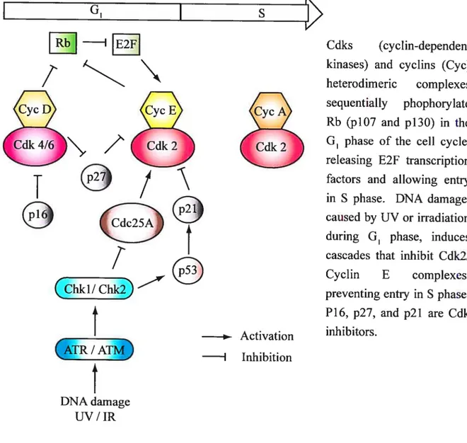
vi 1.8.3 CeH cycle regulation differs between ESCs and differentiated celis 29
1.8.3.1 transition 30
1.8.3.2Sphase 33
1.8.3.3 G2/M transition 33
1.$.3.4TheMphase 34
1.8.3.5 Is ESC cycle regulation an artifact ofcell culture conditions9 34 1.8.4 Telomere maintenance differs between ESCs and their differentiated progenies... 36 1.8.5 Genetic and chromosome anomalies in ESCs: comparison with somatic celis. ..38
1.8.6 Concluding remarks 41
l.8.7Methods 41
1.8.2 Acknowledgments 42
PART III: INTRODUCTION TO REIROVIRUSE$ 43
1.8.9 Structural characteristics 43
1.8.10 Retroviral life cycle 44
1.8.11 Properties ofretroviral vectors 45
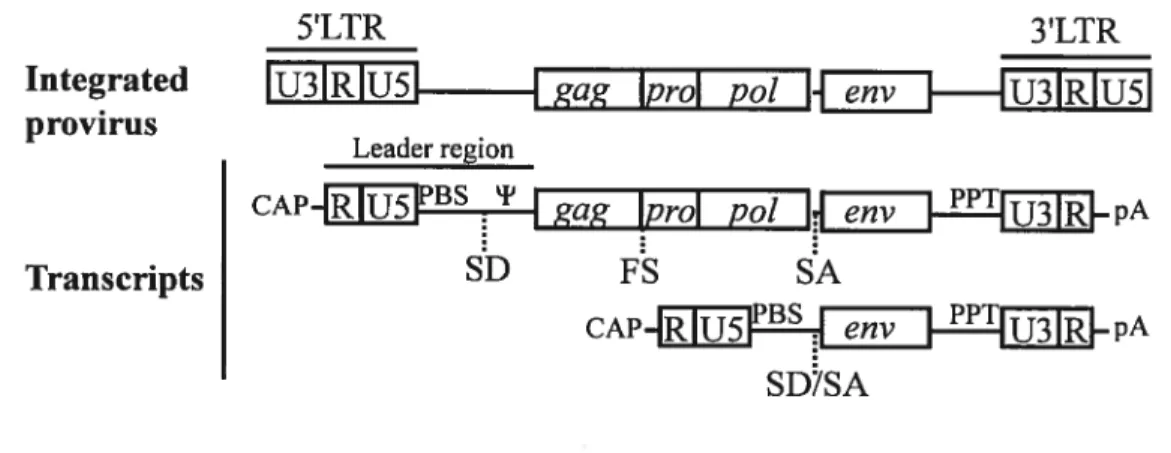
1.8.11.1 Structure of a basic retroviral vector 45
1.8.11.2 Embiyonic stem ce!! viral vectors 47
1.8.12 Properties ofthe packaging celi une 47
1.8.12.1 Principles ofa packaging ceil une 47
1.8.12.2 Tropism and pseudotyping 48
1.8.13 Transduceable ceils 48
1.8.14 Helper viruses, satellite viruses, and satellite RNA 49
1.8.15 Retroviral integration 50
1.2.15.1 Preferential sites of retroviral integration 50 1.8.15.2 Molecular mechanism of retroviral integration 51 1.8.15.3 Determination of retroviral integration sites 51
AIM 0F THE THESIS 53
Chapter 2 APPLICATION 0F A NEW RETROVIRAL SYSTEM 10 CREATE CHRO
MOSOMAL DELETIONS IN ESCs 54
ARTICLE: A retroviral strategy that efficiently creates chrornosomal deletions in
mammalian cells 55
2.1 Author contributions 56
2.2 Abstract 56
2.3 Introduction 56
2.4Results 57
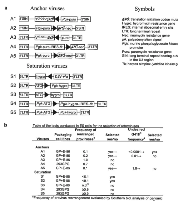
2.4.1 Selection of anchor and saturation proviruses 57
vii
2.4.3 Interchromosomal recombination events .61
2.4.4 In vitro and in vivo differentiation ofrecombined clones 62
2.4.5 Discussion and Conclusions 66
2.5 Methods 67
2.6 Acknowledgments 68
2.7 Supplementary Figures 69
2.8 Supplementary Tables 76
2.9 Supplementary Methods 79
Chapter 3 CREATION 0F A LIBRARY 0F ENGINEERED E$C CLONES SUITED
FOR FUNCTIONAL AS$AY$ 83
ARTICLE 84
DELES: a new library of nested chromosomal DELetions in mouse ES celis suited for
functional screens $4
3.1 Author contributions $5
3.2 Abstract $5
3.3 Introduction $6
3.4 Resuits $7
3.4.1 Generation ofa chromosomal deletion library in ESC clones 87 3.4.2 Presentation of a functional screen performed with puros tertiary clones 95
3.4.3 Global analyses offunctional screens 99
3.4.4 Specific analyses offunctional screens 100
3.5 Discussion and conclusions 108
3.6 Methods 110
3.7 Acknowledgments 113
Chapter 4 DISCUSSION AND PERSPECTIVES 114
4.1 Haploinsufficiency and imprinting 115
4.2 Pending optimizations 116
4.2.1 Complementation approaches 116
4.2.1.1 Identification of a minimal interval conelating with an abnormal phenotype. 116
4.2.1.2 Characterization of deleted segments 117
4.2.1.3 Re-introduction of deleted DNA 117
4.2.1.4 Mapped regions correlating with differentiation anomalies 119 4.2.1.5 Characterization of deletions related to family no.9 120
4.2.2Toward a recessive screen 130
4.2.3 Detection of ESC-derived progenies in situ 131
viii
4.4 Thesis conclusion.133
REFERENCES (FOR CHAPTERS AND APPENDIXES) 134
APPENDIXES xviii
APPENDIX I: Article xviii
Uncovering stemness xviii
Author contributions xviii
Abstract xix
News & Views xix
APPENDIX II: TOWARD THE DESIGN 0F A SUCESSFUL RETROVIRAL SYSTEM xxiii ARTICLE: Shunning pitfalls in retroviral vectors design for functional genomics xxiv
Author contributions xxiv
Abstract xxv
Introduction xxv
Results xxv ii
Rearranged proviruses xxvii
Transmission of undesired retroviral-like particle xxix
Undesired neomycin expression xxxiii
Discussion and Conclusions xxxv
Methods xxxv
Acknowledgments xxxvii
Supplementary Figure xxxviii
Supplementary Methods xl
APPENDIX III: Table xli
Table presenting the genomic features of speculative 3 Mb-deletions anchored to virus Al retroviral integration sites determined by I-PCR (part 1 of 4) xli
Part2of4 xlii
Part 3 of 4 xliii
ix
List of tables
Table I •Advantages and disadvantages of ESC functional genomic approaches 11
Table II • Frequency ofgenetic anomalies detected in mouse ESC clones 40
Table III • Characteristics of independent deletions confirmed by I-PCR and aCGH 61
Table IV • Chimera analysis 66
Table V • Summary of the Cre-mediated recombination around 11 randomly chosen loci
76 Table VI • In vitro differentiation ofprimary and tertiary clones carrying deletions 78
Table VII • Summary of G418’ and G41$R puros tertiary clone generation. (Part 1 of 4) .. 91 Table VIII • Genomic features found in a region spanning a virtual 3-Mb deletion anchored
to the virus Al retroviral integration site related to family no. 5276 109 Table IX • Q-PCR assays employed to detect chromosome 1, 8, 11, and 14 trisomies 112
Table X • Function and expression of RefSeq genes found in family no.9 minimal interval.
125 Table XI • Candidate haploid deletions that could be tested for loss ofheterozygosity. ... 130 Table XII • Potential applications of retroviral-based Cre-ÏoxP recombination 133
X
List of figures
figure 1-1 Early mouse development during the stage of preimplantation 13 figure 1-2 Generation of chimeric mice using micro-injection or aggregation 1$ Figure 1-3 The removal of LIf allows the differentiation of ESCs in EBs in vitro 20 figure 1-4 Effect of seeding density and culture conditions on EBs differentiation 21 figure 1-5 ESC differentiation in mesodermal and hematopoietic lineages on 0P9 stromal
layer 22
Figure 1-6 G1/S transition and G1 DNA damage checkpoint in somatic ceils 30 Figure 1-7 Celi cycle regulation ofundifferentiated and differentiated ESCs 36 figure 1-8 General structure of a simple replication-competent retrovirus 44 figure 1-9 Structures ofretroviral vector plasmids, proviruses, and transcripts 46 figure 1-10 I-PCR allows the determination ofretroviral integration sites 52 figure 2-1 Cre-induced chromosomal rearrangements in mouse ESCs 58 figure 2-2 In vitro and in vivo differentiation ofESC clones with deletions 64
Figure 2-3 Generation of retroviral vectors 69
Figure 2-4 Cre-induced recombination between integrated proviruses 70 figure 2-5 Display showing confirmed chromosomal deletions in ESCs 72 figure 2-6 Evaluation of interchromosomal recombination events 73
figure 2-7 full—length gels and blots 75
Figure 3-1 Schematic representation oftertiary clone generation 89 Figure 3-2 Primary clone retroviral integration sites located by I-PCR 90 figure 3-3 Schematic representation of functional assay approaches 96 figure 3-4 Presentation of ceil counts and performed functional screens 97
Figure 3-5 Ki67 global analyses 100
Figure 3-6 DELES database functionalities 101
figure 3-7 Puros tertiary clones ftom family no. 5077 present normal phenotypes 104 figure 3-8 Puros tertiary clones from family no.5278 present normal phenotypes 105 figure 3-9 Certain puros tertiary clones from family no.5032 present abnormal phenotypes. 106 Figure 3-10 Certain puroS tertiary clones from family no.5276 present abnormal phenotypes. 107 figure 4-1 Determination of a candidate region associated with an abnormal phenotype. 117 figure 4-2 SKY analysis of rare EB cells derived from tertiary clone 9-104 118 figure 4-3 Minimal intervals represented for family no.9 120
xi figure 4-4 Mouse mutant alleles and mapped phenotypes for family no. 9 minimal interval. 123 Figure 4-5 BACs and cDNAs selected to cover clone 9-104 deleted segment 129 figure 0-1 Candidate transcription factors that determine stem celi identity xxii Figure 0-2 Retroviral constmcts were designed to mediate Cre-loxF recombination xxvi Figure 0-3 Rearranged proviruses and transmission of a retroviral-like particle xxx Figure 0-4 Retroviruses associated with unexpected neomycin resistance xxxiv Figure 0-5 Successftil retroviruses and efficient Cre-loxF recombination xxxviii
xii
List of abbreviations
Abbreviation Signification
aCGH Array based comparative genomic hybridization Akt Thymoma viral proto-oncogene
APC Anaphase-promoting complex Apc2 Adenomatosis polyposis cou 2 Apcll Adenomatosis polyposis cou 11 Aprt Adenine phosphoribosyltransferase ASLV Avian sarcoma-leukosis virus ATM Ataxia telangiectasia mutated
aU Attachment sites
BAF Barrier-to-autointegration factor
Bmil B lymphoma Mo-MLV insertion region 1 BMP Bone morphogenic protein
BRCA1 Breast cancer 1
bp Base pairs
BrdUrd 5 -bromouridine
Bub3 Budding uninhibited by benzimidazoles 3 homolog Cdc2O Ceil division cycle 20 homolog
cDNA Complementary deoxyribonucleic acid CDK Cyclin dependent kinase
Cdx2 Caudal-related homeobox 2 Chkl Checkpoint kinase 1 homolog c-myc Myc proto-oncogene protein
CpG C and G nucleotides linked by a phosphodiester bond
DKO Double knockout
DMD Differentially methylated domains DNA Deoxyribonucleic acid
Dpc Days postcoitum
Dppa3 Developmental pluripotency-associated 3 Dppa4 Developmental pluripotency-associated 4
E Embryonic day
EB Embryoid body
xiii EMS Ethylmethanesulphonate
ENU N-ethyl-N-nitrosourea
Env Envelop protein
Eras Embryonic stem cell-expressed Ras ES Embryonic stem (ceils)
ESC Embryonic stem ceil
Esrrb Estrogen-receptor-related receptor beta
EUCOMM European Conditional Mouse Mutagenesis Program Ezh2 Histone methyltransferase enhancer of zeste homologue 2 E4f1 E4F transcription factor 1
Fgf4 Fibroblast growth factor 4
FIAU 1 -(2-deoxy-2-fluoro-f3-D-arabinofumaosyl)-5-iodouracil Gag Group-specific-antigen protein
G0 Gap O phase ofthe celi cycle G1 Gap 1 phase ofthe celi cycle G2 Gap 2 phase ofthe cell cycle
G4 18 Geneticin
G41$R Geneticin resistant
IGIC International Gene Trap Consortium
iPS-MEFs Pluripotent stem cells derived from mouse embryonic fibroblasts iPS-TTFs Pluripotent stem celis derived from adult tail-tip fibroblasts RAT Hypoxanthine Aminopterin Thymidine
HIV- 1 Human immunodeficiency virus HSC Hematopoietic stem ceil
HOX Homeobox
Hoxb4 Homeobox protein Hox-B4
Hprt] Hypoxanthine phosphoribosyl transferase gene hESC Human embryonic stem ceils
Hygro Hygromycin
Hygro’ Hygromycin resistant Hygros Hygromycin sensitive
Hygro Hygromycin sensitive and resistant
IKI\4C International Knockout Mouse Consortium I-PCR Inverse-polymerase chain reaction
Kb kilobase pairs
xiv
KOMP KnockOut Mouse Project
LAP2 ΠLamina-associated polypeptide 2a
LEDGf Lens-epithelium-derived growth factor
11f Leukemia inhibitory factor
LoxP Locus ofcrossover x in P]
LIR Long terminal repeats
M Mitosis phase ofthe ce!! cycle
Mad2 Mitotic arrest deficient 2
Mad3 Mitotic arrest deficient 3
March3 Membrane-associated ring finger (C3HC4) 3
Mb Megabase pairs
MEF s Mouse embryonic fibroblasts
mESC Murine embryonic stem cel!s
MESV Murine embryonic stem ce!1 virus
miRNA Micro ribonucleic acid
MLV Murine leukemiavirus
Mo-MLV Moloney murine leukemiavirus
Mrel 1 Meiotic recombination 11
mRNA Messenger ribonucleic acid
MSCV Murine stem ce!! virus
Nanog Homeobox transcription factor Nanog
Nbsl Nijmegen breakage syndrome 1
NorCOMM North American Conditiona! Mouse Mutagenesis Project
NS Nucleostemin
Oct4 Octamer-binding transcription factor 4
pA Polyadenylation signal
PBS Transfer ribonucleic acid-binding site
PcG Polycomb group genes
PCMV PCC4-ce!1-passaged mye!oproliferative sarcoma virus
PCR Polymerase chain reaction
PIC Preintegration complex
PI3K Phosphatidylinositol-3 -OH kinase
Pol Polymerase
PPT Polypurine tract
Pro Protease
xv PuroR Puromycin resistant
Puros Puromycin sensitive
Puro Puromycin sensitive and resistant
Q-PCR Real-time quantitative polymerase chain reaction Rad5O DNA repair protein RAD5O
Ring lA Ring finger protein 1 Ring lB Ring finger protein 2
RNA Ribonucleic acid
rRNA Ribosomal ribonucleic acid Rtel Regulation oftelomere lenght
RT-PCR Reverse transcriptase-polymerase chain reaction S Synthesis phase of cell cycle
shRNA Small hairpin ribonucleic acid SKY Spectral karyotyping
Smad MAD homolog protein
$ox2 $RY-box2
$tat3 Signal transducer and activator of transcription 3 Suzl2 Supressorofzeste 12
Tbx3 T-box protein 3
Tel] T-cell leukemia/lymphoma 1 Tere Telomerase RNA component Tert Telomerase reverse transcriptase TIGM Texas Institute for Genomic Medicine Tk Herpes simplex tymidine kinase
TKO Triple knockout
TRF 1 Telomeric repeat binding factor 1 TRF2 Telomeric repeat binding factor 2 TRIM5ΠTripartite motif-containing 5 tRNA Transfert ribonucleic acid TTFs Aduit tail-tip fibroblasts UTR Untranslated region
VSV-G Vesicular Stomatitis Virus G
xvi
xvii
Remerciements
J’aimerais d’abord remercier mon superviseur Dr Guy Sauvageau pour son appui inconditionnel tout au long de mes études graduées. Tant son audace que son optimisme auront été des éléments indispensables à la réussite de mon parcours. J’ai bénéficié d’une formation exceptionnelle au sein de son laboratoire, autant conceptuellement que techniquement. Je remercie également mes anciens collègues et mes compatriotes actuels du laboratoire; pour leur aide, leurs encouragements et leur amitié. Je soulignerai la contribution de plusieurs dans le corps de cette thèse. Aussi,je remercie le Dr Richard Martin, ancien étudiant du laboratoire de Dr Trang Hoang, qui m’a initié à la culture des cellules souches embryonnaires. De plus, j’ai profité de l’expertise de plusieurs personnes compétentes que vous découvrirez au fil de la thèse, oeuvrant dans divers Services de l’Institut de recherche en immunologie et en cancérologie (Montréal), de l’Institut de recherches cliniques de Montréal, de la Banque de cellules leucémiques du Québec (Montréal) et du Roswell Park Cancer Institute (Buffalo). Je suis reconnaissante envers toutes les agences de financement qui ont contribué à ma formation soit par des bourses d’étude ou des fonds de recherche alloués à mon projet: les fonds de Recherches en Santé du Québec, les Instituts de Recherche en Santé du Canada, Génome Québec, le Réseau de Recherche en Transgénèse du Québec et les Programmes de Biologie moléculaire de l’Université de Montréal. Finalement, je tiens à remercier ma famille, mon conjoint et mes amis pour leur réconfort, leur humour et leur jugement.
Chapter 1
INTRODUCTION AND LITERATURE
OVERVIEW
Chapter 1 is divided in three sections: the presentation of research objectives (Part I), introduction to embryonic stem celis (Part II), and introduction to retroviruses (Part III). Part II contains two manuscripts related to embryonic stem celi biology: one concerning seif-renewal and pluripotency (published News & Views, Appendix I) and one reviewing selected genetic characteristics (review in preparation). Author contributions to manuscripts are described in the respective sections.
2
PART I: PRESENTATION 0F RESEARCH OBJECTIVES
When I joined the laboratory, the field of genomics was effervescent. International collaborations were underway for the sequencing and the assembly of diverse genomes including human, mouse, and other model organisms. The field of functional genomics was also rapidly developing. Indeed, a combination of approaches was implemented aiming to Iink biological functions to sequence information. We decided to venture in this effort by elaborating a methodology that would be complementary to others that were being developed at that time. Over the years, the genomic knowledge evolved and new functional approaches were designed. Stiil, the methodology described in this thesis subsisted to this active period of time and positioned itself favorably among other expertise.
1.1 The genomic content
The initial analysis of the hurnan genome sequence revealed striking observations. More than 50 % ofthe human genome consists of repeat sequences, often referred as “junk” DNA, which include: transposable elements, processed pseudogenes (retroposed copies of cellular genes), simple sequence repeats, segmental duplications, and blocks of tandemly repeated sequences’. In fact, coding exons and transcript untransÏated regions constitute only 1.2 % and 0.7% ofthe human genome, respectively2. Both for human and mouse, an average of 20 000-25 000 protein-coding genes are predicted (exciuding non protein-coding RNA)2, a number regularly updated with the completion of genome sequencing combined with new computational and experimental data. Ninety-nine percent ofmouse genes have homologues in the human genome; 96% of which are found in syntenic regions3. Ninety percent of mouse and human genomes present conserved synteny along with 40% of alignement3. Several conserved sequences consist of ancestral repeats3. However, comparisons between the genetic material oforganisms such as mouse, human and dog suggested that 2.5-5% ofthe mammalian genome has been under evolutionary selection, thus possibly sustaining biological functions35. These evolutionary conserved elements are though to represent protein-coding genes, untranslated region of protein-coding genes, regulatory elements, non protein-coding RNA, and chromosomal structural elements3. fifty percent of the highly conserved non coding elements cluster in -200 gene-poor regions5. Most of the few genes found in these regions establish or maintain cellular identity (transcription factors involved in differentiation and development, axon guidance receptors)5. Many of these non-coding elements could regulate gene expression by diverse mechanisms, including long-range epigenetic silencing or higher order genome organization5’6. Biological functions and interconnections between
3 most ofthese elements stiil need to be assessed. Obviously, many conserved elements will be acting cooperatively through physical interactions to sustain biological functions. However, it is also expected that some will cooperate functionally toward physiological functions, without physical interactions. This concept, well established in yeast, is referred as synthetic genetic interactions7.
Mammalian genome sequencing and comparative sequence analyses highlight the variable distribution of certain genomic features such as genes, transposable elements, GC content, recombination rate, etc1’3’5. For example, the most repeat-poor region in the human genome is the HOX gene cluster&. Additional conserved repeat-poor regions were identified in mouse and human3. These repeat-poor regions are potential sites of elevated gene regulation3. Another example of non random distribution is the high ftequency of segmental duplications, derived from trans-chromosomal recombination in pericentromeric and subtelomeric regions’. Recombination rates seem higher in distal regions’. According to the genome comparison of different species, synteny block breaks seem to correlate with OC content and might be hot spots ofrecombination, an hypothesis waiting to be addressed5.
f inally, an emerging concept is that some regulatory elements demonstrate conservation, flot primarily at the level of DNA sequence, but at the level of epigenetic marks such as histone modifications6, which can be missed by sequence comparison analysis6. Taking together, these observations suggest that the functional genomics remains largely unexplored.
1.2 Selection of an experimental model
The mouse is an advantageous model to gain insights into human biology and disease. The mouse was already used in our laboratory to study normal hematopoiesis and leukemia. At that time, we wanted to perform a functional screenin vitro,paving the way for an analysis
in vivo,to identi1’ hematopoietic stem ceil regulators.
Mouse embryonic stem cells (ESCs) became our selected model for several reasons. ESCs can be maintained in vitrofor extended period oftime, usually without compromising their euploid karyotype. Also, procedures were already established to differentiate theminvitro
in multiple celi types, particularly into mesoderm derivatives such as hematopoietic lineages. In addition, they could be used in vivo to produce mouse chimeras when re-introduced into embryos or be employed to generate teratocarcinomas when injected subcutaneously into
4 syngeneic mice. Finally, their genome was accessible and modifiable, as illustrated by a growing number ofmutagenesis strategies applied to these ceils. Therefore, ESCs combined both the in vitro and in vivo differentiation potential in addition to the mutagenesis suitability, resulting in an ideal lineage for functional genomics. In fact, they were already prized for such approaches, as described in the upcoming section.
1.3 Functional genomic approaches applied to ESCs
We needed to select a functional genomic strategy that we would apply to ESCs. Some technologies were already optimized at that time, most of them improved over the years and new ones appeared. Different advantages and disadvantages could be recognized for these methodologies. As a preamble, both past and present contexts will be presented to underscore the relevance ofthe selected approach (next section).
1.3.1 Gene targeting
Gene targeting is a methodology that relies on homologous recombination to introduce a modification in a selected region. Typically, a vector containing a selection marker gene flanked by two homology arms is used to abolish the function of a gene, usually by removing the first coding exon. Removal of the selection marker gene is recornmended using Cre loxF or Flp-frt technologies to prevent unspecific effects8. Cre or F lp are site-specific recombinases that catalyze the recombination between two loxP orfrt sequences, respectively. Because various mutations can cause embryonic lethality, precluding analysis later during development and adulthood, conditional gene targeting approaches were designed, again relying on Cre-loxP or Flp-frt technologies. For this purpose, gene inactivation is regulated in a spatio-temporal manner according to the tissue-specific expression or induction of the recombinases.
Gene targeting approaches were well established at the time this project was initiated but remained time consuming. It was laborious to get information about selected loci, to obtain fragments of DNA corresponding to the targeted regions (e.g., physical maps and BAC contigs were largely unavailable), and to create targeting vectors. Today the picture is completely different: the mouse genome sequence is freely available, mapped libraries of BACs and already-made targeting vectors9 are obtainable, new engineering approaches allow easier plasmid and/or BAC modifications’°, etc. Even with these improvements, gene targeting is stili laborious mostly because of the work involving the identification of the
5 proper E$C clones that bear the desired modification(s). However, among the advantages of the methodology are the knowri and precise location of alterations and the accessibility to almost any region, transcribed or not. So far, ‘4000 genes have been targeted in the mouse, with or without a conditional approach11. This number is expected to increase shortly with the targeting of 18 500 additional genes by an international effort (IKMC: International Knockout Mouse Consortium12) conducted by KOMP (KnockOut Mouse Project), EUCOMM (European Conditional Mouse Mutagenesis Program), and NorCOMM (North American Conditional Mouse Mutagenesis Project)11.
1.3.2 Gene trap screens
Gene trap screens are currently the companion of gene targeting with the aim of inactivating every gene in the mouse genome’2. Different trapping vectors have been generated over the years, based either on plasmids or retroviruses. The principle behind the trapping strategy is to catch a complementary genomic feature which is missing for the expression of a selection gene found in the trap vector: promoter, polyadenylation signal (J)A), etc. Depending on the type of vector used, different trapping biases are observed. for example, promoter traps rely on actively transcribed regions while pA traps do flot, some retroviral vectors show preferential integration site (discussed later in section III) sometimes resulting in hypomorphic rather than nul! alleles. Some trap vectors are quite sophisticated, allowing conditional knockdown of gene expressio&3. The International Gene Trap Consortium (IGTC) manages at least 45 000 ESC unes, with integration covering 40% of known mouse genes14. The Texas Institute for Genomic Medicine (TIGM) is currently generating a gene trap library of? 350 000 C57BL/6 E$C clones, expected to cover 1 3 000 genes to completion this year12. This methodology is popular because of its simplicity. Integration sites are mapped by different methods such as plasmid rescue or inverse-PCR (discussed later in section III).
1.3.3 shRNA screens
shRNA-based screens, employing small hairpin RNAs to suppress gene expression, were emerging at the time this project was initiated and are now commonly used in ESCs. Elegant vectors are based on lentiviruses coding both for a shRNA and the corresponding inducible target gene’5. Even if non specific off-target effects and/or partial rather than complete suppression of the gene of interest are frequent, the methodology is relatively efficacious. With the availability of lentivirus-based shRNA libraries16 or microarrays of
6 concentrated lentiviruses spotted on glass slides17, this methodology should be increasingly used in ESCs. for the moment, RNA interference approaches target protein-coding and non protein-coding transcripts, but cannot target untranscribed regions. However, it is now suspected that microRNAs participate in undefined ways to processes such as methylation and heterochromatization’8 and maybe one day, these functions will be exploited in ESCs.
1.3.4 Other insertional mutagenesis screens
Gene targeting, gene trap, and shRNA screens can be viewed as insertional mutagenesis because they rely on vectors integrating in the genome. Additional insertional mutagenesis tools are used in mouse models such as replication-competent retroviruses or retrotransposons, mainly to find proto-oncogenes or tumor suppressor genes19. Although these methodologies could be adapted to ESCs, there are other alternatives that appear more advantageous. for example, replication-incompetent retroviral gene trap vector equipped with a reporter gene can be both mutagenic and be exploited to detect the expression profile of the trapped gene. In the case ofDNA transposons, such as Sleeping Beauty, they act through a eut and paste (excision and integration) mechanism induced by a transposase. Unfortunately, the integrants are subjected to remobilization, leaving behind hardly detectable mutagenic footprints. In addition, these transposons have a tendency to jump in their neighborhood rather than randomly in the genome19.
1.3.5
Chemical screens
Chemical screens can be classified in two broad categories: one relying on chemicals as mutagens and the other on compound libraries that alter the function of ESC without necessarily affecting their genome.
Chemical mutagens such as N-ethyl-N-nitrosourea (ENU) or ethylmethanesulphonate (EMS) were used in ESC to create single base substitutions or alterations in mRNA splicing, transcription, or stability20’21. The frameshifi mutagen ICR191 was also used to induce the addition of guanine stretches 21 According to the loss-of-function experiments evaluated
for the selectable hemizygote locus hypoxanthine phosphoribosyl transferase gene (Hprtl), mutation frequency is in the range of 1 per 1000 to 1 per 1200 cells depending on the conditions tested20’21, implying multiple mutations in each genome. Because of this amount of subtle mutations that do flot contain a landmark for identification, it is necessary to create chimeric mice and proceed through breeding to first dilute the mutation load and then to isolate
7 candidate gene(s) by positional cloning (germ-line transmission is achievable). Different phenotypes have been observed in mice and important genes identified using this system 21
24• Several genome-wide ENU-or EMS-based screens for dominant and recessive mutations are currently ongoing. Major efforts include those conducted by the British, German, Australian, American, Canadian, and Japanese groups25. Transient expression of Bloom in ESC can stimulate homologous recombination between sister chromatids or homologous chromosomes, allowing the recovery ofbi-allelic mutations26. This strategy was used in ESC to study a precise pathway (glycosylphosphatidylinositol-anchor biosynthesis) and defects were complemented by candidate genes (cDNA transfection)26. For clones flot successftilly complemented with known genes, other methods must be used to identify the mutated gene (if it is a gene) or a companion in the same pathway (for example: cDNA library). However, if a combination of determinants is necessary to rescue the phenotypic anomaly, it is difficuit to achieve at the genome-wide level. Focusing on particular chromosomal regions can be an advantageous strategy to use with chemical mutagenesis. Chemical mutagenesis used in combination with heterozygote chromosomal deletions (see beneath for the methodologies to create deletions) or heterozygote chromosome balancers (chromosome containing an inversion suppressing chromosomal recombination for this region, a dominant visible marker, and a recessive gene inducing lethality in a homozygous state25), simplifies the breeding scheme. The mutations caused by chemicals are limited in size and in type according to the mutagen used (for example, single-base substitution involving AI base pairs predominate for ENU20). Importantly, ENU-induced mutations are not bias for any region ofthe genome. Moreover, ENU-mutagenized collection of ESC clones can be screened for mutations in selected genes, allowing the recovery of allelic series27. These ESCs can be reintroduced in developing embryos to create mouse chimeras and these specific mutations can be transmitted in the germ-line27 This procedure allows thein vivo functional evaluation of precise protein
domains27.
Screening using small molecule libraries is an emerging application in the ESC field, which is expected to be employed more extensively in the near future. For example, a library was used to identify compounds able to maintain E$C self-renewal/pluripotency without the use of serum, LIF, or feeders28. The approach was powerful because the team used a pluripotency reporter gene (Oct4-GFP) in ESCs and plated them in 384-well plates28. They characterized one ofthese compounds (SC 1: pluripotin), which allows the maintenance of ESC in a minimal media without compromising their ability to differentiate in vitro and
in vivo in chimeric mice28. Contribution of E$Cs to the gonads of these chimeric mice was proven28, although the proper functionality of the gametes was not assessed by germ-line
8 transmission. Importantly, by immobilizing the compound to an affinity matrix and using mass spectrometry, this group identified two cellular targets of their small molecule (Erkl and RasGAP)28. They further showed that the combined inhibition of both proteins was necessary to maintain self-renewal/pluripotency in the conditions used28.
1.3.6 Mutagenesis with oligonucleotides
The use of single-stranded DNA oligonucleotides to permanently modif’ 1-4 targeted nucleotides in ESC is a recently developed application. Oligonucleotides can be obtained faster than gene targeting vectors not already made. However, both methodologies require the same amount of work to isolate ESC clones and assess the proper targeting. The ftequency of oligonucleotide-based targeting is estimated to be 0.25-1.5 per 106 ceils, as tested on a
limited number ofloci29. Problematically, this methodology is suppressed by DNA mismatch repair mechanisms, thus requiring the transient suppression of proteins such as Msh229. As a consequence of repressing transiently mismatch repair mechanisms, increased frequency of spontaneous mutations is observed on reporter genes29. The distribution of these bystander mutations is unknown, but expected to be lost during mouse breeding since the targeted alteration can be germ-line transmitted29. However, this pitfall should be taken into consideration when designing in vitro screens. Because the mutation is targeted, as opposed to chemical screens, in vitro screens could probably be achieved with the use of independent targeted clones.
1.3.7 Gain-of-function screens
Gain-of-function screens have been applied successfully to ESC. A strategy using episomal transduced cDNA libraiy (derived from E$C) identified Nanog, an homeodomain protein allowing the self-renewal of ESC without leukemia inhibitory factor (LIF)30. Microarray analyses can also be combined to this type ofscreen31. In a way, gain-of-function screens are attractive because of their simplicity and their rapidity. However, to apply the methodology to a genome-wide level, libraries are disadvantageous. For example in home-made cDNA libraries, very long cDNAs are under-represented, more abundant transcripts
areover-represented, some cDNAs are incomplete, etc. b circumvent these drawbacks, one could think about using a BAC library. However, some genes are 50 big that they are not
covered by a single BAC, such as dystrophin (2,2 megabase pairs, 79 exons) (http://genome. ucsc.edu/., Mouse Build 36, 2006)32. fortunately, vast libraries can be purchased, arrayed in multi-well format (example BACs libraries) or spotted in a book (example: Riken cDNA
9 library) allowing fora better aftempt at normalization of each products. Aside from unwanted effects caused by the ectopiclover- expression (toxicity, non physiological expression levels, abnorma cellular localization, etc.), many studies proved that valuable candidate factors can be isolated with this strategy.
1.3.8 Irradiation-based screens
Deletions can be produced in ESCs engineered to express the herpes simplex tyrnidine kinase (tk) gene, by physical irradiation and negative selection (drug FIAU)33. Although anchor sites (tk) introduced in various genomic regions can be identified by plasmid rescue experiments, the mapping of each deletion is difficuit since it requires PCR analysis of numerous simple sequence length polymorphism markers33. The possibility of unidentified genetic lesions also complicates the interpretation of resuits generated with this approach.
1.3.9
Cre-toxP
technology based screens
The Cre-toxP technology has been applied by several groups to create large deletions, but this system is also appropriate to create transiocations, inversions, and duplications. To produce a deletion in ESC, two regions on the same chromosome are successively targeted by homologous recombination using distinct vectors, each canying a lox? site34. $ubsequently, the transient expression of Cre leads to the excision of DNA bePveen the integrated ÏoxF sequences. To isolate ESC recombinants, two nonfunctional halves ofa selection marker gene are inserted in complementary targeting vectors. ESCs are selected in media containing the proper drug(s). Although different combination of marker genes have been developed over the years35, the functional reconstitution ofHprt] is more widespread, presumably because it was the first reported system34. Hprt]-deficient E$Cs need to be ernployed in this case.
A variation of the Hprtl reconstitution method was also elaborated, where the first loxP site was anchored by homologous recombination and the second, delivered by a retroviral vector, avoiding one step of gene targeting36. Another strategy to omit one round of homologous recombination consists in targeting one loxP site, followed by the co-introduction of a ÏoxP-containing plasmid together with the cre, and finally by selecting recombination events by negative selection toward the tk gene incorporated inside the vectors37.
10
1.4 The proposed approach
Several reasons drove the design ofa screen based on chromosomal deletions: several contiguous determinants could be interrogated at the same time, both protein-coding and non protein-coding regions could be screened, potential synthetic interactions could be observed, the alleles were permanently deleted and flot only silenced, and the primary work could 5e done in vitro. Irradiation methodology was not a possibility because the mapping of deletions was not precise and like chemical screens, subject to bystander mutations. A Cre lox? strategy was favored but the laborious step ofhomologous recombination was repelling. We wanted to screen many regions on a genome-wide scale within a reasonable timeframe. In order to overcome these impediments and to bring on an additional tool for functional genomics, two complementary retroviruses were created, each containing a loxP site and capable of rapidly generating deletions in mammalian celis following the addition of Cre. Table I recapitulates the advantages and disadvantages of the methodologies presented in the previous section in addition to the retroviral-based method which I developed in our laboratory. The remaining sections of the introduction will focus on embryonic stem ceils and on retrovirology.
11 Table I •Advantages and disadvantages of ESC functional genomic approaches
Methodologies Advantages Disadvantages
Gene targeting Precise location, precise modification. Labor intensive, time consuming no bias although better frequencies for
some regions
Gene trap Simple, rapid, localization easy Integration bias depending on vectors
RNA interference Simple, rapid Possible non specific off-target effects, variable degree of suppression
Chemical (mutagens) No localization bias reported, Multiple mutations, very subtle hypomorphic, hypermorphic, mutations, laborious identification loss-of-function alleles possible (allelic
series)
Chemical (compounds) Simple, rapid Limited by the library of compounds, dependent on the concentration of compounds, the target(s) might be difficult to identi1’
Oligonucleotides Precise location, simple Possible bystander mutations, characterization of ESC time consuming
Overexpression——— Simple, rapid Depends on libraries coverage, might
be prone to unspecific effect (toxicity, non physiological expression levels, etc.)
Irradiation (deleons) Simple. rapid, can involved large Localization of endpoints difficuit, chromosomal segments possible bystander genetic alterations
Cre-loxP (both loxP Various rearrangements possible A least one round of laborious and targeted or one targeted+ (deletions, inversions, translocation, time-consuming gene targeting, some one introduced by retroviral etc.), precise location, can involved bias might be observed with
gene transfer) large chromosomal segments retroviruses
Our approach: Cre-loxP Various rearrangements possible Some bias might be observed with (both loxF introduced by (deletions, inversions, translocation, retroviruses
retroviral gene transfer) etc.), precise location, can involved large chromosomal segments, avoid one round of gene targeting
12
PART II: INTRODUCTION TO EMBRYONIC STEM CELLS
The in vitro derivation from blastocysts ofthe first mouse embryonic stem celi (ESC) unes was reported in 198138,39 These lineages were holding great promises in developmental
biology because of their ability to differentiate into complex tissues in vivo, to form embryoid bodies (EBs) in suspension culture, and to produce teratocarcinomas when injected subcutaneously in syngeneic mice38’39. As opposed to teratocarcinoma ceils, ESCs have a normal karyotype and contribute more successfully to the germ-line of chirneric mice38’40. The focus of this section is to review the origin and some of the cardinal features of mouse E$Cs: seif-renewal and pluripotency, differentiation, and particular genetic properties.
1.5 The origin ofESC
The protocols currently used to derive E$C unes are similar to those established more than 20 years ago41. Blastocyst stage embryos or isolated muer cell masses (Figure 1-1) are plated on mouse embryonic fibroblast (MEF s) in tissue culture media41. Following several days of culture, the masses are dissociated and replated again on MEFs to generate various differentiated and undifferentiated lineages4t. Colonies with undifferentiated morphology are individuaily isolated and are expanded to generate ESC lines41. Most E$C lines are 4OXY because in XX ESCs both X chromosomes are active, an unstable state (in fact one of the X is frequently loss) that correlates with global reduction of DNA methylation which is flot favorable for the maintenance ofthese cells41’42.
An ongoing debate is the tissue of origin of ESC, the existence of an in vivo counterpart, ami the possibility of being an artifact lineage generated from an adaptation to culture environement43. Cells from both the iimer cell mass and from the primitive ectoderm (Figure 1-1), a tissue derived from the inner cdl mass, can give rise to ESC unes43. However, since flot ail the ceils contained in these tissues can generate ESC lines, ESCs could possibly emerge subsequently from another celi type, such as early germ cells43. ESCs are flot equivalent to inner ceil mass cells because they contribute weakly to extraembryonic endoderm lineages (derivative ofthe primitive endoderm) in vivo (Figure 1-1). A founder population of ceils emerges from the primitive ectoderm (epiblast) soon before gastrulation, and passes through the primitive streak to give rise to many structures of the extraembryonic mesoderm and to germ cells, a process involving dominant local and inductive signals, which might be reproducible in vitro43. ESCs might be related to this founder population43. In fact, mouse primordial germ celis can generate ESC-like colonies that can be maintained
13 for extended period of time in culture and can contribute to chimeras and germ-line transmission41. b complicate the story further, under particular ceil culture conditions, ESCs can be differentiated into primitive ectoderm-like ceils, which can be differentiated in vitro, but are unabie to contribute to chimeric mice44. As expected, ESC and ail the potential parental lineages share several marker genes (Oct4, Nanog, Dppa3, etc.)43, but none ofthem demonstrate in vivo the permanently high proliferation index of ESC observed in culture. Therefore, this sustained proliferation rate might be the resuit of celi culture conditions or ofthe isolation oftransient ceils with this intrinsic property or more possibiy, the artificial combination ofboth. Fortunately, when ESCs leave the in vitro environment toretumin vivo following re-introduction in the mouse embryo, they respond normally to developmental instructions and therefore, do not correspond to transformed ceils. However, if they are not re-introduced in the proper environment, they create teratomas (or teratocarcinomas) instead of contributing adequately to tissues in place.
Figure 1-1 Early mouse deve]opment during the stage ofpreimplantation. Adapted ftom Raiston, A. & Rossant, J., 2OO5.
Two ceil Four ceil One celi fertiIizedegg) Late Compacted morula Motula morula Primitive Primitive 8 ceIl ectoderm endoderm Early Trophoectoderm
14
1.6 Seif-renewal and pluripotency of ESCs
$elf-renewal and pluripotency are key characteristics that define ESCs and are typically discussed together. Seif-renewal is a mechanism that allows the generation of daughter cell(s) with the same characteristics as the parental ce!!. ESCs are thought to generate two identical daughter ceils per division, a process referred to as symmetrical seif-renewai, as well as to preserve immortality41. This state strictly relies on well-defined culture conditions. Pluripotency refers to the in vivo differentiation potential of clonai ESC to contribute to ail lineages derived from the three primary germ layers (ectoderm, mesoderm, endoderm, including the gametes) as well as the extraembryonic mesoderm46. Because ESCs contribute weakly to extraembryonic endoderm and trophoblast lineages, they are considered pluripotent rather than totipotent such as the fertilized egg or the blastomeres. Specific culture conditions allow the preservation ofthe pluripotency. Ironical!y, to characterize this property, ESCs need to lose it, concomitantly with their identity, through in vivo and/or in vitro differentiation. The evaluation of ESC pluripotency in vivo is the most robust assay to observe both the contribution ofESC to all expected lineages and the proper functionalities ofthese progenies. However, this experimentation is expensive. Other assays, although flot as complete, give reasonable insights into the pluripotency ofESCs. The generation ofteratomas or the in vitro differentiation in selected media allows the observation of representative !ineages from the three primary germ layers. More details about in vivo and in vitro differentiation ofESC will be presented in the next section.
What are the factors regulating ESC identity (self-renewal and pluripotency)? A manuscript (News & Views) was written by Mélanie Bilodeau and Guy Sauvageau in 2006, presenting a general overview of the field and two approaches used by independent groups to find regulators of seif-renewal and pluripotency (Appendix I). An update of this area of research will follow.
1.6.1 Update for mechanisms underlying ESC seif-renewal and
pluripotency
It is important to high!ight that both celi extrinsic and intrinsic mechanisms governing ESC self-renewal and pluripotency impiy not oniy positive regulation, but also repression of differentiation and maybe apoptosis. $ometimes, a single factor can act both as a positive and a negative regulator. Cell signaling cascades initiated extrinsically are necessarily linked to ceil intrinsic parts. In addition, epigenetic characteristics such as DNA methyiation and
15 histone modifications seem involved in E$C self-renewaÏ and pluripotency regulation and their roles should be more extensively defined in the coming years.
BMP4 is a celi extrinsic factor acting as a ligand to a ceil intrinsic signaling cascade [BMP receptor-Smad(s)-Id(s)] that suppresses neural determination47. Similarly, LIF is a ceil extrinsic ligand of a celi intrinsic signaling cascade [LIFR-gpl3O-Stat3-target genes] suspected to inhibit non-neuronal differentiation rather than promoting stem celi survival47. Oct4, Sox2, and Nanog are tbree core transcription factors that positively regulate ESC specific genes, but also bind non-expressed tissue-specific transcription factors48. Oct4 and Nanog are specific to pluripotent celis, but Sox2 is not49. Critical levels of Oct4 are required to maintain ESC in an undifferentiated state: repression of Oct4 conveys to loss of pluripotency and formation of trophectoderm, while the overexpression of Oct4 induces differentiation in primitive endoderm and mesoderm50. Nanog positively maintains ESC self renewal in the absence of L1F3° and is thought to suppress differentiation. Nanog is down regulated during differentiation30, inhibits neuroectodermal differentiation when ectopically expressed30, and Nanog-deficient ESCs produce endoderm (possibly primitive)47. Oct4 and Cdx2 transcription factors reciprocally inhibit each other functions for the determination of pluripotent ceils (Oct4 functions expressed, Cdx2 functions repressed) and trophectoderm (Cdx2 function expressed, Oct4 function repressed)51. Nanog, Gata4, and Gata6 might be regulating a balance between the pluripotent state and differentiation in primitive endoderm. The loss ofNanog or the ectopic expression of Gata4 or Gata6 induces ESC to differentiate into primitive endoderm49. In addition, Esrrb, Tbx3, leu, and Dppa4 also control a set of target genes by activation and repression48. Oct4, Sox2, Nanog, Esrrb, Tbx3, Tcll, and Dppa4 also possibly share some target genes48.
The epigenetic level of regulation is expected to be complex and is just starting to be elucidated. In the case ofDNA methylation for example, although neither Oct4 or Nanog genes present annotated CpG islands, their respective promoter present cytosine methylation, correlating with their expression (low level of methylation correlating with expression)6. Methylation is thought to induce repression by preventing the binding of some proteins to DNA (such as transcription factors) and/or by binding methyl-CpG binding proteins that interact with histone deacetylases6.
ESCs present bivalent domains containing the dual repressive (lysine 27 of histone H3 [H3K27] tri-methylation) and activating (lysine 4 of histone H3 [H3K4] tri methylation) histone marks49. These bivalent domains correspond to highly conserved non
16 coding elements49. Several of them are co-occupied by Oct4-Sox2-Nanog and are found in proximity of some (but flot ail) developmentally important genes silenced in E$C, but activated during differentiation49. During differentiation, the bivalent domains presenting activating and repressive histone marks are resolved: expressed genes are associated with H3K4 tri-methylation, tumed off genes are associated with H3K27 tri-methylation, whlle the weakly induced genes keep both signatures49. It is hypothesized that bivalent domains may silence developmental genes in E$Cs, but also keep them poised for activation during differentiation52.
Silencing could be mediated in part by Polycomp group gene (PcG) complexes. Two of the four known PcG complexes are important in ESC, mainly PRCY and PRC248. Methylation of H3K27 is induced by the PRC2 complex, which includes eed (embryonic ectoderm development), Suzl2 (suppressor of zeste 12), and Ezh2 (Histone methyltransferase enhancer of zeste homologue 2)48. H3K27 methylation is a binding site for the PRC 1 complex involving RinglA, Ring]B, and 3mi148. The precise roles of PcG complexes and associated histone modifications in ESC are not completely understood, but likely interfere with nucleosome dynamics and transcription initiation48. Eed- and Ring]3- deficient ESCs present derepressed transcriptional regulators of development48. The recruitment of PRC2 complex to targeted loci may be mediated in part by Oct4, Sox2, and Nanog48.
The mechanisms allowing ESCs to remain undifferentiated and to survive in cell culture conditions are not well defined. Is it the same factors that keep them seif-renewing in an undifferentiated state that prevent their apoptosis? In standard culture conditions (presence of LIF and BMP), few ESC undergo apoptosis53. Are these culture conditions compatible with ESC apoptotic death? The answer is not obvious, because removal of LIF and/or BMP changes the fate ofESC. Altematively, ESCs might have a cell intrinsic machinery preventing their death by apoptosis.
At first sight, deficiency in the gene Zfx uncouples ESC seif-renewal properties (apparently lost) from their pluripotential properties (apparently maintained), while in fact, several celis are lost by apoptosis53. Zfx, located on the X chromosome, encodes a zinc finger protein containing a DNA-binding and a transactivation domains53. ESCs (XY) deficient in Zfx present abnormal morphology and are defective in proliferation because they die from apoptosis, an effect highlighted in serum-free condition in presence of LIF and BMP453. Strangely, Zfx-deficient male embryos (germ-line deletion) develop normally until E9.5 before dying ofuncharacterized extraembryonic tissue anomalies53. When Zfx-deficient ESCs
17 are induced to differentiate in embiyoid bodies, in teratomas, or in chimeric mice, they do so roughly normally (except that they fail to contribute to thymus and bone manow of chimeric mice)53. Zfx overexpression in E$C correlates with massive ceil death in presence of LIF, with abnormal EBs formation (absence of LIF), and with failure to contribute to chimeric mice53. At the molecular level, Zfx deficiency in ESC leads to the up-regulation of stress induced genes53. Also, Zfx binds to the promoters ofTbx3 and Tc1153. Overexpression or deficiency ofZfic increases or reduces the expression ofthese genes, respectively53. Because at least two suspected equilibriums regulate the pluripotency and the extraembiyonic tissue differentiation (Oct4-Cdx2 in the case ofthe trophoectoderm and Nanog-Gata4-Gata6 for the primitive endoderm), the morphology of Zfx-null ESC colonies is altered and Zfx-deficient mice die from extraembryonic defects, it would be interesting to investigate whether dying ceils are in fact differentiated and hardly maintained in ESC-defined culture conditions.
1.7 Differentiation of ESC
1.7.1 Contribution ofESCs to mice
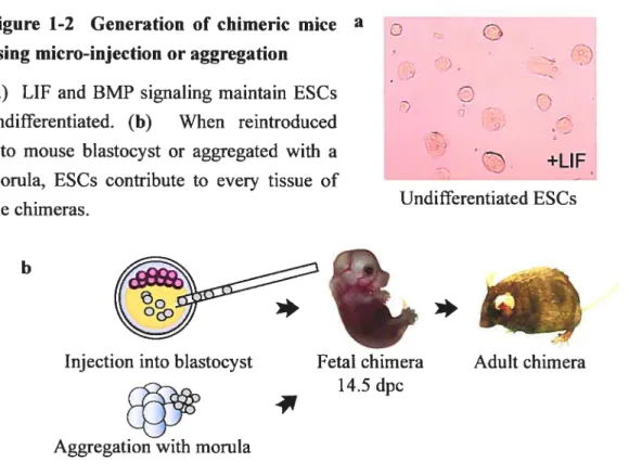
Micro-injection of E$Cs in the blastocoel cavity of mouse blastocyst-stage embryo, followed by transfer to pseudopregnant female, was the first methodology developed to generate chimeric mice with ESCs54 and is stiil currently used today (Figure 1-2). When ESC are injected into blastocysts, they efflciently colonize the tissues that form the fetus and the extraembryonic mesoderm46. Moreover, these ceils contribute very inefflciently to extraembryonic endoderm and trophoectoderm formation46. Initial studies showed that groups of 10-15 ESCs or a single ESC could contribute to chimeric mice, although the percentage of chimerism was systematically lower for the latter46. Today, it is thought that possibly 1 or 2 or occasionally 3 ESC(s) contribute to the chimeric mice55. Interestingly, single ESC selected according to their large Q15tm) or small (10tm) size demonstrate no difference in their contribution potential46.
ESC micro-injection requires expensive equipments, is time-consuming, and necessitates a serious training56. Consequently, the aggregation method was elaborated as an alternative (Figure 1-2). For this technique, ESC clumps are cultivated overnight in proximity of morula-stage embryos (undergoing or just completed compaction, with the zona pellucida removed) in littie depressions56. The following day, aggregated embryos are transfened to pseudopregnant foster mothers56. Competent chimeras for germ-line transmission of ESC-derived gametes are generated efflciently with both the micro-injection
1$ and the aggregation methods56. In the aggregation method, random-bred (like CD1) morulas are used advantageously, since the outbred female mice generate more embryos following superovulation compare to inbred strains56. However, the use of inbred blastocysts (like C57BL/6) is more efficient for micro-injection 56•
Figure 1-2 Ceneration of chimeric mice a using micro-injection or aggregation
(a) LIF and BMP signaling maintain ESCs
O
undifferentiated. (b) When reintroducedinto mouse blastocyst or aggregated with a
+LIF morula, ESCs contribute to every tissue of
-Undifferentiated ESCs the chimeras.
b
A variation of the aggregation technique is to use tetraploid morula stage embryos. Electrofusion is performed on blastomeres of a two-cell stage embryo (diploid), creating the tetraploid ceil that is further maintained until the morula stage57. Altematively, ESC can be injected in the blastocoel cavity ofa tetraploid blastocyst58. Using this set-up, ESCs contribute to the fetus and extraembryonic mesoderm, while the tetraploid celis are generally restricted to the trophectoderm and the extraembryonic endoderm57. During early embryogenesis, the exact moment where tetraploid celis are out-competed by ESC-derived progenies is not known. Although not labeled autonomously, tetraploid ceils have been noticed in ail the analyzed chimeric embiyos during the gastrulation stage (E6.5-7.5), presenting variable contribution (3-80%) to embryonic derivatives of the three primitive genn layers59. When labeled autonomously, tetraploid celis were shown to contribute sporadically to 1% ofcells in a chimeric fetus (E 10), and sometimes to cluster in the hindgut endothelium, the aortic musculature, and the branchial arch vasculature59. Importantly, F1 hybrid ESC unes are crucial for tetraploid complementation assays because ESC derived from inbred embryo engender neonates that die shortly afler birth with respiratory distress58. The molecular
*
*
Injection into blastocyst
Aggregation with morula
Fetal chimera Aduit chimera 14.5 dpc
19 basis underlying the correlation between the limited genetic heterogeneity and the respiratory defect is unknown, but can be bypassed by Iaser-assisted injection of E$Cs in 8-celi stage diploid morulas (method described beneath)58’60. However, tetraploid complementation assay is achievable with ESC unes derived from two related mouse substrains, such as the Ri ESC une (derived from a cross between two 129 mouse substrains)61. Newbom animais derived from this methodology are also referred to as F0 mice because they are derived (almost) completely from the E$Cs, including their gametes, thus bypassing a step of mouse breeding necessary for traditional chimeras to obtain germ-line transmission.
An exciting method recently developed to create F0 mice consists in laser-assisted injection of ESCs in 8-ceil stage morula60. Similar to tetraploid complementation, almost entirely ESC-derived chimeras are obtained, even with lower contamination from the host celis (0.1% instead of 2%)60. Total germ-line transmission (100%) is observed rnost
of the time because of gender conversion60. The most important point is that either inbred or hybrid ESCs and either inbred or outbred host embryos can be used without presenting f0 mice with obvious abnormalities60. It is fascinating that inbred E$Cs, laser-injected in 8-ce!! stage morula, can generate F0 mice free of respiratory distress while injection in tetraploid blastocyst frequently fails to generate normal mice62. Impressive images show the contribution ofinjected ESCs to the totality ofthe inner ce!! mass with this technique, while injection in the blastocyst resuits in a mixture of ESC and host derived ce!!s60. However with both techniques, ESCs fail to contribute notably to the extraembryonic endoderm60, reinforcing the idea that ESCs might be more related to primitive ectoderm than the inner ceil mass.
1.7.2 In vitro
differentiation of ESCs
1.7.2.1 In vitro differentiation methods
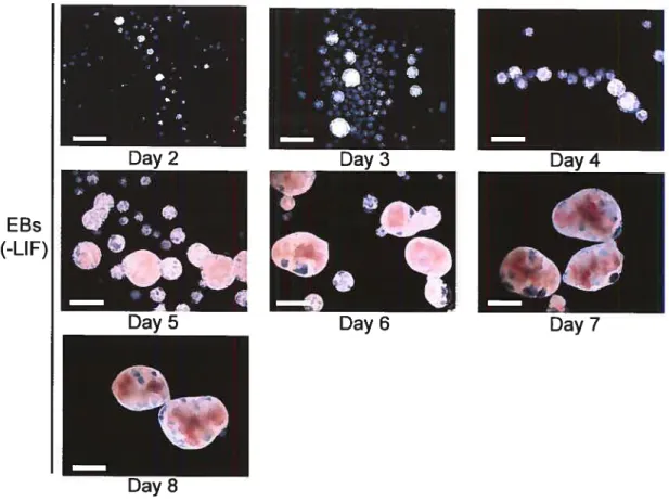
In vitro, three methods are usually used to induce ESC differentiation in absence of LIF. The first one is to grow ESC in !iquid or semi-solid differentiation media to generate three-dimensional aggregates called embryoid bodies (EBs) (Figure 1-3). 11e EBs differentiation allows complex developmental programs to occur, mediated by numerous ceil-ceil interactions44. This complexity can 5e problematic when trying to understand the differentiation into particular lineages, which relies on the proper deve!opment of other lineages.
20
figure 1-3 The removal of LIF allows the differentiation of ESCs in EBs in vitro.
Scale bar: 500 microns.
EBs
(-LIE)
To add to the fact that each serum lot provides an undefined blend of extracellular
factors, the culture media likely becomes rapidly conditioned because the seeding density
makes a significant difference in the differentiation profile ofESCs (Figure 1-4). Depending
on culture conditions, EBs can present fascinating shapes that can be misinterpreted as
phenotypic anomalies. For examples, if debris are present in the culture media, EBs have the tendency to wrap around it or, when plated at high density, to fuse into deformed aggregates (Figure 1-4).
21
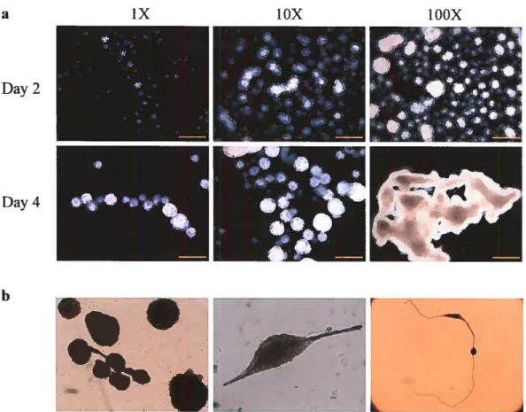
Figure 1-4 Effect of seeding density and culture conditions on EBs differentiation.
(a) The EB differentiation profile is affected by the seeding density. At high density, EBs fuse in bizarre aggregates. Scale bar: 500 microns. (b) EBs’ fascinating shapes when large debris are found in the culture media.
a iX YOX TOOX
In addition, ESCs can be differentiated over a stromal celi layer such as 0P9 celIs63
(Figure 1-5). 0P9 celis were derived from the calvaria of a newbom mouse deficient in the M-CSF gene63. Coculture of ESCs with these strornal ceils allow mesodermal and lymphohernatopoietic differentiation without the addition ofgrowth factors (but in the presence ofserum)63. This might be a simpler way to obtain particular ceil types because differentiation occurs in a monolayer interacting with the stromal ceils. This type of differentiation is also influenced by the seeding density. for particular purposes such as expression studies, stromal ceils need to be separated from ESC-derived progenies. Finally, ESC can be differentiated straight in monolayer or on extracellular matrix, with particular media.
Day 2 Day 4 b -À
.
9%22 Figure 1-5 ESC differentiation in
. mesodermal and hematopoietic
j
hneages on 0P9 stromal layer
-Scale bar: 250 microns.
teb —
1.7.2.2 Additional considerations regarding E SC in vitro differentiation
Although ESC differentiation is a remarkable tool, there are severai pitfal!s. ESC
in vitro differentiation is highly modutated by ce!! culture conditions. Surprisingly, in vitro
differentiation allows ESCs to participate in particular !ineages such as the extraembryonic endoderm64 while they are inefficient to do so in vivo. Additionally, when ESCs are geneticaily modified (for exampie: suppression of Oct4), they form trophoectoderm (tested in presence of LIF), a phenomenon ca!!ed dedifferentiation50. In addition, because of their property to fuse at low frequency with other ceils, ESC can be the unsuspected cause of another process ca!ied transdifferentiation (changeiii ceil fate)65. Fina!ly, although the in vitro differentiation ofESC can be tempora!iy representative ofearly embryogenesis as discussed be!ow, it occurs without an organization such as axis formation.
for ail these reasons, three characteristics were estabiished to conc!ude that an ESC
in vitro differentiation model in a particuiar !ineage is relevant44. First, the system must be efficient and reproducible44. Second, the system should recapituiate the deve!opmental program observedin vivo44. And fina!ly, the differentiated ce!ls should be functional in culture and when transplanted in animal mode!s44.
1.7.2.3 Successfully derived lineages
Primary germ layer induction during ESC differentiation shares pathways that are found in embryogenesis: bone morphogenic protein (BMP) and other transforming growth factor-13 (Nodal/Activin) signa!ings , Wnt signa!ing, and fibroblast growth factor (Fgf) signaling66.
In vitro differentiation produces representative iineages from the mesoderm (hematopoietic, vascular, cardiac, skeletal muscle, osteogenic, chrondrogenic, adipogenic), the
![Figure 1-1 Early mouse deve]opment during the stage ofpreimplantation. Adapted ftom Raiston, A](https://thumb-eu.123doks.com/thumbv2/123doknet/2052065.5456/32.918.215.754.533.1073/figure-early-mouse-opment-stage-ofpreimplantation-adapted-raiston.webp)