Anti-Invasive, Antitumoral, and Antiangiogenic Efficacy of a
Pyrimidine-2,4,6-trione Derivative, an Orally Active and Selective Matrix
Metalloproteinases Inhibitor
Erik Maquoi,1 Nor Eddine Sounni,1 Laetitia Devy,1 Fabrice Olivier,1 Francis Frankenne,1 Hans-Willi Krell,2 Frank Grams,3 Jean-Michel Foidart,1 and Agnès Noël1
1Laboratory of Tumor and Development Biology, University of Liège, Centre Hospitalier Universitaire, Liège, Belgium; 2Roche Diagnostics
GmbH, Division Pharma, Penzberg, Germany; and ³Discovery Technologies, Preclinical Research Pharma, F. Hoffmann-La Roche AG, Basel, Switzerland
Abstract
Purpose: The implication of matrix metalloproteinases (MMPs) in the major stages of cancer progression has
fueled interest in the design of synthetic MMP inhibitors (MMPIs) as a novel anticancer therapy. Thus far, drugs used in clinical trials are broad-spectrum MMPIs the therapeutic index of which proved disappointingly low. The development of selective MMPIs for tumor progression-associated MMPs is, thus, likely to offer improved therapeutic possibilities.
Experimental Design: The anti-invasive capacity of a series of pyrimidine-trione derivatives was tested in vitro
in a chemoinvasion assay, and the most potent compound was further evaluated in vivo in different human tumor xenograft models. The activity of this novel selective MMPI was compared with BB-94, a broad-spectrum inhibitor.
Results: Ro-28-2653, an inhibitor with high selectivity for MMP-2, MMP-9, and membrane type 1 (MT1)-MMP,
showed the highest anti-invasive activity in vitro. In vivo, Ro-28-2653 reduced the growth of tumors induced by the inoculation of different cell lines producing MMPs and inhibited the tumor-promoting effect of fibroblasts on breast adenocarcinoma cells. Furthermore, Ro-28-2653 reduced tumor vascularization and blocked angiogenesis in a rat aortic ring assay. In contrast, BB-94 up-regulated MMP-9 expression in tumor cells and promoted angiogenesis in the aortic ring assay.
Conclusion: Ro-28-2653, a selective and orally bioavail-able MMPI with inhibitory activity against MMPs
expressed by tumor and/or stromal cells, is a potent antitumor and antiangiogenic agent. In contrast to broad-spectrum inhibitors, the administration of Ro-28-2653 was not associated with the occurrence of adverse side effects that might hamper the therapeutic potential of these drugs.
INTRODUCTION
Matrix metalloproteinases (MMPs) are a family of more than 23 zinc-dependent neutral endopeptidases that are collectively capable of degrading structural components of the extracellular matrix, but also growth-factor-binding proteins, growth factor precursors, cell surface receptors as well as cell-adhesion molecules (1, 2). Because of their occurrence in a variety of tissues, MMPs are considered as key enzymes in physiological processes including trophoblast invasion, wound healing, tissue remodeling and repair, bone resorption, and adipogenesis (3, 4). Moreover, MMPs play crucial roles in pathological conditions such as joint destruction and cancer (1, 3). Indeed, MMPs can contribute to virtually all stages of cancer progression, including tumor growth and angiogenesis, apoptosis, as well as tissue invasion and formation of metastasis (2, 5). MMP expression and activity are increased in almost all types of human cancer, and this correlates with advanced tumor stage, increased invasion and metastasis, and shortened survival (2). Overexpression of different MMPs has been detected in the tumor cells and/or in the adjacent stroma (6-8). Altogether, these observations laid down the rationale to develop MMP inhibitors (MMPIs) as anticancer therapy. In the past few years, a large number of small molecules containing both hydroxamate and nonhydrox-amate zinc-binding sites, as well as natural products such as tetracyclines and their derivatives, were developed as inhibitors of MMPs. Numerous studies using broad-spectrum MMPIs, such as batimastat or marimastat, in different preclinical cancer models have demonstrated their ability to delay primary tumor growth and to block metastasis (9,10). However, the majority of Phase III clinical trials using such broad-spectrum MMPIs have been disappointing (11-13). Several reasons, including lack of specificity, toxicity, and the stage of cancer in treated patients, can explain these poor results. It is now recognized that MMPs may have both cancer-promoting and cancer-inhibiting functions (2).
Indeed, MMPs might negatively regulate tumor cell proliferation by generating proapoptotic molecules or producing protein fragments, such as endostatin, that are antiangiogenic (2, 14). Moreover, recent studies have also demonstrated that broad-spectrum MMPIs promote metastasis to the liver of breast carcinomas as well as lymphoma in mice (15, 16). In addition, clinical trials revealed that prolonged treatment with broad-spectrum MMPIs caused musculoskeletal pain and inflammation, thus limiting MMPI dosages administrated (13, 17, 18). Although the precise mechanism of these MMPI-induced side effects remains poorly understood (19), the inhibition of MMP-1 and/or non-MMP sheddase activities has been implicated (10, 17). Therefore, the development of selective inhibitors for tumor progression-associated MMPs, although sparing selected MMPs the activity of which is either beneficial to the host or that, if inhibited, produces side effects, is likely to offer additional therapeutic possibilities. In that context, more specific MMPIs such as BAY 12-9566 (20), BPHA (21), MMI-166 (22) or ONO-4817 (23) have been developed. Pyrimidine-2,4,6-triones (barbiturates) are drugs used for decades as sedatives, narcotics, and antiepileptic agents. Recently, Grams and coworkers identified the pyrimidine-trione template as an efficient zinc-chelat-ing moiety. By using structure-based drug design and combinatorial chemistry, Grams et al. developed pyrimidine-2,4,6-trione derivatives with a high selectivity toward MMP-2, MMP-9, and membrane type 1 (MTl)-MMP (24). These three MMPs are most consistently detected in malignant tissues and are associated with tumor aggressiveness, metastatic potential, and a poor prognosis (2, 13).
In the present study, we demonstrated that members of this new class of nonpeptidic MMPIs, such as Ro-28-2653, are potent anti-invasive, antitumoral, and antiangiogenic agents. Importantly, we show here that the use of broad spectrum MMPIs such as batimastat was paradoxically associated with an up-regulation of MMP-9 secretion by tumor cells as well as with a strong stimulation of angiogenesis in the rat aortic ring assay. These unexpected side effects, the occurrences of which need to be avoided for clinical applications, were not observed in the presence of Ro-28-2653, underscoring the benefit of using more specific rather than broad-spectrum MMPIs.
MATERIALS AND METHODS Cell Culture
Human melanoma A2058 cells overex-pressing the full-length human MT1-MMP cDNA (clone S.I.5) or the empty vector were generated as described previously (25, 26). Human fibrosarcoma HT1080 cells, human breast adenocarcinoma MCF7 and MDA-MB 231 cells, A2058 cells, and normal human fibroblasts were cultured in DMEM supplemented with 10% FCS, 2 mM L-glutamine, and penicillin-streptomycin (100 IU/ml-100 µg/ml).
All culture reagents were purchased from Invitrogen (Merelbeke, Belgium).
MMP Inhibitors
The broad-spectrum hydroxamate MMPIs [GI129471 (27), BB-94 (28), and BB-2516 (29)] were synthesized by Roche Diagnostics according to the PCT Patent Applications WO 90/05719. Recombinant human tissue inhibitor of metalloproteinase-2 was produced and purified as described previously (25). For in vitro assays, synthetic MMPIs were prepared as 10-mM stock solutions in DMSO and used at final concentrations ranging
from 0.01 to 10 µM in 0.1% DMSO.
Measurement of Cellular Metabolic Activity
The influence of the MMPIs on cellular metabolism was evaluated as described previously (30) using the WST-1 assay (Roche Molecular Biochemicals).
In Vitro Invasion Assay
The influence of the MMPIs on HT1080 cell invasiveness was assayed using Transwell cell culture chamber inserts (Costar) as described previously (30). Briefly, uncoated or type IV collagen-coated (22 µg/filter) filters (8 µm pore size) were used for chemotaxis and chemoin-vasion assays, respectively. Lower wells of the chambers contained DMEM supplemented with 20% FCS and 1% BSA (fraction V; ICN Biomedicals,
Doornveld, Belgium) as che-moattractant. The upper wells were seeded with 6 × 10⁴ cells prepared in serum-free DMEM. MMPI solutions (0.01-10 µM) or DMSO alone (0.1%) were added to both lower and upper wells of the chambers. Chambers were incubated in a humid atmosphere at 37°C, for 4 h in a chemotaxis assay or for 48 h in chemoinvasion assay (to maintain a chemotactic gradient, media from both lower and upper wells were renewed after 24 h). After incubation, filters were removed from the chambers, and processed as described previously
(30). Each experiment was performed in triplicate.
Gelatin Zymography
Gelatinolytic activities in conditioned medium were analyzed by gelatin zymography as described previously (25). Conditioned media obtained from phorbol ester-treated HT1080 cells served as standard.
In Vivo Tumorigenic Assays
Matrigel was prepared as described previously (31). The cell suspensions prepared in serum-free medium were mixed with an equal volume of cold liquid Matrigel (10 mg/ml) just before s.c. injection into 6-8 weeks old female athymic nude BALB/c nu/nu mice (Iffa Credo, Les Onci, France). A final volume of 400 µl of mixed Matrigel and serum-free medium (vol/vol)-containing cells (2 × 10⁶ HT1080 cells, 6 × 10⁵ A2058 melanoma cells, or 1 × 10⁶ MDA-MB-231 cells) were injected into one flank of mice. The estradiol-dependent MCF7 cells were similarly injected into nude mice previously implanted with 60 day-release estradiol pellets (Innovative Research of America, Sarasota, FL). MCF7 cells (2.5 × 10⁵ cells in 400 µl of final volume/injection site) were either injected alone or mixed with fibroblasts (1 × 10⁶ cells/injection site) as described previously (32). The synthetic MMPIs (Ro-28-2653 and BB-94) were either suspended by sonication in vehicle (0.01% Tween 20 in PBS; Ref 33) or dissolved in 10% DMSO, 10% chremophore, and 80% PBS. Both preparations of
inhibitors were injected i.p. (100 mg/kg and 30 mg/kg of mouse weight for Ro-28-2653 and BB94, respectively), every 2 days, starting the day of tumor inoculation (day 0) until the end of the assay. For oral administration, Ro-28-2653 was dissolved in 10% dimethylacetamide, 45% polyethylene glycol, and 45% PBS and was administrated orally to the mice by gavage, once a day (90 mg/kg of mouse weight), starting the day of tumor inoculation (day 0) until the end of the assay. The antitumor growth effect of the Ro-28-2653 was similar after either i.p. injection or oral administration.
In each in vivo assay, each experimental group contained at least 5-10 animals. Tumor volumes were estimated two times per week and calculated as described previously (32). Results are expressed as the mean (± SE) of tumor volume. Nodules presenting a volume lower than 50 mm3 were not taken into account because of the technical imprecision of the measurements and because these nodules might correspond to unresorbed Matrigel. Tumor incidence corresponds to the percentage of animal bearing tumors larger than 80 mm3. At the end of the measurement period, mice were killed and their tumors were excised. Tumors were frozen in nitrogen until used.
Immunofluorescence and Morphometry
Cryostat sections (5 µm thick) were processed as described previously (26). Briefly, fixed sections were incubated with antibodies raised against CD31 (rat monoclonal antibody; PharMingen, San Diego, CA) or type IV collagen (rabbit polyclonal antibody produced in our laboratory). Sections were incubated with the
appropriate secondary antibodies conjugated to fluorescein-iso-thiocyanate (Sigma, Bornem, Belgium). Vessel density per tumor field was determined from a minimum of four tumors per group.
The Aortic Ring Assay
Rat thoracic aortas were prepared as described previously (34). One-mm-long aortic rings were embedded in gels of rat tail interstitial collagen (1.5 mg/ml). On day 0, each dish received MCDB131 medium (Invitrogen). When indicated, synthetic MMPIs (0.001-1 µM) or vehicle was added to the medium (three rings per dish, three independent experiments). The cultures were kept at 37°C in a humidified environment for a week and were examined every 2nd day with an Olympus microscope. Quantification of angio-genesis was performed as described previously (34).
Statistical Analysis
Two-sample t test or Mann-Whitney U test were used to evaluate whether differences among groups were significant. Statistical significance was set at P < 0.05.
RESULTS
Development of Synthetic MMPIs with Increased Specificity and Higher in Vitro Anti-Invasive Activity
A high-throughput screening based on the inhibition of the MMP-2-mediated degradation of type IV collagen identified 5-[4-(2-hydroxyethyl)] piperidine-5-phenyl-pyrimidine-2,4,6-trione(compound 1; Fig. 1A, insert) as an inhibitor of MMP-2 activity with an IC50 of 2000 nM (24). To determine the inhibitory activity of compound 1 in a more complex in vitro system, we measured the ability of tumor cells treated or not with this inhibitor to invade through type IV collagen-coated Tran-swell inserts (chemoinvasion assay). In this assay, human HT1080 fibrosarcoma cells (which express high levels of MMP-2, MMP-9, and MT1-MMP) were used. The results were compared with those obtained with GI129471, a broad-spectrum hydroxamate MMPI, used as reference
compound. Treatment of HT1080 cells with GI129471 or compound 1 (both at 1 µM) efficiently blocked the
invasion process (Fig. 1A). In contrast, neither the motility (measured by the chemotaxis assay) nor the metabolic activity (measured by the WST-1 assay) of HT1080 cells were affected (data not shown), indicating that the inhibition of HT1080 invasion by these compounds was most likely mediated through a reduced proteolytic activity. These observations thus laid down the rationale for using the pyrimidine-trione template of compound 1 as an efficient zinc-chelating moiety in the design of a novel class of MMPI with an increased selectivity for gelatinases (i.e., MMP-2 and -9). In that purpose, structure-based drug design and combinatorial chemistry have been used to optimize the residues attached at the 5-position of pyrimidine-trione core (24). To render compound 1 more amenable to chemical manipulations, the piperidine ring was exchanged with a piperazine, resulting in compound 2 (Fig. 1A), without dramatically affecting either its inhibitory activity against MMP-2 (24) or its anti-invasive potential in the chemoinvasion assay (Fig. 1A). Therefore, this compound was used as a starting point for lead optimization. Different series of residues were synthesized and tested in the in vitro invasion assay as exemplified in Fig. 1. The exchange of the phenyl residue of compound 2 for a 4-biphenyl (compound 3), an octyl (compound 4), or a decanoyl (compound 5) decreased its potency to inhibit HT 1080 chemoinvasion (Fig. 1A). In parallel, a different piperazine residue (i.e., 4-nitro-phenyl-piperazine) was attached at the 5-position of pyrimidine-trione of compound 2, replacing the original 4-(2-hydroxyethyl)piperazine (compound 6). This exchange completely suppressed the anti-invasive potency of the initial molecule (compound 2; Fig. 1B). As described above for compound 2, the phenyl residue of
5-[4-(4-nitro-phe-nyl)piperazine]-5-phenyl-pyrimidine-2,4,6-trione (compound 6) was replaced by different residues. Although an octyl (compound 8) failed to restore the inhibitory activity, 4-biphenyl (compound 7) or decanoyl (compound 9) residues both dramatically improved the anti-invasive capacity of the molecule (Fig. 1B). Indeed, when used at 0.1 µM, both compounds led
to an inhibition of chemoinvasion of about 85%. Moreover, the strong potency of compound 7 (referred to as Ro-28-2653) was still observed at a concentration as low as 0.01 µM. None of the above mentioned MMPIs
significantly altered either motility or metabolic activity of HT1080 cells (data not shown).
The characterization of the inhibitory activity of Ro-28-2653 against different metalloproteinases revealed that this compound was very potent against MMP-2, MMP-9, and MTl-MMP, but much less potent against MMP-1, MMP-3, and tumor necrosis factor (TNF)-α converting enzyme (Table 1). This inhibitory profile differed radically from the inhibitory efficacy of broad-spectrum MMPIs such as BB-94 (batimastat).
Because Ro-28-2653 seemed to be a very potent inhibitor of cell invasion, its anti-invasive and antitumoral capacities were further investigated in vitro and in vivo. We first compared, in the in vitro chemoinvasion assay, the inhibitory potential of Ro-28-2653 with other well-established MMPIs, including tissue inhibitor of
metalloproteinase-2, GI129471, BB-94, and BB-2516 (marimastat). As shown in Fig. 2, Ro-28-2653 was the most efficient inhibitor of HT1080 invasion. Indeed, this compound has the highest inhibitory activity, even when used at a 1000-fold lower concentration than the other molecules.
Ro-28-2653 Does Not Stimulate MMP-9 Secretion by HT1080 Cells
It has been previously reported that broad-spectrum hydroxamate MMPIs such as GI129471 and BB-94 increase the expression of MMP-9 in different in vitro and in vivo models (16, 35). To evaluate the impact of Ro-28-2653 on MMP-9 activity, HT1080 cells were incubated during 48 h in the presence of vehicle alone, GI129471, BB-94, or Ro-28-2653, and the resulting conditioned media were analyzed by zymography. In this model, GI129471 and BB-94, but not Ro-28-2653, induced pro-MMP-9 secretion (Fig. 3). In contrast, the secretion of pro-MMP-2 remained unaffected by all of the MMPIs. In accordance with their high inhibitory activity against MT1-MMP, these three MMPIs strongly decreased the MT1-MMP-dependent activation of pro-MMP-2 (Fig. 3).
Fig. 1 Influence of synthetic matrix metalloproteinase inhibitors (MMPIs) on HT1080 invasiveness. HT1080 chemoinvasion was evaluated in Transwell cell culture inserts coated with type IV collagen. HT1080 cells were seeded in the presence of different concentrations of synthetic MMPIs (0.01-10 µM) or vehicle alone (DMSO 0.1%, Control), and the number of cells that have migrated was determined by counting the cells present on the lower side of the filters. Results were expressed as percentages of the migration observed in control condition. GI129471 (1 µM) was used as a reference compound. 4-(2-hydroxyethyl)piperazine (A) and l-(4-nitro-phenyl)piperazine (B) derivatives diversely substituted in the 5-position. *, P < 0.05; **, P < 0.01. Insert, structure of compound 1
{5-[4-(2-hydroxyethyl)]piperidine-5-phenyl-pyrimidine-2,4,6-trione}. R, residue.
Effect of Ro-28-2653 on Tumor Progression in Vivo
We investigated the influence of Ro-28-2653 on the development of tumors induced by the s.c. implantation
into nude mice of human fibrosarcoma, breast adenocarcinomas, and melanoma cell lines.
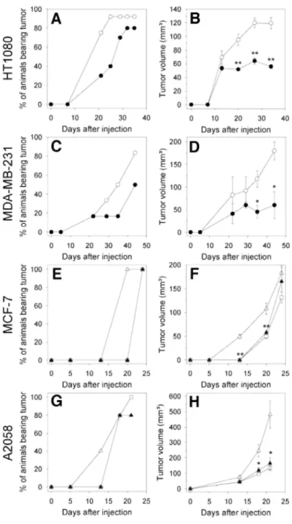
HT1080 fibrosarcoma cells express several MMPs including MMP-2 and -9, as well as MT1-MMP. Thirty-four days after s.c. implantation of HT1080 cells into nude mice, a tumor incidence (percentage of animal bearing tumors larger than 80 mm3) of 90% was reached in vehicle-treated animals and 80% in animals receiving Ro-28-2653 (log-rank test, P = 0.008; Fig. 44). Tumor growth curves show that Ro-28-Ro-28-2653-treatment inhibited the growth of HT1080 tumors by 53% (Fig. 4B). Thus, Ro-28-2653 reduced significantly both the incidence and the growth of tumors induced by HT1080 cell injection.
Fig. 2 Comparison of the anti-invasive activities of different matrix metalloproteinase inhibitors (MMPIs). Chemoinvasion of HT1080 cells was evaluated in Transwell cell culture inserts coated with type IV collagen. HT1080 cells were seeded in the presence of different
concentrations of MMPIs (0.01-10 µM) or vehicle alone [Control, 0.1% DMSO and DMEM alone, for Ro-28-2653, GI129471, BB-94, BB-2516, and recombinant human tissue inhibitor of metalloproteinase-2 (rhTIMP-2), respectively]. The number of cells that have migrated was determined by counting the cells present on the lower side of the filters and was expressed as percentages of the migration observed in control conditions. *, P < 0.05; **, P < 0.01.
.
Table 1 In vitro inhibitory activities of matrix metalloproteinase (MMP) inhibitors against different
metalloproteinases Concentrations of inhibitor at which 50% inhibition of proteolysis occurs (IC50) are indicated.
MMP profiling, IC50,, nM
Inhibitor
MMP-1 MMP-2 MMP-3 MMP-9 MTlα-MMP MT3-MMP TACE Ro-28-2653 16,000 12 1,800 16 10 23 >20,000
BB-94 25 32 67 23 19 29 2,000
α MT1, membrane type 1; TACE, tumor necrosis factor α converting enzyme.
Fig. 3 Modulation of matrix metalloproteinase 2 (MMP-2) activation and MMP-9 expression by synthetic MMP inhibitors (MMPIs). HT1080 cells were incubated during 48 h in the presence of DMSO (0.1%. Control), GI129471 (1 µM), BB-94 (1 µM), and Ro-28-2653 (1 µM). The conditioned media were collected and analyzed by gelatin zymog-raphy. Conditioned medium obtained from
12-O-tetradecanoyl-phorbol-13-acetate-treated HT1080 cells (TPA) was used to identify the different gelatinolytic species. kDa, MI in thousands.
In a second model, MDA-MB-231 cells, highly aggressive human breast adenocarcinoma cells, which express MMP-9 and MTl-MMP but less MMP-2, were used. In this model, Ro-28-2653 reduced tumor incidence from 83% in vehicle-treated mice (44 days after s.c. injection of the cells) to 50% in Ro-28-2653-treated animals (Fig. 4C). Moreover, Ro-28-2653 inhibited by 67% the growth of tumors induced by MDA-MB-231 cell injection (Fig. 4D).
MCF7 human breast adenocarcinoma cells produce very low levels of MMPs and are poorly tumorigenic in vivo in nude mice. Their capacity to develop tumors in vivo is not affected by broad-spectrum MMPIs such as tissue inhibitor of metallopro-teinase-2 or BB-94 (33), but their tumorigenic properties are dependent on the presence of extracellular matrix components, such as Matrigel, and/or on the presence of stromal cells, such as fibroblasts. The coinjection of fibroblasts with MCF7 cells in the presence of Matrigel has been shown to enhance in vivo
tumor growth in a protease-dependent manner (32, 33). Injection of MCF7 cells in the presence of Matrigel resulted in tumor formation with a latency period (time required to obtain 100% of tumors larger than 80 mm3) of about 24 days. When fibroblasts were coinjected with MCF7 cells and Matrigel, the latency period decreased to 20 days (log-rank test, P = 0.003) and the volume of the resulting tumors was significantly increased (Fig. 4, E and F). Although the administration of Ro-28-2653 affected neither the tumorigenicity nor the growth of tumors resulting from the injection of MCF7 cells alone (data not shown), this treatment completely inhibited the tumor-promoting effect of the coinjected fibroblasts. Indeed, in Ro-28-2653-treated animals, the latency period and tumor growth were identical to those observed after injection of MCF7 cells alone (Fig. 4, E and F). Human A2058 melanoma cells produce pro-MMP-2 but very low levels of both MMP-9 and MT1-MMP. Consequently, these cells are unable to activate MMP-2 (25). When inoculated s.c. into nude mice, A2058 cells were poorly tumorigenic. In accordance with our previous observations (25, 26), the transfection of these cells with the cDNA encoding MT1-MMP endowed them with the ability to activate MMP-2 (data not shown) and led to an enhancement of their tumorigenicity (Fig. 4, G and H). Indeed, the s.c. injection of MT1-MMP-transfected A2058 cells (clone S.I.5) resulted in a more rapid tumor take, as well as in larger tumors (P = 0.026), as
compared with mock-transfected cells (clone C.IV.3). This MT1-MMP-promoting effect on tumor growth was completely abolished by Ro-28-2653 (Fig. 4, G and H). Similar results were obtained by using two different clones stably transfected with MT1-MMP cDNA (data not shown).
In a second set of experiments, we evaluated the impact of treatment discontinuation on in vivo tumor growth. Mice inoculated with HT1080 cells were treated daily during 35 days with vehicle or Ro-28-2653 (90 mg/kg of mouse weight per day; oral administration), starting the day of tumor inoculation (day 0) and then left untreated during 2 additional weeks. As observed previously, Ro-28-2653 significantly reduced the tumorigenicity of HT1080 cells (compare Fig. 4, A and B, and Fig. 5). After treatment discontinuation, the tumors continued to grow rapidly in vehicle-treated mice, whereas tumor volumes remained low with modest growth in the group that received the inhibitor (Fig. 5).
Collectively, these results demonstrate that Ro-28-2653 is a potent inhibitor of tumor development. Indeed, this compound significantly reduced tumor growth and inhibited the tumor promoting effect of stromal cells. In all of these models, Ro-28-2653 was well tolerated and did not alter body weights and was as efficient by i.p. injection as by p.o. administration (data not shown).
These data emphasize the usefulness of Ro-28-2653 to target both tumor cells and the stromal compartment.
Comparison of in Vivo Efficacy of Ro-28-2653 and BB-94
To further characterize the antitumoral efficacy of Ro-28-2653, we compared its activity in vivo with that of BB-94 used as a reference compound. The antitumoral activity of these two MMPIs was evaluated in the A2058 melanoma xenograft model. In this model, highly vascularized tumors grew rapidly (Fig. 4H) and reached a volume of 480 ± 88 mm3 3 weeks after the s.c. implantation of A2058 cells overexpressing MT1-MMP (S.I.5 cells). Both of the MMPIs significantly reduced the development of these tumors. However, BB-94 was less efficient than Ro-28-2653 and only partially inhibited the tumor growth, resulting in larger tumors than those observed after 21 days in Ro-28-2653-treated mice (Fig. 6A).
Effect of Ro-28-2653 on in Vivo and ex Vivo Angiogen-esis
To evaluate the potential influence of Ro-28-2653 on angiogenesis, the A2058 melanoma xenograft model and the ex vivo rat aortic ring assay were used. As reported recently (26), the tumors resulting from the s.c. injection of A2058 cells overexpressing MT1-MMP (clone S.I.5) show increased vascularization when compared with mock-transfected cells (clone C.IV.3; Fig. 6B). Microvessel density as a parameter of neoan-giogenesis was analyzed by image analysis in CD31- or antitype IV collagen-stained tumor sections resulting from the injection of C.IV.3 and S.I.5 cells in mice treated for21 days with Ro-28-2653 or with vehicle. This quantification demonstrates that Ro-28-2653 significantly reduced the vascularization of MT1-MMP-expressing tumors (Fig.
6B). In sharp contrast, the administration of BB-94 did not affect tumor vascularization. Similar results were
obtained by using two different A2058 clones stably transfected with MT1-MMP cDNA (data not shown). To more directly assess the effect of Ro-28-2653 on angiogenesis, this compound was tested in the rat aortic ring assay (34). In this ex vivo model, rat aortic expiants were cultured within type I collagen gel during 1 week in the presence of increasing concentrations of the MMPIs (0-1000 nΜ),and microvascular outgrowth was quantified
endothelial cells as branching microvessels with fibroblast-like cells present as isolated cells (Fig. 6B, Control). The addition of Ro-28-2653 (from 1 to 1000 nM)completely blocked neovessel formation (Fig. 6, B and C). In
contrast with Ro-28-2653, the addition of increasing concentrations of BB-94 (from 1 to 1000 ΠM)resulted in a
bell-shaped dose-response curve (Fig. 6C) characterized by a lack of inhibition at the lowest concentration (1 nΜ), a marked proangiogenic activity at intermediate concentrations (10 and 100 nΜ),and an inhibition at the highest concentration (1000 nΜ).
Fig. 4 Effect of Ro-28-2653 on the development of different human tumor xenografts in mice. Human HT1080 fibrosarcoma (A, B), MDA-MB-231 (C, D), and MCF-7 (E, F) adenocarcinomas, and A2058 melanoma (G, H) cells were s.c. injected in the presence of Matrigel in nude mice at day 0. Ro-28-2653 (closed symbol) or vehicle (open symbol) were administrated every 2 days, starting the day of tumor inoculation until the end of the assay. Tumor volumes were estimated two times per week. The evolution of tumor incidence (A, C, E, and G) and tumor volume (B, D, F, and H) was evaluated as a function of time after tumor cell injection. E, F, MCF-7 cells were injected alone (□) or with 1 × 10⁶ fibroblasts (∆). G, H, mice were given injections of mock transfected-A2058 cells (clone CIV.3, □) or membrane type 1
Fig. 5 Effect of the discontinuation of Ro-28-2653 treatment on the growth of HT1080 tumor xenografts in mice. HT1080 cells were s.c. inoculated in the presence of Matrigel in nude mice at day 0. Ro-28-2653 (90 mg/kg,●) or vehicle (○) were administrated p.o. every day, starting the day of tumor inoculation until day 35 (black bars, treatment duration). The evolution of tumor incidence (A) and tumor volume
Fig. 6 Inhibitory effect of Ro-28-2653 on angiogenesis. (A-B) mock transfected-A2058 cells (clone C.IV.3) or membrane type 1 (MT1)-MMP-transfected cells (clone S.I.5) were s.c. injected in the presence of Matrigel in nude mice. BB-94 (30 mg/kg), Ro-28-2653 (100 mg/kg), or vehicle was administrated every 2 days for a period of 21 days, starting the day of tumor inoculation. A, the corresponding tumors were measured and dissected. Tumor vessels were labeled with anti-type IV collagen or anti-CD31 antibodies. B, vessel density was quantified by image analysis. **, P < 0.01 versus C.IV.3; #,P< 0.01 versus S.I.5 + vehicle; §, P < 0.05 versus S.I.5 + BB-94. C, D, Ro-28-2653 blocks the formation of capillary outgrowth from rat aortic rings. C, photomicrographs showing the angiogenic response of collagen-embedded expiants cultured for 7 days in the absence (Control) or presence of Ro-28-2653 (1 µM). Arrows, isolated fibroblast-like cells; arrowheads,
microvessel outgrowths. D, the number of microvessels in expiants treated with vehicle or increasing concentrations of BB-94 (1-1000 nM) or Ro-28-2653 (1-1000 nM) was determined by computer-assisted image analysis. *, P< 0.05; **, P < 0.01.
DISCUSSION
The specific alteration of the MMP expression profile observed in malignant tissues, as well as the implication of these proteinases in the major stages of cancer progression, has fueled interest in the design and evaluation of synthetic MMPIs as a novel anticancer therapy. Despite promising preclinical results in the use of MMPIs as anticancer agents (for review see Refs. 9, 11, and 36), most clinical studies involving these drugs have not shown significant efficacy in patients with advanced cancer (5, 11, 12).
Recent studies have revealed that some MMPs might also play a protective role during cancer progression. Increased expression of 12 by colon carcinoma cells is associated with increased survival (37), and MMP-8-deficient male mice display increased skin cancer susceptibility (38). This dual function of some MMPs during tumor progression can be an obstacle for the use of unselective MMPIs. Indeed, most MMPIs evaluated in vivo up to now target all MMPs, even those that prevent tumor progression, and so can enhance tumor growth by decreasing, for example, the production of angiogenesis inhibitors that are generated by proteolysis of larger molecules. Therefore, additional therapeutic possibilities are likely to be offered by the development of inhibitors targeting selectively tumor progression-associated MMPs, but sparing other MMPs the activity of which is either beneficial to the host or, if inhibited, produces side effects. A new generation of nonpeptidic MMPIs with improved selectivity toward a restricted number of MMPs has been recently described (10). Most of these novel compounds are hydroxamic acid-based inhibitors and most likely bind to the enzyme in a similar way.
To develop a completely new class of MMPI with a different pharmacological profile, Grams et al. (24) used a high-throughput screening to identify the pyrimidine-trione template of 5-[4-(2-hydroxyethyl)]piperidine-5-phenyl-pyrimidine-2,4,6-trione (compound 1) as a new zinc chelator for metalloproteinases. When evaluated in
vitro in a chemoinvasion assay, this compound inhibited the invasiveness of human HT1080 cells as efficiently
as GI129471, a broad-spectrum MMPI used as a reference compound. Structure-based drug design and combinatorial chemistry have been used to optimize the residues attached to the initial pyrimidine-trione core (24). This optimization strategy led to the selection of 5-biphenyl-4-yl-5-[4-(4-nitro-phenyl)-piperazine-1-yl]-pyrimidine-2,4,6-trione (Ro-28-2653). This compound is characterized by a high selectivity toward MMP-2, MMP-9, and MT1-MMP (three MMPs consistently associated with tumor aggressiveness and poor prognosis), but sparing enzymes [MMP-1 and TACE (tumor necrosis factor α converting enzyme)] implicated in the musculoskeletal problems observed during the clinical trials of hydroxamate-derived MMPIs (19). Furthermore, crystallographic studies have revealed that this new class of MMPI binds to the MMPs in a manner that saturates nearly all of the possible interactions of the pyrimidine core moiety to the protein, thus resulting in more
interactions than seen with the typical hydroxamates (24).
When compared with classical broad-spectrum MMPIs such as batimastat or marimastat, Ro-28-2653 showed the highest anti-invasive activity in the in vitro chemoinvasion assay, even when used at a 1000-fold lower concentration. These data thus demonstrate the potency of this novel MMPI to block the capacity of tumor cells to breach extracellular matrix barriers, one of the crucial steps involved in cancer progression (39).
In accordance with these in vitro observations, our in vivo study in different experimental human tumor xenograft models demonstrates that Ro-28-2653 markedly affected tumor growth. It reduced both the incidence and the growth of tumor induced by the injection of HT1080-, MDA-MB-231-, and MT1-MMP-transfected A2058 cells, all expressing different sets of MMPs including MMP-2, -9, and MT1-MMP. In contrast, this compound altered neither the incidence nor the growth of tumor resulting from the inoculation of MCF7 cells that produce very low levels of MMPs, thus supporting a MMP-dependent anti-tumoral activity. In all of these different models, i.p. or p.o. administration of Ro-28-2653 gave rise to similar antitumor effects, was well tolerated, and did not alter the body weight of treated mice.
Accumulating evidence has revealed that carcinomas such as breast adenocarcinomas are composed not only of tumor epithelial cells but also of infiltrating stromal cells arising from the host (40, 41). These stromal cells also secrete MMPs, thereby promoting tumor aggressiveness (8, 33, 42, 43). When mice that had received
coinjections of fibroblasts and MCF7 cells were treated with Ro-28-2653, the tumor-promoting effect of the coinjected fibroblasts was strongly impaired, demonstrating the ability of this compound to target stromal-derived MMPs. Targeting stromal cells rather than cancer cells can be beneficial because stromal cells are less likely to develop drug resistance.
MT1-MMP plays a major role in cancer progression. Indeed, the expression of MT1-MMP correlates with the malignancy of different tumor types (for review, Rev. 44) and its overexpression in tumor cell lines enhanced their invasiveness in vitro as well as their tumorigenicity in vivo (26, 45-47). Moreover, MT1-MMP activity has
been demonstrated to circumvent the growth-suppressive signals embedded within three-dimensional extracellular matrix, thereby conferring tumor cells with an accelerated proliferative capacity (47). Therefore, MT1-MMP represents an attractive target for the development of specific MMPIs. The high in vitro inhibitory activity of Ro-28-2653 against MT1-MMP (IC50, 10 nM)prompted us to undertake a detailed analysis of its in vivo efficacy in the human A2058 melanoma xenograft model. We have previously demonstrated that the
overexpression of MT1-MMP in A2058 cells was associated with enhanced in vitro invasion and increased in
vivo tumor growth (26). Interestingly, the promoting effect of MT1-MMP on tumor growth was completely
abolished by Ro-28-2653, demonstrating its high potency in vivo against MT1-MMP.
To grow efficiently in vivo, tumor cells induce angiogen-esis in both primary tumors and metastatic foci. Accumulating evidence has revealed the involvement of MT1-MMP during both physiological and pathological angiogenesis (44, 48, 49). In the A2058 experimental model, MT1-MMP expression by tumor cells was shown to promote tumor vascularization (26). This MT1-MMP proangiogenic activity was markedly reduced in Ro-28-2653-treated mice, as revealed by a lower blood vessel density. Furthermore, we show that this MMPI was also able to completely block neovessel formation in the rat aortic ring assay, highlighting the usefulness of this MMPI as a powerful inhibitor of angiogenesis. The antiangiogenic activity of Ro-28-2653 can be partly ascribed to its capacity to down-regulate the expression of vascular endothelial growth factor by tumor cells expressing MT1-MMP (50). Collectively, these data suggest that Ro-28-2653 can be envisioned as a potential
antiangiogenic compound for primary tumors and as a therapeutic agent that can help maintain small clusters of metastatic cells in a dormant state.
We reported previously that, despite their ability to block tumor cell invasion in vitro, some MMPIs
paradoxically increased the expression of MMP-9 by these cells (35). In agreement with these observations, we confirmed here that the treatment of HT1080 cells with two different broad-spectrum MMPIs (GI129471 and BB-94) increased MMP-9 secretion. In contrast, such an up-regulation was not observed in the presence of Ro-28-2653. Interestingly, BB-94-treated mice have been shown to display a liver-specific increase of MMP-9 expression that was associated with a higher number of liver metastases (16). When Ro-28-2653 was evaluated in the same in vivo model, MMP-9 level in the liver remained unchanged and the number of liver metastases was strongly reduced (51).
The comparison of the in vivo efficacy of BB-94 and Ro-28-2653 in the human A2058 melanoma xenograft model demonstrated the superiority of Ro-28-2653 as antitumor agent. The lower antitumoral activity of BB-94 can be partly ascribed to its inefficiency in reducing the vascularization of these tumors. In support of this hypothesis, the comparison of the antiangiogenic activities of these two MMPIs in the rat aortic ring assay confirmed the higher efficacy of Ro-28-2653 as antiangiogenic drug. Moreover, BB-94 paradoxically promoted the formation of microvascular outgrowths when used at concentrations ranging from 10 to 100 nM.The exact mechanism of this dose-dependent proangiogenic activity is uncertain. However, the up-regulation of
proangiogenic factors observed in the livers of BB-94-treated mice supports a direct effect of this MMPI on the angiogenic process (16). MMP-9 has been shown to trigger angiogenesis by releasing matrix-bound vascular endothelial growth factor (52). Therefore, the promoting effect of BB-94 on MMP-9 expression can indirectly foster the formation of new blood vessels. Alternatively, BB-94 may also inhibit some proteinases the activity of which generates angiostatic factors such as endostatin. The unexpected proangiogenic activity of BB-94 thus emphasizes the potential detrimental impact of nonspecific MMPIs on cancer progression.
Interestingly, the administration of Ro-28-2653 in mice, rats, and monkeys did not induce any histopathological alteration of the joints, a major adverse effect frequently observed with broad spectrum MMPIs.4
Taken together, these different observations underscore the advantage of the use of more specific rather than broad-spectrum MMPIs, especially in the light of side effects, which need to be avoided for clinical applications. In the light of our in vitro and in vivo results, Ro-28-2653, a pyrimidine-2,4,6-trione derivative, represents an attractive new class of orally available selective MMPIs with potent antitumoral and antiangiogenic activities. In contrast to broad-spectrum MMPIs such as BB-94, the administration of this novel MMPI was not associated with the occurrence of adverse side effects that might reduce the therapeutic potential of these drugs.
4 Unpublished data, Krell, H. W.
Acknowledgments
We thank Guy Roland and Patricia Gavitelli for their technical assistance.
Grant support
This work was supported by grants from the Communauté française de Belgique (Actions de Recherches Concertées), the Commission of European Communities (FP5 no. QLK3-CT02-02136, FP6), the Fonds de la Recherche Scientifique Médicale, the Fonds National de la Recherche Scientifique (FNRS, Belgium), the Fédération Belge Contre le Cancer, the Commissariat Général aux relations Internationales de la Communauté française Wallonie-Bruxelles-F.N.R.S.-INSERM Coopération, the Fonds spéciaux de la Recherche (University of Liège), the Centre Anticancéreux près l'Université de Liège, the FB Assurances, the Fondation Léon Frédéricq (University of Liège), the Direction Générale des Technologies, de la Recherche et de l'Energie from the
"Région Wallonne," the Fonds d'Investissements de la Recherche Scientifique (CHU, Liège, Belgium), the Interuniversity Attraction Poles Programme-Belgian Science Policy (Brussels, Belgium), and Roche Diagnostics GmbH (Penzberg, Germany). The costs of publication of this article were defrayed in part by the payment of page charges. This article must therefore be hereby marked advertisement in accordance with 18 U.S.C Section 1734 solely to indicate this fact.
Note
A. Noël is a Senior Research Associate and E. Maquoi is a Scientific Research Worker from the Fonds National de la Recherche Scientifique (FNRS, Brussels, Belgium).
References
1. Sternlicht MD, Werb Z. How matrix metalloproteinases regulate cell behavior. Annu Rev Cell Dev Biol 2001;17:463-516. 2. Egeblad M, Werb Z. New functions for the matrix metalloproteinases in cancer progression. Nat Rev Cancer 2002;2:161-74.
3. Woessner JF. The matrix metalloproteinase family. In: Parks WC and Mecham RP, editors. Matrix metalloproteinases. San Diego, CA, Academic Press; 1998. p. 1-14.
4. Maquoi E, Munaut C, Colige A, Collen D, Lijnen HR. Modulation of adipose tissue expression of murine matrix metalloproteinases and their tissue inhibitors with obesity. Diabetes 2002;51:1093-101.
5. Overall CM, Lopez-Otin C. Strategies for MMP inhibition in cancer: innovations for the post-trial era. Nat Rev Cancer 2002;2:657-72. 6. Noël A, Emonard H, Polette M, Birembaut P, Foidart JM. Role of matrix, fibroblasts and type IV collagenases in tumor progression and invasion. Pathol Res Pract 1994;190:934-41.
7. Basset P, Bellocq JP, Wolf C, et al. A novel metalloproteinase gene specifically expressed in stromal cells of breast carcinomas. Nature (Lond) 1990;348:699-704.
8. Bisson C, Blacher S, Polette M, et al. Restricted expression of membrane type 1-matrix metalloproteinase by myofibroblasts adjacent to human breast cancer cells. Int J Cancer 2003;105:7-13.
9. Wojtowicz-Praga SM, Dickson RB, Hawkins MJ. Matrix metalloproteinase inhibitors. Investig New Drugs 1997;15:61-75.
10. Hidalgo M, Eckhardt SG. Development of matrix metalloproteinase inhibitors in cancer therapy. J Natl Cancer Inst (Bethesda) 2001;93: 178-93.
11. Zucker S, Cao J, Chen WT. Critical appraisal of the use of matrix metalloproteinase inhibitors in cancer treatment. Oncogene 2000; 19: 6642-50.
12. Coussens LM, Fingleton B, Matrisian LM. Matrix metalloproteinase inhibitors and cancer: trials and tribulations. Science (Wash DC) 2002;295:2387-92.
13. Fingleton B. Matrix metalloproteinase inhibitors for cancer therapy: the current situation and future prospects. Expert Opin Ther Targets 2003;7:385-97.
14. Ferreras M, Felbor U, Lenhard T, Olsen BR, Delaisse J. Generation and degradation of human endostatin proteins by various proteinases. FEBS Lett 2000;486:247-51.
15. Delia Porta P, Soeltl R, Krell HW, et al. Combined treatment with serine protease inhibitor aprotinin and matrix metalloproteinase inhibitor batimastat (BB-94) does not prevent invasion of human esophageal and ovarian carcinoma cells in vivo. Anticancer Res 1999;19:3809-16.
16. Krüger A, Soeltl R, Sopov I, et al. Hydroxamate-type matrix metalloproteinase inhibitor batimastat promotes liver metastasis. Cancer Res 2001;61:1272-5.
17. Drummond AH, Beckett P, Brown PD, et al. Preclinical and clinical studies of MMP inhibitors in cancer. Ann N Y Acad Sci 1999;878: 228-35.
18. Hutchinson JW, Tierney GM, Parsons SL, Davis TR. Dupuytren's disease and frozen shoulder induced by treatment with a matrix metalloproteinase inhibitor. J Bone Joint Surg Br 1998;80:907-8.
19. Renkiewicz R, Qiu L, Lesch C, et al. Broad-spectrum matrix metalloproteinase inhibitor marimastat-induced musculoskeletal side effects in rats. Arthritis Rheum 2003;48: 1742-9.
20. Gatto C, Rieppi M, Borsotti P, et al. BAY 12-9566, a novel inhibitor of matrix metalloproteinases with antiangiogenic activity. Clin Cancer Res 1999;5:3603-7.
21. Tamura Y, Watanabe F, Nakatani T, et al. Highly selective and orally active inhibitors of type IV collagenase (MMP-9 and MMP-2): N-sulfonylamino acid derivatives. J Med Chem 1998;41:640-9.
22. Katori H, Baba Y, Imagawa Y, et al. Reduction of in vivo tumor growth by MMI-166, a selective matrix metalloproteinase inhibitor, through inhibition of tumor angiogenesis in squamous cell carcinoma cell lines of head and neck. Cancer Lett 2002;178:151-9.
23. Yamamoto A, Yano S, Shiraga M, et al. A third-generation matrix metalloproteinase (MMP) inhibitor (ONO-4817) combined with do-cetaxel suppresses progression of lung micrometastasis of MMP-ex-pressing tumor cells in nude mice. Int J Cancer 2003;103:822-8. 24. Grams F, Brandstetter H, D'Alo S, et al. Pyrimidine-2,4,6-triones: a new effective and selective class of matrix metalloproteinase inhibitors. Biol Chem 2001;382:1277-85.
25. Maquoi E, Frankenne F, Baramova E, et al. Membrane type 1 matrix metalloproteinase-associated degradation of tissue inhibitor of metalloproteinase 2 in human tumor cell lines. J Biol Chem 2000;275: 11368-78.
26. Sounni NE, Baramova EN, Munaut C, et al. Expression of membrane type 1 matrix metalloproteinase (MT1-MMP) in A2058 melanoma cells is associated with MMP-2 activation and increased tumor growth and vascularization. Int J Cancer 2002;98:23-8. 27. McGeehan, G. M. Becherer JD, Bast RC, et al. Regulation of tumor necrosis factor-alpha processing by a metalloproteinase inhibitor. Nature (Lond) 1994;370:558-61. GI129471 (27), BB-94 (28) and BB-2516 (29).
28. Inventor: Campion C, Davidson JP, Dickens JP, Crimmin MJ. Assignee: British Biotechnology, Great Britain. Matrix metalloproteinase inhibitor BB-94. Patent Cooperation Treaty Patent Application, WO 90/05719, 5/31/90.
29. Inventor: Miller KM, Bawden LJ, Brown PD. Assignee: British Biotechnology, Great Britain. Matrix metalloproteinase inhibitor BB-2516. Patent WO 93/21942, 11/11/93.
30. Maquoi E, Noël A, Frankenne F, Angliker H, Murphy G, Foidart JM. Inhibition of matrix metalloproteinase 2 maturation and HT1080 invasiveness by a synthetic furin inhibitor. FEBS Lett 1998;424: 262-6.
31. Noël A, Calle A, Emonard HP, et al. Invasion of reconstituted basement membrane matrix is not correlated to the malignant metastatic cell phenotype. Cancer Res 1991;51:405-14.
32. Noël A, De Pauw-Gillet MC, Purnell G, Nusgens B, Lapiere CM, Foidart JM. Enhancement of tumorigenicity of human breast adenocarcinoma cells in nude mice by Matrigel and fibroblasts. Br J Cancer 1993;68:909-15.
33. Noël A, Hajitou A, L'Hoir C, et al. Inhibition of stromal matrix metalloproteases: effects on breast-tumor promotion by fibroblasts. Int J Cancer 1998;76:267-73.
34. Blacher S, Devy L, Burbridge MF, et al. Improved quantification of angiogenesis in the rat aortic ring assay. Angiogenesis 2001;4:133-42.
35. Maquoi E, Munaut C, Colige A, et al. Stimulation of matrix met-alloproteinase-9 expression in human fibrosarcoma cells by synthetic matrix metalloproteinase inhibitors. Exp Cell Res 2002;275:110-21.
36. Nelson AR, Fingleton B, Rothenberg ML, Matrisian LM. Matrix metalloproteinases: biologic activity and clinical implications. J Clin Oncol 2000;18:1135-49.
37. Yang W, Arii S, Gorrin-Rivas MJ, Mori A, Onodera H, Imamura M. Human macrophage metalloelastase gene expression in colorectal carcinoma and its clinicopathologic significance. Cancer (Phila) 2001; 91:1277-83.
38. Balbin M, Fueyo A, Tester AM, et al. Loss of collagenase-2 confers increased skin tumor susceptibility to male mice. Nat Genet 2003;35: 252-7.
39. Hanahan D, Weinberg RA. The hallmarks of cancer. Cell 2000; 100:57-70.
40. Dvorak HF. Tumors: wounds that do not heal. Similarities between tumor stroma generation and wound healing. N Engl J Med 1986;315: 1650-9.
41. Van den Hooff A. Stromal involvement in malignant growth. Adv Cancer Res 1988;50:159-96.
42. Masson R, Lefebvre O, Noël A, et al. In vivo evidence that the stromelysin-3 metalloproteinase contributes in a paracrine manner to epithelial cell malignancy. J Cell Biol 1998;140:1535-41.
43. Huang S, Van Arsdall M, Tedjarati S, et al. Contributions of stromal metalloproteinase-9 to angiogenesis and growth of human ovarian carcinoma in mice. J Natl Cancer Inst (Bethesda) 2002;94:1134-42.
44. Sounni NE, Janssen M, Foidart JM, Noël A. Membrane type-1 matrix metalloproteinase and TIMP-2 in tumor angiogenesis. Matrix Biol 2003;22:55-61.
45. Sounni NE, Devy L, Hajitou A, et al. MT1-MMP expression promotes tumor growth and angiogenesis through an up-regulation of vascular endothelial growth factor expression. FASEB J 2002;16: 555-64.
46. Deryugina EI, Soroceanu L, Strongin AY. Up-regulation of vascular endothelial growth factor by membrane-type 1 matrix metalloproteinase stimulates human glioma xenograft growth and angiogenesis. Cancer Res 2002;62:580-8.
47. Hotary KB, Allen ED, Brooks PC, Datta NS, Long MW, Weiss SJ. Membrane type I matrix metalloproteinase usurps tumor growth control imposed by the three-dimensional extracellular matrix. Cell 2003;114: 33-45.
48. Hiraoka N, Allen E, Apel IJ, Gyetko MR, Weiss SJ. Matrix metalloproteinases regulate neovascularization by acting as pericellular fibrinolysins. Cell 1998;95:365-77.
49. Zhou Z, Apte SS, Soininen R, et al. Impaired endochondral ossification and angiogenesis in mice deficient in membrane-type matrix metalloproteinase I. Proc Natl Acad Sci USA 2000;97:4052-7.
50. Sounni NE, Roghi C, Chabottaux V, et al. Up-regulation of VEGF-A by active MTl-MMP through activation of Src-tyrosine kinases. J Biol Chem 2004;279:13564-74.
51. Arlt M, Kopitz C, Pennington C, et al. Increase in gelatinase-specificity of matrix metalloproteinase inhibitors correlates with anti-metastatic efficacy in a T-cell lymphoma model. Cancer Res 2002;62: 5543-50.
52. Bergers G, Brekken R, McMahon G, et al. Matrix metalloproteinase-9 triggers the angiogenic switch during carcinogenesis. Nat Cell Biol 2000;2:737-44.