Académie Universitaire Wallonie - Europe
Université de Liège
Faculté de Médecine Vétérinaire
Département des Maladies Infectieuses et Parasitaires
Service d’Immunologie et de Vaccinologie
Thèse présentée en vue de l’obtention du grade
de Docteur en Sciences Vétérinaires
Année académique 2013-2014
Ping OUYANG
Etude des rôles du gène ORF134 dans la biologie de
l’infection de l’Herpèsvirus cyprin 3
Study of the roles of Cyprinid herpesvirus 3 ORF134 in
the biology of the infection
Académie Universitaire Wallonie - Europe Université de Liège Faculté de Médecine Vétérinaire Département des Maladies Infectieuses et Parasitaires Service d’Immunologie et de Vaccinologie
Study of the roles of Cyprinid herpesvirus 3 ORF134 in
the biology of the infection
the biology of the infection
Etude des rôles du gène ORF134 dans la biologie de
g
g
l’infection de l’Herpèsvirus cyprin 3
Promotor : Prof. Alain Vanderplasschen
Thèse présentée en vue de l’obtention du grade de Docteur en Sciences Vétérinaires
Année académique 2013-2014
Ping OUYANG
« The way ahead is long and has no ending, yet high and low I’ll search with my will unbending. »
« 路漫漫其修远兮
, 吾将上下而求索. »
Acknowledgments
Acknowledgments
The work presented in this thesis has been carried out in the laboratory of Immunology-Vaccinology, faculty of Veterinary Medicine, University of Liège, Belgium. I am extremely grateful to my promotor Prof. Alain Vanderplasschen for giving me the opportunity to study in his lab. His trust, his permanent support and his passion for science have been an essential source of motivation and inspiration for me. I will always admire him for his constant positive attitude when facing problems and for his trust that the “good must win”.
I would like to express my gratitude to the members of my PhD committee who monitored my work progress and took effort in reviewing and providing me with insightful comments: Prof. Daniel
Desmecht and Dr. François Lieffrig.
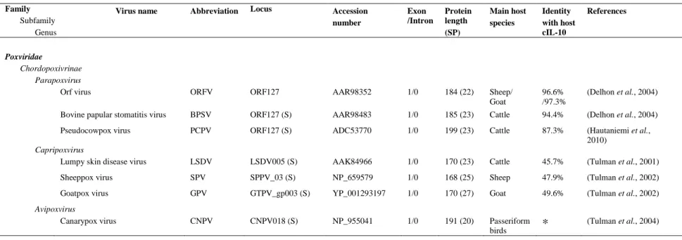
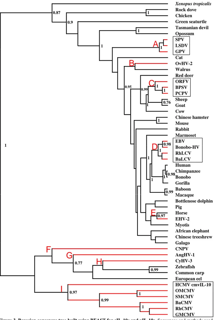
The present work was at the origin of several fruitful collaborations with other laboratories. I would like to thank Prof. Ruddy Wattiez and Dr Baptiste Leroy (Proteomic and Microbiology, University of Mons, Belgium) for their interest in our project and their willingness to perform proteomic analyses. Many thanks to Dr. Andrew Davison and Dr. Derek Gatherer (Centre for Virus Research, University of Glasgow, United Kingdom) for their contribution on the study of viral IL-10 evolution. Many thanks also go to Dr. Adrie Westphal (Department of Agrotechnology and Food Sciences, Wageningen University, The Netherlands) for his contribution on viral IL-10 structural analyses.
The four years spent in the laboratory of Immunology-Vaccinology have not only been my source of scientific experiences, but also of invaluable human relationships. Many thanks go to all the members of the Immunology-Vaccinology lab. I would like to start with the members of the fish group. This very rich environment is composed of 7 persons originating from 6 different countries. Dr.
Krzysztof Rakus and his wife Dr. Joanna Jazowiecka-Rakus are from Poland, thanks for their
endless support and invaluable suggestions on fish immunology. Dr. Anca Reschner comes from Romania, thanks for her constant help and nice discussions. Maygane Ronsmans is a beautiful Belgian girl, thanks for her help during the doctoral formation courses. Maxime Boutier is a very nice French guy and it has been a great pleasure to work with him. Dr. Ma. Michelle Penaranda, the new addition in the lab, comes from the Philippines. Many thanks to these people for their constant help and the very friendly atmosphere they are creating in the fish group office.
I also would like to thank members of the fish group who left the lab. Dr. Guillaume
Fournier, his precious help has been essential to the progress of my thesis. Thanks to Dr. Robert Vrancken, Dr. Stalin Raj, Dr. Benjamin Michel, Dr. Hélène Schroeder and Dr. Bérénice Costes
The laboratory of Immunology-Vaccinology is composed of three sub-groups that are constantly interacting. I would like to thank the people from the two other sub-groups I had the chance to work with. Thanks to the people of the group lead by Prof. Laurent Gillet: Dr Bénédicte Machiels,
Dr Céline Lété, Dr Sylvie François, Dr Sarah Vidick, Bérengère Boutard, Bilal Latif and Mickael Dourcy, as well as the group what lead by Dr Benjamin Dewals: Dr Leonor Plameira, Dr Steven van Beurden, Françoise Myster, Océane Sorel.
The three sub-groups of the Immunology-Vaccinology lab benefit from the work of dedicated technicians and secretaries. I’m thankful to Cédric Delforge, Emeline Deglaire, Christine Thys,
Jérémy Dumoulin, Nathalie Poncelet, Antoine Guillaume, François Massart, Dominique Ziant and Charles Gaspar who contributed directly or indirectly to experiments. Last but not the least, I
would like thank our secretaries Christina Espert and Lorère Dams, who made my life more convenient in the lab.
I would like to thank my master supervisor Prof. Liancheng Lei (College of Animal Husbandry and Veterinary Medicine, Jilin University, China) for his permanent encouragement and constant help. Without him, I would not have the chance to study abroad for 4 years.
I also would like to acknowledge my Chinese friends in Belgium, Xuerong Jiang, Huijun
Cheng, Zhiyan Zhang, Wanbo Li, Qiongzhong Chen, Yongzhen Li, Ji Liu, Ming Fang, Xuewen Xu, Cheng Liu, Xin Zhang, who offered me their help to solve the numerous practical problems that
I had to face when arriving in Belgium. They made also my life in Belgium more colorful.
Many thanks go to my beloved family for their loving consideration and their great confidence in me throughout all these years. Most importantly, I would like to thank my parents for supporting me spiritually throughout my life. I must acknowledge my boyfriend, Dr. Lizi YIN, without his love, his permanent encouragement and constant help, I would not have finished my thesis and life in Belgium would not have been so nice.
Being in Belgium to complete a PhD thesis is definitely one of the most interesting, stimulating and exciting experiences of my live. This experience has been possible thanks to the financial support of the Chinese Scholarship Council (Application No.2009617025).
Liège, 15th September 2013 Ping Ouyang
Abbreviations
List of abbreviations
2D-LC MS/MS: Two-dimensional liquid chromatography tandem
mass spectrometry
α gene: Immediate early gene
aa: Amino acid
ACHV: Atlantic cod herpesvirus
AciHV-1: Acipenserid herpesvirus 1
AciHV-2: Acipenserid herpesvirus 2
AlHV-1: Alcelaphine herpesvirus 1
AngHV-1: Anguillid herpesvirus 1
APCs: Antigen presenting cells
AtHV-3: Ateline herpesvirus 3
Au: Goldfish fin cell
β gene : Early gene
BAC: Bacterial artificial chromosome
BaCMV: Baboon cytomegalovirus
BaLCV: Baboon lymphocryptovirus
BHV-4: Bovin herpesvirus 4
BMDCs: Bone marrow-derived dendritic cells
BoHV-1: Bovine herpesvirus 1
BoHV-5: Bovine herpesvirus 4
BoHV-5: Bovine herpesvirus 5
Bonobo-HV: Bonobo herpesvirus
bp: Base pair
BPSV: Bovine papular stomatitis virus
CaF-2: Carp fin cell
CCB: Cyprinus carpio brain cell
CCG: Cyprinus carpio gill cell
CCMV: Chimpanzee cytomegalovirus
CCV: Channel catfish virus (= IcHV-1)
CD: Cluster of differentiation
cDNA: Complementary DNA
CeHV-2: Cercopithecine herpesvirus 2
CeHV-9: Cercopithecine herpesvirus 9
CHV: Carp herpesvirus (= CyHV-1)
CHX: Cycloheximide
cIL-10: Cellular Interleukin-10
CMV: Cytomegalovirus
CNGV: Carp nephritis and gill necrosis virus
CNPV: Canarypox virus
CRF2: Class II cytokine receptor family
CSIF: Cytokine synthesis inhibitory factor
Ct: Threshold cycle
CXCL10: CXC chemokine ligand 10
CyHV-1: Cyprinid herpesvirus 1(= CHV)
CyHV-2: Cyprinid herpesvirus 2
CyHV-3: Cyprinid herpesvirus 3
DC: Dendritic cells
DC-SIGN: Dendritic Cell-Specific Intercellular adhesion
molecule-3-Grabbing Non-integrin
Del : Deleted
DMEM: Dulbecco’s modified essential medium
DNA: Deoxyribonucleic acid
dUTPase: Deoxyuridine triphosphate pyrophosphatase
E: Early
EBV: Epstein-Barr virus (= HHV-4)
EEDV: Epizootic epitheliotropic disease virus (= SalHV-3)
EGFP: Enhanced green fluorescent protein
EHV-1: Quid herpesvirus 1
EHV-2: Quid herpesvirus 2
EHV-4: Quid herpesvirus 4
ER: External repeats
FBR: Foreign body reaction
FCS: Fetal calf serum
FHM: Fathead minnow cell
FV-4: Frog virus 4 (= RaHV-2)
galK: Galactokinase
GaHV-1: Gallid herpesvirus 1
GaHV-2: Gallid herpesvirus 2
GaHV-3: Gallid herpesvirus 3
γ gene : Late gene
G-CSF: Granulocyte colony-stimulating factor
GFHNV: Goldfish hematopoietic necrosis virus
GMCMV: Green monkey cytomegalovirus
GM-CSF: Granulocyte-macrophage colony-stimulating factor
gp: Glycoprotein
GPCR: G-protein couple receptor
GPV : Goatpox virus
HCMV: Human cytomegalovirus (= HHV-5)
HHV-1: Human herpesvirus 1
HHV-2: Human herpesvirus 2
Abbreviations HHV-4: Human herpesvirus 4 HHV-5: Human herpesvirus 5 HHV-6: Human herpesvirus 6 HHV-7: Human herpesvirus 7 HHV-8: Human herpesvirus 8 HPV: Herpesvirus salmonis
HSV-1: Herpes simplex type 1
HVA: Herpesvirus anguillae (= AngHV-1)
IcHV-1: Ictalurid herpesvirus 1
IcHV-2: Ictalurid herpesvirus 2
IcmHV: Ictalurus melas herpesvirus (= IcHV-1)
IE: Immediate early
IFN: Interferon
IgA, G, M: Immunoglobulin A, G, M
IL: Interleukin
IL-10R: IL-10 receptor
iNOS: Inducible nitric oxide synthase
IPNV: Infectious pancreatic necrosis virus
IR: Internal repeats
IVIS: in vivo bioluminescence imaging system
Jak1: Janus kinase 1
KF-1: Koi fin cell
KFC: Koi fin cell
KHV: Koi herpesvirus
KHVD: Koi herpesvirus disease
L: Late
LAcmvIL-10: Latency associated cytomegalovirus IL-10
LPS: Lipopolysaccharides
LSDV: Lumpy skin disease virus
LTHV: Lucké tumor herpesvirus (= RaHV-1)
LUC: Luciferase
LTR: Left terminal repeats
McHV-1: Macacine herpesvirus 1
McHV-4: Macacine herpesvirus 4
McHV-8: Macacine herpesvirus 8
MDDCs: Monocyte-derived dendritic cells
MeHV-1: Meleagrid herpesvirus 1
MEM: Minimum essential medium
MHC class II B: Major Histocompatibility Complex class II B
MHC: Major histocompatibility complex
mRNA: Messenger RNA
MS: Mass spectrometry
MuHV-1: Murid herpesvirus 1
MuHV-2: Murid herpesvirus 2
MuHV-4: Murid herpesvirus 4
NGF-2: Epithelial-like cell line from fins of coloured carp 2
NGF-3: Epithelial-like cell line from fins of coloured carp 3
NK cells: Natural killer cells
OMCMV: Owl monkey cytomegalovirus
OMV: Oncorhynchus masou virus
ORF: Open reading frame
ORFV: Orf virus
OsHV-1: Ostreid herpesvirus 1
OvHV-2: Ovine herpesvirus 2
PAA: Phosphonoacetic acid
PaHV-1: Panine herpesvirus 1
PBMCs: Peripheral blood mononuclear cells
PBS: Phosphate buffered saline
PCPV: Pseudocowpox virus
PDCs: Plasmacytoid dendritic cells
PeHV-1: Percid herpesvirus 1
p.f.u.: Plaque forming unit
PGE2: Prostaglandin E2
PHA: Phytohaemagglutinin
PrV: Pseudorabies virus
PsHV-1: Psittacid herpesvirus 1
RaHV-1: Ranid herpesvirus 1
RaHV-2: Ranid herpesvirus 2
RELP: Restriction fragment length polymorphism
RhCMV: Rhesus cytomegalovirus
RhLCV: Rhesus lymphocryptovirus
RT-PCR: Reverse transcription PCR
RT-qPCR: Real-time quantitative PCR
RTR: Right terminal repeats
SaHV-2: Saimiriine herpesvirus 2
SalHV-1: Salmonid herpesvirus 1
SalHV-2: Salmonid herpesvirus 2
SalHV-3: Salmonid herpesvirus 3
SD: Standard deviation
SHV: Steelhead herpesvirus
Abbreviations
SMCMV: Squirrel monkey cytomegalovirus
SPV: Sheeppox virus
STAT: Signal transduction and transcription
SuHV-1: Suid herpesvirus 1
SVC: Spring viraemia of carp
Th1: T helper 1
TK: Thymidine kinase
TLEV: Tilapia larvae encephalitis virus
TNFR: Tumor necrosis factor receptor
Tol/FL: Silver carp fin cell
TuHV-1: Tupaiid herpesvirus 1
Tyk2: Tyrosine kinase 2
vIL-10: Viral IL-10
Preamble ……… 1
Introduction ……….. 3
1st chapter: 5
“The order Herpesvirales”
1. Introduction 6
2. Virus structure 6
3. Genomic features 7
4. Common biological properties 7
5. Biological cycle 7
5.1 Lytic infection 7
5.2 Latent infection 9
6. Classification of the order Herpesvirales 9
6.1 The Herpesviridae family 10
6.2 The Malacoherpesviridae family 10
6.3 The Alloherpesviridae family 11
7. References 12
2nd chapter: 16
“Cyprinid herpesvirus 3: an interesting virus for applied and fundamental research”
Abstract 17 1. Introduction 18 2. Characterization of CyHV-3 18 2.1 General description 18 2.2 In vitro replication 21 2.3 Temperature restriction 21 2.4 Geographical distribution 22
2.5 Presence of CyHV-3 in natural environment 22
3. Disease 23
3.1 Disease characteristics 23
3.2 Host range and susceptibility 24
3.3 Pathogenesis 24
3.4 Transmission 26
Table of contents
3.6 Vaccination 27
4. Host-pathogen interactions 27
4.1 Genetic resistance of carp strains to CyHV-3 27
4.2 Immune response of carp against CyHV-3 28
5. Conclusion 30 6. Competing interests 31 7. Author’s contributions 31 8. Acknowledgments 31 9. References 32 3rd chapter: 40
“Interleukin-10s encoded by viruses: a remarkable example of independent acquisitions of a cellular gene by viruses and its subsequent evolution in the viral genome”
Abstract 41
1. Introduction 42
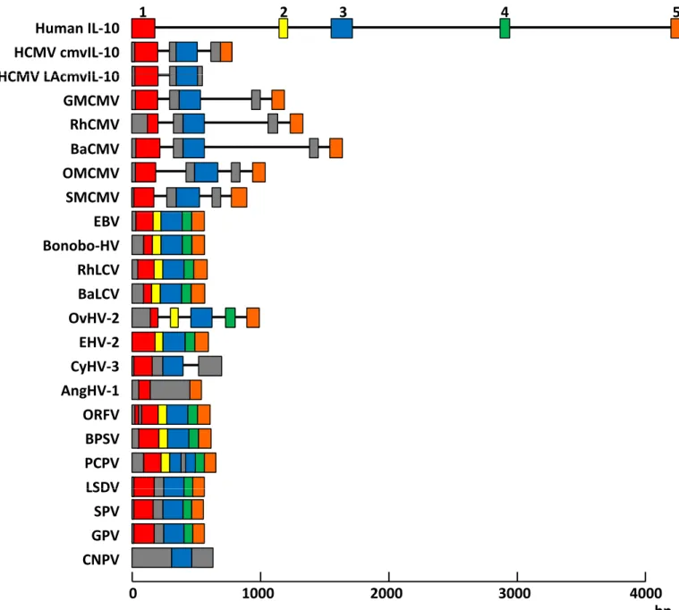
2. Discovery of vIL-10s 42
3. Genetic structure of IL-10 homologues 43
4. Origin and evolution of vIL-10s 43
5. Protein structure of IL-10 homologues 45
6. Transcriptomic and proteomic expression of vIL-10 genes 46
7. Ligand-receptor complexes formed by IL-10 homologues 47
8. Biological activities of IL-10 homologues 48
8.1 Biological activities of cIL-10 48
8.2 Biological activities of vIL-10s 49
9. Viral IL-10s as a topic of applied research 56
10. Concluding remarks 57 11. Acknowledgements 57 12. References 58 Supplementary material S1 70 Supplementary material S2 71 Objectives ……….. 72 Experimental section ……… 74
1st chapter: 75 “The IL-10 homologue encoded by cyprinid herpesvirus 3 is essential neither for viral replication in vitro nor for virulence in vivo”
2nd chapter: 95
“Development of a Safe and Efficacious Attenuated Recombinant Vaccine against Cyprinid herpesvirus 3”
Discussion and perspectives ………. 112
Summary………..………... 119
Preamble
Common carp (Cyprinus carpio) is cultivated for human consumption worldwide. It is one of the most important freshwater species in aquaculture with a world production of 3.4 million metric tons per year (estimation from the FAO for 2010). While the common carp represents a cheap source of animal proteins, its coloured subspecies koi (Cyprinus carpio koi) is cultivated as an expensive pet fish for personal pleasure or worldwide competitive exhibitions. The price of individual collectable subjects fluctuates between 1 to 30 K euros, but can reach much higher prices. In the late 1990s, a highly contagious and virulent disease began to cause severe economic losses in these two carps industries worldwide. The causative agent of the disease was initially called koi herpesvirus (KHV). It has been renamed as cyprinid herpesvirus 3 (CyHV-3) in 2005 and classified in the Alloherpesviridae family of the Herpesvirales order.
In addition to its economic importance, CyHV-3 has several qualities as a fundamental model of infection: (i) It is phylogenically distant from the vast majority of herpesviruses that have been studied so far. (ii) It can be studied in laboratories by infection of its natural host (homologous virus-host model). (iii) The sequence of its genome published in 2007 revealed a fascinating virus with unique properties in the Herpesvirales, such as an extremely large genome (295 Kb), a high number of genes which are not homologous to known viral sequences, and genes that are normally found exclusively in the Poxviridae. (iv) Interestingly, the sequencing of the CyHV-3 genome revealed several genes potentially encoding proteins involved in immune evasion mechanisms. Among these genes, ORF134 encodes a homologue of carp Interleukin 10 (IL-10). The identification of this ORF in the CyHV-3 genome represents an opportunity to study in vitro and in vivo the roles of a viral IL-10 (vIL-10) homologue using a homologue virus-host model. The present thesis was devoted to the study of this viral gene.
The structure of this manuscript is as follows. It starts with an introduction on the Herpesvirales order, on CyHV-3 and on the viral Interleukin-10 homologues encoded by viruses. While the first section is brief and general, the second and the third represent review manuscripts that have been published (in Veterinary research) or accepted for publication (in Journal of General Virology), respectively. After the introduction, the objectives of the thesis are briefly exposed. The results obtained during this thesis are then presented in two chapters. The first chapter has been published in Veterinary Research, while a manuscript including the results of the second chapter is under preparation for publication. In the last section of this manuscript, the main results are discussed and potential perspectives are presented.
Preamble
The introduction of this thesis is structured in three chapters. The first chapter presents a brief description of the order Herpesvirales. The general properties of the viruses belonging to this order and the families it contains are described. This section of the manuscript is an update and adapted version of a text used in most theses of the host lab on herpesviruses. The second chapter of this introduction summarized the knowledge on Cyprinid herpesvirus 3 (CyHV-3) available in the literature when this thesis was printed. It represents a review has been published in Veterinary research. Ping Ouyang is second author of this review. The third and last chapter of the introduction represents a review on viral homologues of Interleukin-10 encoded by viruses (vIL-10). Ping Ouyang is co-first author of this manuscript.
Introduction: The order Herpesvirales
5
Introduction
1
st
chapter:
The order Herpesvirales
1. Introduction
At the border of living and non-living, viruses are submicroscopic biological agents consisting of nucleic acid and protein shell which may be multilayered. They cannot replicate in the extracellular medium and reproduce as obligate intracellular parasites in the host organism. Since the description of the tobacco mosaic virus at the end of the 19th century, thousands of viruses were described in every ecosystem. They infect bacteria, plants and animals (Dimmock et al., 2007). The International Committee on Taxonomy of viruses (ICTV) developed universal systems for classifying viruses. In the current ICTV taxonomy, six orders have been established, the Caudovirales, the Herpesvirales, the Mononegavirales, the Nidovirales, the Picornavirales and the Tymovirales (King et al., 2012).
Members of the order Herpesvirales are enveloped viruses with a linear double-stranded DNA (dsDNA) genome. They share an identical structure. A densely packed DNA core is contained in an icosahedral capsid. The capsid is embedded in a complex proteinaceous layer called the tegument. A lipid envelope containing numerous viral glycoproteins forms the outermost structure of the viral particle (McGeoch et al., 2008). Most of the members of the order Herpesvirales have been shown to realize two distinct phases in their life cycle: lytic replication characterized by a transcription program where immediate-early (IE), early (E), and late (L) genes are expressed successively; and latency, consisting of the maintenance of the viral genome as a non-integrated episome and the expression of a limited number of viral genes and microRNAs (Roizman & Pellet, 2007). Upon reactivation, latency reverses to a lytic replication.
The origin of the order Herpesvirales has been estimated at several hundred million years ago (Davison, 2002). So far, approximately 135 members have been isolated from oyster, fish, amphibian, reptile, bird and mammal species, including human (Davison et al., 2009). Herpesviruses have mainly co-evolved with their host and in most cases are well adapted to them. This adaption is demonstrate that the ability of most herpesviruses to persist in the host species without inducing lethal infection.
The order Herpesvirales contains three families, the Herpesviridae (comprising viruses infecting mammals, birds and reptiles), the Alloherpesviridae (comprising viruses infecting fish and amphibians) and the Malacoherpesviridae (comprising viruses infecting mollusks) families. Below, we will first provide a general and brief description of the structure, the genome, the common biological properties and the replication cycle of the members of the order Herpesvirales. Next, we will discribe briefly the biological specificities of the three families.
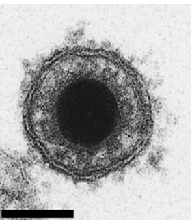
2. Virus structure
Every virus classified in the order Herpesvirales possesses an identical structure (Ackermann, 2004). Their genome is protected by an icosahedral capsid with diameter of approximately 100 nm. The capsid is composed of 162 capsomers (150 hexons and 12 pentons) (Figure 1). This nucleocapsid is surrounded by an amorphous layer of proteins termed tegument, which contains proteins mainly
Glycoproteins Envelope Envelope Genome Capsid Tegument Isomers Examples
Figure 1. Herpesvirus structure. Schematic representation and electron microscopy picture of a viral
particle.
A
LTR RTRB
C
R4 R3 R2 R1 1 1 1 Isomers HHV-6 AlHV-1 BoHV-4 HHV-4 Examples CyHV-3F
D
IR U ER S ULE
2 4 1 UL b’ c’ US c an b a’n an BoHV-1 HHV-1 MuHV-1Figure 2. The order Herpesvirales regroups 6 classes of genome. Horizontal lines represent unique
regions. Rectangles represent left and right terminal repeats (LTR and RTR, respectively) for A group; internal repeats R1 to R4 of the C group and internal and external repeats (IR and ER) for the D group. Terminal repeats of the E group are constituted by two parts. One is composed by n copies of the a sequence near the larger b sequence. The other one is composed by the repeated a sequences followed by a c sequence. Terminal sequences anb and canare inversed and are separated by long (UL) and short (US) unique
sequences. In the B group, terminal sequences are repeated a variable number of times at each extremity. In the D group, Ug p, SScan be inverted compared to the Up LLgiving two different isomers. In the E group, Ug g g p, LLand USS
regions can also be inverted generating four different isomers. Terminal repeats were not described in the F group. Human herpesvirus 1 (HHV-1), 4 (HHV-4) and 6 (HHV-6), Alcelaphin herpesvirus 1 (AlHV-1),
Bovin herpesvirus 1 (BoHV-1) and 4 (BoHV-4), Murin herpesvirus 1 (MuHV-1) and Cyprinid herpesvirus 3
involved in gene expression regulation. Finally, a lipid envelope bearing viral glycoproteins is covering the elements listed above to form a spherical particle of approximately 150 to 300 nm in diameter (Figure 1).
3. Genomic features
Herpesvirus genome is a long dsDNA molecule, linear in the capsid, but circular once it penetrates the nucleus of the host cell (Roizman & Pellet, 2007). Depending of the virus species, the guanine plus cytosine (G+C) percentage varies from 31 to 75% while the genome length varies from 120 to 295 kilo base pairs (kbp) (Aoki et al., 2007; Roizman & Pellet, 2007). The genome contains variable internal and terminal repeated sequences. Based on the arrangement of these sequences, herpesvirus genomes have been classified in 6 different groups (Figure 2) (Roizman & Pellet, 2007). All herpesvirus genomes contain at their termini conserved signals for packaging of the DNA into capsids (Roizman & Pellet, 2007).
4. Common biological properties
Herpesviruses seem to share 4 important biological properties (Ackermann, 2004). Firstly, they encode their own enzymes for nucleic acid synthesis. Secondly, both viral DNA replication and assembly of the nucleocapsid take place in the nucleus of the infected cell. Thirdly, production of progeny viral particles leads to the lysis of the infected cell. Finally, even if this not firmly demonstrated for the Alloherpesviridae and Malacoherpesviridae families, all studied herpesviruses are able to establish a latent infection in their natural host.
5. Biological cycle
Herpesviruses have two distinct phases in their life cycle: lytic and latent infection. The characterization of these two phases is based on the study of members of the Herpesviridae family.
5.1 Lytic infection
The herpesvirus multiplication cycle is illustrated in Figure 3. It starts with the virion attachment on the host cell surface mediated by the interaction of viral glycoproteins with their cellular receptors. For example, human herpesvirus 1 (HHV-1) first binds to the cells through interaction of glycoproteins gC and gB with some cellular proteoglycans such as heparan sulfate (Spear, 2004). A stronger attachment is then mediated by the interaction of gD to its specific cellular receptor (Spear, 2004).
After fusion of the viral envelope with the plasma membrane (or eventually endocytic vesicles), the nucleocapsid and tegument proteins are delivered in the cytoplasm where microtubules bring the nucleocapsid surrounded by the tegument close to the nucleus (Figure 3) (Sodeik et al.,
Attachment of the virion on the cell surface and fusion of the envelope with the plasma membrane
Cytoplasm Viral protein expression and viral DNA replication Immediate Early Early L α β Nucleus Concatemers Late γ Viral egress Enveloppement-deenveloppement model Luminal model
Figure 3. Schematic representation of the lytic infection of herpesviruses (adapted from Flint et al.,
2000). DNA synthesis Viral gene expression α genes β genes DNA synthesis γ1 genes γ2 genes Virion production Time
1997). The genome is then released and enters the nucleus through a pore of the nuclear membrane. As soon as the genome enters in the nucleus, the viral DNA circularizes prior to viral protein synthesis (Garber et al., 1993). This circularization is realized by direct ligation of single unpaired 3’ end nucleotides present at both ends of the genome (Davison, 1984). Tegument proteins migrate with genome into the nucleus where they regulate virus and cellular gene expression.
Herpesvirus gene expression is characterized by a transcription program where immediate-early (IE or α), early (E or β), and late (L or γ) genes are expressed successively (Figures 3 and 4) (Honess & Roizman, 1974; 1975; Jones & Roizman, 1979). IE gene expression is initiated by tegument proteins which interact with cellular transcriptional proteins, such as RNA polymerase II, to activate the transcription. IE genes encode mainly for transcription factors which inhibit IE gene expression and promote E gene expression. The maximum of E gene expression is usually observed between 4 and 8 hours post-infection (Figure 4). They are mainly coding for enzymes involved in nucleotide metabolism and viral DNA replication (Figure 3). Similarly as IE genes, E genes down regulate their own expression while stimulating the expression of L genes. Maximum L gene expression occurs after virus DNA replication (Figures 3 and 4). L genes are further divided in L1 (or γ1) and L2 (or γ2) subclasses. L1 gene expression is increased by viral DNA synthesis genes while L2 gene expression starts only after the synthesis of the viral genome (Figure 4) (Wagner et al., 1998). Most of the L genes code for the proteins incorporated in mature virions; these proteins are called structural proteins. The structural proteome of a virus is defined as all the proteins which enter in the virion composition. Produced capsid proteins encoded by L genes are assembled in the nucleus to form the nucleocapsid containing newly synthesized viral DNA (Figure 3).
The replication of the viral genome is initiated from one or several origins of replication. Specific viral proteins are involved in viral DNA synthesis through a rolling-circle mechanism (Ackermann, 2004; Jacob et al., 1979). This process generates concatemers consisting of complexe structure of high molecular weight made of several genomic units linked head-to-tail (Figure 3). A viral protein complex brings concatemers close to the portal complex of a capsid through which a single genomic unit is internalized and cleaved from the concatemer (Mettenleiter et al., 2009).
Different models were proposed for the egress of the nucleocapsid from the nucleus to the extracellular space (Granzow et al., 2001; Johnson & Spear, 1982; Wild et al., 2005). In the envelopment-deenvelopment model (Figure 3), the temporary enveloped virus in the peri-nuclear space fuses with the external nuclear membrane to deliver the naked capsid in the cytoplasm. Tegument proteins are associated with the capsid before it buds into trans-golgi vesicles to form the envelope (Browne et al., 1996; Granzow et al., 2001; Masse et al., 1999; Smith, 1980). The virion is finally released from the cell by exocytosis or cell lysis (Figure 3) (Flint et al., 2000; Mettenleiter, 2004; Mettenleiter et al., 2009). In the luminal model, the capsids bud in the internal nuclear membrane then migrate in the endoplasmic reticulum (ER). The enveloped virions are then (i) incorporated in a transport vesicle and delivered in the golgi apparatus (vesicular model) or
Figure 5. Acquisition process of herpesvirus envelope. (A) Primary enveloped virions in the perinuclear
space. The electron-dense sharply bordered layer of tegument underlying the envelope and the absence of envelope glycoprotein spikes is noteworthy. (B) After translocation into the cytosol, capsids of HSV-1, PrV and BoHV-4 appear “naked”, whereas those of HCMV and KHV are covered with a visible layer of “inner” tegument. (C) Secondary envelopment and (D) presence of enveloped virions within a cellular vesicle during transport to the plasma membrane. The same stages can be observed for members of the Herpesviridae family and KHV, a member of the Alloherpesviridae family. HSV-1: Herpes simplex type 1; PrV: Pseudorabies virus; HCMV: Human cytomegalovirus; BHV-4: Bovin herpesvirus 4; KHV: Koi herpesvirus. Bars represent 100 nm. Reproduced from Mettenleiter et al. (2009).
(ii) reach the golgi apparatus through connexions between the latter and the ER (intra-cisternal model). Independently of these models, enveloped virions are released by exocytosis (Darlington & Moss, 1968; Johnson & Spear, 1982). Recently, a new model was described for BoHV-1 where capsids present in the nucleus are able to reach the cytoplasm trough enlarged nuclear pore (Wild et al., 2005). The capsids, once in the cytoplasm, bud with golgi-derived vesicles before egress from the host cell by exocytosis.
A recent study by electron microscopy on the morphogenesis of different herpesviruses belonging to the Herpesviridae and Alloherpesviridae families, concludes that the nucleocapsids follow the envelopment-deenvelopment model before being released in the extracellular space by exocytosis (Figure 5) (Mettenleiter et al., 2009).
5.2 Latent infection
Latency is observed in all members of the family Herpesviridae. It consists in the virus maintenance in the host cell without production of viral particles. The mechanisms that induce latency are still poorly understood (Roizman & Pellet, 2007). Latency is supposed to occur when the virus infected specific cell types. The virus can then persist in the host even after the onset of an adaptive immune response able to clear cells supporting a replicative infection. Only few viral genes are expressed during latency. During latency, the genome is maintained as a non integrated episome in the nucleus. When the latent infected cells divide (if they do so), the viral episome is replicated with the cellular genomic DNA. Copies of this episome are then distributed between daughter cells. The latent infection can be interrupted by exogenous stimuli and switched to lytic infection. Latency has been studied mainly in the family Herpesviridae. Regulation of latency seems to be mediated mainly by transcripts (LATs for latency associated transcripts) in alphaherpesviruses (Jones, 2003) while in beta- and gammaherpesviruses latency proteins are expressed (Ballestas & Kaye, 2001; Cardin et al., 2009; Lee et al., 1999).
Recent studies described the presence of microRNAs (miRNA) in the genome of different herpesviruses of the Herpesviridae family (Pfeffer et al., 2005). Ever since, several studies demonstrated miRNA productions amongst the latency transcripts (alphaherpesvirus LATs). They seem to play an important role in cooperation with the beta- and gammaherpesvirus proteins during the viral biological cycle and essentially during the latency where they can modulate cell apoptosis and immune pathways, as well as the viral lytic cycle (Burnside et al., 2006; Cai et al., 2005; Lu et al., 2008; Umbach et al., 2008; Wang et al., 2008).
6. Classification of the order Herpesvirales
The ICTV has classified under the order Herpesvirales viruses encoding the putative ATPase subunit of the terminase (a complex that is responsible for packaging virus DNA into progeny capsids)
Introduction: The order Herpesvirales
10
(Davison, 1992; 2002; Waltzek et al., 2009). This protein is specific to herpesviruses; however, it is also conserved to a lesser degree in the T4-like bacteriophages of the family Myoviridae (Davison et al., 2009). The Herpesvirales order is subdivided in three families: the Herpesviridae, the Alloherpesviridae and the Malacoherpesviridae (Davison et al., 2009; Roizmann et al., 1992).6.1 The Herpesviridae family
The family Herpesviridae is highly studied and is divided into three sub-families: Alpha-, Beta-, and Gammaherpesvirinae (Davison et al., 2009; Roizman & Pellet, 2007). It regroups herpesviruses infecting reptiles, birds and mammals, including humans.
The alphaherpesviruses have a variable host range, a relatively short reproduction cycle, a rapid spread in culture, an efficient destruction of infected cells, and a capacity to establish latent infection in sensory neurons. As example, this subfamily contains the human herpesvirus 1 (HHV-1 or HSV-1) and 3 (HHV-3 or VZV), belonging to the genera Simplexvirus and Varicellovirus, respectively.
In contrast to alphaherpesviruses, betaherpesviruses have a restricted host range. The reproductive cycle is relatively long, and the infection progresses slowly in cell culture. Infected cells frequently become enlarged (cytomegalia). Their latency is established mainly in secretory glands. As example, this subfamily contains the human herpesvirus 5 (HHV-5 or HCMV) and the murid herpesvirus 1 (MuHV-1 or MCHV), belonging to the genera Cytomegalovirus and the Muromegalovirus, respectively.
Gammaherpesviruses have usually a host range restricted to the family or the order of their natural host. In vitro, all members replicate in lymphoblastic cells, and some also cause lytic infections in some types of epithelioid and fibroblastic cells. Viruses in this group are usually specific for either T or B lymphocytes. Latent virus is frequently demonstrated in lymphoid tissue. As example, this subfamily contains the human herpesvirus 4 (HHV-4 or EBV) and 8 (HHV-8 or KSHV), belonging to the genera Lymphocryptovirus and Rhadinovirus, respectively.
6.2 The Malacoherpesviridae family
Until recently, this family consisted in a single virus (Davison et al., 2005): the Ostreid herpesvirus 1 (OsHV-1) infecting the Japanese oyster (Crassostrea gigas). Its genome contains 207 kb and is composed of two unique regions (UL and US; 168 kb and 3 kb, respectively), each flanked by
an inverted repeat (TRL/IRL and TRS/IRS of 7 kb and 10 kb, respectively). The presence of 124 ORFs
are described whose 12 are duplicated in inverted repeats. Interestingly, among all these genes, 38 belong to 12 families of related genes (Davison et al., 2005). Recently, a neurotropic herpesvirus infecting the gastropod abalone (Haliotis spp) was described (Savin et al., 2010). Based on the homology existing between Abalone Herpesvirus (AbHV) and OsHV-1, it has been proposed to
Virus name
(abbreviation) Clade
Common name
(abbreviation) Host(s) Disease
Anguillid HV 1
(AngHV-1) 1
HV anguillae (HVA)
Japanese eel Anguilla japonica
and European eel A. Anguilla Haemorrhages of skin, fins, gills, liver Cyprinid HV 1
(CyHV-1) 1
HV cyprini, carp pox HV, carp HV(CHV)
Common carp
Cyprinus carpio
High losses in fry- exophthalmia haemorrhages, survivors have papilloma Cyprinid HV 2 (CyHV-2) 1 Goldfish hematopoietic necrosis virus (GFHNV) Goldfish Carassius auratus
High mortality at all ages. Necrosis of hematopoietic tissue, spleen, pancreas,
intestine Cyprinid HV 3
(CyHV-3) 1
Koi HV (KHV), carp nephritis and gill
necrosis virus (CNGV)
Common carp
Gill inflammation, hyperplasia, and necrosis, hematopoietic tissue necrosis.
High mortality at all ages Ictalurid HV 1
(IcHV-1) 2
Channel catfish virus (CCV), Channel catfish herpesvirus
Channel catfish
Ictalurus punctatus
Kidney, liver and intestinal necrosis, haemorrhages, high mortality in young
subjects Ictalurid HV 2 (IcHV-2) 2 Ictalurus melas HV (IcmHV) Black bullhead Ameiurus melas
Kidney necrosis, haemorrhages, high mortality at all ages Acipenserid HV
1 (AciHV-1) 2 White sturgeon HV 1
White sturgeon
Acipenser transmontanus diffuse dermatitis, high losses in juveniles
Acipenserid HV
2 (AciHV-2) 2 White sturgeon HV 2 White sturgeon Epithelial hyperplasia Salmonid HV 1 (SalHV-1) 2 HV salmonis (HPV) Steelhead herpesvirus (SHV) Rainbow trout Oncorhynchus mykiss
Mild disease associated with low losses at 10 °C. Adults: female shed virus in ovarian
fluid. Asymptomatic infection Salmonid HV 2
(SalHV-2) 2
Oncorhynchus masou virus (OMV)
Cherry salmon O. masou, coho salmon O. kisutch, sockeye salmon O. nerka, coho salmon
O. keta, rainbow trout,
Viremia, external haemorrhages exophthalmia, hepatic necrosis. High mortality in young subjects. Survivors have oral papilloma. Infected female shed virus in
ovarian fluid Salmonid HV 3 (SalHV3-) 2 Epizootic epitheliotropic disease virus (EEDV)
Lake trout Salvelinus
namaycush, lake trout × brook
trout S. fontinalis hybrids
Epithelial hyperplasia, hypertrophy, haemorrhages on eye and jaw. High mortality in juveniles at 6–15 °C Gadid herpesvirus 1 (GaHV-1) 2 Atlantic cod herpesvirus (ACHV) Atlantic cod Gadus morhua
Hypertrophy of cells in gills. High mortality in adults. Ranid HV 1 (RaHV-1) 2 Lucké tumor HV (LTHV) Leopard frog
Rana pipiens Renal adenocarcinoma
Ranid HV 2
(RaHV-2) 2
Frog virus 4
(FV-4) Leopard frog No known disease Pilchard HV 2 Australian pilchard
Sardinops sagax
Gill inflammation associated with epithelial hyperplasia and hypertrophy. High mortality Tilapia HV Possible Herpesviridae Tilapia larvae encephalitis virus (TLEV) Blue tilapia
Oreochromis aureus Encephalitis in larvae. High mortality
Percid HV 1 (PeHV-1)
HV vitreum, walleye HV
Walleye
Introduction: The order Herpesvirales
11
include the AbHV-1 in the Malacoherpesviridae family (Savin et al., 2010). Despite the lack of similarity with the capsid proteins encoded by other herpesviruses, electron microscopy analysis demonstrates that OsHV-1 and AbHV-1 have a capsid morphology comparable to that of HHV-1 and IcHV-1 (Davison et al., 2005; Savin et al., 2010).6.3 The Alloherpesviridae family
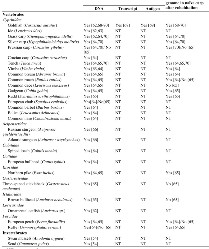
The Alloherpesviridae encompasses viruses infecting fish and amphibians. So far, this family regroups 13 viruses infecting teleost fish, 2 viruses of chondrostean fish and 2 viruses infecting amphibians (Hanson et al., 2011) (Table 1). Phylogenetic studies öbased on the DNA polymerase and the terminase genes led the subdivision of the Alloherpesviridae family into two clades: the first clade comprises large linear dsDNA viruses (245-295 kb) as Anguillid and Cyprinid herpesviruses; the second clade comprises viruses with smaller genome (134-235 kb) as Ictalurid, Salmonid, Acipenserid and Ranid herpesviruses (Davison & Stow, 2005; Waltzek et al., 2009). The genomes of several Alloherpesviridae have been sequenced: Ictalurid herpesvirus 1 (IcHV-1), Cyprinid herpesvirus 3 (CyHV-3), Anguillid herpesvirus 1 (AngHV-1); the Ranid herpesvirus 1 (RaHV-1) and 2 (RaHV-2). Based on these sequences, 12 conserved genes have been identified in the Alloherpesviridae family (Aoki et al., 2007; van Beurden et al., 2010).
Even though Alloherpesviridae are distantly related to Herpesviridae, there are similarities in the way they infect, replicate and persist in the host (Table 1). (i) They display a high level of host specificity, causing disease in only one species or in closely related members of the same genus. (ii) Some alloherpesviruses have been evaluated for long-term latent infections (persistence of viral DNA in survivors without production of infectious particles). Latency has been suggested in CyHV-1, CyHV-3, SalHV-2 and IcHV-1 (Hanson et al., 2011). Much of our knowledge on the biology of Alloherpesviridae is derived from research on two models of infection: IcHV-1 for clade 2 and CyHV-3 for clade 1. CyHV-3 being the subject of this thesis, the remaining part of this introduction has been devoted to this virus.
7. References
Ackermann, M. (2004). Herpesviruses: a brief overview. Methods Mol Biol 256, 199-219.
Aoki, T., Hirono, I., Kurokawa, K., Fukuda, H., Nahary, R., Eldar, A., Davison, A. J., Waltzek, T. B., Bercovier, H. & Hedrick, R. P. (2007). Genome sequences of three koi herpesvirus
isolates representing the expanding distribution of an emerging disease threatening koi and common carp worldwide. J Virol 81, 5058-5065.
Ballestas, M. E. & Kaye, K. M. (2001). Kaposi's sarcoma-associated herpesvirus latency-associated
nuclear antigen 1 mediates episome persistence through cis-acting terminal repeat (TR) sequence and specifically binds TR DNA. J Virol 75, 3250-3258.
Browne, H., Bell, S., Minson, T. & Wilson, D. W. (1996). An endoplasmic reticulum-retained herpes
simplex virus glycoprotein H is absent from secreted virions: evidence for reenvelopment during egress. J Virol 70, 4311-4316.
Burnside, J., Bernberg, E., Anderson, A., Lu, C., Meyers, B. C., Green, P. J., Jain, N., Isaacs, G. & Morgan, R. W. (2006). Marek's disease virus encodes MicroRNAs that map to meq and
the latency-associated transcript. J Virol 80, 8778-8786.
Cai, X., Lu, S., Zhang, Z., Gonzalez, C. M., Damania, B. & Cullen, B. R. (2005). Kaposi's
sarcoma-associated herpesvirus expresses an array of viral microRNAs in latently infected cells. Proc Natl Acad Sci U S A 102, 5570-5575.
Cardin, R. D., Schaefer, G. C., Allen, J. R., Davis-Poynter, N. J. & Farrell, H. E. (2009). The M33
chemokine receptor homolog of murine cytomegalovirus exhibits a differential tissue-specific role during in vivo replication and latency. J Virol 83, 7590-7601.
Darlington, R. W. & Moss, L. H., 3rd (1968). Herpesvirus envelopment. J Virol 2, 48-55.
Davison, A. J. (1984). Structure of the genome termini of varicella-zoster virus. J Gen Virol 65 ( Pt 11), 1969-1977.
Davison, A. J. (1992). Channel catfish virus: a new type of herpesvirus. Virology 186, 9-14. Davison, A. J. (2002). Evolution of the herpesviruses. Vet Microbiol 86, 69-88.
Davison, A. J., Eberle, R., Ehlers, B., Hayward, G. S., McGeoch, D. J., Minson, A. C., Pellett, P. E., Roizman, B., Studdert, M. J. & Thiry, E. (2009). The order Herpesvirales. Arch Virol 154, 171-177.
Davison, A. J. & Stow, N. D. (2005). New genes from old: redeployment of dUTPase by
herpesviruses. J Virol 79, 12880-12892.
Davison, A. J., Trus, B. L., Cheng, N., Steven, A. C., Watson, M. S., Cunningham, C., Le Deuff, R. M. & Renault, T. (2005). A novel class of herpesvirus with bivalve hosts. J Gen Virol 86,
Introduction: The order Herpesvirales
13
Dimmock, N. J., Easton, A. J. & Leppard, K. N. (2007). Towards a definition of a virus. InIntroduction to modern virology, 6 edn, pp. 3 - 17. Edited by N. J. Dimmock, A. J. Easton & K. N. Leppard. Oxford: Wiley-Blackwel.
Flint, S., Racaniello, V. R., Enquist, L. W., Skalka, A. M. & Krug, R. M. (2000). Virology:
molecular biology, pathogenesis, and control. Washington, D. C: ASM press.
Garber, D. A., Beverley, S. M. & Coen, D. M. (1993). Demonstration of circularization of herpes
simplex virus DNA following infection using pulsed field gel electrophoresis. Virology 197, 459-462.
Granzow, H., Klupp, B. G., Fuchs, W., Veits, J., Osterrieder, N. & Mettenleiter, T. C. (2001).
Egress of alphaherpesviruses: comparative ultrastructural study. J Virol 75, 3675-3684.
Hanson, L., Dishon, A. & Kotler, M. (2011). Herpesviruses that infect fish. Viruses 3, 2160-2191. Honess, R. W. & Roizman, B. (1974). Regulation of herpesvirus macromolecular synthesis. I.
Cascade regulation of the synthesis of three groups of viral proteins. J Virol 14, 8-19.
Honess, R. W. & Roizman, B. (1975). Regulation of herpesvirus macromolecular synthesis:
sequential transition of polypeptide synthesis requires functional viral polypeptides. Proc Natl Acad Sci U S A 72, 1276-1280.
Jacob, R. J., Morse, L. S. & Roizman, B. (1979). Anatomy of herpes simplex virus DNA. XII.
Accumulation of head-to-tail concatemers in nuclei of infected cells and their role in the generation of the four isomeric arrangements of viral DNA. J Virol 29, 448-457.
Johnson, D. C. & Spear, P. G. (1982). Monensin inhibits the processing of herpes simplex virus
glycoproteins, their transport to the cell surface, and the egress of virions from infected cells. J Virol 43, 1102-1112.
Jones, C. (2003). Herpes simplex virus type 1 and bovine herpesvirus 1 latency. Clin Microbiol Rev 16, 79-95.
Jones, P. C. & Roizman, B. (1979). Regulation of herpesvirus macromolecular synthesis. VIII. The
transcription program consists of three phases during which both extent of transcription and accumulation of RNA in the cytoplasm are regulated. J Virol 31, 299-314.
King, A., Lefkowitz, E., Adams, M. & Carstens, E. (2012). Order of presentation of virus
taxonomic descriptions. In Virus Taxonomy : Ninth Report of the International Committee on Taxonomy of Viruses, pp. 23-36. Edited by A. King, E. Lefkowitz, M. Adams & E. Carstens. London: Elsevier.
Lee, M. A., Diamond, M. E. & Yates, J. L. (1999). Genetic evidence that EBNA-1 is needed for
efficient, stable latent infection by Epstein-Barr virus. J Virol 73, 2974-2982.
Lu, F., Weidmer, A., Liu, C. G., Volinia, S., Croce, C. M. & Lieberman, P. M. (2008).
Epstein-Barr virus-induced miR-155 attenuates NF-kappaB signaling and stabilizes latent virus persistence. J Virol 82, 10436-10443.
Masse, M. J., Jons, A., Dijkstra, J. M., Mettenleiter, T. C. & Flamand, A. (1999). Glycoproteins
gM and gN of pseudorabies virus are dispensable for viral penetration and propagation in the nervous systems of adult mice. J Virol 73, 10503-10507.
McGeoch, D. J., Davison, A. J., Dolan, A., Gatherer, D. & Sevilla-Reyes, E. E. (2008). Molecular
evolution of the Herpesvirales. In Origin and evolution of viruses, 2 edn, pp. 447-476. Edited by E. Domingo, C. R. Parrish & J. J. Holland. Amsterdam: Elsevier.
Mettenleiter, T. C. (2004). Budding events in herpesvirus morphogenesis. Virus Res 106, 167-180. Mettenleiter, T. C., Klupp, B. G. & Granzow, H. (2009). Herpesvirus assembly: an update. Virus
Res 143, 222-234.
Pfeffer, S., Sewer, A., Lagos-Quintana, M., Sheridan, R., Sander, C., Grasser, F. A., van Dyk, L. F., Ho, C. K., Shuman, S., Chien, M., Russo, J. J., Ju, J., Randall, G., Lindenbach, B. D., Rice, C. M., Simon, V., Ho, D. D., Zavolan, M. & Tuschl, T. (2005). Identification of
microRNAs of the herpesvirus family. Nat Methods 2, 269-276.
Roizman, B. & Pellet, P. E. (2007). The Family: Herpesviridae a brief introduction. In Fields
virology, 5 edn, pp. 2479-2499. Edited by D. M. Knipe & P. M. Howley. Philadelphia: Lippincott Williams and Wilkins.
Roizmann, B., Desrosiers, R. C., Fleckenstein, B., Lopez, C., Minson, A. C. & Studdert, M. J. (1992). The family Herpesviridae: an update. The Herpesvirus Study Group of the
International Committee on Taxonomy of Viruses. Arch Virol 123, 425-449.
Savin, K. W., Cocks, B. G., Wong, F., Sawbridge, T., Cogan, N., Savage, D. & Warner, S. (2010).
A neurotropic herpesvirus infecting the gastropod, abalone, shares ancestry with oyster herpesvirus and a herpesvirus associated with the amphioxus genome. Virol J 7, 308.
Smith, J. D. (1980). An additional role for the outer nuclear membrane in the morphogenesis of
herpes simplex virus. Intervirology 13, 312-316.
Sodeik, B., Ebersold, M. W. & Helenius, A. (1997). Microtubule-mediated transport of incoming
herpes simplex virus 1 capsids to the nucleus. J Cell Biol 136, 1007-1021.
Spear, P. G. (2004). Herpes simplex virus: receptors and ligands for cell entry. Cell Microbiol 6,
401-410.
Umbach, J. L., Kramer, M. F., Jurak, I., Karnowski, H. W., Coen, D. M. & Cullen, B. R. (2008).
MicroRNAs expressed by herpes simplex virus 1 during latent infection regulate viral mRNAs. Nature 454, 780-783.
van Beurden, S. J., Bossers, A., Voorbergen-Laarman, M. H., Haenen, O. L., Peters, S., Abma-Henkens, M. H., Peeters, B. P., Rottier, P. J. & Engelsma, M. Y. (2010). Complete
genome sequence and taxonomic position of anguillid herpesvirus 1. J Gen Virol 91, 880-887.
Wagner, E. K., Petroski, M. D., Pande, N. T., Lieu, P. T. & Rice, M. (1998). Analysis of factors
influencing kinetics of herpes simplex virus transcription utilizing recombinant virus. Methods
Introduction: The order Herpesvirales
15
Waltzek, T. B., Kelley, G. O., Alfaro, M. E., Kurobe, T., Davison, A. J. & Hedrick, R. P. (2009).Phylogenetic relationships in the family Alloherpesviridae. Dis Aquat Organ 84, 179-194.
Wang, F. Z., Weber, F., Croce, C., Liu, C. G., Liao, X. & Pellett, P. E. (2008). Human
cytomegalovirus infection alters the expression of cellular microRNA species that affect its replication. J Virol 82, 9065-9074.
Wild, P., Engels, M., Senn, C., Tobler, K., Ziegler, U., Schraner, E. M., Loepfe, E., Ackermann, M., Mueller, M. & Walther, P. (2005). Impairment of nuclear pores in bovine herpesvirus 1
Introduction
2
nd
chapter:
Cyprinid herpesvirus 3: an interesting virus for applied
and fundamental research
Veterinary Research (2013), 44:85
Krzysztof Rakus
1, Ping Ouyang
1, Maxime Boutier
1, Maygane Ronsmans
1, Anca Reschner
1,
Catherine Vanczok
1, Joanna Jazowiecka-Rakus
1and Alain Vanderplasschen
1*
1
Immunology-Vaccinology (B43b), Department of Infectious and Parasitic Diseases, Faculty of Veterinary Medicine, University of Liège, B-4000 Liège, Belgium * Corresponding author. E-mail: a.vdplasschen@ulg.ac.be. Phone: +32 4 366 42 64
Introduction: Cyprinid herpesvirus 3
17
Abstract
Cyprinid herpesvirus 3 (CyHV-3), a member of the family Alloherpesviridae is the causative agent of a lethal, highly contagious and notifiable disease in common and koi carp. The economic importance of common and koi carp industries together with the rapid spread of CyHV-3 worldwide, explain why this virus became soon after its isolation in the 1990s a subject of applied research. In addition to its economic importance, an increasing number of fundamental studies demonstrated that CyHV-3 is an original and interesting subject for fundamental research. In this review, we summarized recent advances in CyHV-3 research with a special interest for studies related to host-virus interactions.
Key words: Cyprinid herpesvirus 3, CyHV-3, koi herpesvirus, KHV, Alloherpesviridae, Common
1. Introduction
The common carp (Cyprinus carpio) is one of the oldest cultivated fish species. In China, culture of carp dates back to at least the 5th century BC, whereas in Europe, carp farming began during the Roman Empire [1]. Nowadays, common carp is one of the most economically valuable species in aquaculture: (i) it is one of the main cultivated fish for human consumption with a world production of 3.4 million tons per year [2]; (ii) it is produced and stocked into fishing areas for angling purpose; and (iii) its colorful, ornamental varieties (koi carp) grown for personal pleasure and competitive exhibitions represent probably the most expensive market of individual freshwater fish with some prize-winners sold for 104-106 US dollars [3].
Herpesviruses infect a wide range of vertebrates and invertebrates [4]. However, the host-range of individual herpesvirus species is generally restricted revealing host-virus co-evolution. In aquaculture, herpesvirus infections have been associated with mass mortality of different fish species causing severe economic losses [5-7]. In the late 1990s, a new highly contagious and virulent disease began to cause severe economic losses in both koi and common carp industries. Soon after its first known occurrences reported in Israel, USA, and Germany [8, 9], the disease was described in various countries worldwide. The rapid spread of the disease was attributed to international fish trade and to koi shows around the world [10]. The causative agent of the disease was initially called koi herpesvirus (KHV) because of its morphological resemblance to viruses of the order Herpesvirales [9]. The virus was subsequently called carp interstitial nephritis and gill necrosis virus (CNGV) because of the associated lesions [11]. Finally, on the basis of genome homology with previously described cyprinid herpesviruses the virus was renamed cyprinid herpesvirus 3 (CyHV-3) [12].
Because of its worldwide spread and the economic losses it caused, CyHV-3 became rapidly a notifiable disease and a subject of application oriented research. However, an increasing number of recent studies have demonstrated that it is also an interesting subject for fundamental research. In this review, we summarized recent advances in CyHV-3 research with a special interest for those related to host-virus interactions.
2. Characterization of CyHV-3
2.1 General description
2.1.1 ClassificationCyHV-3 is a member of genus Cyprinivirus, family Alloherpesviridae, order Herpesvirales (Figure 1a) [13]. The Alloherpesviridae is a newly designated family which regroups herpesviruses infecting fish and amphibians [14]. It is divided into four genera: Cyprinivirus, Ictalurivirus, Salmonivirus, and Batrachovirus [13]. The genus Cyprinivirus contains viruses that infect common carp (Cyprinid herpesvirus 1 and 3; CyHV-1 and CyHV-3), goldfish (Cyprinid herpesvirus 2; CyHV-2)
A
B
Figure 1. Phylogeny of the order Herpesvirales and the Alloherpesviridae family. (A) Cladogram
depicting relationships among viruses in the order Herpesvirales, based on the conserved regions of the terminase gene. The Bayesian maximum likelihood tree was rooted using bacteriophages T4 and RB69. Numbers at each node represent the posterior probabilities (values > 90 are shown) of the Bayesian analysis.
(B) Phylogenetic tree depicting the evolution of fish and amphibian herpesviruses, based on sequences of the
DNA polymerase and terminase genes. The maximum likelihood tree was rooted with two mammalian herpesviruses (HHV-1 and HHV-8). Maximum likelihood values (> 80 are shown) and Bayesian values (> 90 are shown) are indicated above and below each node, respectively. Branch lengths are based on the number of inferred substitutions, as indicated by the scale bar. AlHV-1: alcelaphine herpesvirus 1; AtHV-3: ateline herpesvirus 3; BoHV-1, -4, -5: bovine herpesvirus 1, 4, 5; CeHV-2, -9: cercopithecine herpesvirus 2, 9; CyHV-1, -2: cyprinid herpesvirus 1, 2; EHV-1, -4: equid herpesvirus 1, 4; GaHV-1, -2, -3: gallid herpesvirus 1, 2, 3; HHV-1, -2, -3, -4, -5, -6, -7, -8: human herpesvirus 1, 2, 3, 4, 5, 6, 7, 8; IcHV-1: ictalurid herpesvirus 1; McHV-1, -4, -8: macacine herpesvirus 1, 4, 8; MeHV-1: meleagrid herpesvirus 1; MuHV - 2, - 4: murid herpesvirus 2, 4; OsHV-1: ostreid herpesvirus 1; OvHV-2: ovine herpesvirus 2; PaHV-1: panine herpesvirus 1; PsHV-1: psittacid herpesvirus 1; RaHV-1, -2: ranid herpesvirus 1, 2; SaHV - 2: saimiriine herpesvirus 2; SuHV-1: suid herpesvirus 1; TuHV-1: tupaiid herpesvirus 1. Reproduced with permission from Waltzek et al. [14].
and freshwater eel (Anguillid herpesvirus 1; AngHV-1). Phylogenetic analyses revealed that the genus Cyprinivirus forms a clade distinct from the three other genera listed above (Figure 1b). Viruses of the Cyprinivirus genus possess the largest genomes (248–295 kb) in the order Herpesvirales.
2.1.2 Morphology
Like all members of the order Herpesvirales, CyHV-3 virions are composed of an icosahedral capsid containing the genome, a lipid envelope bearing viral glycoproteins and an amorphous layer of proteins termed tegument, which resides between the capsid and the envelope [15]. The diameter of CyHV-3 virions is 167–200 nm according to the infected cell type (Figure 2) [15]. Morphogenesis of CyHV-3 is also characteristic of the order Herpesvirales, with assembly of the nucleocapsid and acquisition of the lipid envelope (derived from host cell trans-golgi membrane) that take place in the nucleus and the cytosol of the host cell, respectively [9,15,16].
2.1.3 Genome
The genome of CyHV-3 is a 295 kb, linear, double stranded DNA molecule consisting of a large central portion flanked by two 22 kb repeat regions, called the left and right repeats [18]. To date, this is the largest genome among all sequenced herpesviruses. The CyHV-3 genome has been cloned as a stable and infectious bacterial artificial chromosome (BAC), which can be used to produce CyHV-3 recombinants [19].
The CyHV-3 genome is predicted to contain 155 potential protein-coding open reading frames (ORFs), among which eight (ORF1-ORF8) are duplicated in terminal repeats [13]. Nine ORFs are characterized by the presence of introns [13]. CyHV-3 genome encodes five gene families: ORF2, tumor necrosis factor receptor (TNFR), ORF22, ORF25, and RING gene families [18]. The ORF25 family consists of 6 paralogous sequences (ORF25, ORF26, ORF27, ORF65, ORF148 and ORF149) encoding potential type 1 membrane glycoproteins. Independently of the viral strain sequences, ORF26 is described as a pseudogene; while ORF27 has been characterized as pseudogene in 2 out of 3 sequenced laboratory strains [18]. All non-fragmented members of this family (ORF25, ORF65, ORF148 and ORF149) are incorporated in mature virions, presumably in the envelope [20].
Interestingly, CyHV-3 genome encodes proteins potentially involved in immune evasion mechanisms such as, for example, G-protein coupled receptor (encoded by ORF16), TNFR homologues (encoded by ORF4 and ORF12) and an interleukine-10 (IL-10) homologue (encoded by ORF134) [18].
Among the family Alloherpesviridae, twelve ORFs (named core ORFs) are conserved in all sequenced viruses and were presumably inherited from a common ancestor [13]. The Cypriniviruses (CyHV-1, CyHV-2, and CyHV-3) possess 120 orthologous ORFs. Twenty one ORFs are unique to CyHV-3, including ORF134 encoding an IL-10 homolog [13]. The recently described second IL-10
Figure 2. Electron microscopy examination of CyHV-3 virion. Bar represents 100 nm. Adapted with
permission from Mettenleiter et al. [17].
gp108 p32 p136 gp132 gp131 gp115 gp99 p81 gp149 gp148 gp65 gp25 gp59 p92 p78 p72 p62 p123 p31 p35 p34 p36 p42 p43 p68 p70 p69 p84 p89 p90 p44 p51 p45 p57 p60 p95 p112 p97 p137 <0.25 <0.50 <1 <3 >3 ND
Relative protein abundance
Unclassified structural proteins
p66
Figure 3. Schematic representation of CyHV-3 virion proteome. The viral composition of the envelope
(circle), capsid (hexagon) and tegument is indicated. Membrane proteins of type 1, 2 and 3 are represented by triangles pointed inside, triangles pointed outside and rectangles, respectively. Other proteins are shown as squares. The different fillings indicate the relative abundance of proteins based on their emPAI (< 0.25, < 0.50, < 1, < 3 and > 3). p: protein, gp: glycoprotein, ND: no data. Reproduced with permission from Michel et al. [20].
homolog in the family Alloherpesviridae encoded by AngHV-1 does not seem to be an orthologue of the CyHV-3 ORF134 [21]. CyHV-3 shares 40 orthologous ORFs with AngHV-1 although the total number of ORFs shared by all CyHVs with AngHV-1 is estimated to be 55 [13]. This supports the phylogenetic conclusion that among the genus Cyprinivirus, CyHVs are more closely related to each other than to other members of the family Alloherpesviridae [14]. Interestingly, CyHV-3 also encodes genes with closest relatives in viral families such as Poxviridae and Iridoviridae [18, 22].
2.1.4 Genotypes
Whole genome analysis of three CyHV-3 strains isolated in Israel (CyHV-3 I), Japan (CyHV-3 J) and United States (CyHV-3 U) revealed high sequence identity between the strains [18]. The relationships between these strains revealed that U and I strains are more closely related to each other and form one lineage (U/I), whereas J strain is more distinct and forms a second lineage (J) [18]. The existence of genetic differences between European lineage (including U and I genotypes) and Asian lineage (including J genotype) were later confirmed and suggests independent CyHV-3 introductions in various geographical locations [23, 24]. Furthermore, Kurita et al. demonstrated that the Asian lineage contains only two variants (A1 and A2) while the European lineage has seven variants (E1–E7) [24]. Recently, a new intermediate genetic lineage of CyHV-3 including isolates from Indonesia has been suggested [25]. This hypothesis was later supported by analyses of multi-locus variable number of tandem repeats (VNTR). These analyses also suggested that genetically distinct viral strains can coexist in a same location following various introduction events [26]. Although previous study described presence of both CyHV-3 lineages in Europe [23], an European genotype of CyHV-3 has only been revealed recently in East and Southeast Asia [27]. Recently, Han et al. described polymorphism in DNA sequences encoding three envelope glycoprotein genes (ORF25, ORF65, and ORF116) among CyHV-3 strains from different geographical origins [28].
2.1.5 Proteome
Different groups used mass spectrometry to identify CyHV-3 proteins and to study their interactions with cellular and viral proteins. The structural proteome of CyHV-3 was recently characterized by using liquid chromatography tandem mass spectrometry [20]. A total of 40 structural proteins, comprising 3 capsid, 13 envelope, 2 tegument, and 22 unclassified proteins, were described (Figure 3). The genome of CyHV-3 possesses 30 potential transmembrane-coding ORFs [18]. With the exception of ORF81, which encodes a type 3 membrane protein expressed on the CyHV-3 envelope, no CyHV-3 structural proteins have been studied [20, 29]. ORF81 is thought to be one of the most immunogenic (major) membrane proteins of CyHV-3 [29]. Recently, Gotesman et al. using anti-CyHV-3 antibody-based purification coupled with mass spectrometry, identified 78 host proteins and five potential immunogenic viral proteins [30]. In another study, concentrated supernatant was
Introduction: Cyprinid herpesvirus 3
21
produced from CyHV-3 infected CCB cultures and analyzed by 2D-LC MS/MS in order to identify CyHV-3 secretome. Five viral and 46 cellular proteins were detected [31]. CyHV-3 ORF12 and ORF134 encoding respectively a soluble TNFR homologue and an IL-10 homologue, were among the most abundant secreted viral proteins [31].2.2 In vitro replication
CyHV-3 is widely cultivated in cell lines derived from common carp brain (CCB), gills (CCG) and fin (CaF-2) [32, 33]. Permissive cell lines have also been derived from koi fin: KF-1 [9], KFC [11], KCF-1 [34], NGF-2 and NGF-3 [16]. Non-carp cell lines, such as silver carp fin (Tol/FL) and goldfish fin (Au) were also described as permissive to CyHV-3 [35]. Oh et al. reported the expression of cytopathic effect (CPE) in cell line from fathead minnow (FHM) after inoculation with CyHV-3 [36], but this observation was not confirmed by other studies [9, 35].
In vitro study showed that all annotated CyHV-3 ORFs are transcribed during CyHV-3 replication [37]. Transcription of CyHV-3 genes starts as early as 1 h post-infection and viral DNA synthesis initiates as early as 4–8 h post-infection [37]. Similar to all other herpesviruses, most of CyHV-3 ORF transcripts can be classified into three temporal kinetic classes: immediate early (IE; n = 15 ORFs), early (E; n = 111 ORFs) and late (L; n = 22 ORFs). Seven ORFs are unclassified [37]. Fuchs et al. demonstrated that CyHV-3 ORFs that encode for three enzymes implicated in nucleotide metabolisms: thymidine kinase (ORF55), dUTPase (ORF123) and ribonucleotide reductase (ORF141) are nonessential for virus replication in vitro [38].
2.3 Temperature restriction
Water temperature is one of the major environmental factors that influences the onset and severity of viral infection in fish [39]. This statement certainly applies to CyHV-3 as temperature was shown to affect drastically both viral replication in vitro and CyHV-3 disease in vivo.
2.3.1 In vitro
CyHV-3 replication in cell culture is restricted by temperature. Optimal viral growth in KF-1 cell line was observed at temperatures between 15°C and 25°C. Virus propagation and virus gene transcription are gradually turned off when cells are moved from permissive temperature to the non-permissive temperature of 30°C [40, 41]. However, infected cells maintained for 30 days at 30°C preserve infectious virus, as demonstrated by viral replication when the cells are returned to permissive temperatures [40].