Pépite | Recherche et caractérisation de nouveaux peptides antimicrobiens à partir de bactéries lactiques isolées du meconium
157
0
0
Texte intégral
(2) Thèse de Ahmed Khassaf Al Atya, Lille 1, 2016. Acknowledgements On completion of this thesis, I would like to thank all of those who have accompanied me in this work, but also those, close or far, who influenced positively during my journey. These acknowledgements are always challenging and I apologize in advance to those, who could be forgotten. I owe a great depth of gratitude to my honorable supervisor, Professor Djamel Drider whose dynamic and inspiring supervision and dexterous guidance enabled me to achieve this goal. I am also thankful to him for his concrete contribution through constructive suggestions and providing the research facilities during my whole Ph. D. Also with deep sense of honour, I wish to extend my heartily gratitude to co supervisor Dr. Rozenn Ravallec for giving me the motivation and help me in my study. Also with deep sense of honour, I wish to extend my heartily gratitude to Professor Pascal Dhulster Director of Charles viollette institute for welcoming me and giving me an opportunity to work in this research laboratory. I would like to thank the all my jury members, Professor Abdallah Benachour, Professor Jean Marie Lacroix, Dr. Nathalie Connil, Dr. Isabelle Kempf, Dr. Anne Vachée and Dr. Karima Drider for accepting to evaluate my PhD research project and for their time provision and constructive suggestions to improve this research project. Also with deep sense of honour, I wish to extend my heartily gratitude to Dr. Rabah Boukherroub (IEMN, CNRS) and Professor Sabine Szunerits (IMEN, Lille 1) for their collaboration and SEM assistance. I would like to send cardiac gratitude to Dr. Yanath Belguesmia, Dr. Delphine Caly and Dr. Gabrielle Chataigne, who supported me all the way through this project with their kind collaboration, friendly attitude, timely help and constant encouragement. I would like to express my sincere feelings for my friends Alaa, Ameen, Yazen, Qassam, Mohammed, Amer, Imad, Ayad and all other members of this team who provided me family environment during my stay in this laboratory and never hesitated to offer me any kind of help whenever I needed. I would like to acknowledge the Iraqi Ministry of Higher Education and Scientific Research for giving me the opportunity to do my Ph.D in France by awarding the doctoral scholarship.. II © 2016 Tous droits réservés.. lilliad.univ-lille.fr.
(3) Thèse de Ahmed Khassaf Al Atya, Lille 1, 2016. I found no words on my command to express my gratitude and profound admiration to my my old brother Auda who was always a source of inspiration and encouragement for me. In the end, I feel incomplete if I do not extend my thanks to my affectionate, admiring, friendly and loving wife, Alyaa, my children Ali, Fatimah and Mohmed, sisters and brothers for their unreserved love, affection which enabled me to acquire this long adhered aim.. III © 2016 Tous droits réservés.. lilliad.univ-lille.fr.
(4) Thèse de Ahmed Khassaf Al Atya, Lille 1, 2016. Scientific valorization Publications in peer reviewed journals 1-Al Atya, A.K., Drider-Hadiouche, K., Ravallec, R., Silvain, A., Vachee, A., Drider, D., 2015. Probiotic potential of Enterococcus faecalis strains isolated from meconium. Front. Microbiol. 6. doi:10.3389/fmicb.2015.00227 2-Al Atya, A.K., Drider-Hadiouche, K., Vachee, A., Drider, D., 2015. Potentialization of β-lactams with colistin : in case of extended spectrum β-lactamase producing Escherichia col strains isolated from children with urinary infection. Res. Microbiol. 2-7. Doi: 10.1016/j.resmic.2015.12.002. 3-Al Atya, A.K., Abrioul, H., Kempf, I., jouy, E., Auclair, E., Drider, D., 2016. Effects of colistin and bacteriocins combination on the in vitro growth of Escherichia col strains from swine origin.Research in Microbiology. (Under review). 4-Al Atya, A.K., Belguesmia, Y., Chataigne, G., Ravallec, R., Vachee, A., Szunerits, S., Boukherroub, R., Drider, D., 2016. Anti-MRSA activities of Enterocins DD28 and DD93 and evidences on their role in the inhibition of biofilm formation. (submitted). 5-Caly, D.L., Chevalier, M., Flahaut, C., Cudennec, B., Al Atya, A.K., D’Inca, R., Auclair, E. Drider, D., 2016. The safe enteroccin DD14 shows potent anti-Clostridium perfringens activity. Manuscript in preparation.. Posters 1- Ahmed Khassaf Al Atya, Amadine Silvain, Karima Drider-Hadiouche, Anne Vachee, Rozenn Ravallec, Nour-Eddine Chihib and Djamel Drider. Potential antagonism of Enterococcus faecalis strains isolated from meconium. AMP. 4-6 Juin 2014. Lorient. France. 2- Ahmed Khassaf Al Atya, Karima Drider-Hadiouche, Anne Vachee, Rozenn Ravallec and Djamel Drider. Potentialization of β-lactam antibiotics by enterocins DD28 and DD93 produced by novel strains of Enterococcus faecalis.CBL. 17-19 Juin 2015. Lille. France. 3-Ahmed Khassaf Al Atya, Karima Drider-Hadiouche, Anne Vachee and Djamel Drider. Potentialization of β-lactams with colistin: in case of extended spectrum β-lactamase producing Escherichia coli strains isolated from children with urinary infections. GDR. 26-27 October 2015. Orleans. France 4-Delphine L. Caly, Mickaël Chevalier, Christophe Flahaut, Benoît Cudennec, Ahmed Khassaf Al Atya, Romain D’Inca, Eric Auclair and Djamel Drider. Characterisation of DD14, an enteroccin with antiClostridium perfringens activity. Colloque Adebiotech. 16-17 March 2016. Paris. France.. IV © 2016 Tous droits réservés.. lilliad.univ-lille.fr.
(5) Thèse de Ahmed Khassaf Al Atya, Lille 1, 2016. ABSTRACT In this study, we isolated one hundred and seven (107) bacteria with Gram positive staining and devoid of catalase activity, arguing thereof of their belonging to the group of lactic acid bacteria (LAB). These LAB strains were isolated from samples of meconium of six blind donors at Roubaix hospital (North of France). All these LAB strains were identified by MALDI-TOF as Enterococcus faecalis. Nevertheless, only six isolates including E. faecalis 14, E. faecalis 28, E. faecalis 90, E. faecalis 97 and E. faecalis 101 (obtained from donor 3) and E. faecalis 93 (obtained from donor 5) exhibited antagonism against Gram negative bacteria (GNB) and mainly Gram positive bacilli (GPB). This antagonism was attributed to the production of lactic acid and bacteriocins, which are ribosomally synthesized antimicrobial peptides. The identification of these antagonistic LAB strains was completed by the sequencing of their 16S rDNAs. Besides their antagonism capabilities, these antagonistic strains were studied for a set of characteristics such as antibiotic resistance, hemolytic activity, virulence factor, aggregation and hydrophobicity to design any possibility of probiotic grade. Bacteriocins produced by E. faecalis 28 and E. faecalis 93, designed as enterocins DD28 and DD93, which are issued from two distinct donors, revealed anti-Staphylococcus aureus activity and even anti-methicillin-resistant Staphylococcus aureus (MRSA) activity. They were then purified by a simplified two-step purification procedure including an ion exchange chromatography and reversedphase high-performance liquid chromatography (RP-HPLC). Their molecular masses were determined by mass spectrometry and were shown to be 5,204.48 Da and 5,203.90 Da respectively. Interestingly, these molecular masses do fit with those usually reported in the literature particularly those of enterocins L50A, L50B, MR10A and MR10B. To establish the DNA sequences of enterocins DD28 and DD93, we have amplified the total DNA from the producing strains E. faecalis DD28 and E. faecalis DD93, using the forward (5'-ATG GGAGCA ATC GCA AAA TTA GTG A-3') and reverse (5'-TTA ATG TCT TTT TAG CCA TTT TCA ATT TG-3') primers, that were previously designed to amplify DNA coding for enterocins MR10A and MR10B (Liu et al., 2011).Taking advantage of these primers and PCR conditions, we have obtained DNA fragments (amplicons) from each of the aforementioned strains, cloned into pGEM-T Easy vector and sequenced them at Eurofins MWG operon, Ebersberg (Germany).The obtained DNA sequences displayed perfect alignment (100% identity) with DNA coding for enterocins L50A, L50B and MR10A, MR10B. As mentioned above, the enterocins DD28 and DD93 were remarkable for their activity against S. aureus; a dreadful food poisoning agent and human pathogen. This activity was also evidenced even slightly against methicillin resistant S. aureus (MRSA). The inhibition of MRSA was performed with V © 2016 Tous droits réservés.. lilliad.univ-lille.fr.
(6) Thèse de Ahmed Khassaf Al Atya, Lille 1, 2016. enterocins DD28 and DD93 alone or in combination with erythromycin and kanamycin. The inhibition of MRSA was studied against planktonic cells using the kill curves and checkerboard assays. The inhibition study unveiled a clear synergistic effect. Furthermore, this anti-MRSA activity was also studied and highlighted against biofilm formation using AISI 304 L steel slide and glass devices. Related to bacteriocins and antibiotics associations, we have also established that enterocin DD14, another enterocin obtained in the frame of this project, and which DNA sequence is identical to those of enterocins DD28 and DD93 was able to potentialize colistin, an antibiotic used in the veterinary and human medicines to treat infections caused by GNB. The potentialization of colistin was observed against a set of Escherichia coli strains from swine origin. Indeed, the combination of enterocin DD14 and colistin appeared able to decrease E. coli counts in planktonic and biofilm cultures. Given the importance of the data we have obtained on the potentialization of colistin with enterocins DD14, we investigated whether colistin was also able to potentialize other antibiotics such as those of the -lactams group. To this end, we tested the combinations of colistin and ticarcillin or colistin and cefotaxime, two antibiotics of -lactams group to which six E. coli strains from infant origin were resistant. Indeed these non-nosocomial E. coli strains were isolated at Roubaix hospital from children with urinary infections and were considered based on their antibiogrammes as extended spectrum lactamase (ESBL) E. coli strains. The combinations tested here (colistin-ticarcillin or colistin cefotaxime) have led to synergistic effects, as supported by the FIC index (fractional inhibitory concentration) determinations and killing curves experiments. Indeed, the MIC values of ticarcillin has dropped below its breakpoint, in the case of isolates S2, S3 and S5, while it has almost reached the breakpoint for cefotaxime in the case of isolate S2 and the isolate S1. The data gathered from this ESBL study unveiled a possibility of novel treatment of severe E. coli infections.. VI © 2016 Tous droits réservés.. lilliad.univ-lille.fr.
(7) Thèse de Ahmed Khassaf Al Atya, Lille 1, 2016. RESUME Les bactéries lactiques appartiennent au groupe des firmicutes et sont caractérisées par une coloration différentielle positive et l'absence d'une enzyme respiratoire, la catalase. Le principal objectif de cette thèse était d’isoler et identifier des bactéries lactiques antagonistes à partir de meconium. Ainsi, nous avons isolé 107 bactéries répondant aux critères biochimiques suscités, à partir de six échantillons de méconium, issus de nouveaux nés, à l'hôpital de Roubaix (Nord de France). Toutes les bactéries ont été identifiées par MALDI-TOF comme E. faecalis. Toutefois, seuls les isolats E. faecalis 14, E. faecalis 28, E. faecalis 90, E. faecalis 97 et E. faecalis 101 obtenus à partir du donneur 3, et E. faecalis 93 obtenu à partir de donneur 5, avaient une activité antagoniste contre un panel de bactéries à Gram négatif et à Gram positif. Cet antagonisme est attribué à la production d’acide lactique et de bactériocines, qui sont des peptides antimicrobiens de nature protéique, synthétisés par voie ribosomique. L’identification de ces bactéries antagonistes a été confirmée par séquençage du gène 16S (ADNr 16S). Les souches antagonistes ont été plus amplement étudiées pour établir quelques critères probiotiques. Ainsi les six souches antagonistes sont dépourvues d'activité hémolytique, sensibles aux antibiotiques utilisés pour le traitement des infections à entérocoques, caractérisées par l'absence de facteurs de virulence, majeurs et possèdent des scores d'agrégation et hydrophobie très élevés. Les bactériocines produites par E. faecalis 28 et E. faecalis 93 issues de deux donneurs différents, possèdent une activité anti-S. aureus, y compris anti-S. aureus resistant à la méthycilline (SARM). Ainsi, les bactériocines produites par ces souches et appelées entérocines DD28 et DD93, ont été purifiées par chromatographie échangeuse d’ion et HPLC en phase inverse. Leurs masses moléculaires sont respectivement 5 204,48 Da et 5 203,90 Da. Elles sont compatibles avec celles décrites dans la littérature plus particulièrement celles des entérocines L50A et L50B, MR10A et MR10B. Ainsi, des essais d’amplification des gènes codant pour les entérocines DD28 et DD93 ont été réalisés par des amorces 5'-ATG GGAGCA ATC GCA AAA TTA GTG A-3' et 5'-TTA ATG TCT TTT TAG CCA TTT TCA ATT TG--3' en utilisant le programme PCR suivant : une dénaturation initiale de 5 min à 95 °C, suivie par 30 cycles de 1 min à 95 °C, 1 min à la température de fixation des amorces, et une extension de 10 min à une température de 72 °C. Ces amplifications ont permis d’obtenir des fragments d'ADN (amplicons) de 290 pb. Ces amplicons ont été clonés dans le vecteur pGEM-T Easy vector et séquencés (Eurofins MWG operon, Ebersberg, Germany). L'analyse des séquences nucléotidiques, a permis de montrer, un parfait alignement (100% d'identité) avec les séquences nucléotidiques des entérocines MR10A et MR10B.. VII © 2016 Tous droits réservés.. lilliad.univ-lille.fr.
(8) Thèse de Ahmed Khassaf Al Atya, Lille 1, 2016. Comme indiqué, les entérocines mises en évidence dans cette étude se distinguent par un large antagonisme incluant des souches comme S. aureus; une bactérie pathogène d’intérêt alimentaire et médical. Il faut noter que les entérocines et plus particulièrement les entérocines DD28 et DD93 issues de deux donneurs différents, avaient une activité contre les souches SARM. Pour concevoir de nouveaux concepts thérapeutiques tout en gardant l'efficacité des antibiotiques existants, nous avons réalisé des tests anti-SARM combinant les entérocines DD28 et DD93 avec l'érythromycine et la kanamycine. En effet, après un criblage d'activité anti-Staphylocoque (epidermidis, aureus, SARM), nous avons choisi comme modèle une souche de SARM, appelée SARM-S1, isolée chez un patient âgé de 80 ans à l'hôpital de Strasbourg. Cette souche "SARM-S1" nous a été donnée par le Docteur Gilles Prévost afin de valider l'activité de nos molécules. Par ailleurs, la souche SARM-S1 est résistante à la kanamycine et aux entérocines testées mais elle est sensible à la rifampicine et la vancomycine. Les combinaisons entérocines-kanamycine ou entérocines-érythromycine ont montré un effet synergique avec des index FIC de 0.3. Toutefois, ces effets synergiques sont confirmés par les courbes de bactéricidie avec une perte de population SARM-S1 de plus de 2 Log10, après 3 heures d'incubation avec les antibiotiques suscités. Des essais de protection (fonctionnalisation) de surfaces de types acier ou verre, par les entérocines DD28 et DD93, les antibiotiques et les combinaisons entérocines-antibiotiques ont montré un effet anti-installation de biofilms. Les études ont été appuyées par des observations microscopiques (épifluorescence et microscopie électronique à balayage). Il faut noter que les entérocines isolées dans le cadre de ce travail, à l’instar des entérocines DD28 et DD93, ne sont pas dotées d’une activité contre les bactéries à Gram négatif. Toutefois, notre étude a permis de montrer que l’association de l'entérocine DD14, une bactériocine produite par l'isolat 14 (E. faecalis 14) dont la séquence nucléotidique est identique à celles des entérocines DD28 et DD93, est capable de potentialiser l’activité de la colistine; un antibiotique de nature lipopeptidique utilisé en médecines humaine et vétérinaire pour traiter les infections causées par les bacilles à Gram négatif. Dans cette partie du travail, nous avons établi que la combinaison entérocine DD14-colistine avait un effet synergique sur des souches d'E. coli d'origine porcine. Ces souches appartiennent à la collection Resapath et ont été généreusement données par le Docteur Isabelle Kempf (ANSES, Ploufragan). Certaines des souches étudiées ont une résistance à la colistine, ce qui constitue un défi à relever dans le traitement futur des infections impliquant ces souches. Les effets synergiques observés in vitro sont consolidés par les courbes de bactéricidies et une perte de la population d'environ 2 Log 10. Enfin, les souches E. coli utilisées comme cible dans cette partie expérimentale ont été caractérisées par la technique de RAPD (Random amplified polymorphic DNA) afin d'établir une éventuelle relation clonale. Enfin dans le cadre de la potentialisation des antibiotiques d'importance majeur comme la colistine, nous avons regardé si la colistine elle-même était capable d'avoir un effet sur d'autres antibiotiques VIII © 2016 Tous droits réservés.. lilliad.univ-lille.fr.
(9) Thèse de Ahmed Khassaf Al Atya, Lille 1, 2016. comme ceux de la famille des -lactamines. Ainsi, des souches d'E. coli d'origine infantile ont été isolées à l'hôpital de Roubaix, à partir de patients (enfants) ayant des infections urinaires. Ces souches ne sont pas des souches nosocomiales. Dans un premier travail, nous avons regardé par la technique de RAPD le lien génétique entre ces souches (lien clonal), puis établi leurs antibiogrammes. Ces souches se sont révélées produire une -lactamase et avoir une résistance à ce groupe d'antibiotiques. Des essais combinant la colistine et la ticarcilline ou encore la colistine et la céfotaxime ont montré des activités synergiques in vitro avec des diminutions des CMI respectives de ces antibiotiques en dessous des points critiques. Celles-ci ont été confirmées et consolidées par les courbes de bactéricidies avec une perte de la population bactérienne d'au moins 2Log 10. Les données de cette étude permettent d’espérer de nouvelles possibilités de traitement des infections à E. coli.. IX © 2016 Tous droits réservés.. lilliad.univ-lille.fr.
(10) Thèse de Ahmed Khassaf Al Atya, Lille 1, 2016. Table of content General Introduction… ……………………………………………..………….…………..…….………….... 1 CHAPTER I- LITERATURE REVIEW....................................................................................................... 6 1. Meconium- what is it ?........................................................................................................................ 6 2. Gastrointestinal microbiota of newborn infants……………………………………………………..…. 6 2.1. Factors affecting the development of gut microbiota in infants………………………………...… 7 2.1.1. Mode of delivery……………………………………………………………………………………….... 7 2.1.2. Diet………………………………………………………………………………….………………..……. 8 2.1.3. Antibiotics………………………………………………………………………….……………….…….. 9 2.2. Microbial diversity in meconium…………………………………………………………………….….. 9 3. Lactic acid bacteria (LAB)…………………………………………………………………………………. 12 3.1. General characterization ofLAB…………………………………………………………………...……12 3.2. Classification of lactic acid bacteria……………………………………………………………..…….14 3.2.1. Enterococcus...............................................................................................................................15 3.2.1.1. Habitat....................................................................................................................................... 17 3.2.1.2. Enterococcus species as probiotics...................................................................................... 18 3.3. Bacteriocins.................................................................................................................................... 19 3.3.1. Enterocins.................................................................................................................................... 23 3.3.1.1.EnterocinAS-48..........................................................................................................................24 3.3.1.1.Enterocin L50 A and L50 B....................................................................................................... 25 3.3.2. Application of enterocins........................................................................................................... 25 CHAPTER 2. LAB from meconium and assessment of their probiotic properties..........................27 CHAPTER 3. Anti-MRSA potency of enterocins DD28 and DD93 and inhibition of biofilm formation…………..……………………………………………………………………………….........……… 52 CHAPTER 4. Potentialization of colistin by enterocin DD14: application to E. coli strains from swineorigin............................................................................................................................................81 CHAPTER 5. Potentialization of β-lactams by colistin in the case of extended spectrum βlactamas E. coli producing strains from infant origin..................................................................102 General conclusions and perspectives.............................................................................................120 References...........................................................................................................................................123. X © 2016 Tous droits réservés.. lilliad.univ-lille.fr.
(11) Thèse de Ahmed Khassaf Al Atya, Lille 1, 2016. ABBREVIATIONS ATCC……………………………………….. American type culture collection AMPs ………………………………………. Antimicrobial peptides B. infantis ………………………….…….... Bifidobacterium infantis BLIS………………………………………… Bacteriocin like inhibitory substance C-section …………………………..……… Caesarian section CFS...........................................................Cell free supernatants CFU …………………………………........... Colony-forming-unit CIP …………………………………………. Collection de l'Institut Pasteur CMV ……………………………………..…. Cytomegalovirus CO2 ………………………………..……..... Carbon dioxide Da………………………………………....... Dalton DNA ……………………………………....... Dexo ribonucleic acid E. coli………………………………….…..... Escherichia coli E. faecalis …………………………….….... Enterococcus faecalis ECDC……………………………………..... European Centre for Disease Prevention and Control ESBL…………………………………………Extended-Spectrum beta-lactamases GIT………………………………………...... gastrointestinal tract GPB……………………………………….... Gram positive bacteria GNB……………………………………….... Gram negative bacteria GRAS……………………………………..... Generally recognized as safe H ……………………………………............ hour H2O2…………………………………………Hydrogen peroxide HPV ………………………………………... human papillomavirus Kb …………………………………….......... Kilo base LAB ……………………………………........ Lactic acid bacteria MAS……………………………………........ Meconium Aspiration Syndrome MDR……………………………………....... Multidrug resistant. XI © 2016 Tous droits réservés.. lilliad.univ-lille.fr.
(12) Thèse de Ahmed Khassaf Al Atya, Lille 1, 2016. Min ……………………………………........ Minute MRSA …………………………………........ Methicillin-resistant Staphylococcus aureus PBS………………………………….…....... Phosphate buffered saline PCR …………………………………..….… Polymorphic chain reaction PFGE……………………………………..… Pulsed field gel electrophoresis RAPD………………………………….…… Random amplified polymorphic DNA St. thermophilus ………………………..… Streptococcus thermophilus UTIs………………………………………... Urinary tract infections WHO………………………………………... World Health Organization. XII © 2016 Tous droits réservés.. lilliad.univ-lille.fr.
(13) Thèse de Ahmed Khassaf Al Atya, Lille 1, 2016. List of figures Figure 1. The most commonly detected microorganisms in the amniotic fluid and placenta from preterm and term pregnancies Figure 2.Overview of LAB associations with plants and animals, in human and foods. Figure 3.Generalized scheme for the fermentation of glucose in lactic acid bacteria. Figure 4. Phylogenetic tree of lactic acid bacteria. Figure 5. The Shape of Enterococcus spp. Figure 6. Schematic presentation of the biosynthesis of enterocin Figure 7. Mode of action of bacteriocins produced by lactic acid bacteria. Figure 8. Targets of both antibiotics and bacteriocins, their general location, and examples of each capable of inhibition of these targets. Figure 9. The percentage of LAB isolates in meconium samples Figure 10 Identification of LAB isolates by MALDI-TOF Mass Spectrometry Figure 11 Antibacterial Staphylococcal activity of enterocin DD28 Figure 12 Structure of polymyxins Figure 13 Mode of action of polymyxin B against Gram negative bacilli targeting the external envelope Figure 14 Modular structure of NRPS with different domains. List of tables. Table 1. The factors affecting colonization and diversity of infant’s microbiota Table 2. Enterococcus species and their allocation into species groups Table 3. Comparison of classification systems for bacteriocins Table 4. Application of enterocin in human and animal health Table 5. Applications of enterocins in natural or fermented food products Table 6. Number of lactic acid bacteria (LAB) isolates in the meconium samples Table 7. Amplification of putative genes coding for known virulence determinants Table 8. Determination of minimum inhibitory concentration (MIC). XIII © 2016 Tous droits réservés.. lilliad.univ-lille.fr.
(14) Thèse de Ahmed Khassaf Al Atya, Lille 1, 2016. General introduction Bacteriocins have been widely used in many fields such as food preservation and have been proposed for medical applications such as the treatment of infections caused by the multidrug-resistant (MDR) bacteria (Karpiński and Szkaradkiewicz, 2016). In the past 70 years, significant developments in the diagnosis and treatment of infectious diseases have been achieved and conducted to major reduction of the associated morbidity and mortality (Magnussen et al., 2004). Since the discovery of penicillin, and until recent past, antibiotics have been relevant and efficient for the treatment of different bacterial infections (Gould and Bal, 2013; Tenover, 2006). Nevertheless, several studies reported a high number of bacterial pathogens exhibiting resistance to antibacterial agents. Moreover, organizations such as the European Centre for Disease Prevention and Control (ECDC), and the World Health Organization (WHO), are considering infections caused by MDR bacteria as a major public health concern and social burden (Roca et al., 2015). Many pathogens including Staphylococcus aureus, Enterococcus spp, Enterobacteriaceae, Pseudomonas aeruginosa, and Acinetobacter baumanii are considered as MDR bacteria because of their aptitude of resistance to various antibiotics (Magiorakos et al., 2012). The mechanisms of antibacterial resistance are complex. Moreover, scientists stated that microorganisms embedded in biofilms could cause many infections in animals and humans, leading to complex clinical profiles. The relationship between the biofilm formation and antimicrobial resistance have been established in many types of pathogenic bacteria such as E. coli, Klebsiella pneumoniae, Moraxella catarrhalis, Proteus mirabilis and P. aeruginosa (Franci et al., 2015). Remarkably, there are many community-acquired and nosocomial infections caused by resistant bacteria such as those involved in the respiratory tract infections, gastro-intestinal infections (GTIs) and urinary tract infections (UTIs) (Gupta et al., 2011; Lee et al., 2013). UTI are important cause of death (Raghubanshi et al., 2014). The most common causes of UTIs are the extended-spectrum -lactamase (ESBL) producing bacteria, including E. coli and K. pneumoniae but also K. oxytoca, and Proteus spp. These bacteria were shown to be resistant to antibiotics of different groups including penicillins, cephalosporins and monobactams (Kumar et al., 2015). The occurrence of ESBL-producing bacteria has increased worldwide (Lee et al., 2007; Briongos-Figuero et al., 2012). The most ESBL- production is occurring in E. coli strains, which are responsible for over 80% of children UTIs (Yoon et al., 2011). Despite the emergence of infections caused by antibiotic-resistant bacteria, there are many attempts to find effective treatments that could have essential role in the inhibition of the growth of the aforementioned bacteria. These inhibitions could be conducted through potentialization of existing antibiotics, determining novel insights in the pharmacokinetics and pharmacodynamics. The concepts of antibiotics combinations provide social outcomes by reasonable use of antibiotics, control of their toxicity and. 1 © 2016 Tous droits réservés.. lilliad.univ-lille.fr.
(15) Thèse de Ahmed Khassaf Al Atya, Lille 1, 2016. management of resistance development (Cassir et al. 2014). Nowadays, there is a need for novel molecules or alternative therapeutics to treat bacterial infections mainly those ascribed to GNB (Albers et al., 2014). To this end, chemistry and genomics have been increasingly applied to uncover new potential therapeutics by using antimicrobial peptides (AMPs) (Anthouard and DiRita, 2015; Lombardi et al., 2015). AMPs, such as bacteriocins have been suggested to be a valid alternative to “traditional” antibiotics (Balciunas et al., 2013; Cotter et al., 2013; Cavera et al., 2015). Bacteriocins are proteinaceous ribosomally synthesized, which are mainly active against closely related species (Ge et al., 2016). Notably, all classes of bacteriocins are ribosomally synthesized, and their coding DNA are usually organized in operon structures containing the structural gene, as well as genes coding for immunity proteins, ABC transporters and accessory proteins. In the case of lantibiotics (class I bacteriocins), they are secreted onto the extracellular medium through an ATP Binding Cassette, formed by proteins encoded by lanf, lanE and lanG genes. Notably, lanI gene codes for a lipoprotein (immunity protein), which binds to the outer surface of the membrane and interacts thereof with the lantibiotic to prevent pores forming (Franz et al., 2007). Genetic and regulation of non lantibiotics bacteriocins showed some similarities with lantibiotics group including the arrangement of the structural, transport, regulatory and immunity genes. However, the immunity system for the non-lantibiotic bacteriocins is specific (Drider et al., 2006), Genes coding for bacteriocins are either located on the chromosome or DNA plasmids (Miller et al., 2005). Bacteriocins belonging to class IIa group are produced under immature form with the leader peptide, then the gene coding for the pre-bacteriocin is co-transcribed with the immunity gene. Moreover a second operon including genes coding for proteins involved in the transport of bacteriocin, and a third one, located nearby comprises the genes of the regulatory system, which are necessary to transport and secrete the mature bacteriocin (Miller et al., 2005). Class IIb bacteriocins are leaderless peptides. Seemingly, there is a specific immunity system allocated this group. In this case, the immunity was conferred by the ABC transporter system, by pumping the bacteriocin out of the cell (Cintas et al., 2000; Criado et al., 2006b).. Another group of bacteriocins have particular structure, including cyclic or circular bacteriocins, as the well studied enterocin AS-48, with head-to-tail ligation and are genetically organized in operon comprising the gene coding for the peptide, and those coding for the immunity proteins, ABC transporter system and hydrophobic basic proteins located at the membrane (Ito et al., 2009). In the case of enterocin AS-48, the complete expression of fully operative bacteriocin and immunity require not less than 10 ORFs (as-48A, B, C, C1, D, D1,E, F, G and H) (Franz et al.,2007) Recent studies have shown that bacteriocins produced by LAB might be active against unrelated species such Campylobacter jejuni, E. coli and Salmonella Thyphimurium (Embaby et al., 2014; Rumjuankiat et al., 2 © 2016 Tous droits réservés.. lilliad.univ-lille.fr.
(16) Thèse de Ahmed Khassaf Al Atya, Lille 1, 2016. 2015) . LAB found naturally in the human microbiota are generally considered as safe and could be thereof considered as serious candidate for probiotic applications. Bacteriocins produced by enterococci, so far called enterocins, interesting models because of their antagonism against food-spoiling or pathogens. Enterocins are synthesized by different Enterococcus species mainly E. faecalis, E. faecium, E. mundtii, E. avium and E. hirae strains, which are isolated from different sources (Birri et al., 2010; Goto and Yan, 2011; Saavedra et al., 2004; Sánchez et al., 2007). Moreover, several studies indicated that many enterocins, such as enterocin AS-48 produced by E. faecalis AS-48, displayed antagonism against GNB and GPB (Grande Burgos et al., 2014). Moreover, several studies underlined the possibilities of using enterocins in human and animal health and also in food preservation (Birri et al., 2010; Todorov et al., 2010). Related to this, the combination of antibiotics and bacteriocins offers a promising strategy to design novel therapeutic options, at time when the potency of traditional antibiotics is fading (Naghmouchi et al., 2013). The combination of antibiotics have been always used to treat severe infections. However, the combinations of antibiotics with bacteriocins are becoming interesting alternatives aiming at reducing the amount of antibiotics used in both human and veterinary medicine. These novel formulations are anticipated to reduce the overall general level of bacterial resistance. Potential achievements with combination of antimicrobial compounds, compared to monotherapy, include a broader antibacterial spectrum, synergistic effects and reducing risk for emerging resistance (Tängdén, 2014). Because of the increasing resistance to broad-spectrum -lactams in Gram-negative bacilli and absence of no new antibiotics in the pipeline, we assume that combination of antimicrobials therapy should be further explored, as previously suggested by G. Weiss (2003). Antimicrobial agents already used including colistin, which has been recently reintroduced in the human therapy as drug of last resort. Indeed, colistin was combined to different classes of antibiotics including carbapenems, tigecycline, or aminoglycosides, leading thereof to successful treatments on clinical cases (Bassetti and Righi, 2015). The combination of colistin with other antibiotics against MDR strains appeared to be successful (Kumar et al., 2010; Traugott et al., 2011). Moreover, the combination of colistin with azithromycin, doxicycline and rifampin, has revealed a synergistic activity against P. aeruginosa isolated from an intensive care unit (López-Fabal et al., 2008). Another report combining colistin and nisin, which is a class I bacteriocin produced so far by Lactococcus lactis (nisin A, Z, F) and Streptococcus uberis (nisin U) exhibited synergistic action against a set of GNB, among which E. coli O157, P. aeruginosa and S. Thyphimurium. Besides, the antagonism displayed by this combination, the use of colistin and nisin formulation, has led to the drop of colistin cytotoxicity on mammalian Caco-2 cells (Naghmouchi et al. 2013). Interestingly, enterocin AS-48, which is described as a perfect bacteriocin was tested in combination with 2-nitro-1-propanol (2NPOH) against S. aureus strains in oat and soya drinks (Burgos et al., 2015). Huang et al. (2013) reported that enterocin RM6 produced by E. faecalis was active against MRSA. In direct line, Cotter et al. (2013) and Allen et al. (2014) considered the combination of bacteriocins and phenolic compounds as good concepts for the control of resistant strains in the food chain. Data involving bacteriocins in the medical field as novel 3 © 2016 Tous droits réservés.. lilliad.univ-lille.fr.
(17) Thèse de Ahmed Khassaf Al Atya, Lille 1, 2016. therapeutic agents (Cavera et al., 2015) are in continuing enhancement, which advocates their potential and timely utility to help in overcoming the antibiotic resistance burden. Recent study reported that bacteriocin use as nisin in combination with conventional antibiotics such as ceftriaxone triggers the outer membrane permeation of Salmonella spp (Singh et al., 2013). Currently, bacteriocins stand as viable and sustainable alternative to traditional antibiotics in the infection treatments (Cotter et al., 2013). 4 © 2016 Tous droits réservés.. lilliad.univ-lille.fr.
(18) Thèse de Ahmed Khassaf Al Atya, Lille 1, 2016. Main objectives of this project Use of bacteriocin as alternative therapeutics to treat bacterial infections when the potency of antibiotics is fading is indeed an interesting option that needs to be deeply studied. The data related to the use of bacteriocins as safe antibacterial agents to replace the traditional antibiotics or enhance their activity must pay attention of the public authorities, agencies of regulation and governments. Different options have been designed to fight against the phenomenon of bacterial resistance. These options include the use of probiotics, essential oils, phage therapy (Wang et al., 2015), and more recently the use of bacteriocin (Ghodhbane et al., 2015). The bacterial resistance is a scaring situation that we need to master and avoid to threaten the next generations. Any concept and any evidence aiming at diminishing the bacterial resistance should be welcome in human and veterinary medicine. To this end, different objectives were designed and achieved throughout the present PhD project. The present manuscript is organized into the following chapters: Chapter 1 is a dissertation on the microbial content and diversity of the meconium. Here, the role of lactic acid bacteria, in particular that of Enterococcus genus is highlighted. Chapter 2 is a research chapter dedicated to isolation and characterization of LAB mainly those displaying antagonism and probiotic properties. Chapter 3 is a research chapter focused on the purification of enterocins DD28 and DD93, determination of their molecular masses and coding DNA sequences. In this chapter, the anti-MRSA activity is unveiled on planktonic and biofilm formation. Chapter 4 is research chapter focused on the potentialization of colistin by enterocin DD14; another bacteriocin purified and characterized in our laboratory. The activity of colistin and enterocin DD14 was tested against a set of E. coli strains from swine origin, which were grown under planktonic and biofilm cultures. Chapter 5 is research chapter dealing, in turns, with the potentialization of antibiotics of -lactams group by colistin. This potentialization was performed on the extended spectrum β-lactamase (ESBL) producing E. coli strains, isolated from children with urinary infections. Chapter 6 is a general conclusion and draws some prospects.. 5 © 2016 Tous droits réservés.. lilliad.univ-lille.fr.
(19) Thèse de Ahmed Khassaf Al Atya, Lille 1, 2016. Chapter I: Literature Review 1. What is meconium? Meconium comes from the Greek word “meconiumarion” meaning poppy juice or opium-like (Romero et al., 2013). It is the first intestinal discharge or the earliest feces of infants, and consists of material ingested or secreted by the gastrointestinal tract (GIT) during the time that the fetus spends in the uterus (Kumagai et al., 2007). It is a sticky dark green substance containing epithelial cells, blood, amniotic fluid, lanugo, bile, bile acid, pancreatic juice, water, and mucus (Swarnam et al.2012). The green coloration of meconium is due to the bile pigments, which are products of heme catabolism. It is the first to be detected in the bile of the fetus after 14 weeks of gestation, then the concentrations of bile increase during gestational period (Blumenthal et al., 1980; Yamaguchi and Nakajima, 1995). Several reports indicated that many drugs are metabolized in the liver, secreted into the bile and thereafter enter into the small bowel, and therefore can be detected in the meconium of the neonate at the time of birth (Wingert et al., 1994). At birth the newborn will evacuate about 60-200 g of meconium (Lee and Kim, 2013). In some cases, the meconium passes when the fetus is still in-utero, staining the amniotic fluid. Meconium stained amniotic fluid is seen in 20% of deliveries and likely to develop respiratory distress (Mundhra, 2013). Because meconium is seldom found in the surrounding amniotic fluid and when a newborn baby breathes a mixture of meconium and amniotic fluid, Meconium Aspiration Syndrome (MAS) generally occurs in term or post-term infants and may be associated with severe respiratory failure (Dargaville, 2012; Haakonsen Lindenskov et al., 2015). 2. Gastrointestinal microbiota of newborn infants Microorganisms found in the meconium are closely related to the microbiota of the gastrointestinal tract (GIT) of newborns. GIT is a complex ecosystem influenced by many biotic and abiotic factors playing a fundamental role in maintaining homeostatis equilibrium. Ninety per cent of the human body is composed of prokaryotic cells, and this group of microorganisms is defined as microbiota (Tsabouri et al., 2014). The GIT is the largest source of microbes, as the human gut microbiome contains up to 10 14 bacteria, which is 10 fold the number of cells in the human body (Munyaka et al., 2014). The bacteria living in the GIT are known as gut microbiota or gut flora and play an important role in human health through promoting the intestinal homeostasis, the development and the maturation of the immune system and protecting against pathogens (Gerritsen et al., 2011; Hattori and Taylor, 2009). Several studies realized formerly demonstrated dynamic changes in the bacterial composition in the gut during pregnancy and childhood development (Johnson and Versalovic, 2012; Palmer et al., 2007). Other studies suggested that the GIT of infants is sterile and the initial bacterial colonization starts after baby transit through the birth canal (Thum et al., 2012) or immediately after the birth (Biasucci et al., 2008). After a vaginal delivery, the GIT of the infant is colonized by the vaginal and fecal. 6 © 2016 Tous droits réservés.. lilliad.univ-lille.fr.
(20) Thèse de Ahmed Khassaf Al Atya, Lille 1, 2016. microbiota of the mother. Infants born by caesarean section are exposed to the skin microbiota of the mother instead. However, the first microbial exposure of newborn delivered by caesarean section perhaps arise from the surrounding environment, hospital staff, and/or other neonates (Satokari et al., 2009; Schwiertz et al., 2003). Studies on intestinal microbiota should include analysis of the microbial ecology and the complexity of the metabolism of the microbial community, as various host–microbial interactions occur at the interface between microbes and host intestine (Ley et al. 2006). Gut microbes start their colonization as soon as the infant’s oral mucosa is exposed to the environment (Mackie et al. 1999). More recently, especially in relation to bacteria colonizing the placenta, evidence has been presented suggesting spread of microorganisms from the mother to the amniotic cavity (Fardini et al., 2010). Some data reported that C-section delivered newborns have lower gut microbial diversity, compared to the vaginally delivered infants (Song et al., 2013). Recently, Groer et al. (2014) demonstrated that bacterial strains from the maternal gut were transmitted onto child intestine during the pregnancy (Groer et al. 2014). It is further admitted that the microbial colonization starts with facultative anaerobic bacteria belonging to genus of Enterococcus or Streptococcus and Enterobacteriaceae, which are followed by anaerobic bacteria such as Lactobacillus, Bifidobacterium, Clostridium, Bacteroides (Makino et al., 2011). 2.1. Factors affecting the development of gut microbiota in infants The first colonization of intestinal microbiota in newborns appeared to be influenced by external and internal factors, which may consequently affect the infant’s health. The type and number of microbial species of infants GIT were not determined by chance, but by a combination of factors such as mode of delivery, gestational age, host genetics, diet, antibiotic use, and environmental factors (Musso et al., 2010). Some factors, more than others, could affect the colonization of GIT and the composition of the gut microbiota in newborns (Munyaka et al. 2014). 2.1.1. Mode of delivery The bacterial community colonizing the GIT of infant is from the environmental origin, mainly from the mother. The mode of delivery (vaginally or Caesarian section) determines the nature of infant gut microbes (Thum et al., 2012). Through normal vaginal delivery, an infant is exposed to the mother’s vaginal and fecal microbes, which results in the colonization of bacteria such as Bifidobacterium, Lactobacillus, Enterococcus and E. coli (Marques et al., 2010; Morelli, 2008). Makino et al. (2013) showed that several Bifidobacteria originating from the mother origin were transmitted and resulted in the colonization of the infant’s intestine shortly after the birth through vaginal delivery. It has been reported that vaginally delivered newborns acquired bacterial communities closely related to their own mother’s vaginal microbiota, dominated by Prevotella, Lactobacillus and Sneathia spp (Goulet, 2015). On the other hand, a number of studies have described altered intestinal. 7 © 2016 Tous droits réservés.. lilliad.univ-lille.fr.
(21) Thèse de Ahmed Khassaf Al Atya, Lille 1, 2016. microbiota profiles in infant delivered by Caesarian section (Scott et al., 2015). Caesarian-section babies harbor microbes that are similar to those of the skin including, Corynebacterium, Staphylococcus, and Proionibacterium (Dominguez-Bello et al., 2011; Song et al., 2013). (Table 1) Table 1. The factors affecting colonization and diversity of infant’s microbiota (Munyaka et al., 2014) Factors affecting colonization of gut microbiota before birth. Factors affecting colonization of gut microbiota during or at birth. Factors affecting colonization of gut microbiota after birth. 1-Intra-uterine environment. 1-Mode of delivery (Caesarean section vs vaginal delivery). 1- Breast feeding vs formula feeding. 2-Maternal exposures or practices such as 2-The environment at the time of delivery stress, antibiotic use and smoking 3-Length of gestation period (term vs preterm). 2-Weaning or food supplementation. 3-Contact with the mother or health care staff 3-Antibiotic exposure 4-Home structure (contact with the mother and other family members including siblings and close contact relative). 2.1.2. Diet The mother needs a diet during pregnancy period and breastfeeding to provide adequate energy and nutrients to support her metabolism and the fetus development, and subsequent milk production. Diet is considered as one of the primary drivers shaping the changes in the structure of the microbiome during infancy, and eventually in the establishment of the adult one, as it has been shown for the microbiome after the exposure to the components of food (Voreades et al., 2014). Moreover, dynamic balance between the host physiology, diet, and GIT microbiota, directly influences the stability of the gut ecosystem and microbial diversity (Munyaka et al., 2014). Previous studies underlined the differential exposure of the infant to fecal, vaginal and skin bacteria from the mother. The mode of birth, and the type of feeding during the first months of life (breastfeeding or formula), are major factors affecting the richness, composition and diversity of the GIT microbial community (Vallès et al., 2014). Furthermore, a recent publication advocated that the type of feeding influences the microbiota composition directly, through providing the substrates for bacterial proliferation and functions (Le Huërou-Luron et al., 2010). It has been reported an increase of more than twice of the number of bacterial cells in breastfed newborns, compared to formula-fed ones (Guaraldi and Salvatori, 2012). The origin of found Lactobacilli and Bifidobacteria or their DNA in the placenta remains speculative. Lactobacilli and Bifidobacteria are also found in the breast-milk (Repa et al., 2015). Notably, Bifidobacterium bifidum, B. longum,. 8 © 2016 Tous droits réservés.. lilliad.univ-lille.fr.
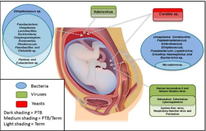
(22) Thèse de Ahmed Khassaf Al Atya, Lille 1, 2016. B. breve and B. adolescentis were isolated from both breast-fed and formula-fed infants, whereas B. infantis is typical of breast-fed, and Bacteroides fragilis of formula-fed infants (Fallani et al., 2011; Mackie et al., 1999). 2.1.3. Antibiotics The GIT microbiota composition of neonates varies between diseased and healthy individuals. Thus, the use of antibiotics during pregnancy could lead to a delayed colonization or reduced abundance of beneficial bacteria (Of et al., 2002; Russell et al., 2012). Indeed, antibiotics can cause an imbalance in the gut microbiota resulting in the repression of both pathogenic and beneficial species, permitting thereof the overgrowth of antibiotic-resistant strains. However, it is now established that effects of antibiotic treatments on GIT microbiota can differ with the dose and type of antibiotic administered (Voreades et al., 2014). The reduction of microbial diversity is often detected after ingestion of antibiotics in infants of less than one year, and the complete recovery of the initial bacterial composition is seldom achieved (Ferrer et al., 2013). Several studies indicated that the use of antibiotics in the prenatal period (during pregnancy) were associated with a delayed colonization by some microbes especially those belonging to Bifidobacteria and Lactobacillus genera (Zwielehner et al., 2011). Moreover, the antibiotic exposure, which is common in preterm infants, extremely decreases the microbial diversity and promotes the growth of pathogens such as Klebsiella, Clostridium and Veillonella spp, which are associated with infants sepsis (Madan et al., 2012). 2.2. Microbial diversity in meconium Historically, the fetus in the intrauterine environment was considered as sterile, with the initial microbial exposure occurring at the birth by vaginally delivering or Caesarian section through contacting maternal vaginal or skin microbiota (Dominguez-Bello et al., 2010; Palmer et al., 2007). However, the presence of microorganisms in meconium suggests that an internal route may transport them to the fetal GIT during pregnancy. Such route might proceed through the bloodstream of the placenta, from where bacteria could pass into the amniotic fluid or fetal circulation (DiGiulio, 2012; Jiménez et al., 2008). During the different stages of pregnancy, the infants swallow large quantities of amniotic fluid, suggesting that the maternal digestive tract may be the origin of microbes found in the amniotic fluid and leads to microbial colonization of the fetal gut (Dasanayake et al. 2005; Jiménez et al. 2005). However, numerous studies reported the presence of different microbes in the umbilical cord, amniotic fluid and placenta (DiGiulio et al., 2008; Hitti et al., 1997; Oh et al., 2010) (Figure 1).. 9 © 2016 Tous droits réservés.. lilliad.univ-lille.fr.
(23) Thèse de Ahmed Khassaf Al Atya, Lille 1, 2016. Figure 1. The most commonly detected microorganisms in the amniotic fluid and placenta from preterm and term pregnancies (Payne and Bayatibojakhi, 2014). In fact, several reports have shown that meconium is not sterile and gut colonization may start before the birth (Jiménez et al., 2008; Madan et al., 2012). On the other hand, meconium microbiota differs from those present in the feces of adults (vaginal and skin), but presents similarities to the fecal microbiota samples from young infants (Gosalbes et al., 2013),although most of studies have focused on the bacterial diversity in the amniotic fluid and meconium. Furthermore, studies reported the presence of fungi, viruses, and protozoa in these two specific environments (Zauli et al., 2013). Remarkably, the eukaryotic microorganisms can be found in the intestinal microbiota. Analysis of the fungal diversity in the GIT unveiled that a majority of fungal phylotypes belongs to Basidiomycota and Ascomycota, including Candida and Saccharomyces genera (Ott et al., 2008; Scanlan et al., 2008). Different species belonging to the Candida genus appeared in the amniotic fluid (Kim et al., 2010). Candida albicans is certainly the predominant species recovered directly from the amniotic fluid, but C. parapsilosis, was reported to cause fetal infection, and C. glabrata was isolated from the amniotic cavity (DiGiulio, 2012). In addition, some authors highlighted the presence of many types of viruses in placenta and amniotic fluid such as human papillomavirus (HPV) and cytomegalovirus (CMV) (Srinivas et al., 2006). Moreover, Villanueva et al. (2000) reported that the presence of CMV in the meconium of infected neonates can be detected by PCR. Other viral taxa were also detected by PCR in amniotic fluid including adenovirus, including enterovirus, Epstein-Barr virus, and respiratory syncytial virus (Baschat et al., 2003). However, the 10 © 2016 Tous droits réservés.. lilliad.univ-lille.fr.
(24) Thèse de Ahmed Khassaf Al Atya, Lille 1, 2016. most recent studies have focused on the bacterial diversity in the meconium of healthy and ill neonates. The presence of pathogenic species, or absence of beneficial species, in early childhood has been suggested to play an important role in the initiation of preterm birth, autism or other immunological deficiency, and development of asthma or eczema allergy (Vaishampayan et al., 2010; Wang et al., 2011; Yap et al., 2011). Another evidence that supports the in-utero origin of the meconium microbiota is the fact that microbial composition in feces changes particularly during the first week from the birth, including many types of bacteria such as Serratia, Klebsiella and Lactobacillus appearing (Moles et al., 2013). In a previous study the microbial composition of meconium showed high concentrations of staphylococci, enterococci, and enterobacteria colonizing the GIT of vaginally and caesarean-delivered term and preterm neonates even from the first day of life (Adlerberth et al., 2006; Borderon et al., 1996). Furthermore, bacterial transfer from the mother to the fetus was established on a mice model. Oral administration of labeled bacteria such as E. faecium to pregnant mice allowed the isolation of the same bacteria from the fetal meconium after birth by caesarian section (Jiménez et al., 2008). After birth, the microbiota of the infants gut resembles to the maternal vaginal microbiome and skin, with Enterococcaceae, Streptococcaceae, Closteridiaceae, Lactobacillaceae, and Bifidobacteraceae predominating bacteria taxa (Aires et al., 2011; Lahtinen et al., 2009). A report suggested that the composition of oral microbes of pregnant women showed that some bacteria, such as Actinomyces naselundii, were associated with low birth weight and preterm delivery, while others, such as lactobacilli, were related to a higher birth weight and term delivery date (Dasanayake and Li, 2005). An additional illustration of the link between the meconium microbiota composition and infant health has been provided by Gosalbes et al. (2013), who found several types of meconium microbiota in healthy newborns containing various potential pathogens as Staphylococcus, E. coli, and Shigella spp., but dominated by LAB including Enterococcus spp., Leuconostoc spp., and Lactococcus spp. Moreover, some studies showed that E. faecalis strains were predominant in the meconium (Al Atya et al., 2015). Interestingly Bifidobacteria have been detected in healthy, full- term birth babies (Lewis et al., 2013). The composition and diversity of the GIT microbiota play an important role in the human health, providing efficient defense against pathogens by various mechanisms such as colonization resistance and production of antimicrobial compounds (Gerritsen et al., 2011).. 11 © 2016 Tous droits réservés.. lilliad.univ-lille.fr.
(25) Thèse de Ahmed Khassaf Al Atya, Lille 1, 2016. 3. Lactic acid bacteria (LAB) 3.1. General characterization of lactic acid bacteria LAB are Gram-positive non-spore-forming, catalase-negative, anaerobic or facultative aerobic cocci or rods bacteria, producing lactic acid as the main fermentation metabolite derived from carbohydrates (O’Sullivan et al. 2002). LAB are found in different ecological niches including soil (Chen and Yanagida, 2006), swine (Mélançon and Grenier, 2003) and water (Hwanhlem et al., 2014). Some LAB isolated from another sources such as fermented foods, plants and the gastrointestinal tracts of many animals and humans (Rubio et al., 2014), where some species can live as commensal microorganisms (Steidler and Rottiers, 2006). LAB species are indigenous microorganisms in many food-related habitats, including plant (fruits, vegetables, and cereal grains) and milk, they are also naturally present in the mucosal surfaces of animals such small intestine, colon, and vagina (Makarova et al., 2006) (Figure 2). Moreover, it is widely known that LAB can excrete different metabolites. Metabolism of LAB is associated with the production of many beneficial compounds like antimicrobial compounds, organic acids and enzymes implicated in bioconversion of complex organic compounds into simple functional compounds (Corsetti et al., 1998). The lack of catalase in LAB causes an accumulation of hydrogen peroxide (H2O2), which is considered as the one of the most important inhibitory metabolites acting against both bacteria and fungi (Caplice, 1999). Kang et al. (2005) reported that some strains of Lactobacillus fermentum were able to inhibit the growth of enterotoxigenic E. coli that causes diarrhea in piglets by producing hydrogen peroxide.. Figure 2. Overview of LAB associations with plants and animals, human and foods ( Douillard and Vos 2014).. 12 © 2016 Tous droits réservés.. lilliad.univ-lille.fr.
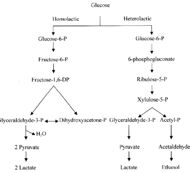
(26) Thèse de Ahmed Khassaf Al Atya, Lille 1, 2016. The main source of energy for bacterial growth is carbohydrates. Metabolisation of carbohydrates by LAB into different useful compounds (mainly lactic acid), is achieved by fermentation. Based on the end-product formed during the fermentation of glucose, LAB can be mainly divided into two main groups (Figure 3): (i) Homofermentative LAB, which produce lactic acid as the major or sole end-product of glucose fermentation, such as Lactococcus, Streptococcus, Pediococcus, and some of lactobacilli. (ii) Heterofermentative LAB, which produce equimolar amounts of lactate, CO2 and ethanol from glucose via the hexose monophosphate or pentose pathway, like Weissella and Leuconostoc and some lactobacilli (Prückler et al., 2015) (Figure 3). The production of organic acids (lactic acid, acetic acid, propionic acid) by LAB has an antimicrobial effect against pathogenic bacteria through the inhibition of active transport processes, enzymes-leaded reactions and modification of their membrane potential (Cleveland and Montville, 2001). Production of organic acid, such as lactic acid, causes a decrease of the pH, contributing to the food preservation by inhibiting the growth of most food spoilage microorganisms (Yang et al. 2012).. Figure 3. Generalized scheme for the fermentation of glucose in LAB (Caplice 1999).. 13 © 2016 Tous droits réservés.. lilliad.univ-lille.fr.
(27) Thèse de Ahmed Khassaf Al Atya, Lille 1, 2016. 3.2. Classification of lactic acid bacteria The organization of LAB taxonomy into different genera was proposed based on their morphologies, growth at different temperatures, glucose fermentation aptitudes, ability to grow at high salt concentrations, acid or alkaline tolerance and configuration of the lactic acid produced. Recently, classification of LAB has suggested new genera, which now include the following ones: Carnobacterium, Dolosigranulum, Aerococcus, Alloiococcus, Enterococcus, Globicatella, Lactococcus, Oenococcus, Tetragenococcus, Vagococcus, and Weissella. Lactobacilli, Carnobacteria and some Weissella are rods while the remaining genera are cocci (Liu et al., 2009). Another interesting study was performed by Stiles and Holzapfel (1997), who defined four major phylogenetic groups of LAB (Figure 4): . The first group consists of Streptococcus and Lactococcus. In the Streptococcus genus, one specie. (Streptococcus thermophilus) is used in the food industry. . The second group contains Oenococcus, Leuconostoc and Weissella (Collins et al., 1993). These. bacteria are widely used in industry food for wine production (Oenococcus oeni), and fermentation of certain vegetables (Leuconostoc citreum). . The third group includes the genera Enterococcus, Tetragenococcus, Melissococcus, Carnobacterium,. Vagococcus, Aerococcus, and Lactosphaera The fourth group includes Lactobacillus and Pediococcus.. 14 © 2016 Tous droits réservés.. lilliad.univ-lille.fr.
(28) Thèse de Ahmed Khassaf Al Atya, Lille 1, 2016. Figure 4. Phylogenetic tree of LAB (Schleifer and Ludwig, 1995).. The morphology is considered as a major characteristic in bacterial taxonomy, and it is still important in the current description of LAB genera (Menconi et al., 2014). Thus, LAB can be divided into rods (Lactobacillus and Carnobacterium) and cocci (all other genera). However, the genus Weissella can include cocci and rods (Collins and Samelis, 1993). Recently, molecular based approaches, such as 16S rDNA sequencing were developed and allowed a more accurate identification of bacteria (Tamang et al., 2008). Calo-Mata et al. (2007) identified genera Carnobacterium, Enterococcus, Tetragenococcus and Vagococcus upon the sequencing of their genes coding for ribosomal RNA 16S and 23S.. 3.2.1. Enterococcus The Enterococcus genus is composed of bacteria exhibiting morphologies such as cocci, occurring alone or in pairs (Figure 5). They are Gram-positive, facultative anaerobic, catalase-negative, oxidase-negative, growing at 10°C, 45°C and able to grow in 6.5% NaCl (Devriese et al., 1993; Facklam and Elliot, 1995). Most species of Enterococcus can hydrolyze esculin in the presence of 40% bile salts (Oulquié Moreno et al., 2006). The heat resistance of enterococci depends not only on the temperature but also on the growth phase (Martinez et al., 2003). Enterococci are usually homo fermentative, the lactic acid is the end product of glucose fermentation, without production of gas (Klein, 2003). 15 © 2016 Tous droits réservés.. lilliad.univ-lille.fr.
(29) Thèse de Ahmed Khassaf Al Atya, Lille 1, 2016. Figure 5. The shape of Enterococcus spp.. Until 1984, Enterococcus spp. were classified as Group D streptococci, with Streptococcus faecalis and Streptococcus faecium as major species. Considering physical and biochemical characteristics, these species were classified as Enterococci rather than Streptococci (Schleifer and Kilpper-Balz, 1984). Reclassification of the species belonging to the Streptococci into genera Streptococcus, Lactococcus, and Enterococcus was based on genetic, chemical and biochemical analyses (Devriese et al., 1995; Schleifer and Kilpper-Balz, 1984). Recent reports have focused on the identification of the Enterococcus species, by using biochemical analysis and molecular identification techniques. Based on 16 rRNA gene similarities, there are 37 species of Enterococcus, which fall into seven species groups (Table 2).. Table 2. Enterococcus species and their allocation into species groups (Franz et al., 2011) Species group based on16S rRNA gene similarity. Species. E. avium group. E. avium, E. devriesei, E. gilvus, E. malodoratus, E. pseudoavium.. E. raffinosus. E. cecorum group. E. cecorum, E. columbae. E. dispar group. E. dispar, E. asini, E. canintestini, E. hermanniensis, E. Pallens. E. faecalis group. E. faecalis, E. caccae, E. haemoperoxidus, E. moraviensis, E. silesiacus, E. termitis, E. ureasiticus, E. quebecensis. E. faecium group. E. faecium, E. canis, E. durans, E. hirae, E. mundtii, E. phoeniculicola, E. ratti, E. villorum, E. thailandicus. E. gallinarum group. E. gallinarum, E. casseliflavus. E. saccharolyticus group. E. saccharolyticus, E. acquimarinus, E. camelliae, E. italicus, E. sulfurous. 16 © 2016 Tous droits réservés.. lilliad.univ-lille.fr.
(30) Thèse de Ahmed Khassaf Al Atya, Lille 1, 2016. Another study reported that the genus Enterococcus includes more than 40 identified different species (Santagati et al., 2012). DNA relatedness, assessed by fingerprints approach allowed the identification and differentiation of the enterococcal species (Baele et al., 2000). The phylogenetic position in Enterococcus species group was determined by 16S rDNA sequençing. For example, the phylogenetic analysis of E. pseudoavium showed the closest relatedness with E. avium species group (Švec et al., 2005). The phylogenetic analysis based on 16s rDNA gene sequencing also provided evidence that Enterococcus strains are more closely related to the Tetragenococcus, Vagococcus and Carnobacterium rather than to Streptococcus and Lactococcus (Holzapfel et al., 2001). 3.2.1.1. Habitat Enterococci are ubiquitous bacteria found in the environment, they are found in a wide variety of environments such as water, soil, sewage, plants, and animals (Goto and Yan, 2011; Layton et al., 2010). However, several studies on ecology of Enterococcus species showed predominance of E. faecium and E. faecalis from different sources like fish, cheese, sausages, minced beef and pork (Foulquié Moreno et al., 2006; Klein, 2003). Interesting work performed by Leclerc et al. (1996) established the presence of E. faecalis, E. faecium, E. durans, E. hirae and E. cecorum in the gastrointestinal tract of animals including poultry, cows, sheep, and pigs. Enterococci are also considered as a part of the natural gastrointestinal microbes of healthy human (Ben Said et al., 2015). High concentrations of Enterococcus species are present in the human feces, reaching about 104 to 106 of bacteria per gram of wet weight (Layton et al., 2010). In consequence enterococci are used as indicators of drinking water fecal contamination. Some data reported that the species of E. faecalis and E. faecium may be more abundant in the feces of human than other species of enterococci, while E. casseliflavus and E. mundtii may be more prevalent than the other species in the environmental biotopes like plants (Ferguson et al., 2005; Wheeler et al., 2002). Many strains of enterococci colonize the human body, especially the GIT, vagina, skin, oral cavity, the upper respiratory tract as “normal” commensals (Hayashi et al., 2005; Jamet et al., 2012). Sometimes these bacteria can be involved in the nosocomial infections located in the urinary tract, blood stream, abdomen, biliary tract, endocardium, and surgical site (Peel et al., 2012). Many Enterococcus species are intrinsically resistant to different classes of antibiotics, including penicillins, cephalosporins, and lincosamides, to a lesser extent to aminoglycosides. In some cases, the therapeutic option becomes limited due to the increasing levels of acquired resistance to multiple antibiotics in the Enterococcus species such as E. faecalis and E. faecium (Heuer et al., 2006). Thus, it is essential to investigate not only the antibiotic resistance profile but also the assessment of the virulence genes of enterococci isolates from the environment and those which are used in the fermentation of meat and vegetable products or used as dairy starter cultures, there are many types of virulence genes found in enterococcal species (Carlos et al., 2010).. 17 © 2016 Tous droits réservés.. lilliad.univ-lille.fr.
Figure


+7


Documents relatifs
Pour imprimer qu’une partie de feuille, on sélectionne le tableau à imprimer puis on choisit le Menu FICHIER – IMPRIMER et on coche impression de la sélection.. L'exercice
En rappelant à cette place l’œuvre que nous avons réalisée il y a deux ans, en consa crant à Ferdinand Hodler deux numéros exceptionnels, nous n ’obéissons qu’au
Ferroelectric Composite Ceramic Probe for MRM Marine Moussu, Stanislav Glybovski, Elizaveta Nenasheva, Redha.. Abdeddaim, Stefan Enoch,
Transfer, 42:311–317, October 1989. Greenhouse models of Venus’ high sur- face temperature, as constrained by Pioneer Venus measurements. The case for a wet, warm climate on early
However, an inclusive framework is still required to organize vast amounts of molecular data, and to provide information about two other key ques- tions of biodiversity studies:
Previously published studied have shown the role of several parameters on the leaching of salt during the water uptake [22]: the initial diffusion coefficient of
This hypervisor is built inside Anaxagoros, a real-time microkernel designed to execute safely hard real- time and non real-time tasks.. This allows the execution of hard
The FODA formalism is intuitive, precise and unambiguous, as confirmed by Gliss [33]. The great popularity of the method is in part due to its ease of use. All methods
