Université de Montréal
STUDY 0F THE ADHERENCE OFACTINOBACILLUSPLEUROFNEUMONIAE TO IMMORTALIZED PORCINE EPITHELIAL CELLS
PAR
ELIANE AUGER
Département de pathobio logie et microbiologie Faculté de medicine vétérinaire
Mémoire présenté à la Faculté des etudes supérieures en vue de l’obtention du grade
Maître ès sciences (M.Sc) en sciences vétérinaires
option microbiologie
Avril, 2006
de Montré al
Direction des bibliothèques
AVIS
L’auteur a autorisé l’Université de Montréal à reproduire et diffuser, en totalité ou en partie, pat quelque moyen que ce soit et sut quelque support que ce soit, et exclusivement à des fins non lucratives d’enseignement et de recherche, des copies de ce mémoire ou de cette thèse.
L’auteur et les coauteurs le cas échéant conservent la propriété du droit d’auteur et des droits moraux qui protègent ce document. Ni la thèse ou le mémoire, ni des extraits substantiels de ce document, ne doivent être imprimés ou autrement reproduits sans l’autorisation de l’auteur.
Afin de se conformer à la Loi canadienne sur la protection des
renseignements personnels, quelques formulaires secondaires, coordonnées ou signatures intégrées au texte ont pu être enlevés de ce document. Bien que cela ait pu affecter la pagination, il n’y a aucun contenu manquant.
NOTICE
The author of this thesis or dissertation has granted a nonexciusive license allowing Université de Montréal to reproduce and pubhsh the document, in part or in whole, and in any format, solely for noncommercial educational and research purposes.
The author and co-authors if applicable retain copyright ownership and moral rights in this document. Neither the whole thesis or dissertation, nor substantial extracts from it, may be printed or otherwise reproduced without the author’s permission.
In compliance with the Canadian Privacy Act some supporting forms, contact
information or signatures may have been removed from the document. While this may affect the document page count. it does flot represent any loss of content from the document.
Ce mémoire intitulé
STUDY 0F THE ADHERENCE 0F ACTINOBACILLUS PLEUR OPNE UMONIAE TO IMMORTALIZED PORCINE EPITHELIAL CELLS
Présenté par
ELIANE AUGER
À
été évalué par un jury compose des persomies suivantesMicha1 Mourez, président-rapporteur
Mario Jacques, directeur de recherche
Nous avons standardisé un modèle in vitro d’adhérence pour l’étude des
pathogènes respiratoires du porc en utilisant deux lignées cellulaires porcines nouvellement établies, notamment la lignée de cellules de trachée Newbom Pig Trachea (NPTr) et la lignée de cellules de poumon St. Jude Porcine Lung (SJPL). Avec l’aide de ce modèle, nous avons étudié les propriétés d’adhérence et d’invasion de l’agent étiologique de la pleuropneumonie porcine, Actinobaciltus pïeuropneumoniae, ainsi que d’autres Pasteureltaceae du porc comme Haemophihts parasuis, Actinobacillus suis et Pasteurella multocida. Nous avons observé des différences d’adhérence entre les souches ainsi qu’entre les lignées. La croissance d’ A. pÏeuropneumoniae serotype I sous conditions restreinte en fer ou en NAD n’a pas affecté son adhérence aux deux lignées. Au niveau de l’invasion, la souche d’ A. pleuropneumoniae testée n’envahie pas les cellules dans notre modèle tandis que les autres Pasteurellaceae ont la capacité d’envahir les cellules. La production de cytokines pro-inflammatoires par les detix lignées suite à une induction par des bactéries A. pleuropneumonicte inactivées a démontré une augmentation de la production de IL-8 par les cellules NPTr, mais aucune production d’autres cytokines n’a été détectée pour les deux lignées. Une analyse des cellules SJPL en contact avec A. pleuropneumoniae suggère une surexpression de protéines impliquées dans l’apoptose et une répression de protéines impliquées dans la croissance cellulaire. Ces résultats démontrent le grand potentiel de ces lignées pour l’étude de la pathogenèse des pathogènes respiratoires du porc.
MOTS-CLÉ : porc, adhérence, invasion, cytokines, lignée cellulaire, poumons, trachée.
n
We have developed an in vitro adherence model for the study of respiratory
pathogens of swinc using two newly established porcine respiratory tract ceil unes, namely the Newbom Pig Trachea (NPTr) and the St. Jude Porcine Lung (SJPL) ce!! unes. Using this model, we studied the adherence and invasion abilities of the
etiological agent of porcine pleuropneurnonia, Actinobaciltus pleuropneumoniae, as well as other porcine Pasteureltaceae such as Actinobaciltus suis, Heamophilus parasuis, and Fasteurelta multocida. We obseiwed differences in adherence between strains and between ce!! unes. Growth of A. pleuropneumoniae serotype I under iron or NAD-deprived conditions did not affect the adherence to both celi unes. The strain of A. pÏeuropneumoniae tested did not invade porcine epithelial celis in our in vitro mode! whule the other porcine PasteureÏtaceae have the capacity to invade. The production of pro-inflarnmatory cytokines by both celi unes following an induction with killed A. pleuropneumoniae revealed an increased production of IL-8 by the NPTr ceils, while no detectable levels were noticed for the other cytokines in either celi unes. Protein profihing of flic SJPL celis revealed a tendency for the up regulation of apoptosis proteins and for the down-regulation of cel! growth proteins. Altogether, these resuits dernonstrate the great potential and versatility of these ceil unes in the study of the pathogenesis of porcine respiratory tract pathogens.
TABLE 0F CONTENTS
RÉSUMÉ
ABSTRACT iii
TABLE 0F CONTENTS y
LIST 0F TABLES viii
LIST 0F FIGURES ix
LIST 0F ABBREVIATIONS xi
DEDICATION xiii
ACKNOWLEDGEMENTS xiv
I. INTRODUCTION
II. LITERATURE REVIEW 3
A. PLEUROPNEUMONIAE 4
1. HISTORY 4
2. CHARACTERISTIC$ 4
3. CLASSIFICATION 5
4. ETHIOLOGY 0F PORCINE PLEUROPNEUMONIAE 5
5. TREATMENT ANI PREVENTION 6
6. VIRULENCE FACTORS 0F A. PLEUROPNEUMONIAE $
6.1. EXOTOXINS 9
6.2. CAPSULE 12
6.4. BIOFILM .16 6.5. HESS .17 6.5.1.LPS 17 6.5.2. TYPE IV FIMBRIAE 18 6.5.3. OMP 0F 60 kDa 19 6.5.4. OMP 0F 50 kDa 20 6.5.5. PGA 21
6.6. IRON ACQUISITION SYSTEMS 21
6.6.1. SDEROPHORES 22
6.6.2. TRANSFERRTN-BINffNG PROTEN 22
6.6.3. HEME COMPOUNOS 23
6.6.4. TRANSPORT ACROSS THE CYTOPLAMSIC
MEMBRANE 24
6.7. OTHER ViRULENCE FACTORS 25
6.7.1. HEMAGGLUTNATION 25 6.7.2. HEMOLYSN 25 6.7.3. PROTEASES 26 6.7.4. SUPEROXDE DISMUTASE 26 6.7.5. UREASE 27 7. EXPEREVIENTAL MODELS 27
8. TECHNIQUES FOR THE IDENTIFICATION 0F NOVEL
VIRULENCE FACTORS 28
8.1.IVET 28
8.2. STM .29
8.3. SCOTS 29
8.4. MICROARRAY 30
VIRULENCE FACTORS 0F OTHER FASTEURELLACEAE 32
1. HAEMOFHILUSPARASUIS 32
2. A CTINOBA CILL US SUIS 33
3. FASTE URELLA MULTOCIDA 33
OBJECTIVES 35
III. MATERIALS & METHODS 36
IV. DISCUSSION 70
V. CONCLUSION 78
VI. REFERENCES 80
LIST 0F TABLES
Literature Review
Table 1. Distribution of the APX toxins genes in the different serotypes of A.
pleuropneumoniae 10
Article
Table 1. Bacterial strains used in the present study 56
Table 2. Proteins of $JPL ceils that are up-regulated following an infection with A.
pleuropneunzoniae serotype 1 S4074 61
Table 3. Proteins of SJPL celis that are down-regulated following an infection with
LIST 0F FIGURES
Literature Revîew
Figure 1. The capsular polysaccharide from A. pteuropneumoniae serotype I 12
Figure 2. Structure of lipopolysaccharides 13
Figure 3. Structure of the core oligosaccliaride region of A. pteztropneurnoniae 14
Figure 4. Structure ofthe 0-antigen of A. pleuropneumoniae serotype 1 15
Figure 5. The type IV fimbrial operon of A. pleuropneuinoniae 19
Figure 6: Diagrammatic representation of the iron uptake systems of A.
pteuropneurnoniae 24
Article
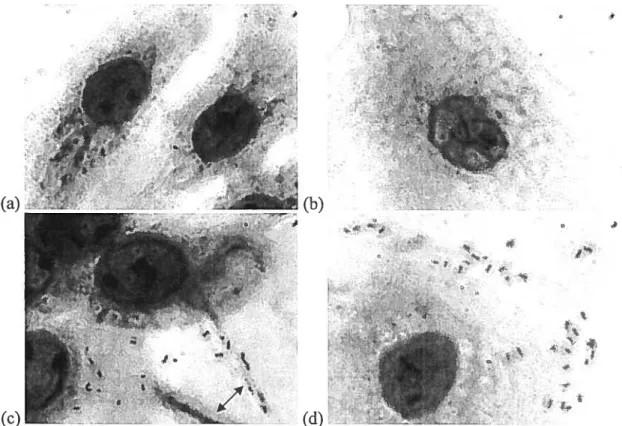
Figure 1. NPTr and SWL celis stained with Giemsa stain in the presence or absence of A. pleuropneumoniae serotype 1 S4074 seen through a Leica DMR microscope at
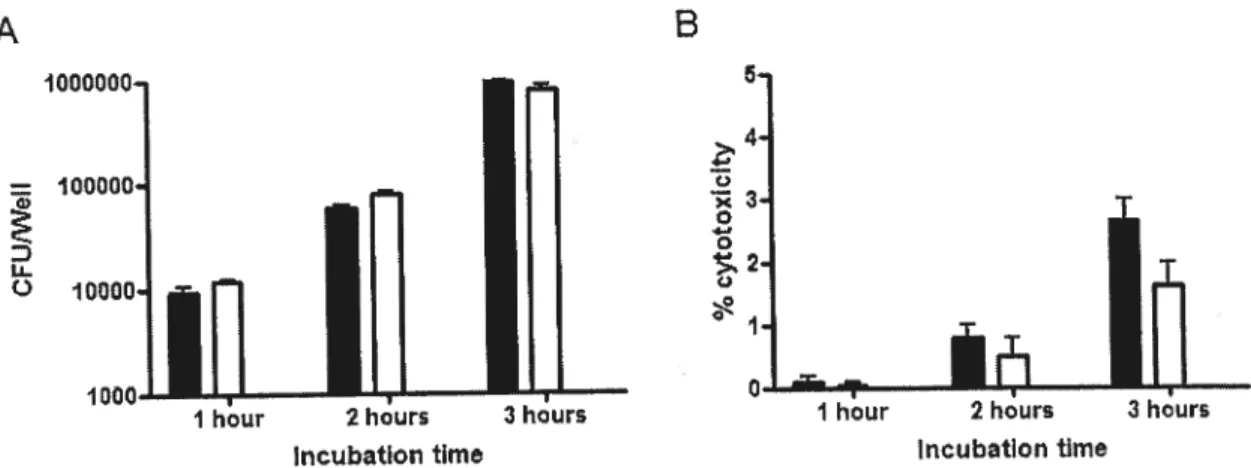
Fïgure 2. Adherence of A. pleuropneumoniae serotype I $4074 to $JPL and NPTr from 1 to 3 hours % cytotoxicity of $JPL and NPTr following infection with A. pleuropneuinoniae serotype I $4074 from I to 3 hours 58
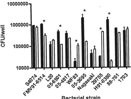
Figure 3. Adherence of twelve Pasteurellaceae to the $JPL and NPTr ceil une after
3h of infection 59
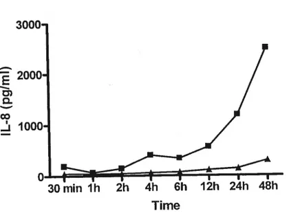
Figure 4. Production of IL-8 by NPTr celis following an induction with killed A.
LIST 0F ABBREVIATIONS
A. actinoinycetenicoiliitaits Actinobacithts actinomycetemcomitans
A.pteuropneumoniae Actinobacitius pteuropneunzoniae
APX A. pteuropneumoniae RTX
A. suis Actinobacillus suis
Bifi Brain Heart Infusion
DMEM Dubelcco’s Modifled Eagle Medium
DPBS Dublecco’s Phosphate Buffer Saline
E.coti Escherichia cou
EDDUA Ethylenediamine dihydroxyphenyl acetic
acid
ELISA Enzyme linked immunoabsorbent assay
EPS Exopolysaccliaride
FBS fetal Bovine Serum
flttt ferric hydrosamate uptake
H. iiifluenzae Haenzoph ilusinfluenzae
H.parasuis Haemophitus parasuis
HgbA Hemoglobin binding protein A
IVET In vivo expression technology
LDH Lactate dehydrogenase
LPS Lipopolysaccharide
MEM Minimum Essential Medium
NAD Nicotinamide adenine dinucleotide
NPTr Newbom Pig Trachea
ORF Open reading frame
PGA Poly-beta- 1 ,6-N-acetyl-D-glucosamjne
PIA Polymer ofbeta-1,6-Iinked N acetylgiucosamine
P. lituttocida Pas teurelta muttocida
PPLO Pleuropneumonia-like organisms medium
RTX Repeats in the structural toxin
SCOTS Selective capture offranscribed sequences SJPL St. Jude Porcine Lung
b my parents Lyne Guittot & Marc Auger
for believingin me ciiid Forproviding me with alt the toots to succeed
ACKNOWLEDGEMENTS
These iast two years will definitely stay engraved in my memory for the rest of my life, and that is due to ail the people that sunounded me and supported me in this joumey. I owe these two years of realisations, achievement and growth to Dr. Mario Jacques, rny research director, for giving me the opportunity to live this experience in his laboratory amongst a great group of peopie I’m 110W proud to cali
my friends. He also provided me with great insight and knowledge of the research world.
I wouÏd also like to acknowledge the support I received by ail the members of the laboratory. Geneviève, Isabelle, Josée, Julie, Julien, Kavi, Lara and Vincent, through your points of view and advices, you have ail participated in the creation of this document.
I address a big thank you to ail members of the research group of infectious diseases of the swine for their support of ail nature, especially to Dr. Michael Mourez and Dr. Marcelo Gottschalk for participating to my advising comity as well as to the jury ofthis document.
I also want to acknowledge the support given to me by my family; my parents, grand-parents (especially Gerard Auger for his support and encouragement), my god-parents (especially Diane Auger for lier tremendous heip), my brother Vincent Auger and my partner Jason Lane. I appreciate everything you ail did for me.
Porcine respiratory diseases have heavily impacted the economy of the pig rearing industry worldwide (103). Actinobacittus pleuropneumoniae, exemplar of these porcine respiratory pathogens, causes porcine pleuropneumonia by colonising the respiratory tract of swine (103). The colonisation of the host is known to be the first step of infection and is initiated by the adhesion of the bacteria to the host celis (92).
For this reason, numerous models have been used to study the pathogenesis of this porcine pathogen, like in vivo models, primary ceil models including tracheal, bucal and lung epithelial celis as well as tissue section including frozen tracheal and lung cross-sections (15, 45, 51, 109). These models and other experiments led to the discovery of LPS as the only known adhesin of A. pleuropneumoniae as well as the observation ofmany putative adhesins (7, 11, 31, 48, 51, 62, 82, 90, 109, 114). To the best of our knowledge, however, no models using irnrnortalised celis unes, which are known to be very effective and advantageous in other models, have been used to study A. pÏeuropneuinoniae.
The goal ofthis present study is therefore: (1) to standardise irnmortalised ceil une infection model to study the adherence and the invasive capacities of A. pleuropneurnoniae and other porcine Pasteurellaceae using two newly established porcine respiratory tract ceil unes; narnely the Newbom Pig Trachea (NPTr) (33) and the St. Jude Porcine Lung (SJPL) (94) cell unes; and (2) to study the response of the celis to their interaction with A. pleuropneumoniae by calculating pro-inflammatory cytokine production and by profihing protein expression using antibody microarray technology.
Actiizobacittus pteuropnetuitoniae
1. Uistory
Formerly known as Haeinophilus pleuropizeumoniae or Haemophilus parahaeinoÏyticus, Actinobacillus pïeuropneumoniae is a member of the Pasteurellaceae family and the etiologic agent of porcine pleuropneumonia (103). The disease was first identified in the United States in 1957 and then made its way around the world, helped by the industrialization of swine production (25, 83). It is now present in most pig-keeping countries including many European countries, the United States, Mexico, South America, Canada, Japan, Korea, Taiwan aiid Australia (103, 112).
2. Characteristics
A. pïeuropnettmoniae is a small Gram-negative capsulated rod with coccobacillary morphology and is highly specific to its host, the swine (43, 112). A high dose however, has been shown to be able to infect rats and mice (45). A. pÏeuropneumoniae is a non-motile facultative anaerobe that does not produce spores (13). Biochemically, it is a beta-hemolytic pathogen (103). 1n addition, it can degrade urea, reduce nitrates and nitrites, acidify fructose, glucose, maltose, manitol, xylose and mannose, but cannot produce gas during glucose fementation and inositol and mutine acidification (13).
3. Classification
The two biotypes of A. pteuropneunioniae are detennined by their NAD
requirement; biotype 1 regrouping NAD-dependent serotypes, and biotype 2 regrouping the serotypes capable of synthesising NAD from specific pyridine nucleotides or their precursor (1$). Surface polysaccharide antigens determine the different serotypes of each biotype. Biotype 1 includes serotypes 1 to 12 and 15, while biotype 2 includes serotypes 2, 4, 7, 9, 13 and 14. It is important to note that two biotype 1 strains belonging to the serotype 13 have been isolated recently. Serotypes 5 and 1 are subdivided in serotype la, lb, 5a and 5h based on minor
differences in the polysaccharides. The serotypes 1, 5, 9, 10, and 11 seem to be more virulent than the other serotypes (103). The most prorninent serotypes in North America are serotypes 1, 5 and 7. In Europe, the primary serotype found is serotype 2 while serotype 15 predominates in Australia (14, 112).
4. Etiology of porcine pleuropneumonia
Porcine pleuropneumonia is a very contagious and often fatal disease. A. pleuropneunzoniae colonises the respiratory tract of pigs to cause this disease, characterised by necrotic ami hemoragic lung lesions, coughing, severe respiratory distress and more (18). four stages of the disease have been identified; peracute, acute, subacute and chronic (103). During infection, cytokines, including IL-l, IL-8 and TNF-Œ, are detected in alveolar fluid and tissue lesions (103).
In the peracute phase, sudden onset of the disease is observed in one or more pigs in the same or different pens. Although fever is present (41.5°C), no major
respiratory signs are noticed. Slight diarrhea and vomiting as weli as apathy and anorexia are the major symptoms ofthis phase ofthe disease (103).
In the acute phase, a high morbidity and mortality are often observed as well as severe cardiac and respiratory distress. Many pigs in the same or different pens are usually affected (103). Lung lesions are characteristic ofthe acute and preacute phase and lead to oedema, inflammation, hemorrhage and necrosis (18).
In the subacute phase as well as in the chronic form ail the acute signs have disappeared and the pigs are left with oniy spontaneous or intermittent cough (103). Weight loss due to loss of appetite is also a characteristic feature of chronic cases (112).
After the infection has established in a herd, continuous apparition of these symptoms occurs as well as, periodically, a reappearance of acute cases (25).
5. Treatments and Preventïon
The use of penicillin and other penicillin analogs are usually effective against A. pleuropneurnoniae infections, even though piasmid-mediated antibiotic resistance is being identified more frequently, especially in serotypes 1, 3, 5, and 7 (32, 103). Resistance to ampicillin, streptomycin, sulfonamides, tetracyclines and chloramphenicol is of great concem. Indeed, low MIC values have been observed in vitro for penicillin, ampicillin, cephalosporin, chioramphenicol, tetracylcines, colistin, suiphonamide, cotrimoxazole and gentamicine while high MIC values were observed for streptomycin, kanamycin, spectinomycin, spiramycin and lincomycin. The major problem in using antibiotic treatments is the pattem of herd infection.
Antibiotics are only effective in the initial phase of the disease and since the animais are anorexic during infection, the antibiotic must 5e given by injection and should be given repeatedly to insure high concentrations in the blood (103). Mass treatment is therefore needed and the rapid course of the disease renders the treatment a matter of timing and of limited value (32). In addition, aithough ciinicai signs may cease, chronic infections in lung abscesses or on tonsils of carriers persist after treatment and are an important source of infection for other animais (103).
Many vaccine formulations have been produced with the goal of generating efficient protection against A. pteuropneumoniae infections. Whole-ceIl bacterins reduced mortality, but do flot protect against heterologous serotypes. SuS-unit vaccines seem to confer a Setter cross-protection than bacterins, decreasing clinical symptoms and increasing performance of the animais. Apx toxins have been shown to be an important part of sub-unit vaccine but are not sufficient on their own. Other factors are evidently involved in protective immunity (44). Extra-cellular products, iron-binding proteins, and outer membrane proteins are other antigens which have been found to induce protection from A. pleuropneumoniae infections (103). Adhesins wouid 5e great vaccine candidates as they are immunogenic and since adhesion is the first step in the pathogenesis of porcine pleuropneumonia. The predicament however, is that the virulence factors responsible for adhesion of A. pleuropneurnoniae are pooriy known (103). LPS, the oniy known adhesin of A. pleuropneuinoniae, have been shown to induce protection (89).
Prevention is without a doubt the key solution to avoid outbreaks and consequentiy avoiding death losses as well as monetary losses. Thorough cleaning of barns and rooms as weII as an ail-in and ail-out management of animal is of great importance and has contributed to a decrease in infection of younger animais by virulent strains of older animais (32). New animais should be derived or originate from a disease-free herd and should be held in quarantine prior to introduction to the new herd (103). Canadian workers implemented a program in which animais of common immune status are comruingled so that populations are compatible in an immunologicai point of view. Three categories have been formed; (1) seroiogicaiiy positive for A. pteuropneumoniae without a history of clinical disease, (2)
serologically negative and clinicaliy free of A. pÏeui-opneumoniae and (3) history of ciinicai disease caused by A. pleuropneumoniae which has been pathoiogically and microbioiogicaliy confirmed. This program greatiy decreased the risk of porcine pleuropneumonia and showed growth performance improvement as well as a decrease in disease outbreaks (32).
6. Virulence Factors of A.pteuropneuiizoniae
The virulence of A. pleuropneumoniae is accomplished by the heip of many factors including exotoxins, endotoxin, capsule, outer membrane proteins, adhesins, transferrin binding proteins and other iron-acquisition systems. These virulence factors which contribute to the different stages of A. pleuropneumoniae infection are discussed in detail below.
6.1 Exotoxins
A. pleuropneumoniae produces four different type oftoxins which are ail part of the RTX (repeat in the structural toxin) farnily of toxins and are consequently called Apx (A. pleuropneurnoniae RTX toxin). RTX toxins are pore-forming proteins that are greatly cytolytic and common in Gram-negative pathogens (35).
ApxI is the rnost hernolytic and cytolytic of ail Apx and therefore greatÏy cytotoxic for phagocytic cells (35). It was formerly named cytolysin I or hemolysin I (43). The apxl operon contains four genes place in the order apxICABD (35). ApxIC being the activator gene, apxlA the pretoxin structural gene, and apxlB and apxlD the secretion apparatus genes (35). The transcriptional activity of the apxl operon can be greatly enhanced by the addition of calcium in the growth medium (36). The ApxI is a 105 kDaprotein and is expressedby serotypes 1, 5a, 5b, 9, 10 and 11(35).
ApxII is weakly hemolytic and cytotoxic and is a also 105 kDa protein (35). The ApxII toxin was formerly known as cytolysin II or hernolysin 11(43). It is expressed by ail serotypes except serotypes 10 and 14, and is encoded by the apxll operon which has the activator gene and the structural gene but lacks the secretion apparatus genes. ApxII seerns to use the products of the apxIBD genes for export (35).
Encoded by the apxIII operon, ApxIII is a 120 kDa protein which is strongly cytolytic but flot hemolytic (60). None of the biotype 2 strains express this toxin and only serotypes 2-4, 6 and 8 of biotype 1 express it (35). It was formerly known as pleurotoxin (43).
ApxW is weakly hemolytic and has a co-hemolytic (CAMP) effect similarly to the effects of ApxII but is only expressed in vivo. Other than the unusuai
C-terminal end, ApxWA has ail the structural characteristics ofRTX toxins (93).
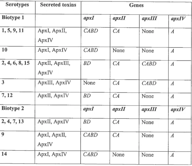
Table 1. Distribution of the APX toxins genes in the different serotypes of A. pleuropneuinoniae (35, 102).
Serotypes Secreted toxïns Genes
Biotype I apxl apxll apxIII apxlV
1, 5, 9, 11 ApxI, ApxII, CABD CA None A
ApxW
10 ApxI, ApxW CABD None None A
2,4, 6, 8, 15 ApxII, ApxIII, BD CA CABD A
ApxW
3 ApxIII, ApxIV None CA CABD A
7, 12 ApxII, ApxIV BD CA None A
Biotype 2 apxl apxll apxIII apxlV
2, 4, 7, 13 ApxII, ApxIV BD CA None A
9 ApxI, ApxII, CABD CA None A
ApxW
14 ApxI, ApxW CA3D None None A
The toxins secreted in vivo as weli as the presence of the different apx genes
ApxI and ApxII have both shown to be maximaliy expressed when ceil density increases and when growth rate decreases, indicating that a mechanism could be used by the bacteria to prevent high-ievei expression of these toxins until an infection is established (57). It was shown, in A. pleuropneunzoniae serotype 1, that Apx toxins are released from the celis into the culture medium by small vesicles of about 20 to 200 nm. The authors also mention the presence of many proteases in these vesicles and that other serotypes of A. pleuropneumoniae may use the same system to release their Apx toxins (77).
The pore forming activities of ApxI, ApxII and ApxIII were studied and revealed similarities between ApxI and ApxIII in that they both created the sarne amount of pores in aqueous-phase-bathing lipid bilayer membranes, but the pores of ApxIII were smaller than that of ApxI; 1.8 nm as cornpared to 2.4 nm for ApxI. ApxII were able to produce pores of similar size to those ofApxl (2.5 nm) but with a 10 times weaker formation ability (70).
The virulence of the different serotypes coincides greatly with the presence of the Apx toxins, especially ApxI and ApxII. In fact, serotypes 1, 5, 9 and 11 are known to be especially virulent and ail express both ApxI and ApxII. Apx toxins are a major virulence factor as they hinder the host’s immune defences and render the strain virulent (35). They are essentiai in pathogenesis of porcine pleuropneumonia (43).
6.2 Capsule
A. pÏeuropneumoniae is an encapsulated pathogen. The capsule, responsible for the iridescence of the colonies on clear agar medium, is present on ail strains and is betwcen 18 to 230 nm thick depending of the serotype (30, 43). Serotypes 5a, 5b and 10 consist of repeating oligosaccharide units whule serotypes 2, 3, 6, 7, 8, 9 and 11 consist of teichoic acid polymers joined by phosphate diester bounds and serotypes 1, 4 and 12 consist of oligosaccharide polymers joined through phosphate bounds. The structure of the serotype 1 capsular polysaccharide is illustrated in Figure 1.
f
4)*n G IcNAcp-(1 —6)-n-Ga1p-(l_QJ—Qï
infAc)0;5
Figure 1. The capsular polysaccharide from A. pleuropneurnoniae serotype 1. Adapted from Altman et al., 1985.
The genetic locus responsible for capsular poiysaccharide export contains four genes ananged in the order cpxDCBA, while the synthesis genes are arranged in the order cpsABCD (30). The capsule is not as major an immunologic factor as the Apx toxins but is certainly required for virulence (43). In fact, a correlation lias been observed between the thickness of the capsule and the virulence of the strain (30). Indeed, capsule-deficient mutants of serotypes I and 5 were less pathogenic than the
parental strains. An acapsular mutant of serotype 1 also showed increased adherence to tracheal cross sections indicating that the capsule does not have a role in adhesion (91). Essentially, this information reveals that although the capsule may rnask certain adhesins, rendering the bacteria less adherent, the capsule protects the bacteria greatly from host defences (30, 91).
6.3 Endotoxin: Lîpopolysaccliaride
This surface polysaccharide is a complex molecule composed of three distinct regions; (j) lipid A, (ii) core-oligosaccharide and (iii) 0-antigen (Figure 2).
The 0-antigen, the most variable part of the LPS, is responsible for the characteristic smooth, semi-rough or rough phenotype of the different serotypes of A. pleuropiteumoniae which depends on the amount of repeated polysaccharide chains
present. Serotypes 2, 4 and 7 are srnooth, serotype 1 and 5 are semi-rough, while serotypes 3 and 6 are rough (11, 21). It is the core oligosaccharide region, however, which seems to be necessary for optimal adherence (see section 6.5.1) and is composed of 3-deoxy-D-mano-2-octulosonic acid (49). It is linked covalently to the
::; LdA
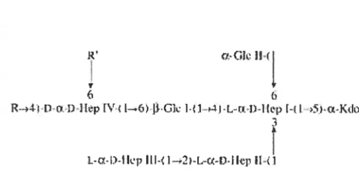
lipid A and is made up of an outer and an inner core (48). Two types of core oligosaccharide have been suggested; type I and 11(55, 73). Based on electrophoresis mobility, serotypes 1, 6, 9, and 11 have a core of type I whule the other serotypes have a core of type 11(55). The structure of this region is represented in Figure 3 below. R’ JIr T 6 R->4:i n•l 1 kp [V [—.( [Gl L.- Dl tp l L j L-C-l)-tlip fl-l-2—[ -fl-[kp Il-Li
Figure 3. Structure of the core oligosaccharide region of A. pteuropneumoniae.
The R and R’ region differ in different serotypes. for serotype 1, R is (1S)-GalaNAc-(1—* 4,6)-ŒGa1 II-(1—-* 3)-f3-Gal I-(1S)-GalaNAc-(1—*, and R’ is H. Adapted from St. MichaeÏ et aï.
(2004).
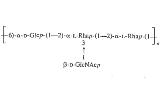
The serotype 1 0-antigen is composed ofbranched tetrasaccharide repeating units composed of 4 residues; 2 a-L-rhamnopyranosyls, 1 Œ-D-glycopyranosyl and 1 2-acetamido-2-deoxy-3-D-g1ucose (3). The structure is illustrated in Figure 4. The O antigen does not however appear to play a role in virulence but seems to be implicated in serum resistance and in binding to phosphatidylethanolamine (58, 63, 90). Jnterestingly, a field isolate of A. pleuropneumoniae serotype 1 with a truncated outer core and no 0-antigen was stiil virulent in pigs (53).
6)-a-DGIcp-(1—2)-a-L-Rhap-( 1—2)-L-Rhap-(
L 3
13-D-GIcNACP
Figure 4. Structure of the 0-antigen of A. pleuropneumoniae serotype 1. Adapted from Altman et al. (1986).
The lipid A consists of about 9.2% of the total lipopolysaccharide and is predominantly formed ofCl4:0, C16:0 and 3-hydroxy C14:0 fatty acids (30). The A. pleuropneumoniae lipid A, as well as its fatty acid components, are able to bind porcine hemoglobin yet the significance of this molecule in an in vivo iron acquisition system is not clear (18). Liposomes containing lipid A were shown to provide protection from death and severe lesions adding support to the role of the lipid A in the pathogenesis of A. pteuropneumoniae (30).
Purified LPS are known to be able to cause damage to ceils and tissues. However, A. pleuropneumoniae LPS are not the cause of the typical lesions seen in porcine pleuropneurnonia, although they may contribute to their formation by increasing the effects ofthe Apx toxins (43).
6.4 Biofilm
A. pleuropneurnoniae lias first been reported to create biofilm in 2004 by Kaplan et at.(62). Biofiims are stmctured community of bacterial celis enclosed in a self-produced polymeric matrix. They are formed by initial attaciuuent of bacterial celis to a solid surface, followed by the arrangement of micro-colonies which then differentiate into exopolysaccharide-encased mature biofiims (22). This exopolysaccharide (EP$) encasernent traps nutriments, helps prevent detachrnent and protects the bacteria from biocides amifrom the immune system of the host (68). One of the best studied EPS is a hexosarnine-containing polymer called PIA in $taphylococcus species and PGA in E. cou. (11 1). ActinobacilÏus actinornyceterncornitans and A. pÏeuropneurnoniae both produce PGA, a linear polymer ofN-acetyl-D-glucosarnine residues in !3 (1, 6) linkage (62).
Following the observation of biofiims in A. pÏeuropneumoniae, Kaplan et al. studied the prevalence of this phenotype in other strains. Out of the 15 reference strains tested only the L20 reference strain of serotype 5b and the 56513 reference strain of serotype 11 exhibited biofilm formation but about haif of ail field isolates of serotypes 1, 5 and 7 where observed to produce biofiims. It was also observed that after one or two passages in fresh broth, the phenotype became irreversibly lost (61).
6.5 Adhesîns
The A. pteuropneumoniae adhesion mechanism is stili poorly known. LPS are the only known adhesins of A. pleuropneumoniae, ail others being putative adhesins. Below, the adhesin and putative adhesins of A. pleuropneumoniae are described in
detail.
6.5.1 Lipopolysaccharide (LPS)
folowing the observation that A. pleuropneumoniae is able to adhere to frozen lung sections as well as porcine tracheal sections, Jacques et al. was able to show that the LPS are the major molecule responsible for this adhesion phenomenon (11, 12, 48, 51, 82). Isogenic mutants of the serotype 1 reference strain 4074 have also been created by Jacqties et aï. using mini-TnlO transposons. These mutants were then characterised to investigate which region of the LPS is responsible for the adhesion. The core region, especially the galactose and D,D-heptose residues ofthe outer core, was shown to be essential for adherence (39, 81, 86, 88). A mutation in the core region caused a decreased adherence whiÏe a mutation in the 0-antigen did not, demonstrating that indeed, the core region and not the 0-antigen is responsible for adherence. As for the lipid A region, purified LPS that did flot contain the region were stili able to block adherence of A. pleurop;zeuinoniae to frozen tracheal cross-sections demonstrating that the lipid A is not necessary for adherence ($2).
Although a 38.5 kDa putative LPS-binding receptor has been identified for A. pleuropneumoniae, no definite receptors have been described to date ($2).
6.5.2 Type IV fimbriae
fimbriae were first noticed on A. pÏeuropneumoniae by Utera et al., but were suspected since 1988 (107). These fimbriae were later shown to be group A type IV fimbriae or pili (114). Type IV fimbriae are present on many Gram-negative bacterial pathogens and can serve many functions like adhesion and twitching motility. In addition, it has been shown that proteins of extremely high similarity with the type IV pilins are essential components of biological processes such as extracellular protein secretion. Type IV fimbriae are characterized according to sirnilarities in amino acid sequence of the pilin polypeptide and the occurrence of N-methylated amino acids in
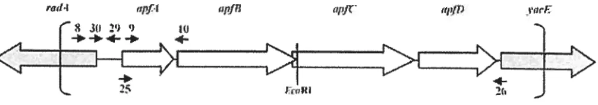
the first amino acid of the mature pilin structural subunit (104). The firnbriae are only expressed by A. pleztropneumoniae under specific conditions which indicates that the expression is regulated by specialised regulatory elements (114). In fact, a study by Overbeke et aï. showed that fimbriae couM be detected on serotypes 2, 5a, 9 and 10 when grown under NAD-restricted condition while they were only detected on serotype 2 and not serotype 5a, 9 and 10 when grown under NAD-rich conditions (109). The fimbrial subunit of A. pÏeuropneurnoniae, called ApfA is a 17 kDa protein and highly similar to type IV fimbrial subunit of other bacterial pathogens and highly conserved between 12 representative serotypes of A. pleuropneumoniae (101, 114). The genes allowing biosynthesis of these fimbriac are ananged in an operon with the order apfABCD as shown in Figure 5 (16).
Figure 5. The type IV fimbrial operon of A. pleuropneumoniae. White arrows represent genes of the type IV fimbriae. Adapted from Boekerna et al. (2004).
The operon is under the regulation of a promoter which is controlled by environmental conditions, such as growth phase and nutrirnent avaiÏability. Host celi contact has also been observed to be correlated to strong firnbriae prornoter activity (16).
The type IV fimbria is an immunogenic molecule as well as a putative adhesin which is expressed in different serotypes. This makes it a great potential vaccine candidate and potentially a major virulence factor of A. pteuropneuinoniae (101).
6.5.3 Outer membrane protein of 60 kDa
A putative adhesin of 60 kDa has been discovered by Enriquez-Verdugo et al. in 2004 (31). After showing that ail A. pleuropneumoniae serotypes tested were capable of adhering to collagen, the authors suggested that an outer membrane protein of 60 kDa would be the collagen binding protein responsible for this adhesion. The authors made this observation using overlay assays of outer membrane proteins (OMP) electroblotted onto a nitrocellulose membrane. They were abie to
show that a 60 kDa protein was able to recognize four types of pig-lung collagen. Serotypes 1 to 12 including 5a and 5b were tested and only serotypes 6 and 11 did flot express this protein. Following this observation, the authors purified the protein and did an inhibition assay using an anti-0MP60. Ccli binding was inhibited in a concentration-dependent effect. Using electron microscopy, the putative protein was observed to be located on the bacterial surface. The authors are working on the identification of the gene encoding this 60 kDa protein which wiil hopefuily lead to the creation of 0MP60 mutants (31).
6.5.4 Outer membrane proteïn of 55 kDa
In 2002, Overbeke et al. observed a 55 kDa hand on a SDS-PAGE gel ofthe sacrosyl-insoluble OMP fraction of A. pleumpneuntoniae serotypes 2, 5a, 9 and 10 strains when grown in NAD-restricted condition (109). This hand was absent or weaker when bacteria were grown in NAD-rich conditions. Since an increased in adhesion was observed when bacteria were grown in NAD-restricted condition, the authors concluded that this OMP of 55 kDa could be involved in adherence. The adhesion scores of serotype 2 did not differ betwcen the NAD-rich and NAD restricted conditions. furthennore, the 55-kDa protein was expressed by this serotype under both conditions. The N-terminai arnino acid sequence of this protein was obtained for serotypes Sa and 10 showing no hornology with other proteins sequences (109). 0MP55 mutants should be created and tested in an adhesion model to prove its role in adherence.
6.5.5 PGA
PGA is an exopolysaccharide and a major component of biofiim matrix encoded by thepgaABCD locus (62). This linear polymer ofN-acetyl-D-glucosarnine residue in 3 (1, 6) linkage is well characterised as PIA in StaphyÏococcïts species and PGA in Escherichia cou (62, 111). As aforementioned (section 5.4), it is a hexosamine-containing polymer that was recently observed in A. pÏeuropneurnoniae (62). Numerous studies revealed PIA or PGA as being a polysaccharide intercellular adhesin (111). Kaplan et al. suggests that A. pÏeuropneunzoniae biofiims produce a hexosarnine-containing intercellular adhesin highly related to the pgaC-dependent polysaccharide of A. actinomyceterncomitans which is stnicturally and functionally related to the E. cou PGA and Staphylococcus epidennidis PIA. PGA does not, however, seem to have an important rolein adhesion to surfaces (62).
6.6 Iron acquisition systems
Iron, an essential nutriment for A. pleuropneuinoniae and almost ail living organisms, is scarce in the host’s environrnent since it is usually complexed to
glycoproteins such as transferrin and lactoferrin (43). Bacteria have evolved many different systems to acquire iron. Below are explanations of the ones used by A. pÏeuropneuntoniae as well as a schematic representation of these systems (Figure 6).
6.6.1 Siderophores
Siderophores are iron chelators that can bind iron present at very low concentrations (68). A. pleuropneumoniae lias been recognised to secrete a yet unkown type of siderophore when grown in iron depleted medium. It is also believed to express receptors for hydroxamate and catechol siderophores secreted by other rnicroorganisms (18, 28). A 77 kDa protein called FhuA has been identified as the receptor for ferric hydroxarnate, a hydroxarnate siderophore. FhuA has also been shown to act as a felTichrome receptor which is not regulated by iron (75). This receptor is coded by thej7iu operon and is hornologous to theJhuACDB cluster of E. coil. The rest ofthefliu operon is involved in the transport ofthe iron source across the cytoplasmic membrane. FhuD being the periplasmic protein that transiocate ferric hydroxamate from the outer to the inner membrane and fhuB and FhuC the cytoplasmic-membrane-assosiated proteins and part of the ABC transporter that internalises ferric hydroxarnate (74).
6.6.2 Transferrin-binding proteïns
A. pteuropneumoniae is able to use transferrin, afthough solely coming from swine (18). This fact lias been suspected to play a role in the high host specificity of A. pleuropneurnoniae (43). TbpA and TbpB are the two transferrin-binding proteins expressed by A. pÏeuropneumoniae in iron-restricted conditions and are co transcribed with the exbBD genes (18). The tbpA and tbpB genes are located on the same operon and express the highly immunogenic TbpA and TbpB proteins (106). TbpA is a 100 kDa protein which likely forms a transmembrane channel for transport
across the outer membrane, while TbpB is a 60 kDa protein suspected to 5e a lipoprotein anchored to the outer membrane. Both TbpA and TbpB bind the C-lobe of porcine transferrin and are necessary in the utilisation of iron from transferrin as a sole source. TbpB has also been shown to be abie to bind hemoglobin (18). ExbB and exbD are linked to the TbpA and TbpB proteins and form an inner membrane complex in association with TonB which enabies transport of iron across the outer membrane by providing energy to high affinity receptors (106). This mechanism is of great importance in virulence as is shown by exbB and toizB mutants which are avinilent (18).
6.6.3 Heme Compounds
Ail serotypes of A. pÏeuropneumoniae are known to use free heme, hemin, hematin and haemoglobin. Herne is flrst bound by TonB-dependent heme/hemoglobin receptors and is then transported into the peripiasm. Archambault et aï. suggested that A. pÏeuropneumoniae syrithesizes potential hernin and hemoglobin-binding proteins that could be implicated in the iron uptake from porcine hemin and hemogiobin and that iron-restriction increases expression of A. pÏeuropnewnoniae hemoglobin receptors (4, 6). HgbA, an outer membrane protein
present in A. pleuropneumoniae, has been suggested to play a major role in heme acquisition. Indeed, it interacts with hemogiobin to import heme into the celi. The sequence Asp592 to Pro807 of HgbA contributes to heme transport foiiowing hemoglobin binding to HgbA. It has been shown that HgbA may 5e self-sufficient as hemoglobin receptor, but it may also function simuitaneously with another protein to
mediate heme import (100). An HgbA model lias been produced by comparative modeling and by a Hidden Markov Model and showed a globular N-terminal cork dornain contained within a 22-stranded beta barrel domain. This model implicates loop 2 and 7 in recognition and binding of hernoglobin or the heme ligand (84). Lipopolysaccharides and outer membrane proteins have also been suggested to bind porcine hemoglobin, but it is yet unknown if they play a role in iron acquisition (5, 6,
10, 18).
6.6.4 Transport across the cytoplasmïc membrane
After iron has crossed the outer membrane, it is transported across the cytoplasmic membrane with the help of a periplasmic binding-protein-dependent transport system encoded by the afuCDBA operon (18, 73).
Figure 6: Diagrammatic representation of the iron uptake systems of A. pleuropneunzoniae. Adapted ftom Bosse’ et al. (2002).
p
6.7 Other virulence factors
Other virulence factors of A. pleuropneumoniae incÏude hemaggiutins, as welÏ as the secretion of a hernolysin, an urease, proteases and a superoxide dismutase. These different factors are detailed below.
6.7.1 Hemagglutinins
Different strains of A. pleuropneurnoniae have shown hemaggiutinating properties although no hemaggiutinins have been described to date. In fact, seven different pattems were found depending on the origin of the agglutinated erythrocytes, though no coffelation was found between the pattems of agglutination and the serotypes. Fimbriae and hydrophobie interactions are flot believed to play a role in hernaggiutination (56).
6.7.2 Hemolysin
The hemolysin of A. pïeuropneunzoniae, named H1yX, contains four iron sulfur clusters (42, 99). The use of a htyl mutant showed that, under anoxic conditions, the H1yX protein is responsible for the upregulated expression of both DMSO reductase and aspartate ammonia lyase. furthermore, an hlyX mutant showed a reduction in virulence compared to the parent strain (8).
6.7.3 Proteases
A. pleuropneumoniae has the ability to secrete proteases which can degrade porcine gelatine, IgA, actin, hernoglobin and more (43). Metailoproteases have been shown to be responsibie for the degradation of porcine IgA and IgG. One of the metalloprotease observed is a multimeric protein of appromaximately 101 kDa which can resist to heat and chemical denaturation (41). Another metalloprotease of more thari 200 kDa was also observed in ail serotypes of A. pleuropneumoniae (40). The role of these proteases in pathogenesis is flot yet certain but it is speculated that the cleavage of IgA couid facilitate the spread of A. pÏeuropneumoniae through mucus while the cleavage of hemoglobin could be an iron acquisition system of A. pteuropnewnoniae (43).
6.7.4 Superoxide Dïsmutase
Copperzinc superoxide dismutases are widely found in Gram-negative bacteria including in A. pteuropneumoniae (64). Superoxide dimutases are metailoproteins that prevent the accumulation of cytotoxic oxygen and nitrogen free radicals produced by the reduction of molecular oxygen by catalyzing the dismutation of superoxide radical anion to hydrogen peroxide and oxygen (96). The A. pleuropneumoniae superoxide dismutase is encoded by the sodC gene and produces a periplasmic protein (64). A sodC mutant of A. pleuropneumoniaeserotype
I created by Sheehan et aï. showed no decrease in virulence and created lesions which were indistinguishable from those of the wiid type. This may be due to the presence ofthe potent Apx toxins which are highly cytotoxic (96).
6.7.5 Urease
An urease enzyme, encoded by the operon ureABC, has been identified in A. pÏeuropneumoniae and is formed of 3 subunits (19). This operon is believed to be
regulated but this assertion stiil remains to be determined (17). Urease-negative mutants were used in in vivo studies and were flot able to cause infection. This indicates that urease activity may be needed for infection and that urease is indeed a virulence factor of A. pteuropneumoniae (20). Conversely, contradicting resuits were published by Baltes et aï. who observed that an ureC mutant was stiil able to create infections undistinguishable from those of the parent strain. In addition, although the mutant could not be isolated from healthy lung tissues like the parent strain three weeks postinfection, higher number of A. pleuropneumoniae-specific B ceils were found in the bronchoalveolar lavage fluid of pigs infected with the mutant (9). ClearÏy, more studies have to be done on the A. pÏeuropneumoniae urease to demonstrate its role in pathogenesis.
7. Experïmental models
Many experirnental models have been used to study the infection mechanisms of A. pteuropneumoniae including models using primary celi or tissues as well as in vivo models in pigs or models of adherence to biological substances like collagen and fibronectin (15, 29, 31, 45, 51). The studies done using these models greatly increased our knowledge of the infection mechanism of A. pleuropneumoniae. Indeed, LPS as adhesins as well as ail the putative adhesins of A. pleuropneurnoniae were found using these models (see section 6.5). The effect of different growth
conditions on adherence has also been elucidated using these models. In the present literature, there is no report of infection models using immortalized celi lunes and the first report of the establishment of porcine respiratory tract ceil unes dates of October 2001 (94). Two porcine respiratory tract epithelial celi unes have now been estabhshed and reported in the literature, namely the Newbom Pig Trachea (NPTr) and the St-Jude Porcine Lung (SJPL) ceil unes. The NPTr celi une were established from a 2-day-old piglet from a pathogen ftee herd while the $JPL celi une was spontaneously established from the lung of a nom-ial 4-week old female Yorkshire pig (33, 94). The use ofthese celi unes has the possibility to generate a great amount of information on the infection rnechanism of A. pleuropneumoniae as well as that of other swine respiratory tract pathogens.
8. Techniques for the identification of novel virulence factors
Many techniques have now been developed to permit the identification of virulence factors required for infection and survival of bacterial pathogens in the host. They include, IVET, STM, SCOTS and more recently microarrays. These techniques and how they have been used to study the virulence factors of A. pleuropneurnoniae are explained below.
8.1 In vivo expression technology (IVET)
The IVET system is based on complementation of a mutated strain and uses a tandem set ofin vivo and in vitro prornoterless reporter to identify promoters that are tumed on in the host (69, 97). Fuller et aï. developed an WET system for A.
pleuropneurnoniae based on an attenuated riboflavin mutant. The mutant strain is complemented with fragments of the genome 0f A. pÏeuivpneurnoniae which are
inserted upstream of luciferase and riboflavin genes. The riboflavin genes are therefore only expressed when the genomic fragment inserted contains a functional promoter, permitting survival of the bacteria in the host. Ultimately, mutants of the genes of interest have to be constnicted in order to insure the role of the genes in in vivo survival. Using this system in an experimental infection model in swine, fuller et al. have identified ten genetic loci that are specifically induced in vivo. Amongst
these, a loci coding for LPS biosynthesis has been identified (38).
8.2 Signature-tagged mutagenesis (STM)
STM allows for screening of multiple transposon mutants by tagging each mutant in a pool with a unique DNA sequence. A screen is then performed to find mutants which are unable to survive in vivo and ultimately, the mutated genes are identified (37).
InA. pteuropneurnoniae, many virulence factors have been identified through this technique. Amongst others, a gene coding for OmpA as well as numerous genes coding for LPS biosynthesis have been detected (37, 95).
8.3 Selective capture of transcribed sequences (SCOTS)
The SCOTS technique aims to identify genes that are upregulated in vivo. To do this, transcribed sequences of bacteria present in the host and of culture-grown bacteria are captured using biotinylated chromosomal DNA coupled to streptavidin
coated paramagnetic beads. Using a PCR-based subtractive hybridization with the transcripts from the culture-grown bacteria, genes that are upregulated can be identified. Southem dot biot signais which are weaker or absent in the culture specific cDNA biot compared to those of the in vivo-specific cDNA represent genes that are upregulated in vivo. This technique is very sensitive and pennits the isolation ofbacterial genes expressed in tissues ofinfected animais.
In A. pÏeuropneuinoniae, SCOTS has been used to identify 46 genes transcribed by the bacteria in necrotic porcine lung tissue. Otit of these 46 genes, 20 had previousiy been associated with virulence or in vivo expression either in A. pteuropneumoniae or in other organisms. Amongst other, an autotransporter adhesin
similar to Hsf in Haemophilus influenzae as weli as a fimbriae-Iike protein and an outer membrane protein simiiar to PomA of Pasteurella multocida have been shown to be upregulated in vivo (7).
8.4 Mïcroarray
Sequencing of the genome of different A. pÏeuropneumoniae serotypes has enabled researchers to use DNA microarrays to detect changes in gene expression. Presentiy, genome sequencing projects are in progress for the three most prevalent serotypes of A. pÏeuropneumoniae in North America, nameiy serotypes 1, 5 and 7. The genomic sequence of serotype 5h is being completed in Dr. John Nash’s iaboratory at TBS-NRC (Ottawa) using the L20 strain (49). An ORF DNA microarray was constnicted from these sequences. The microarray contains spots representing 2025 ORFs of the genome in duplicate as weli as controls; A. pÏeuropneumoniae L20
and porcine DNA (www.ibs-isb.nrc-cnrc.gc.cal glycobiology/appchipse.html). Complementary DNA obtained from bacteria grown in conditions mimicking those of the host are labelled with different fluorescent dye than cDNA from control bacteria grown in normal nutrirnent rich medium (green for one sample and red for the other). The labelled cDNA are then co-hybridized on the microarray and scanned in a dual laser scanner. The intensity of fluorescence of the spots is directly proportional to the expression of the gene. Spots with high intensity for host like condition-specific cDNA and low control-specific cDNA represent up-regulated genes (46).
A study using the A. pleuropneumoniae 5h L20 microarray has been submitted for publication and compares gene expression of A. pÏeuropneumoniae serotype 1 grown under iron-rich and iron-restricted conditions. A restriction in iron mimics in vivo conditions as iron is a scarce elernent in the host. Gene identified as being up-regulated in iron-restricted conditions are therefore genes that may be required for survival in vivo and consequently virulence factors. In this study, 92 genes were up-regulated due to this restriction. In most cases, genes involved in iron acquisition and transport were up-regulated, including tonBl, exbB], exbD], tbpA ÏzugZ and hgbA. New putative iron acquisition systems were also highlighted through this study. Aside from iron acquisition systems, a gene from the Apx toxins, apxlC, was also observed to be up-regulated as well as a putative autotransporter similar to the Ssal protein of Mannhei,nia huemolytica. Many genes with unknown function were also identified and will be further characterized to show their putative role as virulence factors (27).
Vïrulence Factors of Other Pasteurettaceac
1. Haemophitus parasuis
H. parasuis is an important pathogen of the swine causing fatal polyserositis, Glasser’s disease which is characterised by severe inflammation of serous membranes as well as meningitis, arthritis and pneumonia (71). H. parasuis colonizes the upper respiratory tract to cause infection, but the mechanism by which this process is conducted is poorly known (80). H. parasuis bas been shown to adbere
and invade porcine brain microvascular endothelial ceils in a recent study by Vanier et al. (110). High levels of adhesion and invasion were observed for the serotype 5 reference strain as well as for most field strains tested. Interestingly, higher levels of invasion were observed for serotypes 4 and 5 than for other serotypes tested (110). H. parasuis is also able to cross the mucosal barrier and enter the blood stream to create systemic infections, but yet again the process by wbich this is done is not known (108). Capsular polysaccharides are thought to provide phagocytosis resistance enabling the bacteria to survive in the blood stream but this notion is controversial (80). No toxins have been detected in the 15 serotypes of H. parasuis but firnbriae and transferin-binding proteins are present in at least some serotypes (67). Other putative virulence factors of H. parasuis include lipooligosaccharides, a capsule, fimbriae-like structures and neurarninidase activity (66, 76, 80, 115).
2. Actinobacittus suis
A. suis is a swine pathogen associated to many clinical signs including sudden death, dyspnea, cough, fever, neurological signs, hemorrhagic lesions on the skin ears and abdomen and gross pathological lesions. A. suis produces toxins similar to ApxI and ApxII of A. pleuropnettmoniae and also express different types of LPS and capsules (67). Using PCR-based STM, Ojha et aï. was able to identify new genes necessary for A. suis colonization of the respiratory tract of swine. Two have a putative attachrnent function; OmpA and a hemaggiutinin (79). The OmpA homologue in H. influenzae plays an important role in the initiation of H. influenzae
infection while the E. cou OmpA is known to recognise many ligands on eukaryotic target molecules and is irnplicated in the invasion ofthese cells (85, 87).
3. Pasteuretta muttocida
F. multocida is the causative agent of many diseases in varions species including, in swine, atrophic rhinitis by the toxigenic capsular type D strains and pneumonia by the non-toxigenic capsular type A strains (50). P. rnuïtocida is known as a poor colonizer and no known adhesin has been clearly identified even though the attachrnent ofthis bacterial pathogen has been studied in various models (1, 23, 24). LPS, hemaggiutinins, capsular hyaluronic acid and a major outer membrane protein have all been suggested to play a role in adhesion. The capsule is also believed to be an important virulence factor of F. inultocida but in iron-restricted conditions, as encountered in vivo, the bacteria are poorly capsulated (50). Capsulated isolates are more virulent in mice and piglets but the capsular material does flot seem to be
involved in adherence (52). In addition to adhesion, avian strains of P. muttocida have been shown to invade porcine and feline epithelial celis (54). Other virulence factors of P. multocida include phagocyte resistance, exotoxin, fimbriae and iron regulated factors (34).
Objectives
Although a great deal of research has been done on the infection mechanisms of A. pteuropneumoniae, much is stiil left to investigate. Indeed, littie is known about
the adhesion mechanism of A. pleuropneumoniae and rnuch is stili to leam on the bacterial and cellular response to infection. The goal of the present study is to increase our knowledge of the infection mechanism of A. pÏeuropneurnoniae. Using immortalized porcine respiratory tract ceil une infection models, we expect to generate a large amount of information on the adhesion and invasion abilities ofA. pteuropneumo;ziae and other porcine PasteureÏlaceae. Using microarray teclrnology, we will also investigate the change in expression of SJPL ccli proteins upon contact with A. pteuropneurnoniae.
Interactions of Actiitobacittiis pteuropizeuinoitiae and Other Pasteurettaceac with Porcine Lung and Tracheal Epithelia] Celis
Eliane Auger, Mahendrasingli Ramjeet, Marcelo Gottschalk aiid Marlo Jacques
Groupe de Recherche sur les Maladies Infectieuses du Porc, Faculté de Médecine Vétérinaire, Université de Montréal, Saint-Hyacinthe, Québec, Canada, 12$ 7C6
Abstract
We have developed in vitro adhesion models for the study ofrespiratory pathogens of
swine using two newly established porcine respiratory tract ceil unes, namely the Newborn Pig Trachea (NPTr) and the $t. Jude Porcine Lung (SJPL) celi unes. Using these ccli unes, we studied the adherence and invasion abilities of the etiologicai agent of porcine pleuropneurnonia, A ctinobacitÏus pÏeuropneumoniae. Our resuits showed that ail A. pleuropneumoniae strains tested adhered to both ceil unes while no invasion was observed. Growth of A. pÏeuropneuinoniae serotype 1 under iron- or NAD-deprived conditions did not affect the adherence to both ceil unes. The adherence and invasion of other porcine Fasteuretlaceae such as ActinobaciÏtus suis, Haemophilus parasuis, and Pasteurella inuttocida have aiso been studied and showed different leveis and pattems of adherence as weil as some invasion abilities. The analysis of proinflarnmatory cytokines production by both ccii unes fouiowing stimulation with heat-kiil cd A. pteuropneumoniae revealed an increased production of IL-8 by the NPTr ceils. A protein profihing of 608 different ccli signalling proteins was performed on the SJPL cells following an interaction with A. pleuropneurnoniae and resuÏted in a tendency for up-regulation of proteins involved in apoptosis. Altogether, these resuits demonstrate the great potential and versatility of these ccli unes in the study ofthe pathogenesis of porcine respiratory tract pathogens.
Introductïon
Porcine respiratory diseases have heavily impacted the economy of the pig rearing industry worldwide (24). The study of the infectious rnechanisms of the pathogens responsible for these diseases is therefore ofgreat importance to perniit the discovery ofnew ways to treat and prevent infections.
Actinobacillus pteuropneuinon iae, exemplar of these porcine resp iratory p athogens, causes porcine pleuropneumonia by colonizing the respiratory tract of swine (24). The colonjzatjon of the host is known to be the first step of infection and is initiated by the adhesion of bacteria to host cells (16). Thus, a substantial arnount of research has been conducted on the adhesion mechanisms of this pathogen. Diverse models such as frozen tracheal and lung cross-sections as well as a variety of models using primary celis such as tracheal, bucal and lung epithelial ceils have been used to study the adherence capacities of A. pteuropneumoniae (4, 8, 11, 27). These models led to
many observations about the adhesion rnechanisms of A. pleuropneurnoniae. In particular, our group demonstrated that lipopolysaccharides (LPS), principally the core region, are involved in adhesion of A. pteuropneuinoniae to frozen porcine tracheal and lung sections (3, 10, 11, 19, 21). In addition, using diverse techniques, different groups have identified many putative adhesins, including type IV fimbriae (30), a poly-beta-l,6-N-acetyl-D-glucosarnine (PGA) polymer (13), as well as a few outer membrane proteins, one of which is an autotransporter similar to Hsf of H.
Two celi unes have been established recently; the newbom pig trachea (NPTr) (7) and the St. lude porcine lung (SJPL) (22) ceIl unes. The NPTr celi une was established following serial culture of primary celis of a pathogen free 2-day-old piglet, while the SJPL celi une was spontaneously established from the lung of a 4-week-old female Yorkshire pig (7, 22).
In this study, we characterized infection models using both the NPTr and the SJPL ccli unes. We tested the adhesion of A. pÏeuropneurnoniae in different conditions as well as its ability to invade epithelial celis. Other swine Pasteurellaceae, narnely Actinobacillus suis, Pasteurella inultocida and Haemophilus parasuis, have also been tested. The models were also used to evaluate epithelial ceils activation by A. pleuropneumoniae.
Materials & Metliods
Bacterial strains and culture conditions. Ail strains used in this study are enumerated in Table I. The A. pÏettroneuinoniae serotype 1 reference strain S4074 was used in ail tcsts. Ail A. pleuropneuinoniae strains as well as the P. muttocicta capsular type A and D strains (Table Ï) were grown in brain heart infusion (BHI) broth and/or agar (Gibco, Burlington, VT) suppiemented with 15 rg/m1NAD at 37°C in 5% C02. The A. suis strain (Table 1) was grown in the same condition with the addition of 25 ig/m1 nalixidic acid and 5 tg/m1 chloramphenicol. Both H. parasuis strains (Table 1) were grown on pleuropneumonia-like organisms medium (PPLO) broth and/or agar at 37°C without C02.
Ccli culture. The Newborn pig trachea (NPTr) celi une (Instituto zooprofflattico Sperimentai, Bresceia, Italy) (7) was grown at 37°C in 5% CC2 in Minimum essential medium (MEM) (Gibco), supplemented with 10% fetal bovine serum (FB$) (Gibco), and 1% sodium pyruvate (Gibco). The St. Jude Porcine Lung (SJPL) ceil une (St. Jude Children’s Hospital, Memphis, TN) (22) was grown at 37°C in 5% CC2 in Du{beco’s rnodified Eagle’s medium (DMEM) (Gibco), supplemented with 10% FBS, 1% sodium pyruvate and 1.5% MEM non-essential amino acids solution (Gibco).
Microscopy. Ccli were seeded to semi-confluence in wells of 4-well LabTekil
chamber microscopy siides ÇNunc, Naperville, IL) and incubated ovemight. One ml ofa 2.5 X 10⁶ CFU/mi suspension of A. pÏeuronpneurnoniae $4074 was added to the
weils. The siides were then incubated 2 hours at 37°C. Four washes were perfoi-med to rernove non-adherent bacteria. The celis were then fixed 10 minutes in methanol and stained 30 minutes with Giernsa stain (Sigma, St. Louis, MS). Four washes were perforrned to remove the excess stain and the slides were ieft to dry. Non-infected ceils were also stained as controls. Observation was donc at a bOX magnification on a Leica DMR microscope.
Adherence assay. To quantify the adherence of the different strains on both ccli unes, 2.5 X epitheiial ceils were seeded into wells of24-well tissue culture plates
(Sarstedt, Numbrecht, Germany) and incubated overnight. 500 jil of an ovemight culture of the bacteria to be studied was inocuiated in a fresh 5 ml broth and incubated until an OD600nm of 0.6 was reached. The bacteria were resuspended in the
appropriate ccii culture medium to a concentration of 2.5 X 10⁶ CFU/ml. One ml of
this suspension was added to each well containing the epithelial ceils and the plates were incubated from 1 to 3 hours. Non-adherent bacteria were removed by rinsing the wells four tiines with Dubelco’s phosphate-buffer saline (DPBS) buffer (Gibco). The bacteria-associated celis were released from the wells by adding 100 pi of 1X trypsin-EDTA (Gibco) and resuspended in 900 pi DPBS buffer. Serial dilutions of the samples were perforrned and plated on agar plates to determine the number of
bacteria that adhered to the epithelial ceils. Extra wells were used to perform bacterial counts at every incubation time in order to track bacterial growth.
Iron- and NAD-dcprived adherence assay. To study the influence of iron restriction on adherence, tests were performed with A. pÏeuropneuinoniae grown in BHI broth containing 50 jig/ml of ethylenediarnine-N,N’ -bis(2-hydrosyphenylacetic acid) (EDDHA) as well as with A. pleuropneumoniae grown in iron-rich conditions (no EDDHA). To study the influence of NAD restriction on adherence, tests were perfomied with A. pÏeuropneumoniae grown in BHI broth containing only 0.15 jig/rnl NAD, as well as with A. pteuropizeumoniae grown in NAD-nch conditions (15 tg/m1 NAD).
Invasion assay. 2.5 X i0 epithelial celis were seeded into wells of 24-well tissue
culture plates and incubated overnight. One ml of a 2.5 X i0 CFU/ml bacterial
suspension was added to each well containing the epithelial celis and in wells containing no epithelial ceils as negative controls. Plates were incubated for 1 to 3 hours. Nonadherent bacteria were removed by rinsing the wells four times with DPBS buffer. To remove any extracellular bacteria, I ml of culture medium containing 100 j.ig/ml of gentamicin and 100 ig/ml of penicillinlstreptomycin was added to each well followed by a 1 hour incubation period at 37°C in 5% C02. Killed bacteria were removed by rinsing the wells two times with DPBS buffer. Cells were then lysed by adding 100 il ofsterile dH2O. The water solution containing the lysed
ceils as well as any invading bacteria was then plated on agar plates and incubated ovemight to determine the total number ofbacteria that invaded the epithelial celis.
Stimulation of cytokine production. Induction assays were performed with both ceil unes as described by Ramjeet et al. (20). Briefty, 1 ml culture medium containing 1 x io A. pleuivpneurnoniae $4074, killed by a heat treatment at 60°C for 45 mm, was added to wells of a 24-well tissue culture plates containing a monolayer of epithelial ceils. The plates were incubated from 30 min to 48 tirs at 37°C in 5% C02. The supernatant was then collected and analyzed by ELISA to detect the amount ofIL-1f3, TNF-a, IL-6 and IL-8 produced by the stirnulated epithelial ceils. ELISAs were performed using the sarne technique and antibodies as described by Ramjeet et al. (20). The stimulation tests were also performed using 35 to 3500 endotoxin units/rnl of A. pÏeuropneumoniae serotype 1 $4074 LPS. These LPS molecules were shown to induce a response in porcine alveolar macrophages (20).
Protein profiling of SJPL celis in contact with A. pteuropneuinouiae. Two 175 cm2 tissue culture flasks were seeded with a confluent monolayer of SJPL celis. 500
tl of an ovemight culture of A. pÏeuropneuinoniae $4074 was used to inoculate a
fresh 5 ml broth which was incubated until an OD600nm of 0.6 was reached. 25 ml of
DMEM culture medium with or without 1 X 1 CFU/ml of bacteria was added to the
flasks. Both flasks were then incubated 3 hours at 37°C in 5% C02. Following incubation, the flasks were washed 3 times with DPB$ and 500 jil of a lysis solution containing 20 mM MOPS, 0.5% triton and inhibitors was added. Using ceil
scrappers, the ceils were removed from the flasks and put into microcentrifiige tubes on ice. Sonication treatments equalling j—180 joules were performed using a Cole
Parmer ultrasonic processor in order to lyse the ceils. The samples were then ultracentrifugated at 50,000 rpm for 30 minutes using a Sorvail RC M100
ultracentrifuge. The supematant was conserved and analyzed for protein concentration using the Bradford assay (Bio-Rad, Hercules, CA). Samples were diluted to 2 mg/rnl and frozen at -80°C. The samples were then analyzed using an antibody microarray that tracks changes in protein expression of 608 different ceil signaling proteins in duplicate including phospho-sites and kinases (Kinexus, Vancouver, BC). The samples were differentially labelled and hybridized side-by side on the same microarray. Qualitative and semi-quantitative analyzes of the expression and phosphorylation states of the celi signaling proteins were performed.
Cytotoxicity assay. The cellular cytotoxicity was measured in the different assays using the LDH measurement assay CytoTox 96® Non-Radioactive Cytotoxicity Assay (Promega, Madison, WI). Briefly, after each incubation tirne, supematants to be studied were recuperated to evaltiate the release of LDH by the epithelial celÏs. Supematant ofnon-infected ceil was used as a negative, while total lysis ofcells by a treatment with 2% triton was used as a 100% cytotoxicity positive control. The samples were read in a Power Wave X340 (Biotek Instruments mc, Winooski, VT, USA) microplate reader at a wavelength of 490 nrn. The resuits were used to calculate the percentage of cytotoxicity.
Statistical analysis. Statistical analyzes were performed using a general linear model, an extension of the ANOVA models. P<O.005 was considered statistically different.