Effect of Knockdown of Giα Proteins Using Antisense
Oligodeoxynucleotides Encapsulated in Cationic Liposomes
on the Development of Hypertension in Spontaneously
Hypertensive Rats
par
Yousra Ali El-Basyuni
Département de Physiologie Faculté de Médecine
Mémoire présenté à la Faculté de médecine en vue de l’obtention du grade de M.Sc.
en Physiologie
Novembre, 2013
Faculté des études supérieures et postdoctorales
Ce mémoire intitulé:
Effect of Knockdown of Giα Proteins Using Antisense
Oligodeoxynucleotides Encapsulated in Cationic Liposomes
on the Development of Hypertension in Spontaneously
Hypertensive Rats
Présenté par Yousra Ali El-Basyuni
a été évalué par un jury composé des personnes suivantes :
Michèle Brochu, président-rapporteur
Madhu B. Anand-Srivastava, directrice de recherche Lucie Parent, membre du jury
Résumé
L'hypertension artérielle est l'une des principales causes de morbidité et de mortalité dans le monde. La compréhension des mécanismes qui sont à la base du développement de l'hypertension offrira de nouvelles perspectives pour un meilleur contrôle de l'hypertension. Nous avons précédemment montré que le niveau des protéines Giα-2 et Giα-3 est augmenté chez les rats spontanément hypertendus (SHR) avant l'apparition de l'hypertension. Le traitement avec les inhibiteurs de l’enzyme de conversion de l’Angiotensine (IEC) est associé à une diminution de l’expression des protéines Gi. De plus, l'injection intrapertoneale de la toxine de la coqueluche inactive les deux protéines Giα et empêche le développement de l'hypertension chez les SHR. Cependant, la contribution spécifique des protéines Giα-2 et Giα-3 dans le développement de l'hypertension n'est pas encore connue. Dans la présente étude, l’Anti-sens oligodésoxynucléotide (AS-ODN) de Giα-2 et Giα-3 (1mg/Kg en poids corporel) encapsulé dans des liposomes cationiques PEG / DOTAP/ DOPE ont été administrés par voie intraveineuse aux SHR pré-hypertendus âgé de trois semaines et aux Wistar Kyoto (WKY) rats de même âge. Les contrôles des WKY et SHR non traités ont été injectés avec du PBS stérile, liposomes vides ou oligomères sens. La pression artérielle (PA) a été suivie chaque semaine en utilisant la technique manchon caudal. Les rats ont été sacrifiés à l'âge de six semaines et neuf semaines. Le cœur et l'aorte ont été utilisés pour étudier l'expression des protéines Gi. Le knockdown des protéines Giα-2 par l’injection de Giα-2-AS a empêché le développement de l'hypertension à l'âge de six semaines. Par la suite, la PA a commencé à augmenter rapidement et a atteint le niveau que l'on retrouve dans les groupes témoins à l'âge de neuf semaines. D'autre part, la PA du groupe traité avec le Giα-3-AS a commencé à augmenter à l'âge de quatre semaines. Dans le groupe des SHR-Giα-3-AS, la PA a augmenté à l’âgé de six semaines, mais moins que celle de SHR-CTL. Le cœur et l'aorte obtenues des SHR Giα-2-AS et Giα-3-AS à partir de l’âgé de six semaines ont eu une diminution significative de l’expression des protéines Giα-2 et Giα-3 respectivement. Dans le groupe des WKY Giα-2-AS et Giα-3-AS l'expression des protéines Giα-2 et Giα-3 respectivement a diminué malgré l'absence de changement dans la PA par rapport aux WKY CTL. À l'âge de neuf semaines, les SHR traités avec du Giα-2-AS et Giα-3-AS avaient la même PA et expression des protéines Gi que le SHR CTL. Ces résultats suggèrent que les deux
protéines Giα-2 et Giα-3 sont impliqués dans le développement de l'hypertension chez les SHR, mais le knockdown de Giα-2 et pas de Giα-3 a empêché le développement de l'hypertension. Mots-clés : L’hypertension, Protéine Giα-2, Protéine Giα-3, Anti-sens, Liposomes, rats SHR.
Abstract
Hypertension is one of the leading causes of morbidity and mortality in the world. Understanding the mechanisms underlying the development of hypertension will provide new insights into better control of hypertension. We have previously shown that the levels of Giα-2 and Giα-3 proteins were augmented in SHR before the onset of hypertension. Antihypertensive (ACE) inhibitor is associated with decreased Gi-proteins. In addition, intrapertoneal injection of pertussis toxin (PTX) inactivated both Giα proteins and prevented the development of hypertension in SHR. However, the specific contribution of Giα-2 and Giα-3 proteins in hypertension development is still not known.
In the present study, Giα-2 and Giα-3 Antisense oligodeoxynuleotide (AS-ODN) (1 mg/Kg body weight) encapsulated in PEG/DOTAP/DOPE cationic liposomes were administrated intravenously to three-week-old pre-hypertensive SHR and their age-matched WKY while control WKY and SHR were injected with sterile PBS, empty liposomes or sense oligomer. Blood pressure (BP) was monitored weekly using tail-cuff technique. The rats were sacrificed at the age of six weeks and nine weeks. Heart and aorta were used to study Gi proteins expression. The knockdown of Giα-2 protein by Giα-2-AS injection prevented the development of hypertension up to the age of six weeks; thereafter the BP began to increase rapidly and reached the same level found in control groups at the age of nine weeks. On the other hand, the BP of the Giα-3-AS treated group began to increase at the age of four weeks. The SHR Giα-3-AS had augmented BP at six weeks but lower than that of SHR-CTL. The heart and aorta obtained from six week-old SHR Giα-2-AS and Giα-3-AS had significant decrease in Giα-2 and Giα-3 proteins expression respectively. WKY Giα-2-AS and Giα-3-AS had decreased in Giα-2 and Giα-3 protein expression respectively despite having no change in BP compared to CTL WKY. At the age of nine weeks, the SHR Giα-2-AS and Giα-3-AS had the same BP and Gi protein expression as the control SHR. These results suggest that both Giα-2 and Giα-3 proteins are implicated in the development of hypertension in SHR but the knockdown of Giα-2 not Giα-3 has prevented the development of hypertension.
Dedication
To my dear parents for their powerful spiritual encouragement & their faith in my abilities To my beloved sisters Sondos and Alaa and my adorable brother Ahmed
for their optimism & support
To my precious little princess Tasnim, thanks for understanding that Mommy is busy doing her masters.
I want you to be proud of me.
To my little prince Yassin who has just joined our happy family
Thanks for waiting until Mommy submits her thesis.
Last but not least to my beloved husband Mohamed I owe more than what I can ever express; thank you for your endless love, thoughtfulness and support.
Acknowledgements
First and foremost, praise to Allah almighty for guiding me along my path and giving me the courage and the patience to finish my master studies.
I would like to express my deepest gratitude and boundless appreciation to my mentor and supervisor Dr. Madhu Anand-Srivastava for her sincere guidance and for the insightful discussions that made this master’s thesis come true. I am highly indebted to her not only for giving me the opportunity to join her lab but also for directing me to this interesting and promising research project.
I would like to extend my sincere thanks to Dr. Rikard Blunck for his valuable advice and guidance during liposome formulation and to my committee members, Dr. Michèle Brochu and Dr. Lucie Parent for their invaluable comments in revising my thesis.
Special thanks are owed to our lab members Yuan Li, Mohammed Emehdi Atef, Svetlana Gusan and Nathan Mbong for lending me a helping hand during my studies and for making the lab environment such a friendly one.
Heartfelt thanks go also to my friends Basma Ahmed, Sherin Ali and Amina Soukrati for their scientific assistance and moral support, and to our faithful friends Inge Reed, Fred Reed and Sherifa Kuşpinar for their moral support.
Finally, I would like to extend my deepest gratitude to my parents, my sisters Sondos and Alaa, my brother Ahmed, my sweet daughter Tasnim, my little prince Yassin and my husband Mohamed Gaber and all my friends in Canada and Egypt for their encouragement and support. I couldn’t have done it without you.
Table of Contents
Résumé ... III Abstract ... V Dedication ... VI Acknowledgements ...VII List of Figures ...XII List of Tables ... XIV List of Abbreviations ... XV
Chapter 1 ... 1
Introduction and literature review ... 1
Introduction ... 2
1. Cardiovascular Diseases ... 2
1.1 Hypertension... 2
1.1.1 Classification of Hypertension ... 3
1.1.2 Blood pressure ... 3
1.1.3 Mechanisms of Blood pressure Regulation ... 4
1.1.4 Role of Renin-Angiotensin system in Hypertension ... 4
1.1.5 Role of Endothelin-1 in Hypertension ... 5
1.1.6 SHR as a Model of Hypertension ... 6
1.1.7 Complications of Hypertension ... 6
1.1.8 Antihypertensive Drugs ... 6
2. Recent approaches in hypertension research ... 8
2.1 Antisense oligodeoxynucleotides ... 8
2.1.1 Mechanism of action ... 9
2.1.2 Chemical Modification ... 10
2.1.3 Antisense Approach in hypertension research ... 12
2.2 Delivery systems ... 14
2.2.1 Liposomes... 15
2.2.1.1 Cationic lipids ... 17
2.2.1.2 Helper lipids ... 18
2.2.1.3 Polyethylene glycol (PEG) ... 19
2.2.1.4 Intracellular mechanism and pathway of the lipoplexes ... 19
2.2.2 Virosomes ... 20
2.2.3 Polymerosome ... 21
3. G proteins ... 21
3.1 Discovery of G proteins... 21
3.2 Structure of the Heterotrimeric G proteins ... 21
3.3 Activation of Heterotrimeric G proteins ... 23
3.4 Classification of G-protein α-subunits ... 25
3.4.1 Gsα proteins ... 26
3.4.2 Giα proteins ... 26
3.4.2.1 Giα-2 protein ... 27
3.4.2.2 Giα-3 protein ... 28
3.5 The adenylyl cyclase system ... 28
3.5.1 G protein coupled receptors ... 29
3.5.2 Adenylyl cyclase enzyme ... 29
3.5.3 Mechanism of signal transduction in Adynylyl cyclase system ... 31
4. Role of G proteins in the development of hypertension ... 32
4.1 Regulation of Giα proteins ... 33
4.1.1 Vasoactive peptides ... 33
4.1.1.2 Implication of Angiotensin II in Hypertension ... 34
4.1.1.3 Endothelin -1 and G protein expression ... 35
4.1.1.4 Implication of Endothelin -1 in Hypertension ... 36
4.1.1.5 Arginine-Vasopressin ... 36
4.1.1.6 Arginine-Vasopressin and G protein expression ... 37
4.1.1.7 Implication of Arginine-Vasopressin (AVP) in Hypertension ... 38
4.1.1.8 Neuropeptide Y ... 38
4.1.1.9 Neuropeptide Y and G protein signaling ... 39
4.1.1.10 Implication of Neuropeptide Y in hypertension ... 40
4.1.2 Oxidative stress ... 41
4.1.2.1 Oxidative stress and G protein signaling ... 41
4.1.2.2 Implication of Oxidative stress in hypertension ... 43
4.1.3 Growth factors ... 43
4.1.3.1 Growth factor receptors ... 44
4.1.3.2 Activation of Growth factor receptors signaling pathways ... 45
4.1.3.3 Transactivation of Growth factor receptors by GPCR ... 47
4.1.4 Epinephrine, Norepinephrine and Hypertension ... 48
4.1.4.1 G proteins and norepinephrine ... 48
4.1.4.2 α2-adrenoreceptors and Gi protein signaling ... 49
4.1.4.3 β2-adrenoreceptors and Gi protein signaling ... 49
Objective and Hypothesis ... 50
Chapter 2 Scientific Article ... 52
Chapter 3 ... 87
Discussion, Conclusion &Perspective ... 87
2. General Conclusion: ... 91 3. Perspectives and Future work ... 93 References ... 95
List of Figures
Chapter 1Figure 1.1: The role of Renin-Angiotensin-Aldosterone system in Hypertension. ... 5
Figure 1.2: The basic principle of Antisense therapy.. ... 9
Figure 1.3: Mechanisms of antisense action. (A) RNase H cleavage induced by antisense-oligodeoxynucleotides. (B) Translational arrest by blocking the ribosome. ... 10
Figure 1.4: Examples of chemical modifications of AS ... 11
Figure 1.5: Liposome structure; lipid bilayer and central hydrophilic core. ... 15
Figure 1.6: Schematic presentation of 4 major liposome types.. ... 16
Figure 1.7: Chemical structure of DOTAP.. ... 18
Figure 1.8: Chemical structure of DOPE.. ... 18
Figure 1.9: A- Illustration of the lipoplex -mediated transfection. B- Transition from lipid bilayer (lamellar pahse) into inverted hexagonal structure. ... 20
Figure 1.10: Three-dimensional structure of the heterotrimeric G protein. ... 23
Figure 1.11: The activation cycle of the heterotrimeric G protein. ... 24
Figure 1.12: Coupling of the ligand to the seven-transmembrane protein receptor (GPCR) and activation of Giα /o proteins ... 27
Figure 1.13: Structure of G proteins coupled receptor (GPCR). ... 29
Figure 1.14: Structure of Adenylyl cyclase enzyme. ... 30
Figure 1.15: Illustration of the three main components of the Adenylyl cyclase system. ... 31
Figure 1.16: Summary the possible mechanism by which endogenous angiotensin II increases the expression of Giα-2 and Giα-3 proteins.. ... 34
Figure1.17: Bimodal fashion of intracellular signaling pathways involved in NPY-induced VSMC Proliferation via activation of Gi proteins. ... 40
Figure 1.18: Illustration of the objective and hypothesis of the current study. ... 51
Chapter 2 Figure 2.1: Measurment of PEG-cationic liposomes and polydespersity index ... 74
Figure 2.2: Antisense encapsulation inside the liposomes ... 75
Figure 2.3-B: Effect of Giα-2 knockdown on Diastolic Blood pressure ... 77
Figure 2.3-C: Effect of Giα-2 knockdown on Mean Blood pressure ... 78
Figure 2.4-A: Effect of Giα-3 knockdown on Systolic Blood pressure ... 79
Figure 2.4-B: Effect of Giα-3 knockdown on Diastolic Blood pressure ... 80
Figure 2.4-C: Effect of Giα-3 knockdown on Mean Blood pressure ... 81
Figure 2.5: Effect of Giα-2 and Giα-3 knockdown on Heart rate ... 82
Figure 2.6: Effect of Giα-2-AS treatment on Giα-2 protein expression at six weeks. ... 83
Figure 2.7: Effect of Giα-3-AS treatment on Giα-3 protein expression at six weeks ... 84
Figure 2.8: Effect of Giα-2-AS treatment on Giα-2 protein expression and Effect of Giα-3-AS treatment on Giα-3 protein expression at nine weeks ... 85
Figure 2.9: Effect of Giα-2-AS treatment on Giα-3 protein expression and Effect of Giα-3-AS treatment on Giα-2 protein expression at six weeks ... 86
Chapter 3 Figure 3.1: Summary of the results of the current study.. ... 92
List of Tables
Table 1.1: Preclinical Data of Gene Therapy for Hypertension Vasoconstrictor Genes: AS-ODNs ... 13 Table 1.2: Classification of Heterotrimeric G proteins according to major subunits. ... 25
List of Abbreviations
1K1C 1 1 kidney 1 clipACE Angiotensin-converting enzyme ADH Antidiuretic hormone
ADP Adenosine Diphosphate
Ang Angiotensin
AQP2 Aquaporin 2
ARBs Angiotensin receptor blockers AS-ODN Antisense oligodeoxynucleotides AT1 Angiotensin II type 1 receptor AT2 Angiotensin II type 2 receptor ATP Adenosine triphosphate
AVP Arginine vasopressin AVP Arginine vasopressin
cAMP Cyclic Adenosine monophosphate CCB Calcium-channel blockers
CO Cardiac output
CVD Cardiovascular diseases CYP4A Cytochrome P-450 4A
D2 Dopaminergic receptors type 2 Dahl/SS Dahl Salt Sensitive rat
DBP Diastolic blood pressure DLS Dynamic light scattering DNA Deoxyribonucleic acid DOCA Deoxycorticosterone acetate
DODAB/C Dioctadecyldimethyl ammonium bromide/chloride DOPC Dioleylphosphatidyl choline
DOPE 1,2-di-[cis-9-octadecenoyl]-sn-glycero-3-phosphoethanolamine
DOTMA N-[1-(2,3-dioleyloxy)propyl]-N,N,N-trimethyl ammonium ENaC epithelial sodium channel
eNos endothelial nitric oxide synthase ERKs Extracellular signal-regulated kinases
ET Endothelin
ETA Endothelin receptor type A ETB Endothelin receptor type B
FSK Forskolin
G proteins Guanine nucleotide regulatory proteins GDP Guanosine diphosphate
Giα G protein : α subunit inhibitory of the adenylyl cyclase
GNAI1 Guanine nucleotide-binding protein G(i), alpha-1 subunit gene GNAI2 Guanine nucleotide-binding protein G(i), alpha-2 subunit gene GNAI3 Guanine nucleotide-binding protein G(i), alpha-3 subunit gene GPCR G proteins coupled receptors
Gsα G proteins : α subunit stimulatory of the adenylyl cyclise GTP Guanosine triphosphate
Gαgust G proteins : α subunit gustidin
Gαt-c G proteins : α subunit Cone transducin Gαt-r G proteins : α subunit Rod transducin H2O2 Hydrogen peroxide
H4B Tetrahydrobiopterin
HR Heart rate
HTN Hypertension
IP3 Inositol 1,4,5 trisphosphate JG cells Juxtaglomerular cells JNK c-Jun N-terminal kinases
kDa Kilodalton
L-NAME Nω-nitro-L-arginine methylester
MAP Mean arterial pressure
MAPK Mytogen activated protein kinase mmHg Millimetre mercury
MPS Macrophages
mRNA messenger Ribonucleic acid NAD Nicotinamide adenine dinucleotide
NADPH Reduced Nicotinamide adenine dinucleotide phosphate
Nm Nanometre
NO Nitric oxide
NPR-C Natriuretic peptide receptor type C
NPY Neuropeptide Y
O2- Superoxide radical PBS Phosphate buffered saline PDGF Platelet-derived growth factor
PDGF-R Platelet derived growth factor receptors PEG Polyethylene Glycol
Pi Free inorganic phosphate PI3K Phosphatidylinositol 3-kinase PKA Protein Kinase A
PKB Protein Kinase B
PLA2 Phospholipase A2
PLC Phospholipase C
PTX Pertussis Toxin
RAS Renin-angiotensin system Redox Reduction-oxidation ROS Reactive Oxygen Species
RTK Receptor protein-tyrosine kinase SA node Sinoatrial node
SBP Systolic Blood pressure
SHR Spontaneously Hypertensive rats
SHR-SP Stroke prone Spontaneously Hypertensive rats
TM Transmembrane
VSMC Vascular smooth muscle cells WHO World health organization WKY Wistar-Kyoto rats
Chapter 1
Introduction
G proteins are considered as vital signal transducers that are implicated in many physiological functions including arterial tone and reactivity. Abnormal G-proteins levels, particularly of Giα-2 and Giα-3 proteins, have been shown to be responsible for augmented vascular resistance observed in hypertension. The current study was undertaken to specify the role of Giα-2 and Giα-3 proteins in the early development of hypertension in spontaneously hypertensive rats using antisense oligodeoxynucleotides to knockdown each protein separately. Liposome offered an interesting and safe approach for antisense delivery. This study not only distinguishes the difference between the role of Giα-2 and Giα-3 proteins in the development of hypertension but might also open the door for gene therapy in hypertension and other cardiovascular diseases.
1. Cardiovascular Diseases
Cardiovascular disease (CVD) is one of the leading causes of morbidity and mortality in the world. According to World Health Organization (WHO) 2011 statistics, cardiovascular diseases became the number one cause of death throughout the world: 7.25 million people died from ischemic heart disease, 6.15 million from stroke or other forms of cerebrovascular diseases. High blood pressure or hypertension is the number one risk factor for stroke and a major risk factor for various kinds of heart diseases. Consequently, studying prevention and treatment of hypertension appears crucial in cardiovascular disease prevention.
1.1 Hypertension
Defined as increased systemic arterial blood pressure, hypertension is a serious health problem that results in major mortality and morbidity rates. Hypertension is considered a well-established risk factor for all forms of CVD as well as a major cause of renal, cerebrovascular and ocular diseases. Uncontrolled hypertension is closely associated with end-organ diseases including coronary artery disease, congestive heart failure, stroke, renal failure, hypertensive retinopathy and peripheral arterial disease. As a result, it is a great socioeconomic burden for the community (Weir c2005).
1.1.1 Classification of Hypertension
Hypertension could be classified according to a number of different parameters. According to etiology, hypertension is classified into primary (essential) and secondary. Essential hypertension constitutes approximately 90 – 95 % of hypertension cases. In essential hypertension, the causes behind the increased blood pressure remain unknown, whereas, in secondary hypertension the blood pressure increases as a result of an identified pathology like hyperthyroidism, pheochoromocytoma, aortic coarctation, corticoadrenal disorders and many other diseases (Weir c2005).
Essential hypertension has a major impact on health worldwide due to its renal, cardiovascular and retinal complications, thus control of blood pressure seems to be a key component in prevention of many diseases. Despite considerable advances in the treatment of hypertension, effective management remains poor and new strategies to control high blood pressure and cardiovascular risk reduction are required. The prevention and appropriate management of hypertension continue to pose a challenge for doctors and researchers. Understanding the underlying mechanisms behind the development of hypertension will provide new insights into better control of blood pressure.
1.1.2 Blood pressure
Blood pressure is the force exerted on the walls of the arteries by the circulating blood pumped by the heart. This pressure allows the blood flow to deliver oxygen and nutrients to different organs of the body. The two main determinants of the arterial blood pressure are the cardiac output and the vascular resistance. Cardiac output (CO) is the volume of blood being pumped by the heart in the time interval of one minute.
CO = Stroke Volume x Heart rate
Stroke volume is the amount of blood pumped per cycle whereas heart rate (HR) is the number of beats /min. On the other hand vascular resistance is described as resistance to the flow of blood determined by the tone of the vascular musculature and the diameter of the blood vessels. Increased vascular resistance results from disturbance of the balance between vasodilators and vasoconstrictors in favor of vasoconstrictors (Izzo et al. 2008). In order to exert their vasodilator or vasoconstrictor effect at the cellular level, there is an urging need for
secondary messengers or transducers like G proteins which are the core of this study and it will be discussed later in detail.
1.1.3 Mechanisms of Blood pressure Regulation
Maintaining normal blood pressure is crucial for normal tissue perfusion. Basically there are two mechanisms involved in the control of blood pressure.
(1) Short-term mechanisms, which act through regulating blood vessel diameter, heart rate and contractility.
(2) Long-term mechanisms, which act through regulating blood volume.
Short term regulation involves nervous and chemical responses. It starts by stimulation of baroreceptors in carotid sinus, aortic arch, and other large arteries of the neck and thorax.
Long term regulation is via juxtaglomerular cells (JG cells) in the kidney which monitor alterations in the blood pressure. Long-term blood pressure regulation involves renal regulation of blood volume via the renin-angiotensin-aldosterone system, pressure natriuresis and release of antidiuretic hormone (ADH) (Cowley 1992; Hall 2003).
1.1.4 Role of Renin-Angiotensin system in Hypertension
The renin-angiotensin-aldosterone system (RAAS) is a coordinated hormonal cascade that plays a key role in the regulation of blood pressure as illustrated in Figure 1.1. Renin, produced by the kidney, cleaves liver derived angiotensinogen to form angiotensin I (Ang I). Subsequently, Ang I is converted by angiotensin-converting enzyme (ACE) into angiotensin II (Ang II) which is a potent vasoconstrictor and increases systemic pressure (Carey c2007). In addition, Ang II induces aldosterone secretion by the adrenals, and thereby increases blood volume, again leading to increased systemic blood pressures. Ang II also stimulates catecholamine release from the adrenal medulla and sympathetic nerve endings, stimulates thirst center in the hypothalamus and enhances myocardial contractility (Akhtar et al. 1997). Ang II activates voltage-dependent L-type calcium channels and increases intracellular calcium which in turn leads to renal vasoconstriction (Ruan et al. 1996). These effects result in increased blood volume through salt and water retention and increased vascular resistance, which consequently leads to increased blood pressure.
Figure 1.1: The role of Renin-Angiotensin-Aldosterone system in Hypertension (Ref: Rad A 2006).
1.1.5 Role of Endothelin-1 in Hypertension
Endothelin (ET) was discovered by Yanagisawa and co-workers in 1988 (Yanagisawa et al. 1988) who also characterized and cloned it from porcine aortic endothelial cells (Yanagisawa et al. 1988). ET is a 21 amino acid polypeptide which exists in at least three isoforms, ET-1, ET-2 and ET-3 (Inoue 1989). ET-1 exhibits inotropic and mitogenic properties. It influences salt and water homeostasis and stimulates both the renin-angiotensin-aldosterone and sympathetic system (Bobik et al. 1990; Rabelink et al. 1994; Schiffrin 1995; Iglarz et al. 2003). The overall effect of ET-1 is to increase vascular tone and consequently increase blood pressure. In addition, ET-1 is believed to play an important role in vascular remodeling associated with experimental and human hypertension (Bobik et al. 1990; Rabelink et al. 1994; Schiffrin 1995; Iglarz et al. 2003). ET-1 levels was enhanced in hypertension (Hanehira et al. 1997; Lu et al. 2003). ET-1, as discussed later in detail, is an extremely potent vasoconstrictor inducing increase in vascular resistance and consequently increases in blood pressure.
1.1.6 SHR as a Model of Hypertension
The spontaneously hypertensive rat (SHR) is one of the most commonly used genetically hypertensive rat models according to the number of publications (Pinto et al. 1998). It is also used to study various cardiovascular diseases. The SHR strain was obtained during the 1960s by Okamoto and colleagues, who mated Wistar Kyoto (WKY) males with a marked elevation of blood pressure with females with slightly elevated blood pressure. As a result of crossbreeding, researchers were able to amplify the related elevation of BP until the rats become downright spontaneously hypertensive. These Wistar-Kyoto rats with high blood pressure have been named Spontaneously Hypertensive rats (SHR) (Okamoto et al. 1963).
1.1.7 Complications of Hypertension
Hypertension places stress on several organs, including the kidneys, eyes, and heart, causing them to deteriorate over time. The risk of developing complications of hypertension becomes more likely in the presence of significant elevation of blood pressure with other risk factors like increasing age, smoking, abnormal cholesterol, family history of premature heart disease, obesity and diabetes. Heart complications include stroke, coronary artery disease, cardiac arrhythmias, and heart failure. Some complications such as encephalopathy, renal failure, aortic dissection, cerebral aneurysm are very expensive to treat and can be fatal if left untreated (Weir 2005).
1.1.8 Antihypertensive Drugs
Arterial blood pressure can be reduced by decreasing cardiac output, systemic vascular resistance, or central venous pressure. There are four major classes of antihypertensive drugs: diuretics, vasodilators, cardioinhibitory drugs and centrally acting sympatholytics (Klabunde 2007).
Diuretics act by reducing blood volume which not only reduces central venous pressure, but even more importantly, reduces cardiac output by the Frank-Starling mechanism due to the reduction in ventricular preload. This class includes three main categories: loop diuretics, thiazide diuretics, and potassium-sparing diuretics. Basically, loop diuretics inhibit sodium and chloride reabsorption via inhibition of Na+-K+-2Cl- co-transporter. It has been shown that loop diuretics also inhibit the arginine vasopressin (AVP)-sensitive adenylate cyclase activities directly and indirectly (Osajima et al. 1992). AVP exerts its action via G protein coupled
receptors (GPCR) and Giα proteins as discussed later in this chapter in detail. Thiazide diuretics inhibit the NaCl co-transporter in the distal tubule. Unlike the first two classes that are potentially hypokalemic, potassium-sparing diuretics block the epithelial sodium channel (ENaC). Some drugs in this class antagonize the actions of aldosterone receptor (cytoplasmic mineralocorticoid receptor) at the distal segment of the distal tubule (Klabunde 2007).
As the name implies, vasodilator drugs relax the smooth muscle in blood vessels, which causes the vessels to dilate. Vasodilator drugs can be classified based on their primary mechanism of action. Some of these drugs exert their action by antagonizing specific GPCR such as alpha-adrenoreceptor antagonists (alpha-blockers), angiotensin receptor blockers (ARBs) and beta-adrenoreceptor agonists (β-agonists). Endothelin receptor antagonist (ERA) is another GPCR blocker and it is mainly used for treatment of pulmonary hypertension. Another class of vasodilators is that of angiotensin converting enzyme (ACE) inhibitors. ACE is an important component of the Renin-Angiotensin-Aldosterone system, which is discussed previously in Figure 1.1 ACE inhibitors produce vasodilatation by inhibiting the formation of Ang II as they prevent the conversion of Ang I into Ang II. Decreasing Ang II, a potent vasoconstrictor acting on AT1 receptors (GPCR), leads to vasodilatation and thereby decreases systemic pressures (Carey c2007; Klabunde 2007).
Other vasodilators include calcium-channel blockers (CCB). Currently approved CCBs bind to L-type calcium channels located on the vascular smooth muscle, cardiac myocytes, and cardiac nodal tissue. These channels are responsible for regulating the influx of calcium into muscle cells, which in turn stimulates smooth muscle contraction and cardiac myocyte contraction. CCBs cause vascular smooth muscle relaxation (vasodilation), decrease myocardial force generation (negative inotropy), decrease heart rate (negative chronotropy), and decrease conduction velocity within the heart, particularly at the atrioventricular node (Klabunde 2007).
Direct acting arterial dilators such as hydralazine exerts its vasodilator action by causing smooth muscle hyperpolarization through the opening of K+-channels. It may also cause vasodilatation by inhibiting IP3-induced release of calcium from the smooth muscle sarcoplasmic reticulum. Finally, hydralazine stimulates the formation of nitric oxide by the vascular endothelium which also leads to vasodilatation (Klabunde 2007).
2. Recent approaches in hypertension research
Despite the presence of several antihypertensive drugs, hypertension remains a major risk factor for many cardiovascular diseases including stroke, myocardial infarction, congestive heart failure and end stage renal failure. Only 29% of treated patients reached adequate correction. In addition to their short duration of action with the need for repeated frequent doses, most of the currently available antihypertensive drugs possess several systemic side effects such as dizziness, lightheadedness and coughing especially with long term use which might be contributing to low patient compliance. Unfortunately, there are still many patients with poorly controlled blood pressure.
Most of the conventional drugs act by binding to specific proteins or specific receptors and thereby modulate their function. In the upcoming section, the light will be shed on a novel approach that is the use of antisense oligodeoxynucleotide.
2.1 Antisense oligodeoxynucleotide
Antisense oligodeoxynucleotide (AS-ODN) is a short fragment of single strand DNA containing 13 to 25 nucleotide bases. AS-ODN provides a valuable and highly specific therapeutic tool. It inhibits the expression of a target gene in a sequence-specific manner and consequently inhibits target protein synthesis.
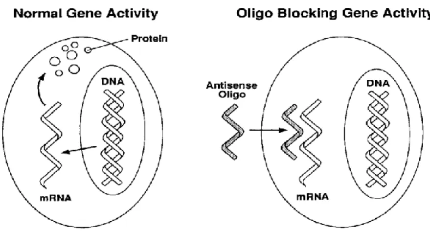
In 1978, Paul Zamecnik and Mary Stephenson reported the first experiments on antisense mechanisms of gene silencing, using short synthetic antisense oligodeoxynucleotides to inhibit replication of the Rous sarcoma virus. The main concept, as briefly described in Figure 1.2, is that if an AS-ODN with a sequence complementary to specific mRNA encoding of the protein of interest is introduced into a cell, it hybridizes with its target mRNA and thus blocks expression and synthesis of that protein (Zamecnik et al. 1978).
Figure 1.2: The basic principle of Antisense therapy. Normal protein synthesis is blocked by inhibiting mRNA transcription by Antisense oligodeoxynucleotide (M.Ian Phillips 2001).
2.1.1 Mechanism of action
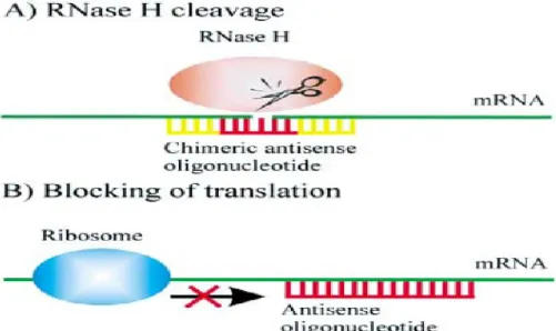
AS-ODN strategies have been employed in a variety of studies both to understand normal gene function and to knockdown gene expression. AS-ODN enter the cell and hybridize target mRNA leading to mRNA degradation and/ or inhibiting transcription by blocking ribosomes as illustrated in Figure 1.3. AS-ODN- dependent mRNA degradation is performed by RNase H enzyme, which is normally present to digest RNA primers during the process of replication fork. This mRNA degradation results in inhibition of protein synthesis (Skoblov et al. 2009). The difference between antisense approaches and conventional drugs is that most of these drugs bind to proteins and thereby modulate their function. In contrast, antisense act at the mRNA level, preventing its translation into protein. In recent years, considerable progress has been made through the development of novel chemical modifications to stabilize AS-ODN against nuclease degradation and to enhance their target affinity, cellular uptake as well as improving their pharmacokinetic and pharmacodynamic properties (Kurreck 2003; Skoblov 2009).
Figure 1.3: Mechanisms of antisense action. (A) RNase H cleavage induced by antisense-oligodeoxynucleotides. (B) Translational arrest by blocking the ribosome (Kurreck 2003).
2.1.2 Chemical Modification
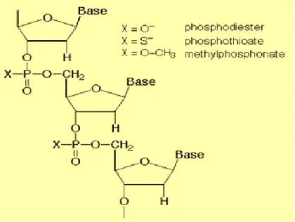
Phosphodiester antisense oligodeoxynucleotides (PO-ODN) "natural form" is rapidly degradable by endonuclease and exonuclease enzymes in the 3' direction. Moreover, PO-ODNs are not serum stable (Crooke et al. 2000; Eder et al. 1991). Because of nuclease activity and serum instability, PO-ODNs possess a short half-life in tissue culture media and in the serum. To avoid degradation by nucleases, chemical modifications of antisense backbone has been introduced. In general, three generations of these nuclease-resistant modified oligodeoxynucleotides can be distinguished. Figure 1.4 describes two examples of chemical modification of the first generation where one of the non-binding oxygen atoms in the phosphodiester backbone was replaced by sulfur group (phosphorothioate), methyl group (methylphosphonates). The second generation (combination of phosphorothioation and methylphosphonation) and the third generation (closed nucleic acids, morpholino phosphoroamidites) despite passing clinical trials phase I and phase II, are not as popular as first generation phosphorothioation because of their high cost and poor membrane permeability (Skoblov 2009; Goel et al. 2003).
2.1.2.1 Phosphorothioation
Phosphorothioation is replacement of non-bridging oxygen atom by sulphur group in the oligodeoxynucleotide chain (Eder et al. 1991). This is the AS-ODN modification most widely used/studied in research and clinical trials. The advantage of phosphorothioated antisense was that it is neuclease stable, exerted excellent antisense activity and is capable of activating RNAase H dependent activity. The main disadvantage was that phosphorothioate backbone induces sequence of non-specific independent effects including activation of heparin-binding growth factors, such as acidic fibroblast growth factor, basic fibroblast growth factor, platelet-derived growth factor, and vascular endothelial growth factor leading to cellular toxicity (Guvakova et al. 1995; Stein 1997; Dias et al. 2002). End-capped phosphorothioation has been shown to have effective transfection with marked reduction in cellular toxicity. Endcapped phosphorothioation is where the replacement by sulfur contents took place only at the sides (Hebb et al. 1997; Skoblov 2009)
Figure 1.4: Examples of chemical modifications of AS; phosphorothioate and methlphosphonate. (Ref. http://www.copewithcytokines.de/cope.cgi?key=Antisense)
2.1.3 Antisense Approach in hypertension research
The most intensively studied AS-ODNs are phosphorothioated. It is important to point out that internalization of naked DNA is usually inefficient. Delivery systems have been developed to mediate a highly efficient cellular uptake and protect AS-ODN against degradation in biological fluids as described later in detail.
In 1998, the first phosphorothioated antisense drug fomivirsen (vitravene) was approved by the US Food and Drug Administration (Marwick 1998). The phosphorothioate DNA is intravitreally injected to treat cytomegalovirus-induced retinitis in patients with AIDS. Approval of vitravene was a milestone for companies involved in the antisense field. Further antiviral or anticancer Phosphorothioated AS-ODNs are being investigated in Phase I or II trials. Most of the antisense molecules currently being tested are intravenously or subcutaneously injected. In addition, respirable antisense oligonucleotide (RASON) targeting the adenosine A1 receptor has been developed to treat asthma (Sandrasagra. et al. 2002). RASON has duration of action of approximately one week, giving it the potential to be the first once-per-week treatment for this disease.
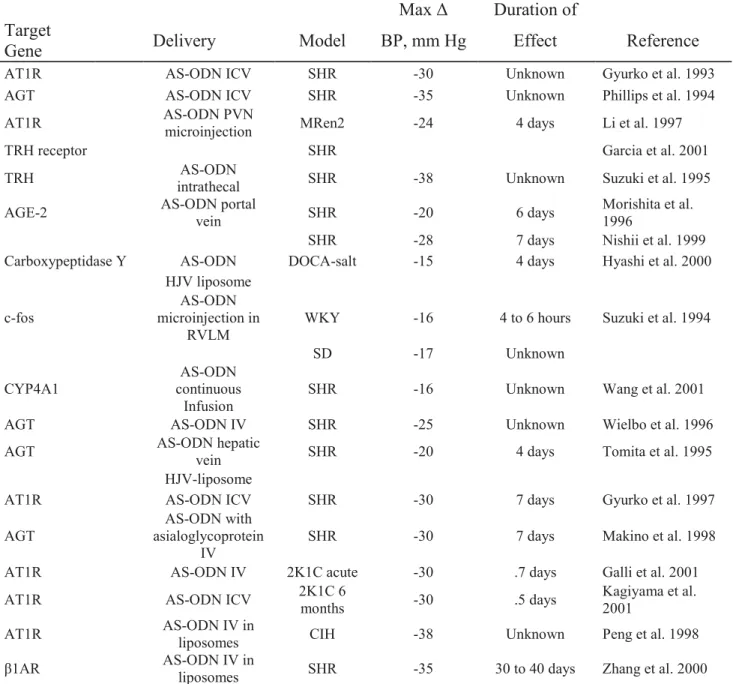
In the field of hypertension research, antisense has been successfully used in hypertension research targeting the components of the RAS like angiotensinogen, angiotensin receptor type 1(AT1), angiotensin-converting enzyme (ACE), angiotensin gene acting element. AS-ODN targeting components of RAS have effectively reduced high blood pressure in several animal models of hypertension including SHR, a surgical model (2KIC), and an environmental model (cold-induced hypertension). Antisense could be administrated centrally within the brain by I.C.V. or peripherally by I.V administration (Phillips et al. 2005). On the other hand, antisense targeting β1 adrenoreceptor exhibited profound and prolonged reduction in hypertension (Zhang et al. 2000). Table 1.1 demonstrates several studies conducted in the field of hypertension using antisense approach (Phillips 2001).
Table 1.1: Preclinical Data of Gene Therapy for Hypertension Vasoconstrictor Genes: AS-ODNs
Max Δ Duration of Target
Gene Delivery Model BP, mm Hg Effect Reference AT1R AS-ODN ICV SHR -30 Unknown Gyurko et al. 1993 AGT AS-ODN ICV SHR - 5 3 Unknown Phillips et al. 1994 AT1R AS-ODN PVN microinjection MRen2 -24 4 days Li et al. 1997
TRH receptor SHR Garcia et al. 2001
TRH intrathecal AS-ODN SHR -38 Unknown Suzuki et al. 1995 AGE-2 AS-ODN portal vein SHR -20 6 days Morishita et al. 1996
SHR -28 7 days Nishii et al. 1999 Carboxypeptidase Y AS-ODN DOCA-salt -15 4 days Hyashi et al. 2000
HJV liposome c-fos microinjection in AS-ODN
RVLM WKY -16 4 to 6 hours Suzuki et al. 1994
SD -17 Unknown
CYP4A1 continuous AS-ODN
Infusion SHR -16 Unknown Wang et al. 2001 AGT AS-ODN IV SHR -25 Unknown Wielbo et al. 1996 AGT AS-ODN hepatic vein SHR -20 4 days Tomita et al. 1995
HJV-liposome
AT1R AS-ODN ICV SHR -30 7 days Gyurko et al. 1997 AGT asialoglycoprotein AS-ODN with
IV SHR -30 7 days Makino et al. 1998 AT1R AS-ODN IV 2K1C acute -30 .7 days Galli et al. 2001 AT1R AS-ODN ICV 2K1C 6 months -30 .5 days Kagiyama et al. 2001 AT1R AS-ODN IV in liposomes CIH -38 Unknown Peng et al. 1998 β1AR AS-ODN IV in liposomes SHR -35 30 to 40 days Zhang et al. 2000
ICV: intracerebroventricular; Unknown, recovery of pressure not recorded; THR, thyrotropin-releasing hormone; AGE, angiotension gene-activating element; IV, intravenous; and CIH, cold-induced hypertension. (M. Ian Phillips 2001).
In the same prospective, antisense targeting growth factor receptors also exhibited decreased blood pressure. Epidermal growth factor receptor antisense (EGFR-AS) treatment attenuated Ang-II induced cardiac hypertrophy and hypertension in SHR (Kagiyama et al. 2002). EGFR-AS reduced the BP but did not normalize it. This reduction of BP by EGFR-AS can be partially explained by an inhibition of EGFR/MAP kinase (ERK)–mediated vasoconstriction
and/or a disappearance of the receptor for EGF-mediated contraction (Kagiyama et al. 2002). Furthermore, administration of insulin-like growth factor-I (IGF-I) receptor antisense in SHR lowered resting blood pressure, produced a profound reduction in responses to vasoconstrictor agents such as Ang II and noradrenaline and reduced the vascular expression of IGF-IR and AT1R (Nguyen et al. 2006). In addition, antisense targeting vasoactive peptide receptor, human neuropeptide Y (NPY) Y1 receptor, markedly attenuated the contractile response to neuropeptide Y in both arteries and veins after treatment with hY1-AS (Erlinge et al. 1993). More recently, ET-1 AS effectively suppressed the ET-1 production and the Ang-II-stimulated proliferation of mesangial cells, and therefore may offer treatment for proliferative glomerulonephritis. Proliferative glomerulonephritis, which is a disorder of the glomeruli and small blood vessels in the kidneys resulting in increased blood pressure, oliguria, hematuria, has devastating complications such as renal failure and hypertensive encephalopathy (lee et al. 2007). It has been shown that the transfection efficiency of antisense could be greatly increased if antisense was encapsulated or conjugated with delivery system as discussed in the following section.
2.2 Delivery systems
Despite the encouraging prospects of nucleotide chemistry discussed in the previous section, cellular uptake of antisense is still considered as an important hurdle that must be overcome for successful antisense applications. In cultured cells, internalization of naked DNA is usually inefficient, as the charged AS-ODNs have to cross a hydrophobic cell membrane. A number of methods have therefore been developed for in vitro and in vivo delivery of AS-ODNs (Hughes et al. 2001, Liang et al. 2002).
Antisense has a negative charge, hydrophilic character and anionic backbone, reducing its transfer across the cell membrane. Only a small fraction leaves the endosome-lysosome system and binds to the complementary mRNA. In the light of the preceding data, much research has been devoted to develop vectors for antisense delivery. These vectors play a crucial role in antisense protection and increasing cellular uptake. As a result, they can also prolong the dose interval (Crooke 2008). The most widely known vectors for delivery of nucleic acids are: viral vectors (virosomes), lipid vectors (liposomes) and polymer (polymerosome). By far the most commonly and successfully used delivery system is liposomes. Positively charged lipids can either encapsulate nucleic acids within their aqueous center or form lipid–nucleic acid complexes as a result of opposing charges. For efficient release of the ODNs from the endosomal
compartment, many transfection reagents contain helper lipids that disrupt the endosomal membrane and help to set the antisense free (Kurreck 2003).
2.2.1 Liposomes
Liposomes were first described by British hematologist Alec D. Bangham in 1961 (published 1964). The word liposome is derived from two Greek words: lipo "fat" and soma "body". The primary composition of liposomes is phospholipids, which are naturally occurring molecules that tend to self-assemble in aqueous media into spherical vesicles (Bangham et al. 1964).
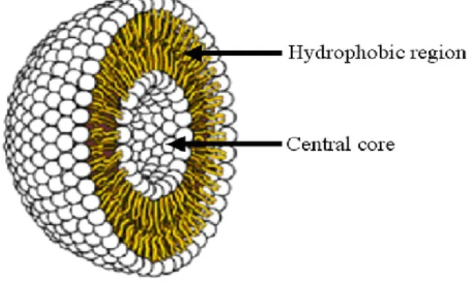
Liposomes are composed of lipid bilayer with a hydrophilic core as demonstrated in Figure 1.5. Their diameter generally ranges from 50 nm to a few micrometers; membrane thickness is around 4 nm. They are usually classified according to the number of lipid bilayers into unilamellar and multilamellar vesicles. Normally, both hydrophilic and lipophilic drugs can be loaded into the core and lipid bilayer of liposomes, respectively. However, the loading of hydrophobic drugs can be limited by the space in the hydrophobic lipid layers (Bangham et al. 1964; Simard et al. 2007).
Figure 1.5: Liposome structure; lipid bilayer and central hydrophilic core (Vanniasinghe et al. 2009).
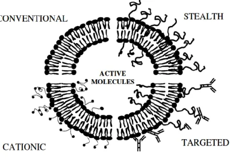
Liposomes could be classified according to lipid charge into cationic, anionic and neutral. In addition, there are stealth and targeted liposomes as seen in Figure 1.6. Steric stabilization (stealth liposomes) is usually required to avoid the rapid clearance by macrophages (MPS) and is usually achieved by grafting hydrophilic polymers (i.e. PEG) to the surface of the liposomes. Active targeting (immunoliposomes) can also be carried out by attaching targeting antibodies to the surface coating. Liposomes have been employed as delivery vehicles for various
chemotherapeutic agents. Thanks to the pioneering efforts of G. Gregoriadis, D. Papahadjopoulos and others, as well as the work of those inspired by them, the liposomal based drug doxorubicin (DoxilTM) has been approved for the treatment of ovarian cancer (Simard et al. 2007; Ross et al. 2011). Liposome-based drugs showed extended circulation lifetime and enhanced accumulation in tumors, compared to the free drug. In addition, liposomes have been used successfully for encapsulating antisense in hypertension research (Phillips 2001).
Figure 1.6: Schematic presentation of 4 major liposome types. Conventional liposomes are either neutral or negatively charged. Sterically stabilized (stealth) liposomes carry polymer coatings to obtain prolonged circulation times. Immunoliposomes (antibody-targeted) may be either conventional or stealth. For cationic liposomes, the several different means of imposing a positive charge (Simard et al. 2007).
Lipid vectors have low toxic and immunogenic reactions, thus bypassing the potential hazards and immune reactions associated with viral vectors. Liposomes are biodegradable, safe with repeated dosing with no known limitation for loading capacity and can be used for targeted delivery. In addition, liposomes are easy to handle and can be produced in large quantities (MacLachan 2007).
2.2.1.1 Cationic lipids
Cationic lipids such as dioctadecyldimethyl ammonium bromide/chloride (DODAB/C), 1, 2-dioleoyloxy-3-[trimethylammonio]-propane (DOTAP) and N-[1-(2, 3-dioleyloxy) propyl]-N, N, N-trimethyl ammonium (DOTMA) gained great attractiveness as drug vectors for nucleic acids. Electrostatic attraction between the positively charged cationic liposomes and the negatively charged antisense facilitated encapsulation. Moreover, they exerted high drug efficiency and facilitated uptake by the cell membrane. Anionic liposomes are considered demanding as they require high lipid concentration to formulate liposomes. On the other hand, neutral liposomes have good intracellular biodistribution but they are poorly internalized by the cells. Antisense delivered by cationic liposomes has a higher tendency to enter the nucleus as it affects the intracellular distribution which was seen by fluorescent and nuclear staining which in turn facilitates the access target mRNA (MacLachan 2007).
The term ″Lipoplexes″ is used to describe complexes between cationic lipids and nucleic acids. Electrostatic interactions occur between the positive charged cationic lipid head group and the negative charged phosphate backbone of nucleic acid. The net positive charge is important both to prevent their rapid aggregation and to promote their binding to the negatively charged cell membranes (MacLachan 2007).
The growing knowledge of the characteristics and functions of lipoplexes has led to the realization that the structure and the type of cationic lipids used are not the sole parameter involved in the design of a successful gene delivery system. The addition of a co-lipid has been shown to enhance transfection efficiency and improve liposomes stability. The molar ratio of the positive charge of cationic lipid nitrogen (N) to the negative charge of antisense phosphate (P) greatly influences the characteristics of the liposomes. The positive to negative charge ratio (+) / (-) which is also referred to as (N/P ratio) was found to be crucial parameter in the optimization of cationic lipid-based gene delivery systems (Simard et al. 2007).
In this section, special attention is paid to cationic lipid DOTAP. DOTAP, which is widely known as transfection lipid, consists of a monocationic trimethylammonium head group and two unsaturated hydrocarbon chains, derived of oleic acid. Phase transition temperature of DOTAP is low (less than 5 ˚C) due to its two unsaturated hydrocarbon chains; this low phase transition temperature of DOTAP makes it more stable. Mono cationic lipids such as DOTAP are less
sensitive to serum than polycationic lipids andtherefore have better transfection rates (Regelin et al. 2000).
Figure 1.7: Chemical structure of DOTAP ([1-(2, 3-dioleoyloxy) propyl]-N, N, N-trimethylammonium methylsulfate).
2.2.1.2 Helper lipids
Helper lipids such as cholesterol, dioleylphosphatidyl choline (DOPC), and 1, 2-di-[cis-9-octadecenoyl]-sn-glycero-3-phosphoethanolamine (DOPE) are usually incorporated with cationic lipids to help the release of the antisense from the endosomal system and also to enhance transfection efficiency. DOPE has a cone-shaped molecule with a tendency to form inverted hexagonal phases at pH ≥ 8. The inverted hexagonal shape destabilizes the endosomal compartment and helps the release of AS-ODN from the endosomes before being digested by the lysosomal system. DOPE is also called colipid or fusogenic lipid as it helps fusion with the cell membrane and facilitates entry of lipoplexes in the cell; this fusion also helps release from the endosomes. Due to their neutral charge, helper lipids make the liposomes more stable and decrease the toxic effects of positively charged lipids and thereby increasing their circulation lifetime (MacLachan 2007).
Figure 1.8: Chemical structure of DOPE (1, 2-di-[cis-9-octadecenoyl]-Sn-glycero-3-phosphoethanolamine).
2.2.1.3 Polyethylene glycol (PEG)
Polyethylene glycol (PEG) is a hydrophilic polymer. Incorporation of PEG into cationic liposomes (stealth lipoplexes) leads to prolonged stability and greatly enhances the circulation lifetime of liposomes by providing a protective, steric barrier against interactions with plasma proteins and cells. Moreover, PEG prevents liposomal aggregation as it inhibits calcium-induced fusion between LUVs. It is important to point out that 10% of PEG prevents leakage of entrapped contents upon mixing. Neutral charge of PEG reduces toxicity caused by high positive charges of the vector (Simard et al. 2007; MacLachan 2007).
2.2.1.4 Intracellular mechanism and pathway of the lipoplexes
Over the past decade, significant progress has been accomplished in understanding the cellular pathways and mechanisms involved in lipoplex-mediated gene transfection. Despite the fact that the main steps that are required for DNA to travel from the cellular environment to the nucleus have been studied, the molecular mechanisms involved in some steps remain to be elucidated. In particular, escape of lipoplexes from endosomes and DNA entry into the nucleus are not yet fully understood.
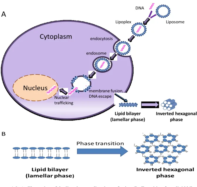
Lipoplexes enter the cells through endocytosis as illustrated in Figure 1.9-A. Serum proteins bind to the surface of the lipoplexes, which in turn bind to specific receptors on the cell membrane. Endosomes bring the lipoplexes to the perinuclear region so the released antisense will have greater chance of entering the nucleus (Elouahabi et al. 2005). Escape of lipoplexes / DNA from the endosomes occurs in gratitude to helper / fusogenic lipids (DOPE). DOPE fuse with the endosomal membrane leading to transition from bilayer into inverted hexagonal structure resulting in release of lipoplexes/DNA from the endosomes (Figure 1.9-B). Subsequently, these ″flip-flop″ movements lead to weakening of the electrostatic interaction between DNA and cationic lipid, inducing the release of DNA in a step known as ″lipid transfer″. It is important to point out that phosphorothioation is crucially important at this stage as it protects antisense in the cytoplasm before entering the nucleus. Most of the released DNA enters the nucleus for binding to target mRNA resulting in high transfection efficiency. Entry into the nucleus might be an active process through nuclear pores or passive process during cellular mitosis as the nuclear membrane becomes temporarily disintegrated (Elouahabi et al. 2005).
Figure 1.9: A- Illustration of the lipoplex -mediated transfection. B- Transition from lipid bilayer (lamellar pahse) into inverted hexagonal structure resulting in release of lipoplexes/DNA from the endosomes and the release of DNA form the lipoplexes (Adapted from Chen et al. 2010; Seong et al. 2004)
2.2.2 Virosomes
Viruses are composed of an envelope or capsid that contains genetic material (DNA or RNA) in a compact form. A variety of these viruses have been converted to vectors to deliver genes to cells (e.g. adenoviruses, retroviruses, influenza virus and adeno-associated viruses). Virosomes protect pharmaceutically active substances from proteolytic degradation and can be used for targeted delivery (Bhattacharya et al. 2011). Although viral vectors offer superior transfection efficiency compared to other delivery systems, their use is limited because of
Nucleus
endocytosis membrane fusion, DNA escape endosomeCytoplasm
Lipoplex DNA Liposome Nuclear traffickingA
Lipid bilayer (lamellar phase) Inverted hexagonal phase Phase transition Lipid bilayer (lamellar phase) Inverted hexagonal phaseB
inherent safety concerns. The use of viral vectors for gene therapy can be associated with severe inflammation and immunological problems (Verma et al. 1997; Lehrman 1999). The toxicity of viral vectors is usually due to random integration of the transported genes (Waehler et al. 2007; Young et al. 2006). In addition, the size of the DNA and the type of the genetic material that can be encapsulated into viral vectors restrict their applicability. Hence, there is a need for alternative synthetic approaches for the delivery of nucleic acids.
2.2.3 Polymerosome
Cationic polymers (e.g. polyethylenimine (PEI) and poly (amidoamine) (PAMAM)) as well as neutral polymers poly (ethylene glycol) (PEG) have been studied as non-viral gene carriers because of their ability to protect DNA / RNA from enzymatic degradation and to increase cellular uptake by endocytosis. Detailed discussion of polymersome vectors in nucleic acid delivery is beyond the scope of this introduction, it is worth mentioning that polymersomes demonstrated a significant enhanced antitumor efficacy and improved safety in preclinical studies and advanced to phase III clinical trials (Auzenne et al. 2002).
3. G proteins
3.1 Discovery of G proteins
In 1994, Alfred G. Gilman and Martin Rodbell won the Nobel Prize in Physiology or Medicine jointly for their great discovery of "G-proteins and the role of these proteins in signal transduction in cells". G-proteins or Guanine nucleotide regulatory proteins form a group of membrane proteins that is responsible for transduction of cell signaling into a cascade of cellular responses.
3.2 Structure of the Heterotrimeric G proteins
The crystal structure of heterotrimeric G proteins has provided a framework for understanding the biomechanics of G proteins activation (Cabrera-Vera et al. 2003; Sprang 1997). All members of the G proteins family share a common structural core. G-proteins are heterotrimeric proteins composed of three distinct subunits; α, β, and γ.
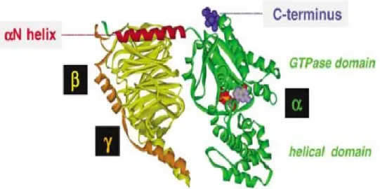
The Gα-subunits is involved in the coupling specificity, hydrolyze GTP and contains ADP-rybosylation factor 1(ARF-1). As illustrated in Figure 1.10, Gα subunits contain two domains; a GTPase domain that is involved in the binding and hydrolysis of GTP and a helical domain that buries the GTP within the core of the protein (Mixon et al. 1995; Sprang 1997). The GTPase domain is composed of six stranded β-sheets surrounded by five α helices. This domain contains five loops that act in consensus for guanine nucleotide binding: the diphosphate-binding (P-) loop, two Mg2+ -binding domains and two guanine ring-γ-phosphate binding motifs. Comparison of the inactive (GDP bound) and active (GTP) bound crystal structures has revealed the presence of three flexible regions, designated switches I, II and III which become more rigid in the GTP-bound active form (Lambright et al. 1994; Mixon et al. 1995). The GTPase domain not only hydrolyses GTP but also contains sites for binding to βγ dimer, GPCR and downstream effector proteins. The helical domain may play a role in GPCR selectivity and effector protein selectivity. It is composed of six α-helices that form a lid over the nucleotide binding site burying it in the core of the protein (Liu et al. 1995; Liu et al. 1998; Remmers et al. 1999). Little is known about the structure of the extreme amino (N-) and carboxy (C-) terminal domains of Gα-subunits because in the isolated G protein crystal structures solved thus far, the N and C termini of Gα were either removed from the protein or disordered. The N-terminal of the Gα subunit is responsible for interaction with the β-propeller of the Gβ subunit. N-terminal and C-terminal play a key role in the activation process (Cabrera-Vera et al. 2003).
Five different β-subunits of 35–36 kDa have been revealed. They share 50-90% of their structural sequence and have a propeller structure. Each blade of the propeller binds to the next strand in the next blade. The N-terminal adopts α-helical conformation that is essential for interaction with Gγ subunit (Sondek et al. 1996; Cabrera-Vera et al. 2003).
Members of Gγ are encoded by 12 genes, between 7 and 10 kDa and share 30-80% sequence identity (Downse et al. 1990). They are composed of two α helices connected by a loop. The N- terminal interacts with the N-terminal of Gβ whereas, the C- terminal binds to blades 5 and 6 of Gβ. The Gβγ dimer acts as one functional unit that is tightly associated and can only be dissociated by denaturation (Sondek et al. 1996; Cabrera-Vera et al. 2003).
Figure 1.10: Three-dimensional structure of the heterotrimeric G protein showing Gα (green) β (yellow) γ (orange). The N-terminal αN helix (red) is required for binding of Gα to the other subunits and the C-terminal receptor contact region (blue) convey GPCR specificity. The GDP molecule (purple) is buried between the GTPase and helical domain (Milligan 2006).
3.3 Activation of Heterotrimeric G proteins
Guanine nucleotide regulatory proteins (G-proteins) are a large family of guanosine triphosphate (GTP) binding proteins that play a crucial regulatory role as transducers in a variety of signal transduction systems. These include the adenylyl cyclase / cAMP system (Gilman 1987)the receptor-mediated activation of phospholipase C and A2 (Crockcroft et al. 1985).
As illustrated in Figure 1.11, the inactive GDP-bound form binds tightly α to βγ subunits, whereas the GTP-bound form of α dissociates from βγ and serves as a regulator of effector proteins. Upon ligand binding and receptor activation, the receptor interacts with the heterotrimeric G protein to promote conformational changes and dissociation of GDP from the guanine nucleotide binding site. GDP is released and replaced by GTP. Binding of GTP to Gα induces a conformational change and promotes the dissociation of hormone receptor complex and dissociation of G-protein into α and βγ. Both α-GDP and βγ-subunits can interact with effectors. All α-subunits possess intrinsic GTPase activity and hydrolyze the terminal phosphate of bound GTP to yield bound GDP and free inorganic phosphate (Pi), which in turn terminate the activation cycle. Regulators of G protein signaling (RGS) also known as
GTPase-activating proteins play a crucial role in controlling the activity of G proteins. The rate-limiting step in G protein activation is the release of GDP from the nucleotide-binding pocket. GDP is spontaneously released from the heterotrimeric G-protein at a rate that varies depending on the Gα-subunit. The GDP-bound form of α-subunit has high affinity for βγ and then re-associates with the βγ dimer to form the heterotrimeric in the basal resting state. Both the Gα and Gβγ dimer mediate G-protein signaling (Gilman 1984; Gilman 1987; Mixon et al. 1995; Sprang et al. 1997; Cabrera-Vera et al. 2003; Srivastava et al. 2008).
Figure 1.11: The activation cycle of the heterotrimeric G protein. When a ligand binds to the GPCR, a conformation change occurs in the receptor that exchanges GDP for GTP on the α subunit and this triggers dissociation of the α subunit from βγ dimer and the receptor. After the free a unit works on target proteins, GTP will be hydrolyzed to GDP. The GTPase activity is enhanced by binding of the RGS (Adapted from: Bao et al. 2010). α βγ α βγ βγ βγ βγ α α α
3.4 Classification of G-protein α-subunits
The family of G-protein α-subunits can be subclassified according to their functional and structural relationship. Four major subfamilies exist according to amino acid homology and are represented as follows: Gsα, Giα, Gqα / α11, and Gα12 / α13. Despite their similarity, the Gα families can elicit different functions and have distinct and sometimes overlapping functions for their binding partners (Neves et al. 2002).
Table 1.2: Classification of heterotrimeric G proteins according to major subunits.
Family
Subfamily
Subtype
Effector
I Gs Gαs(S) Gαs(L) Gαolf ↑ AC ↑ GTPase of tubulin ↑ src ↑ AC II Gi G0 Gz Gt Gα i1 Gα i2 Gα i3 GαoA GαoB Gαz Gαt-r Gαt-c Gαgust ↓ AC ↑ GTPase of tubulin ↑ src, MAPK ↑K+ channels ↓ AC ↑K+ channels ↓ AC ↑cGMP-PDE Unknown III Gq Gαq Gα11 Gα14 Gα15or α16 ↑ PLCβs
↑ Bruton’s tyrosine kinase (Gαq) IV G12 Gα12 Gα13 ↑ NHE-1 ↑ PLD ↑ iNOS
Adapted from: ″Insights into G Protein Structure, Function, and Regulation.″ Cabrera-Vera 2003.
3.4.1 Gsα proteins
Molecular cloning has revealed four different forms of Gsα ;1, 2, 3and Gsα-4 having molecular weights of Gsα-42, Gsα-45, Gsα-47 and 52 kDa respectively and resulting from alternative splicing of exon 3 of the Gsα gene. Gsα-1 and -2 contain exon 3, whereas exon 3 is spliced out in Gsα-3 and -4 (Zou et al. 1996). Gsα is associated with adenylyl cyclase stimulation and increased cAMP production (P Bray 1986 ; Robishaw 1986; Murakami T 1988). Cholera toxin can lead to ADP rybosylation of Gsα and persistent activation. The Gαs family also includes GαsL which is expressed in the neuroendocrine cells and Gαolf which was initially discovered in the olfactory system and is responsible for olfactory signal transduction (Dean MK 2001). AC induces cAMP formation and results in the activation of protein kinase A (PKA), which modulates gene transcription.
3.4.2 Giα proteins
Activated Giα proteins inhibit adenylyl cyclase (AC). As illustrated in Figure 1.12, Giα proteins have a variety of effects other than AC inhibition. These effects include mitogen-activated protein kinase (MAPK) and phosphatidylinositol 3-kinase (PI3K) pathways. Activation of the enzyme phospholipase A2 (PLA2) may also occur, which induces the release of arachidonic acid (AA), as well as inhibition of the Na+/H+ exchanger in the plasma membrane, and the lowering of intracellular Ca2+ levels. Subsequent activation of the MAPK and PI3K pathways results in the phosphorylation of extracellular signal-regulated kinases (ERKs) and protein kinase B (PKB), cellular proliferation respectively (Leurs et al. 2005).
The Giα proteins subfamily includes Giα-1, Giα-2, Giα-3. Gαo, Gαt, Gαgust and Gαz share high structural homology with Giα proteins (Gilman 1995). All members of this family are pertussis toxin (PTX) sensitive and contain a cysteine residue at C-terminal, except Gαz. Giα proteins are responsive to ADP-ribosylation by PTX (Hsia et al. 1984). Gαo is present mainly in the central nervous system and has two isoforms A and B. Members of the subfamily Giα and Gαo are involved in the inhibition of adenylate cyclase, regulation of ion channels and regulation of phospholipase C (Stryer et al. 1986; Spiegel 1987).
Gαt has two isoforms that are present in the retina, namely Rod transducin (Gαt-r) and Cone transducin (Gαt-c). Gαz is mainly expressed in blood platelets. While Gα-gustidin
(Gαgust) are present in the taste buds and are responsible for transducing sweet and bitter signalling. (Devi 2005).
The three distinct forms of Giα, namely, Giα-1, Giα-2, and Giα-3 are cloned and encoded by three different genes (Itoh et al. 1986, Jones et al. 1987 and Itoh et al. 1988). Giα-1 protein has a molecular weight of 41kDa and is encoded by the gene GNAI1. Giα-1 is present predominantly in the brain neural tissue (Patel et al. 2001). In this study, great attention is paid to the study of Giα-2 and Giα-3 owing to their imperative implication in the development of hypertension.
Figure 1.12: Coupling of the ligand (Histamine) to the seven-transmembrane protein receptor (GPCR) activates Giα /o proteins leading to modulation of several signaling pathways (Adapted from Leurs et al. 2005).
3.4.2.1 Giα-2 protein
Giα-2 has a molecular weight of 40 kDa encoded by the gene GNAI2 (Patel et al. 2001). Giα-2 is involved in several signaling pathways including regulation of immune responses against microbial and non-microbial stimuli and regulation of cardiovascular signal transduction.

![Figure 1.7: Chemical structure of DOTAP (N-[1-(2, 3-dioleoyloxy) propyl]-N, N, N- N-trimethylammonium methylsulfate)](https://thumb-eu.123doks.com/thumbv2/123doknet/2064892.6263/37.918.109.795.809.954/figure-chemical-structure-dotap-dioleoyloxy-propyl-trimethylammonium-methylsulfate.webp)