Université de Montréal
“Redox regulation ofET-1-induced activation ofERK1/2, PKB and Pyk2 signaling in A-10 vascular smooth muscle ceils”
par Grace Bou Daou Département de Physiologie
Faculté de médecine
Mémoire présenté à la Faculté des études supérieures envue de l’obtention du grade de
Maître ès sciences (M.Sc.) en physiologie
Septembre, 2003
o
X1L
\], û3E)
dl)
de Montréal
Direction des bibliothèques
AVIS
L’auteur a autorisé l’Université de Montréal à reproduire et diffuser, en totalité ou en partie, par quelque moyen que ce soit et sur quelque support que ce soit, et exclusivement à des fins non lucratives d’enseignement et de recherche, des copies de ce mémoire ou de cette thèse.
L’auteur et les coauteurs le cas échéant conservent la propriété du droit d’auteur et des droits moraux qui protègent ce document. Ni la thèse ou le mémoire, ni des extraits substantiels de ce document, ne doivent être imprimés ou autrement reproduits sans l’autorisation de l’auteur.
Afin de se conformer à la Loi canadienne sur la protection des renseignements personnels, quelques formulaires secondaires, coordonnées ou signatures intégrées au texte ont pu être enlevés de ce document. Bien que cela ait pu affecter la pagination, il n’y a aucun contenu manquant.
NOTICE
The author of this thesis or dissertation has granted a nonexclusive license allowing Université de Montréal to reproduce and publish the document, in part or in whole, and in any format, solely for noncommercial educational and research purposes.
The author and co-authors if applicable retain copyright ownership and moral rights in this document. Neither the whole thesis or dissertation, flot substantial extracts from it, may be printed or otherwise reproduced without the authot’s permission.
In compliance with the Canadian Privacy Act some supporting forms, contact
information or signatures may have been removed from the document. While this may affect the document page count, it does flot represent any loss of content from the document.
Université de Montréal Faculté des études supérieures
Ce mémoire intitulé:
“Redox regulation ofET-1-induced activation ofPyk2, ERK1/2 and PKB/Akt signaling in A-10 vascular smooth muscle celis”
Présenté par: Grace Bou Daou
a été évalué par un jury composé des personnes suivantes:
Président-rapporteur: Josette Nol
Directeur de recherche: Ashok K. Srivastava
Membre du jury: Ryszard Grygorczyk
SUMMARY
Reactive oxygen species (ROS) have been shown to mediate the effect of several growth factors such as angiotensin II (Ail), epidermal growth factor (EGf) and platelet-derived growth factor (PDGF). Endothelin-1 (ET-1) is an important growth factor for vascular smooth muscle ceils (VSMC) which is beheved to contribute to the pathogenesis of vascular abnormalities such as atherosclerosis, hypertension and cardiac hypertrophy. However, a possible role
of ROS generation in mediating the ET-1 response on ERK1/2 and PKB, key
components of growth promoting and proliferative signaling pathways, has flot been examined in detail. Therefore, the aim of the present study was to investigate the involvement of ROS in ET-1-mediated activation of ERK1/2 and PKB as well as Pyk2 in A-10 VSMCs. These cefis are obtained from rat ernbiyonic thoracic aorta. Pyk2 is a non-receptor Ca2-dependent protein tyrosine kinase and an upstream regulator of MAPK signaling. ET-1 stimulated the phosphorytation of ERKÎ/2, PKB and Pyk2 in a dose and time-dependent fashion with maximum response being elicited at 10 nM winch peaked at 5 min. Treatment of VSMC with ET-1 resulted in an increase in the generation of ROS that could be blocked with diphenyleneiodonium (DPI), an inhibitor ofNADPH oxidase. furthermore, DPI pretreatment of ceils prior to stimulation with ET-1, attenuated ET-1 enhanced phosphorylation of ERK1/2, PKB and Pyk2. N acetylcysteine (NAC), another ROS scavenger, also exhibited a sirnilar response. Moreover, DPI caused a decrease in the protein synthesis stimulated by ET-1. These resuits demonstrate that ROS is a critical mediator of ET-1-induced signaling events linked to hypertrophic and growth promoting pathways inVSMC.
There is an emerging evidence suggesting that nitric oxide (NO), a vasoactive substance, contributes to the regulation of several hormone-mediated responses such as EGF, PDGF as well as ET-1 and exerts an anti-mitogenic and anti-proliferative effect in vitro. However, the mechanism by winch NO
antagonizes ET-1 effect remains unknow. Therefore, the aim ofthis study was to determine if NO generation would modify ET-1 -induced signaling pathways involved in cellular growth and proliferation in A-10 VSMC. NO effect has been evaluated by measuring phosphorylation levels of ERKY/2, PKB and Pyk2 by immunoblot. Treatment of A-10 ceils with S-nitroso-N-acetylpenicfflamine ($NAP), a NO donor, attenuated the ET-1-enhanced phosphorylation of ERK1/2, PKB and Pyk2. Since, NO mediates principally its effect through a cycic GMP/soluble guanylate cyclase pathway, we investigated the role of 8-Br-cGMP, a non-metabolizable and ceil permeable analogue of cGMP, winch exhibites a sirnilar effect to SNAP on ET-1-induced ERK1/2, PKB and Pyk2 phosphorylation. Furthermore, ODQ, an inhibitor of guanylate cyclase activity, reversed the inhibitory effect of NO on ET-1-induced responses. SNAP also appeared to decrease the protein synthesis induced by ET-1. Taken together, these data demonstrate that NO attenuates selectively ERK1/2, PKB and Pyk2 phosphorylation induced by ET-1 via cGMP, winch antagonize the growth promoting and pro liferative effects of ET-1.
SOMMAIRE
Les espèces réactives oxygénées modulent l’effet de plusieurs facteurs de croissance comme l’angiotensine II (AIT), l’EGF (“epidermal growth factor”) et le PDGf (“platelet-derived growth factor”). L’endotheline-l (ET-1) est un important facteur de croissance pour les cellules du muscle lisse vasculaire (“V$MC”) et contribue aux anormalités vasculaires, entre autre, l’athérosclérose, l’hypertension ainsi que l’hypertrophie cardiaque. La génération des espèces réactives oxygénées pourraient aussi jouer un rôle dans l’activation des voies de signalisation activées par l’ET-l, en particulier l’activation de ERKT/2 et de PKB. Ces deux composantes clés sont impliquées dans la croissance et la prolifération cellulaire. Ainsi, le but de la présente étude est d’évaluer la participation des espèces réactives oxygénées dans l’activation de ERK1I2, PKB et Pyk2 induite par ET-1 dans les “VSMC” de lignée A-10. Ces cellules sont obtenues à partir de l’aorte thoracique embryonnaire de rat. Pyk2 est une protéine tyro sine kinase dépendante du calcium et constitue ainsi un régulateur ascendant de la voie de signalisation des MAPKs. ET-1 a permis de stimuler la phosphorylation de ERK1/2, PKB et Pyk2 de façon dépendante de la concentration et du temps d’où une réponse maximale a été obtenue à 10 nM et après une stimulation de 5 min. Le traitement des “VSMC” avec ET-1 a augmenté la production des espèces réactives oxygénées, alors qu’une diminution a été observée en présence du diphenyleneiodonium (DPI), un inhibiteur de la NADPH oxydase. En outre, le prétraitement des cellules avec DPI a permis d’atténuer la phosphorylation de ERK1/2, PKB et Pyk2 induite par ET-1. Une réponse similaire a été observée en utilisant un autre antioxidant, le N-acetylcysteine (NAC). De plus, DPI a inhibé la synthèse protéique stimulée par ET-1. Ces résultats démontrent que les espèces réactives oxygénées sont des médiateurs importants dans l’activation des composantes de la voie de signalisation de ET-1, ce qui contribue à la croissance et à l’hypertrophie cellulaire.
Il a été suggéré que l’oxyde nitrique (NO), une substance vasoactive, contribue à la régulation des réponses induites par plusieurs hormones comme l’EGF, le PDGf et l’ET-l en exerçant un effet anti-mitogénique et anti prolifératW in vitro. Cependant, le mécanisme par lequel NO antagonise l’effet de ET-1 est à ce jour inconnu. Le but de cette étude est donc d’évaluer si la génération de NO pourrait modifier les composantes de la voie de signalisation de ET-1. L’effet de NO a été déterminé en mesurant les niveaux de phosphorylation de ERK1/2, PKB et Pyk2 à l’aide d’immunobavardage. Le traitement des cellules A-10 avec S-nitroso-N-acetylpenicifiamine (SNAP), un donneur de NO, a permis d’atténuer la phosphorylation de ERK1/2, PKB et Pyk2 induite par ET-1. Comme NO médie principalement son action par la voie de GMPc/guanylate cyclase soluble, on a donc étudié le role du $-Br-GMPc, un analogue du GMPc non-métabolisable et perméable à la cellule. Un effet similaire à celui de SNAP a été observé. En outre, ODQ, un inhibiteur de l’activité de la guanylate cyclase, a renversé l’effet de NO sur la phosphorylation des composantes de signalisation stimulées par ET-l. SNAP a permis aussi de diminuer la synthèse protéique induite par ET-1. Finalement, les résultats démontrent que NO atténue sélectivement la phosphorylation de ERK1/2, PKB et Pyk2 de la voie de signalisation de ET-1 par l’intermédiaire du GMPc, ce qui antagonise les effets de ET-l soit la croissance et la prolifération cellulaire.
TABLE 0F CONTENTS
Summaiy iii
Sommaire V
Table ofcontents vii
List offigures t
List ofabbreviations xii
Acknowledgements XV
CHAPTER 1: INTRODUCTION 1
1.1 Endothelin 2
1.2 Structures ofendothelins 2
1.3 Molecular genetics and regulation of generation ofendothelln 4
1.4 Bio synthetic pathway 6
1.5 Sites ofgeneration 6
1.6 Plasma concentrations of endothelins 7
1.7 Biological actions ofendothelin-1 7
1.8 Endothelin receptors $
1.9 Activation ofthe phosphoinositide cascade by ET-1 9 1.10 Mechanisms of ET-1 -induced activation of MAPK cascade 11 1.11 Phosphatidylinositol-3 kinase cascade 16
1.11.1 ClassilicationofPl-3Ks 16
1.11.1.1 ClassIPI-3Ks 17
1.11.2 Protein kinase B signaling pathway 1$ 1.12 Role ofendothelin in cardiovascular diseases 19
1.12.1 Endothelln in human hypertension 19 1.12.2 Endothelin in experimental hypertension 22
1.13 Reactive oxygen species and its implications in ET-1 signaling
24
1.14 Nitric oxide 26
1.14.1 formation ofnitric oxide 27
1.14.2 NOS isoforms 2$
1.14.3 Nitric oxide function 29
1.14.4 Guanylate cyclase 30
1.14.5 Regulation ofcGMP production 32
1.14.6 NO in signal transduction 35
1.15 Objectives ofthe present study 36
CHAPTER 2: ARTICLE 1 38
Abstract 40
Introduction 42
Materials and Methods 44
Resuits 4$ Discussion 51 Figure legends 55 figures 59 References 65 CHAPTER 3: ARTICLE 2 74 Abstract 76 Introduction 7$
Materials and Methods $1
Resuits $4
Discussion $7
Figures .93
References.9$
CHAPTER 4 :GENERAL DISCUSSION 106
CHAPTER 5 :CONCLUSION 114
LIST 0F FIGURES
CHAPTER I INTRODUCTION
Figure 1.1 : Structures ofendothelins 3
Figure 1.2 :Regulation of ET-1 synthesis and
its
pathway ofgeneration 5 Figure 1.3 : Special case ofGq-protein-coupled receptor in intracellularsignaling 10
Figure 1.4 : Schematic model showing key steps in ET-1-induced
activation of MAPK
and
P13-KIPKB signaling 12Figure 1.5 Potential pathways ofendothelin-1 in the pathophysiology
of hypertension or its complications 20
Figure 1.6 : Key steps in the production ofreactive oxygen species 25
Figure 1.7 The nitric oxide synthase (NOS) reaction 2$
Figure 1.8 : Schematic representation of a soluble guanylate
cyclase
&t3
heterodimer 32Figure 1.9 : The nitric oxide/cGMP signal transduction 34
CHAPTER 2
ARTICLE 1
Figure 1 : ET-1-induced dose-responses ofERKI/2, PKB and Pyk2
phosphorylation in A-10 VSMCs 59
Figure2 : lime-course ofET-1-induced ERKY/2, PKB and Pyk2
phosphorylation in A-10 VSMCs 60
Figure 3 : Dose-dependent effect ofNADPH oxidase inhibitor, DPI on ET-l-induced ERK1/2, PKB and Pyk2 phosphorylation
Figure 4 : Effect of DPI on ET-1-induced ROS generation in A-10
VSMCs 62
Figure 5 : Effect ofsuperoxide scavenger, NAC on ET-1-induced ERK1/2, PKB and Pyk2 phosphorylation in A-10 VSMCs 63
Figure 6 : Effect of DPI on ET-1-induced [3Hileucine incorporation into
proteins 64
CHAPTER3 ARTICLE 2
Figure 1 : Dose-dependent effect ofthe NO donor, SNAP on ET-1-induced ERK1/2, PKB and Pyk2 phosphorylation in A-10 VSMCs 93
Figure 2 : Effect ofa stable analogue ofcGMP, 8-Br-cGMP on ET-1-induced
ERKI/2 and PKB phosphorylation in A-10 VSMCs 94
Figure 3 : Effect ofthe inhibitor ofthe soluble guanylate cyclase, ODQ on ET-I-induced ERKY/2, PKB and Pyk2 phosphorylation
inA-1OVSMCs 95
Figure 4 Effect ofSNAP on ET-1-induced [3H]leucine incorporation into
proteins 97
CHAPTER 5 CONCLUSION
Figure 5.10: Hypothetical model showing nitric oxide interaction
LIST 0F ABBREVIATIONS
A II Angiotensin II
ACE : Angiotensin converting enzyme ATF-2 : Activation transcription factor 2 BU4 : 6(R)-tetra-hydro-L-biopterin CaM : Calmodulin
cAMP : Cycic adenosine monophosphate cGMP : Cycic guanosine monophosphate DG : Diacylglycerol
DNA : Deoxyribonucleic acid
ECE : Endothelin converting enzyme
EDCF : Endothelium-derived contracting factors EDHF : Endothelium-derived hyperpolarizing factors EBRF : Endothelium-derived relaxing factors
EGF : Epidermal growth factor
ERK : Extracellular signal-regulated kinases
ET : EndotheUn
FADH : flavin adenine dinucleotide, radical form FMN : Flavin mononucleotide
GC : Guanylate cyclase GDP : guanosine diphosphate
GPCR G-protein-coupled receptors
Grb2 : Growth factor binding protein 2
GTP : Guanosine triphosphate 1P3 : Inositol triphosphate
JNK/SAPK : c-Jun NI-12-terminal kinase/stress-activated protein kinase
kDa : Kilodaltons
mRNA : Messenger ribonucleic acid
MAPK : Mitogen-activated protein kinases
MEK/MKK: MAPK kinase
MEKKJMKKK: MAPK kinase kinase
NADY : Nicotinamide adenine dinucleotide, phosphate,oxidized form
NADPH : Nicotinamide adenine dinucleotide, phosphate,reduced form
NO : Nitric oxide
NOS : Nitric Oxide Synthase
Pl3OCas : Crk-associated substrate, protein of 130 kDa
PUE Phosphodiesterase E
PDGF Platelet derived growth factor
PDK : PI(3,4,5)P3 dependent protein kinase
PI : Phosphatidylinositol
PIP2 : Phosphatidylinositol-4,5-bisphosphate
P13-K : Phosphatidylinositol 3-kinase
PKB : Protein kinase B
PKG : Protein kinase G PLC : Phospholipase C
Pyk2 : Protein tyrosine kinase 2 ROS Reactive oxygen species
SUS-PAGE : Sodium dodecyl sulfate -polyacrylamide gel
Shc : Src homology SOS : Son ofthe sevenless Src : Sarcoma rous virous
ACKNOWLEDGEMENTS
With a sense of humble compliance I feel great honor to express sincere gratitude to my supervisor Dr. Ashok K. Srivastava for molding me capable enough to understand the subject, conveying the knowledge and ski! to accomplish this study. It was his constant inspiration, kind guidance, healthy criticism and suggestions that I am able to present tins work; I shail always remain grateftul to his guidance, advice and help.
I express my unfathomable sense of reverence to Dr. Madhu B. Anand Srivastava rendering her valuable suggestions and encouragement in the completion ofthis study.
My immense gratitude are due to my colleagues, especially to Mohammed Mehdi and Dr. Nihar R. Pandey for the help, suggestions and healthy critics throughout. I would like to thank Dr. Yuan Li and Dr. Sheffla Hashim for the needffil technical support.
Words are inadequate to express my sincere feelings and indebtedness for my family without their moral support and encouragement it would have been impossible for me to carry out tins study.
Last, but flot the least, I wish to record my heartfelt feelings for immense cooperation of a special person my husband, Don Dagher, who supported me throughout during tins critical time.
1.1 Endothelin
Endothelin, one of the most potent vasoconstrictors, was discovered by Yanagisawa and co-workers (1) in 1988. It was characterized and cloned from porcine aortic endothelial ceils (1) and exerts inotropic and mitogenic properties, influences homeostasis of sait and water and stimulates the renin-angiotensin aldosterone and sympathetic nervous systems (2,3,4). In fact, the overail effect of the actions of endothelin is usually to increase vascular tone and biood pressure. Endothelin also play an important role in the pathophysiology of cardiac, vascuiar and renal diseases associated with regional or systemic vasoconstriction (5) such as hypertension and atherosclerosis.
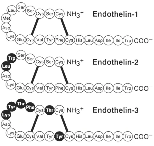
1.2 Structures of endothelins
Endothelin (ET) is a 21 aminoacid peptide winch exists in at least three isoforms: ET-1, ET-2 and ET-3 (6,7). Ail ET isopeptides share a common structure: two disuffide bonds (Cys’-Cys’5 and Cys3-Cys”), a cluster of tbree polar charged side chains on aminoacid residues s-10 and a hydrophobic C-terminus (residues 16-21) containing the aromatic indole side chain at Trp21. ET-2 contains two aminoacid substitutions (Trp6-Leu7) and shares 90% sequence homology with ET-1. ET-3 contains six aminoacid substitutions (Thr2, Phe4-Thr5-Tyr6-Lys7 and Tyr’4) and shares 71 % sequence hornology with ET-1 and ET-2 (8,9). The hydrophobie C-terminus of ET is essential for its
bioactivity, as well as the ioop configuration (9). ETs share close sequence homology
(-
67%) and similar bioactivities with the sarafotoxins, a group of peptide toxins isolated from venom of some scorpions and snakes (8). Thedisulfide bonds, polar side chains and hydrophobic C-terminus of ETs are largely conserved in sarafotoxins (9) (Fig. 1.1).
NH3
Endothelin-1
EndotheUn-2
Endothelin-3
Figure 1.1 : Structures of endothelins. Dark circles indicate where
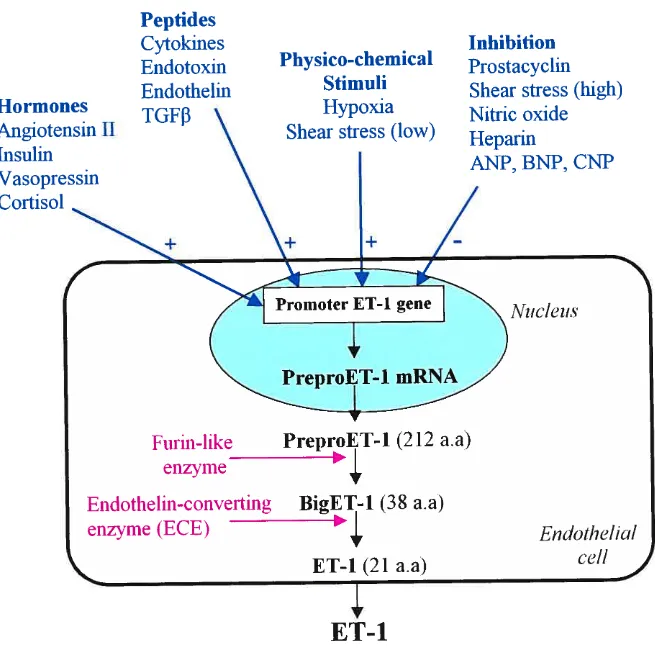
1.3 Molecular genetics and regulation of generation of endothelin
Each member of the endothelin family possesses a separate gene that
encodes a specific precursor for the mature isoform (5). The 5’ upstrearn
promoter region of the genes contains binding sites for activating protein 1 and
nuclear factor 1, which are involved in transcriptional induction of mRNA for endothelin- 1 by angiotensin II and transforming growth factor 3 (5,10). The 3’
untranslated region (3’-UTR) of the mRNA contains adenine-uracil-rich (AU
rich) sequences that regulate the stability of preproendothelin- 1 mRNA (5).
Generation of endothelin-1 is induced by many stimuli, including vasoactive
hormones, growth factors, hypoxia, shear stress, lipoproteins, free radicals,
endotoxin and cyclosporin (11) (Fig. 1.2). Production of endothelin-1 is
inhibited by stimuli that act to increase intracellular level of cyclic guanosine
monophophate (cGMP), including endothelium-derived nitric oxide,
Hormones
Angiotensm II
Insuim
Vasopressm
Cortisol
Peptides
Cytokines
Endotoxm
Endotheim
TGFf3
\\\\
Physico-chemical
Stimuli
Hypoxia
Shear stress (low)
‘j.
ET-1
Inhibition
Prostacyclin
Shear stress (high)
Nitric oxide
Heparm
ANP, BNP, CNP
Ï
Figure 1.2 Regulation of ET-1 synthesis and
its
pathway of generation.ANP = atrial natriuretic peptide; BNP = brain natriuretic peptide; CNP =
C-type natriuretic peptides; a.a = aminoacids.
(Based on ref 5)
r
Furin-like
enzyme
—PreproET-1
(212
a.a)
Endothelin-convertmg
BigET-1 (38 a.a)
enzyme (ECE)
ET-1
(21
a.a)
Endothelial
ccli
1.4 Biosynthetic pathway
The initial product of the human endothelin- 1 gene is preproendothelin- 1, a 212 aminoacid immature peptide (Fig. 1.2). Preproendothelin-1 is proteolytically cleaved by a furin-like enzyme to form biologïcally inactive intermediate, a 3$-aminoacid peptide termed big endothelin-1 (1,5). A protease called, endothelin-converting enzyme (ECE), then cleaves Trp21-Va122 of big endothelin-1 to form the mature 21-aminoacid ET-1 peptide (1,5,13). Processing
of big ET-1 to ET-1 is essential for its biological activity (1,5,14). Endothelin is
secreted by a constitutive pathway, but evidence also suggested that in some ceils endothelin can be secreted by a pathway via secretory granules (15). The
pathway of secretion involves the rough endoplasmic reticulum, golgi cisternae,
golgi small exocytic vesicles directly beneath the plasma membrane (15).
1.5 Sites of eneration
Endothelial ceils are the major site of generation of endothelin- 1, tins tightly correlates with the high expression levels of mRNA for
preproendothelin- 1 and the presence of intracellular converting enzyme in these
ceils (6,8,12). Human aortic vascular smooth muscle ceils express mRNA for endothelin-1, but its production is 100 fold less than that in endothelial ceils (5). Also, endothelin- I is produced by the afrway epithelial ceils, macrophages, fibroblasts, cardiomyocytes, posterior pituitaly and central nervous system
(6,8,11). Endothelin-2 is produced by the kidney and intestinal epithelial cefis, and endothelin-3 by brain neurons and renal tubular epithelial cefls (6,8).
1.6 Plasma concentrations ofendothelins
Plasma concentrations of ET-1 is in the range 1-10 pmoVL in healthy subjects (5,6). Plasma ET-2 and ET-3 are found at even Iower concentrations (6). Therefore, under normal physiological conditions, endothelins are flot circulating hormones; rather they act as autocrine and paracrine factors at multiple sites in the body (6).
1.7 BioIoical actions ofendothelin-1
When injected in vivo, ET is the most potent vasoconstrictor agent yet identffied (1,9) particularÏy in the brain (16), renal (17) and pulmonary
vasculature (18,19). Intraventricular injection of ET-1 induces a transient
increase in arterial pressure, respiratory rate and renal sympathetic nerve activity followed by a long-term depression of these parameters (6,20). While, intravenous bolus administration of ET-1 in different species leads to a short lived decrease in vascular resistance followed by the long-term increase, implicating a balancing act of dilator and pressor ffinctions for endothelins (6). ET-1 exerts a positive chronotropic (21) and inotropic (22) action on human heart, and also mediate cardiac hypertrophy (23) and rernodeling in congestive
heart failure through its mitogenic properties (24). ET-1 has also mitogenic
effects on smooth muscle ceils (25), fibroblasts (26), macrophages (27), mesangial ceils (31), and increases as well ceil proliferation (28,29,30,31).
1.8 Endothelin receptors
Endothelins exert their biological actions through the activation of two
receptor subtypes, ET-A and ET-3 (14,32). Both receptors beÏong to a large
family of transmembrane guanine nucleotide-binding protein-coupled receptofs (GPCRs) (33). They contain seven transmembrane domains of 22-26
hydrophobic aminoacids in their --400-aminoacid sequences (14,32). Thefr N-terminal region is extracellular and their C-N-terminal region is intracellular (7). Type A receptors exist mainly in vascular smooth muscle ceils but is also found in cardiomyocytes, fibroblasts, hepatocytes, adipocytes, osteoblasts and brain
neurons (22,3 2,34) and present higher affinities for 1 and 2 than for
ET-3 (6,14). Type B receptors exist predominantly in endotheial ceils and smooth muscle ceils but is also found in cardiomyocytes, hepatocytes, fibroblasts,
osteoblasts, different epithelial celis and neurons (22,32,34) and have equal
subnanomolar affinities for ail endothelin peptides (6,14). Therefore, ET-1 binding to ET-A and ET-B receptors on srnooth muscle produces vasoconstriction, celi growth and cefi adhesion (14,35). The binding of ET-1 to endotheial ET-B receptors stirnulates the release of NO and pro stacydlin winch
prevents apoptosis, inhibits ECE-1 expression in endothelia] celis and plays an
important role in ET-1 clearance (14,35).
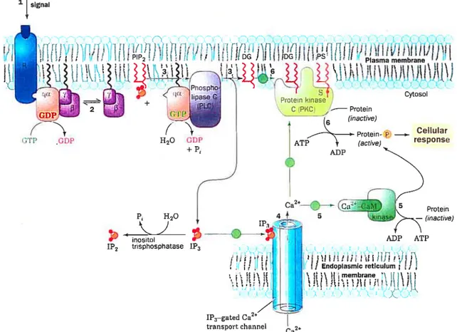
1.9 Activation ofthe phosphoïnositide cascade by ET-1
following the binding of ET-1 b ils receptor, the hormone-receptor complex activates Gq-protein which is the best characterized signaling interaction with ET-A receptor (6,7). As with ail heterotrimeric G-proteins, Gq consists of an Œ-subunit (aq, or related a-subunit, such as al 1), a member ofthe
-subunit family as weil as a member of the y-subunit family and is associated to
the membrane (36) (Fig. 1.3). In the inactive Gq heterotrimer, aq is ligated to GDP. Exchange of GDP for GTP on aq leads to the dissociation of aq(GTP) and y and both remain associated with the membrane (33). Thefr dissociation leads to activation of phosphoinositide-specific phospholipase C (PLC
)
(37,3$), winch then hydrolyzes the membrane phospholipid, phosphatidylinositol-4’,5’-bisphosphate [Ptdlns(4,5)P2J to two second messengers: hydrophobic diacylglycerol (DAG), winch remains in the plane of the membrane, and soluble inositol- 1 ‘,4’,5 ‘-trisphosphate [Jns( 1 ,4,5)P3J (37,3$). Ins( 1 ,4,5)P3 diffuses into the cytoplasm and activates some calcium channels of the sarcoplasmic reticulum, winch leads 10 an increase of Ca2 levels in the sarcoplasma and ceilcontraction (7,8). DAG together with Ca2 activates the phosphatidylserine dependent protein kinase, protein kinase C (PKC) (39) (Fig. 1.3).
10 Plasma membrane Cytosol D PKC Protein TÇ(actwe) — tO L ADP HO 1P3
:
___
inositol 1P2 trisphosphatase 1P3 AT?/11
1P3—gated Ca2” transport channel Ca’Figure 1.3 : Special case of Gq-protein-coupled receptor in intracellular signaling. (1) The binding of an agonist to a surface receptor, R, activates phospholipase C through the intermediacy of what is shown here as (2) a Gq protein. Phospholipase C catalyzes the hydrolysis of PIP2 to 1P3 and DG (3). The water-soluble 1P3 stimulates the release of Ca2 sequestered in the endoplasmic reticulum (4) winch in tum activates numerous cellular processes through the intermediacy of calmodulin and its homologs (5). The non-polar DG remains associated with the membrane, where it activates protein kinase C to phosphorylate and thereby modulate the activities of a number of cellular proteins (6). Tins latter activation pro cess also requires the presence of the membrane lipid phosphatidylserine (PS) and Ca2. PIP2= phosphatidylinositol bisphosphate; 1P3
inositol triphosphate; DG = diacylglycerol. The green circles fflustrated in the
figure do flot apply to the special case of Gq-protein-coupled receptor. (Based on
ref 134) External signal
ftf)
ii!!I
(PP CDP i t t. j Endoplasmic reticulum ) t 1 membrane tilt itt,flt t!’t L APKC is a family of serine/threonine kinases that is subdivided into three
groups. The classicai or conventionnai PKCs winch require DAG and Ca2 include the isoenzyrnes ci, f3 and ‘ (40). Novel PKC (nPKC) where the activities
are DAG-dependent though probably Ca2tindependent include the isoenzymes
, g, r, O, a and y and atypical PKC (aPKC) are independent of DAG and Ca2
and include and 7. isoenzymes (39).
1.10 Mechanisms ofET-1-induced activation ofMAPK cascade
The next process associated with exposure to ET-1 is the activation of a member of the small GTP-binding protein family, Ras winch involves exchange
of GDP for GTP (41). Once activated, Ras-bound to membrane recruit the first
member of the mitogen-activated protein kinases (MAPK) calied Raf or MAPKKK (42,43). Raf phosphorylates MEK/MAPKK at specilic serine/threonine residues, winch in turn, phosphorylates ERK1/2 (MAPK42/44)
on threonine and tyrosine residues (42,43). MAPK are serine/threonine protein
kinases, winch are also activated in response to a variety of extemal stimuli such as growth factors, hormones and stress (23,42,43). In a variety of ceil types, activation of ERK1/2 leads to the phosphorylation of downstream cytosolic
regulatory proteins, such as p90’ winch phosphorylates ribosomai proteins and participates in protein synthesis (44) (Fig. 1.4). Mso, ERK1/2 migrates from the cytosoi to the nucleus and phosphorylates many transcription factors winch lead
n
Figure 14 : Schematic model showing key steps in ET-1-induced activation of MAPK and PI-3KIPKB signaling. PTKs = protein tyrosine kinases.
ET-1 Plasma Membrane
(Nudear events leadin to gene activation, ccli growth, proliferation)
Several reports have demonstrated that ET-1 activates ERK1/2 signaling pathway in many ceil types including cardiomyocytes (45), fibroblasts (2$), glomerular mesangial ceils (31) and vascular smooth muscle ceils (VSMC) (46). Activation of the ERK1/2 cascade is the best characterized. Although, ]NK and p3$mapk are most strongly activated by cytotoxic cellular stress and are better classffied as stress-activated protein kinases (SAPK). ET-1 also activates JNK (c-Jun-NH2 terminal kinase) and p3 $mapk cascades to a lesser degree than ERK in cardiomyocytes (45,47), VSMC (4$) as well as in mesangial ceils (49). In addition, several other smafl G-protein families have been stimulated including Rho, Rab and Ran (41). ET-1 activates members of the Rho family in cardiomyocytes (50) and fibroblasts (51) which are positive regulators of p3$mapk pathway (52). The remaining smafl G-proteins and thefr expression characteristics have flot been investigated systenmticafly.
There is some evidence supporting the involvement of PKC in Ras activation in many ce!! types including cardiomyocytes (53) and rat myometrial celis (40). The nature of the connection between the two processes is obscure and other pathways may operate. A possible role of a calcium-regulated cytop!asmic proline-rich tyrosine kinase 2, Pyk2 (also known as related adhesion focal tyrosine kinase (RAFTK), focal adhesion kinase-2 (FAK-2) and ceil adhesion kinase [3 (CAK
[3),
calcium-dependent tyrosine kinase (CADTK)), in the activation of MAPK has been suggested in primary astrocytes (54,55) and rat kidney mesangial ceils (56). In severa! other ceil types, the Ca2- and PKCdependent Pyk2 activation (40,57) has been shown to link GPCRs to upstream regulators of ERK1/2 MAPKs, such as Src, Shc, Grb2, son of the sevenless (SOS) and the Ras guanosine nucleotide exchange factor (27,56,58,59). Previous data had shown that ET-1 -induced association of Pyk2 through the binding of its autophosphorylated Tyr-402 to the SH2 (Src-homology 2) domain of c-$rc lead to c-Src activation in many ceil types including mesangial cefis (56,5$) and cardiomyocytes (47,56,60). Activated c-Src bound to Pyk2 rnight directly phosphorylate adjacent cellular proteins, such as pl3OCas. Once tyrosine-phosphorylated, pl3OCas has been shown to act as docking protein to recruit its effectors able to activate JNK (61,62) in cardiomyocytes (47).
Evidence suggested that ET-1 mediates EGf receptor (EGFR) transactivation winch predominantly contributes to ERK activation, while Pyk2 contributed less in cardiomyocytes (60). Both kinases activation are mediated tbrough PKC signaling (60). Conversely, a recent report demonstrated that ET 1-induced JNK activation is preferentially regulated by Pyk2, c-Src and the pl3OCas/Crk complex but flot by EGfR (47). However, the regulation of transactivation of EGFR is signfficantly different arnong ceil types. In VSMC, EGFR transactivation has been shown to mediate the angiotensin II-induced ERK activation (63). ET-1-induced transactivation of EGFR contributes to the activation of Shc adapter molecule, leading to its interaction with Grb2 (64), winch could then associate with SOS. $hc-associated Grb2ISOS induces exchange of GDP for GTP on Ras (59) and ultimately activate ERK signaling
(60,65). However, the invoïvement of Pyk2 in ET-1-induced signaling in
1.11 Phosphatidylinositol-3 kinase cascade
Mother effector of Ras species, phosphatidylinositol-3 kinase (PI-3K) is
an enzyme that is implicated in a myriad of celiular processes. PI-3K activity has been Iinked to cefi growth and transformation, differentiation, motffity and survival (66,67). Ihe PI-3K group of lipid kinases catalyze the transfer of
phosphate from ATP to the 3’position of the inositol ring of the membrane
localized phosphoinositides. PI-3K phosphorylates at least three substrates: phosphatidylinositol (Ptdlns), phosphatidylinositol-4-phosphate (Ptdlns-4-P)
and phosphatidylinositol-4,5-bisphosphate (Ptdlns-4,5-P2) and generates the
reaction products: Ptdlns-3-P, Ptdlns-3,4-P2 and Ptdlns-3,4,5-P3 respectively
(68). These phospholipids act as second messengers to activate several proteins like PDK, PKB/Akt and p7OS6K (67,6$).
1.11.1 Classification ofPI-3Ks
PI-3Ks are divided into three classes based on their structure and
mechanism of regulation (69). Class I PI-3Ks generate Ptdlns-3-P, Ptdlns-3,4-P2, Ptdlns-3,4,5-P3 and are activated by receptor tyrosine kinases and G-protein
coupled receptors (70). Class II PI-3Ks generate Ptdlns-3-P and Ptdlns-3,4-P2
and possess a lipid binding domain, whereas, Class III PI-3Ks generate Ptdlns 3-P only (70). Ptdlns-3-P is constitutively present in ail ceils and its levels do not change following stimulation (70).
1.11.1.1 Class I PI-3Ks
Class I PI-3Ks are famfly 0f heterodimeric proteins, each of which
consisis of a catalytic subunit of 110-120 kfla and a regulatory subunit of $5
kDa (66). Three mammalian catalytic PI-3Ks sharing 42-48% aminoacid
sequence identity have been cloned and are designated pli OŒ, pli
013,
pli 06.Each of these proteins interacts with the p85 regulatory subunits at the
N-terminal region, and contains a domain that binds to the smafl G-protein Ras, a «PIK domaim> homologous to a region found in other phosphoinositide kinases and a C-terminal catalytic domain (66,68). The catalytic p110 subunit possesses
both intrinsic kinase serine/threonine and phosphoinositide kinase activities
(69,71).
Two isoforms of p85, p$5Πand p$5f3, have been purffied and cloned (71,72). P85 subunits do flot possess any known enzymatic activity but are
composed of several domains with homology to those found in other docking
proteins. P85a and p85J3 contain an N-terminal Src-homology 3 (SH3) domain,
two or three proline-rich segments, a region of homology to GTPase-activating proteins for the rho family of small G proteins (rho-GAPs) and two Src homology 2 (SH2) domains (71). The inter-SH2 domain located between the
two SH2 domains is necessary and sufficient for interaction with the N-terminal
1.11.2 Protein kinase B signaIin pathway
Several targets of PI-3K have been identffied, however, most widely studied target is protein kinase B (PKB), also known as Akt ta product of akt proto-oncogene). PKB is a serine/tbreonine kinase and three isoforms have been identilied in mammalian system: PKBŒIAkt1, PKB[3/Akt2 and PKB1’/Akt3 (67,73,74). They are activated by dual phosphorylation on threonine (Thr308) and serine (5er473) residues (73). AIl family members contain a central kinase domain with specfficity for serine or threonine residues in substrate proteins (73,74). N-terminal of PKB possesses a pleckstrin homology (PH) domain that binds phospholipids. A short glycine-rich region that bridges the PH domain to the catalytic domain follows the PH domain. The C-terminus of PKB is hydrophobie and possesses a proline-rich domain (75).
The lipid products of PI-3K bind with high affmity and specfficity to the PH-domain ofPKB with a preference ofPtdlns-3,4-P2 over Ptdlns-3,4,5-P3 (76). Tins binding induces transiocation of PKB to the plasma membrane where phosphorylation of Thr3O$ by Ptdlns-3,4,5-P3 dependent protein kinase-1 (PDK-1) and Ser473 by the hypothetical site PDK-2 is required for the activation of PKB (76). Phosphorylation of both sites is mitogen- and PI-3K-dependent (76). Several different targets of PKB have been identffled and include members ofthe apoptotic cascade such as Bad (73,75), caspase (77) and glycogen synthase kinase 3 (G$K-3) (7$) (Fig. 1.4).
In mesangial ceils, ET-1 receptor activation has been shown to stirnulate PI-3K phosphorylation through Ras (59). Also, in rabbit internai carotid artery
vascular smooth muscle ceils (ICA V$MCs), PI-3K appeared to be involved in
ET-1-induced Pyk2 tyrosine phosphorylation (79). Conversely, studies using
angiotensin II (Ail), a vasoactive peptide with similar effects to ET-1, have
suggested that Pyk2 regulates PI-3K cascade specffically via interaction of
Pyk2 with pl3OCas winch lead to their association with PI-3K in VSMC (65). Recently, ET-I has been shown to slightly increase PKB phosphorylation in cardiomyocytes (80). However, no direct activation ofPKB in response to ET-1 has been demonstrated in VSMC.
1.12 Role of endothelin in cardiovascular diseases 1.12.1 Endothetin in human hypertension
The haDmark of hypertension is an increase in peripheral vascular resistance winch is considered to be related to an increase in tone of resistance arteries as well as to structural changes or vascular remodeling ofthe blood vessels ($1,82). Several forms of hypertension are mediated by high endothelin (ET) levels in the circulation or by alterations in response to ET at the receptor level ($2,83). Besides the abifities of ET to increase vascular tone, it also induces hypertrophy in smooth muscle ceils and fiinctions as mitogen as well (8 1,84,85) (Fig. 1.5).
t Generation t Sensitivitv t Clearance t Nitric oxide
‘7
tENDOTHELIN ACTWflY
Figure 1.5 : Potential pathways of endothe]in-1 in the pathophysio]ogy of hypertension or its complications. VSM, vascular smooth muscle; RAAS, renin-angiotensin-aldosterone system; SNS, sympathetic nervous system. (Based on ref 5) acrophage Mitogenesis activation t SN S activitv Peripheral Cerebrovascular Chronic Ileart Ischacmic Heart
vascular disease failure Dtseasc
Disease 4—
Enhancement of generation of endothelin- 1 (ET-1) plays a rote in hypertrophic remodeling of arteries in moderately to severely hypertensive patients ($3). Also, this enhancement of ET-1 generation contributes to elevate blood pressure and may explain the reduced responsiveness of arteries to ET-1 through downregulation of ET receptors. Recently, it lias been shown that sait-sensitive hypertensive patients oflen have Iow plasma renin activity and thefr endothelin in plasma responds in an exaggerated fashion with an increase afier sodium depletion, in association with enhanced plasma catecholamines ($6). This suggests a reiationship of the sympathetic system, sodium sensitivity and reactivÏty of the endothelin system that may contribute to blood pressure elevation in these subjects (81,82,87).
Proliferative effects of ET have been demonstrated in vascular smooth muscle ceils (81,84,88) as well as in renal ceils such as mesangial ceils (81,88,89). Thus, the capacity of endothelin to regulate contractile responses and proliferation of vascular smooth muscle and its capacity to profoundly affect renal ffinction make it a primary candidate as a mediator of hypertension.
1.12.2 Endothelin in experimental hypertension
Most hypertensive animal models have normal or only silghtly increased plasma endothelin levels (90). $everal studies have reported that in deoxycorticosterone acetate (DOCA)-salt and aldosterone-sait hypertensive rats (91,92), DOCA-salt treated spontaneously hypertensive rats (SHR) (93), DaM sait-sensitive rats (94), one-kidney-one-chp Goldblatt hypertensive rats (95) and stroke-prone SHR (96), there is overexpression of preproET- 1 mRNA in the endotheium (97). In SHR (91,93,98), endothelin doesn’t seem to play an important role, although increased vasoconstrictor response to ET-1 has occasionally been reported in SHR (99).
Elevated vessel ET mRNA mediate structural effects such as vascular hypertrophy due to its growth promoting properties (82). DOCA-sait hypertensive rat arteries show severe vascular hypertrophy with prorninent medial thickening (100) and overexpression of the ET-1 gene (101). However, in SHR, littie vascular hypertrophy and no ET-1 gene expression are reported (102). Overail, it seems that in the DOCA-salt hypertensive rat and in rats with malignant hypertension, ET plays a more important role than in other models. Tins suggests that ail animal hypertensive models are not the same. The different hypertensive diseases have different etiologies in winch endothelin plays different roles, but in more severe forms of hypertension, such as malignant, ET plays a clear central role.
1.12.3 Endothelin in atherosclerosis
Atherosclerosis involves injury of endotheial ceils, inflammation with macrophage and monocyte infiltration of the vessel wall, release of cytokines
and growth factors, migration of smooth muscle celis to the intima, and lipid accumulation in foam ceils (82). Evidence lias recently accrued suggesting involvement of ET-1 in these processes leading to atherosclerosis development and progression ($2). ET-1 is chemoattractant for monocytes and macrophages and acts as a comitogen for vascular smooth muscle ceils together with growth
factors (103,104). Plasma and tissue ET are elevated in proportion to the extension of atherosclerosis in patients with advanced disease (105). ET-B receptors are upregulated in atherosclerotic human coronary arteries (106). However, no change in ET-A and ET-B proportions in the media of coronary
arteries was detected (107). Rossi et al. (108) showed that atherosclerotic and hypertensive individuals exhibited increased immunoreactive ET in the arteries. Many components of human atherosclerotic lesions such as endothelial ceils, macrophages, and smooth muscle ceils express ET-1 (109). Mechanisms whereby increased ET-1 may contribute to atherosclerosis include stimulation of migration of smooth muscle ceils (110) into the intima of vessels, activation of inflammation in the vessel wall by stimulating cytokines and by increasing oxidative stress (111).
1.13 Reactive oxygen species and its implications in ET-1 signaling
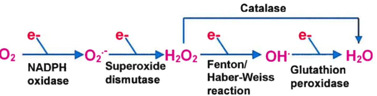
During the last few years, evidence has accumulated to suggest that the generation of reactive oxygen species (ROS) play a crucial role in the development and the progression of vascular dysftinction (112). Under oxidative stress conditions, excessive endogenous formation of ROS overcomes cellular antioxidant defence mechanisrns, which results in ROS-iriltiated modification of lipids, proteins, carbohydrates and DNA (112). ROS are very srnall, rapidly diffusible, highly reactive molecules and include hydroxyl radicals (0H), superoxide anion (0/) and non-radical derivative such as hydrogen peroxide
(H202) (Fig. 1.6). Endogenously, the main source of ROS is the mitochondria winch converts 1-2 % of consumed molecular oxygen into superoxide anion (113). In VSMCs and endothelial ceils, NADHINADPH oxidases represent the most important source of 0/ (114). NADPH oxidase catalyzes the NADPH dependent reduction of oxygen to 0/, winch is converted to H202 either by a protonation reaction or by the action of superoxide dismutase (SOD). H202 is reduced to F120 by catalase or glutathion peroxidase. Under certain conditions and in presence of metals, I-1202 can generate the extremely active 01f via Fenton or Haber-Weiss reaction (115) (Fig. 1.6).
Cata lase
I
e
02
02
“H202
“0H
‘H20
NADPH Superoxide Fenton!
• Glutathion
oxidase dism utase Haber-Weiss peroxidase
reacti on
figure 1.6 : Key steps in the production of reactive oxygen species.
NADPH oxidase is a multicomponent enzyme. Several isoforms have been found in the vascular wall (114). The plasma membrane-associated flavocytochrome b55$ consists of two subunits: gp9lphox and p22phox (114), and is the key catalytic component responsible for the transfer of electrons from NADPH to molecular oxygen (116). P47phox, p67phox and a smafl GTP binding protein, Rac, are the cytosolic components that transiocate from the cytosol to the membrane during NADPH oxidase assembly (117). ET-1 has been shown to activate NADPH oxidase, thereby increasing O{ levels in endothelial ceils (11$) and stimulates O2 production in pulmonary smooth muscle ceils
(119). Recent findings also suggest that ET-1 can increase 0f levels via activation ofNADPH oxidase in DOCA-sait rats (120).Whereas, growth factors such as AH and PDGf have been shown to generate ROS in VSMCs (114,121).
Increased ROS generation has bcen associated with a variety of cardiovascular pathologies (122) including hypertension (123) and athero sciero sis (124). Pathogenesis of cardiovascular diseases by activating
ROS are thought to participate in the cellular signaling pathways responsible for
promoting ceil growth (125) and proliferation (30,126,127). It has been demonstrated that ET-1 induces JNK and p38mapk activation through ROS generation but not ERK1/2 (30). These flndings are consistent with those of Fei
et aL (128), who demonstrated that JNK activation but flot ERK1I2 activation by ET-1 was signfflcantly inhibited by antioxidants in rat smooth muscle ceils. Conversely, a recent study demonstrated the involvement of ROS in ET-1-induced activation of ERKI/2 pathway as well as JNK and p3$mapk in cardiac fibroblasts (127). However, the contribution of ROS in ET-1-induced ERKY/2 activation is controversial and remain to be claril’ in VSMC. In addition, ROS have been shown to regulate PKB signaling pathway in human hepatorna ceil (126) as well as in VSMC (115). But whether ROS stimulate PKB activity in
response to ET-1 has flot been yet elucidated. More evidence have suggested that Pyk2 is activated in presence of11202 (129). However, a possible role of
ROS in mediating the ET-1 response on Pyk2 and whether NADPH oxidase is involved in the generation of ROS by ET-1 are questions that stifi need to 5e answered.
1.14 Nitrïc oxide
Nitric oxide (NO) is a free radical that was previously described as a non
prostaglandiri, endothelium-derived relaxing factor (EDRF) (14,130,131) and is involved in the regulation of a large number of biological processes (14,130).
Initial efforts to understand the role of NO in the nervous, cardiovascular and immune systems expanded to numerous cellular events and pathologies including apoptosis, inflammation, kidney ffinction, diabetes, oxidative stress and aging (131,132).
1.14.1 Formation of nitric oxide
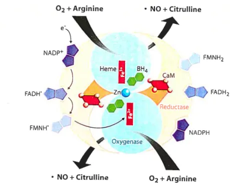
NO is formed from the aminoacid, L-arginine, in an oxidative reaction that consumes molecular oxygen and reducing equivalents in the form of NADPH
+
(lil,132,133) (Fig. 1.7). Reaction products are NO, NADP and citruilme. Since NO is a signaling hydrophobie molecule small enough to pass across the target-ceil plasma membrane, NO cannot be stored and released as needed (130,134). NO is produced by the enzyme nitric oxide synthase (NOS), by the deamination of L-arginine. NOS is an enzyme requfring FAD, FMN, heme, Ca2, calmodulin and 6(R)-tetra-hydro-L-biopterin (BI-I4) as cofactors (133) (Fig. 1.7). NO acts locally because it has a short half-life (5-10 seconds) in the extracellular space before it is converted to nitrates and nitrites by oxygen and water (130,131,134).
Figure 1.7 : The nitric oxide synthase (NOS) reaction. CaM = catmoduhn; Zn = Zinc. (Based on ref 135)
1.14.2 NOS isoforms
Three distinct NOS enzymes, each a product of a unique gene, have been identffied and characterized (130,131,135,136). The neuronal form (nNOS or NOS-1) is a Ca2-dependent enzyme found in neuronal tissue and skeletal muscle. Four splice variants of full lengh nNOS (nNOSŒ) have been identffied recently (nNOSf3, nNOSy, nNOSi and nNOS-2). The second isoform of NOS (iNOS or NOS-2) is inducible in a variety of ceils and tissues in response to cytokine or endotoxin activation. The third form, first found in vascular
02 ÷ Arginine • NO+Citrulline
t
FMNH2 FADH2 NADPH FADH1))
FMNH1
OxyqenaseI
NO+Citrulline 02+Arginine2+
endothehal celis (eNOS or NOS-3), 15 also Ca -dependent, but differs from the
neuronal form by its smailer size. eNOS is rnyristoylated and palmitoylated at the N-terminus. Those modifications are required to localize to the plasmalemmal caveolae of endothelial ceils. Human enzymes exhibit approximately 51-57 % homology at the aminoacid level (130,135). Structuraily, ail NOS isozymes consist of a carboxy-terminal reductase dornain winch binds the flavin cofactors. A Ca27ca1modu1in binding domain lies in the center foilowed by an oxygenase domain where binding of heme, 02, BR1 and
arginine substrate take place (135).
1.14.3 Nitric oxide function
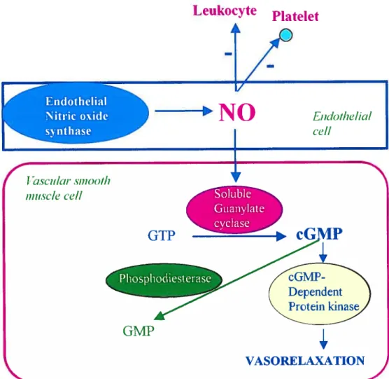
NO ceilular signaling involves the regulated synthesis of NO by eNOS in the vascular endothelium, diffusion of NO into adjacent smooth muscle ceil and activation of the soluble isoforrn of guanylate cyclase (sGC) (137). When NO binds to the pentacoordinate ferrous heme of the sGC that appears to be uniquely tuned to interact with NO, conformational changes occur in the enzyme, stimulating the reaction (138). NO causes relaxation of the smooth muscle by mediating the formation of cGMP that acts as a second messenger and activates the cGMP-dependent protein kinases (protein kinase G) (137,139), which in tum, facilitates the phosphorylation of various proteins as weil as the reduction of intraceilular calcium concentrations by different mechanisms (140). Moreover, NO also targets many proteins either by nitrosylation of thiol
residues, nitration of tyrosine or oxidizing DNA and protehis (140). Increasing evidence indicates also that NO may inactivate NADPH oxidase by inhibiting
its
assembling process, thus reducing the ROS levels (141). In higher concentrations, NO can react rapidly with superoxide (O2) to form peroxynitrite(0N00), a potent oxidant with the potential to disrupt protein structures by nitrating the protein tyrosine residues (142). Although NO signaling is complex as a resuit of its interactions with ROS, heme groups on proteins, sullhydryl groups,
and
other cellular targets, the activation of guanylate cyclase remains the most important pathway in mediating NO function (137).1.14.4 Guanylate cyclase
Guanylate cyclase is an enzyme that catalyses the conversion of the guanosine triphosphate (GTP) to 3’-5’-guanosine monophosphate (cGMP). The guanylate cyclase is found in many cellular compartments (140). Two major forms of guanylate cyclase
are
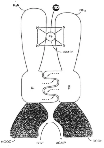
known, the particular guanylate cyclase and the soluble guanylate cyclase. It is generally conceded that activation of soluble guanylate cyclase (sGC) is the principal intraceflular event that hiitiates relaxation (143,144). The activity of the sGC is regulated by nitrovasodilators, oxidation products of fatty acid and free radicals (134,145). sGC is a heterodimer of two subunits a and13.
Each subunit is divided in three different domains: the heme-binding domain, the catalytic domain and the dimerization domain (145).N-terminal of each subunit contains heme as a prosthetic group winch serves as a site for NO binding (145). sGC lacking the heme moiety, is flot able to be activated by NO (145). Heme is attached to the protein portion of the enzyme by an imidazole axial ligand and binding of the heme is specific to the [3 subunit ofthe N-terminal region (146,147). C-terminal ofeach subunit possesses a catalytic domain with a high homology sequence between the monomers (146,147). Coexpression of the catalytic domain of both subunits is necessary for GC activity. There is the dimerization domain between both domains described above that mediates the association of the heterodimer winch is essential for the catalytic subunit (146,147). NO binding to the berne oftlie sGC resuits in the formation of a complex penta-coordinate heme-nitrosyl that breaks the axial histidine Iink (147). Tins conformational change exposes the catalytic site to GTP, leading to the activation of the enzyme and conversion of GTP to cGMP by sGC in the presence ofMg2 or Ï4n2 ions (137) (Fig. 1.8).
H2N
1.14.5 Reulation ofcGMP production
In most tissues, the intracellular concentration of cGMP is determined by the rate of formation which is regulated by agonist-induced stimulation of a cyclase and hydrolysis of cGMP by a related group of phosphodiesterase E (PDE) (14$) (Fig. 1.9). There are at least seven known distinct mammalian PDE families. Each one differs from each other in
HOOC
Figure 1.8 : Schematic representation of a soluble guanylate cyclase & heterodimer.
biochemical and physical properties, responses to specific effectors, inhibitors and regulatory control mechanisms (14$). Type V PDE has been isolated from a number of tissues including human platelets (149), trachea (150) and VSMC (151) and is commonly referred to as cGMP-specffic PDE. PDE V is characterized by selectively hydrolyzing only cGMP, independently of Ca2/ca1modulln. Inhibitors of PDE V such as A02 131-1 have vasodilating and anti-aggregating properties, which may protect the vascular wail against arteriosclerotic changes (149).
r»EndotheIiaI Nitric oxide synthase
Leukocyte
Ptatelet
—p
NO
f
J ;scuÏcir Ç111t9C)t/1 Inhiscic’ ec’lI Endothelicil ce!!r
Soluble Guanylate cyclase GTP Phosphodiesterase -cGJVIP
cG Dependent Proteinkinase
VASORELAXATIONGMP
1.14.6 NO in si2nal transduction
The endotheium serves as the principal physiological source of NO in blood vessels (152). As evidenced, NO contributes to the regulation of several hormone-mediated responses (153,154,155). In addition to its vasodilating effect, NO can also inhibit atherogenesis (130), thrombocyte aggregation (156) and VSMC proliferation (148,152) and migration (157). There is also increasing body of evidence suggesting that NO is opposed to the physiological and pathophysiological effects (14,137) of growth factors and vasoactive peptides such as EGF (14$), PDGF (15$) and bFGF (157). Tins is probably achieved by inhibiting one or several of the signaling events induced by these factors (130,152,157,159,160). According to several studies, mitogens such as ET-1 stimulate the synthesis of DNA and ceil proliferation by activating the phosphorylation cascade of MAPK (28,161,162,163). The potential mechanism that could modulate VSMC proliferation is the release of NO by the endothelium either via a cGMP-dependent (137,14$) or a cGMP-independent mechanisms (164,165,166,169). In cardiomyocytes, ET-1 -induced protein synthesis (170,171) has also been shown to be inhibited by NO (167). Furthermore, NO was recently found to suppress the AII-induced activation of three major MAPKs, ERK1/2, p38mapk and JNK (16$) as weil as Pyk2 (155) in cardiac fibroblasts. However, it is flot known whether NO similar to ils effect on growth factor and MI-induced responses can also modulate signaling events triggered by ET-1 receptor activation in VSMC. In smooth muscle-derived A7r5
ceils, NO has been shown to regulate PDGF-induced activation of PKB (154). These data implicate the PKB signaling cascade as an important mitogenic pathway that is subjected to modulation by NO in VSMC (154). However, the role of NO/cGMP in modulating PKB signaling pathway in response to ET-1 has not yet been investigated in any ceil type.
1.15 Obiectives ofthe present study
As described above, ROS play an essential role in propagating the signais ofseveral growth factors such as EGF, PDGF and Ail. The contribution ofROS in ET-1-induced MAPK activation remain controversial and there are no reports documenting the activation of PKB as well as Pyk2 by ET-1 in VSMC. Therefore, we have undertaken tins study to elucidate a role of ROS on key components of ET-1 signaiing system as well as protein synthesis in V$MC, and have examined whether ROS generation contributes towards thefr response.
Furthermore, since NO is an important modulator of intracellular signaling system by many growth factors such as PDGF and EGF in VSMC as well as MI in cardiac fibroblasts. We have also investigated a possible role of NO on ET-1-sensitive signaling systems in VSMC. We first elucidated the role of NO on key components of ET-1 signaling systems ERK1/2, PKB and Pyk-2 as well as protein synthesis in VSMC. We then used 8-Bromo-cGMP, a cycic GMP analogue and ODQ, an inhibitor of sGC, to examine whether NO is acting
through a cGMP-dependent mechanism. These studies have utilized standard protocols of ceil biology such as ceil culture, SDS-PAGE, western bloting as wefl as radioisotopes.
ARTICLE 1
Reactive oxygen species mediate
EndotheÏin-1-induced activation ofERKYz, PKB and Pyk2
andprotein synthesis in vascular smooth muscle
Reactive oxygen species mediate E ndothelin- 1 -induced
activation
of ERK%, PKB
and Pyk2
and protein
synthesis in vascular smooth muscle celis
Grace Bou Daou and Ashok K. Srivastava
Centre de recherche, Centre hospitalier de l’Université de Montréal - Hôtel
Dieu, Department of Physiology, Université of Montréal, Montreal, Quebec, Canada
Running titie: Role ofROS in ET-l-induced activation ofERK Y2, PKB, Pyk2 and protein synthesis
Address for correspondence: Ashok K. Srivastava, Ph.D Centre de recherche CHUM, Hôtel-Dieu 3840, rue St. Urbain Montréal (Québec) H2W 1 T7 Tel: 514-$90-8000 ext.12917 Fax: 514-412-7152
Abstract
Reactive oxygen species (ROS) have been shown to mediate the effect of several growth factors such as angiotensin II (MI), epidermal growth factor
(EGf) and platelet-derived growth factor (PDGf). Endothefin-1 (ET-1) is an
important growth factor for vascular smooth muscle ceils (VSMC) which is
beieved to contribute to the pathogenesis of vascular abnormalities such as
atherosclerosis, hypertension and cardiac hypertrophy. However, a possible roTe
of ROS generation in mediating the ET-1 response on ERK1/2 and PKB, key
components of growth promoting and proliferative signaling pathways, has flot
been examined in detail. Therefore, the aim of the present study was to investigate the involvement of ROS in ET-1-mediated activation of ERKÎ/2 and PKB as well as Pyk2 in A-10 VSMCs. Pyk2 is a non-receptor protein tyrosine kinase and an upstream regulator of MAPK signaling. ET-1 stimulated the
phosphorylation of ERK1/2, PKB and Pyk2 in a dose and time-dependent
fashion with maximum response being eicited at 10 nM winch peaked at 5 min.
Treatment ofV$MC with ET-1 resulted in an increase in the generation ofROS that could be btocked by diphenyleneiodonium (DPI), an inhibitor of NADPH
oxidase. furthermore, DPI pretreatment of ceils prior to stimulation with ET-1,
attenuated ET-1 enhanced phosphorylation of ERK1/2, PKB and Pyk2. N
acetylcysteine (NAC), another ROS scavenger, also exhibited a similar response. Moreover, DPI also caused a decrease in the protein synthesis stimulated by ET-1. These resuits demonstrate that ROS is a critical mediator of
ET-1-induced signahng events linked to hypertrophic and growth promoting pathways in VSMC.
Introduction
Endothelin-1 (ET-1), a 21-amino acid peptide hormone, exhibits vasoconstrictor (1) and mitogenic (2) properties. These effects of ET-1 are elicited through the activation of 2 receptor subtypes, ETA and ETB, which
belong to a family of heptahelicai G-protein-coupled receptors (GPCRs) (3-5). ET-1 receptor activation is coupled to multiple signaling pathways, such as phospholipases C and D (6), Ca2 (7), mitogen-activated protein kinases (MAPKs), including extracellular signal-reguiated kinases 1 and 2 (ERK1/2), c-Jun-NH2-terminal kinase (JNK), and p3SMAPK (8-11) as well as phosphatidylinositol 3-kinase (12). Extensive studies, carried out predominantiy in cardiomyocytes (6, 8, 2$) and glomerular mesangial ceils (11, 12), have implicated both receptor and non-receptor protein tyro sine kinases (PTKs) in transducing ET-1 -evoked signaling responses (11, 15-18). Various PTKs activated by ET-1 include c-Src (13-15), epidermal growth factor (EGF) (17) and a Ca2tdependent PTK, Pyk2 (11, 17).
Recent experiments have indicated that reactive oxygen species (ROS) play an essential role in propagating the signais of several growth factors, peptide hormones and cytoldnes, such as piatelet-derived growth factor (19), EGf (20), angiotensin II (AIl) (21), insulin (22), interleuldn-1 (23) and tumor necrosis factor-Π(24). Increased ROS generation has been linked to the pathogenesis of several cardiovascular diseases, such as hypertension, atherosclerosis, restenosis and congestive heart failure (29-31). ET-l lias also
been shown to augment ROS production in various ceil types (25-27), and a role
of reduced nicotinamide adenine dinucleotide phosphate (NADPH) oxidase in
the ET-1 -induced elevation of vascular ROS production has been suggested recently (32).
Although it has been reported that ROS mediate ET-1 -stimtttated activation of ERK1/2 and JNK (27), to the best of our knowledge, a detailed
investigation into the contribution of ROS in other ET-1 -evoked growth
promoting signaling pathways has not been carried out. Moreover, despite the
fact that ET-1-induced vascular smooth muscle celi (VSMC) proliferation (26, 33) and fibrogenesis (15) may contribute to vasculature remodeling leading to vascular disease, flot much information is available on ET-1 -evoked signaling in these ceils. Therefore, in the present studies, we have investigated the effect of ET-1 on ERKÎ/2, protein kinase B (PKB) and Pyk2, key regulators of the
proliferative signaling pathway in A-10 VSMCs, and examined whether ROS
Materials and Methods
MaterialsET-1 was purchased from Peninsula Laboratories (Belmont, CA, USA), and N-acetyl-L-cysteine (NAC), diphenyleneiodonium (DPI) and bis-N methylacridinium nitrate (lucigenin), from Sigma (St. Louis, MO, USA). Monoclonal phospho-specffic-Tyr204-ERKY/2 antibody, polyclonal ERK1/2 antibody and horseradish peroxidase-conjugated goat anti-mouse immunoglobulin were from Santa Cruz Biotecbnology Inc. (Santa Cruz, CA, USA). Polyclonal phospho-specffic-Ser473-PKB and total PKB as well as phospho-specfflc-Tyr402-Pyk2 and total Pyk2 antibodies were procured from New England Biolabs (Beverly, MA, USA). The enhanced chemiluminescence (ECL) detection system kit and L-(4,5-3H) leucine were from Amersham Pharmacia Biotech (Baie d’Urfé, QC, Canada).
Methods Cet! culture
VSMC derived from embryonic rat thoracic aorta A-10 ceils were maintained in culture with DMEM containing 10% fetal bovine serum at 37°C in a hurnidffled atmosphere of 5% C02, as described earlier (34). The ceils were grown to $0-90% confluence in 60-mm plates and incubated in serum-free DMEM 20 h prior to the treatments.
Cet! tysis and Western btotting
Cefis incubated in the absence or presence of various agents were washed twice with ice-cold PBS and lysed in 200 t1 of buffer (25 mlvi Tris-HC1, pH 7.5, 25 mM NaC1, 1 mM Na orthovanadate, 10 mM Na fluoride, 10 mM Na pyrophosphate, 2 mM benzamidine, 2 mM ethylenebis(oxyethylenenitrolo) tetraacetic acid, 2 mM ethylenediamine tetraacetic acid, I mM phenyhnethylsulfonyl fluoride, 10 pg/ml aprotinin, 1% Triton X-100, 0.1% sodium dodecyl sulfate (SDS) and 0.5 lg/ml leupeptin) on ice. The ceil lysates were centrifuged at 12,000g for 10 min at 4°C. Protein concentrations were measured by Bradford assay. Equal amounts of prote in were subjected to 10% SDS-polyacrylamide gel (SDS-PAGE), transfened to PVDF membranes (Mifiipore, MA, USA) and incubated with respective primary antibodies (monoclonal phospho-specffic-Tyr204-ERK1 /2 antibody (1:2,000), polyclonal phospho-speciflc-Ser473-PKB antibody (1:4,000), phospho-specffic-1yr402-Pyk2 antibody (1:1,000)). The antigen-antibody complex was detected by a horseradish peroxidase-conjugated second antibody (1:4,000), and protein bands were visualized by ECL. The intensity of specific bands was quantffied by NIH Image software as described previously (35).
Measurement afROS generalion
ROS production was measured by the lucigenin method (21) with minor modifications (36). Briefly, the ceils were preincubated in the absence or
presence of 10 !IM DPI in DMEM for 30 mm, then treated with ET-1 (10 nM for 5 mm). They were trypshuized, collected by centrifugation, and the peflet was washed in modffied Krebs buffer containing NaC1 (130 mM), KCT (5 mM), MgC12 (1 mM), CaC12 (1.5 mM), K2HPO4 (1 mM), and HEPES (20 mM), pH 7.4. Aller washing, the cefis were resuspended in Krebs buffer, and the ceil concentration was adjusted to 1 x 1 in 900 d buffer. To measure ROS production, the ceil suspension was transferred to plastic tubes and assessed in a luminometer (LB 9507, Berthold, Wildbad, Germany). Measurernent was started by an injection of 100 il lucigenin (linal concentration 5 x 10 M) at time zero. Photon emission was counted every 5 min for up to 20 min. The emission in relative light units was corrected for nonspecific luminescence in the absence of ceils. Modified Krebs buffer was used as a control (blank). Solutions containing DPI in the absence of ceils did flot display any significant interference in the lucigenin assay.
Measurement of [3HJteucine incorporation
A-10 ceils were treated for 20 h with endothelin- 1 (10 nM; Belmont, CA, USA). Protein synthesis was assessed by the addition of 2 .iCi!mL of
[3Hjleucine (ICN Biomedicals, Inc., Costa Mesa, CA, USA) for a period of 20 h. To assess the role ofreactive oxygen species (ROS), ceils were pretreated for 30 min with DPI (5 .tM; Sigma, St-Louis, MO, USA), a specific inhibitor of flavoprotein, NADH!NAD(P)H oxidase (32,37). Following the completion of
the experimental protocol, A-10 ceils were washed twice with cold PBS, and 1 ml of cold 5% trichioroacetic acid was added for 30 min to precipitate protein. The precipitates were subsequently washed twice with cold water and resuspended in 500 d of 0.4 M NaOH. Aliquots were counted in a scintillation counter.
$tatistics
$tatistical analysis was performed by one-way, repeated-measures analysis of variance (ANOVA) followed by a Fisher post hoc test. AU data are reported as means ± SE. The differences between means were considered significant at P< 0.05.
Resuits
Efftct of ET-1 on ERK1/2, PKB and Pyk2 phosphorytation in A-10 V$MCs Our hiltial experiments were aimed at analyzing the effect of ET-1 on the phosphorylation of 3 key elements of the signaling pathway, namely, ERK1/2, PKB and Pyk2. Their activation was assessed by using phospho-specific antibodies against each of these kinases. As shown in Figure 1, ET-1 concentration-dependently enhanced the phosphorylation of ERK1/2 (Fig. lA), PKB (Fig. lB) and Pyk2 (Fig. 1C). In each case, maximum phosphorylation occurred at ET-1 concentrations of 1-10 nM. Next, we analyzed the time dependence ofthe ET-1 response at 10 nM. As seen in Figure 2, ET-1 treatment of A-10 ceils rapidly increased the phosphorylation of ERK1/2 (Fig. 2A), PKB (fig. 2B) and Pyk2 (Fig. 2C). A significant increment was detected within 1 min of treatment and peaked at about 5 mm, then gradually declined to submaximal levels at 30 to 60 min. No alteration in the total amount of ERK1/2, PKB or Pyk2 was observed under these experimental conditions.
Effect of DPI on ET-1 -induced phosphorytation of ERK1/2, PKB and Pyk2 in A-10 VSMCs
To investigate whether ROS generation was involved in ET-1-induced activation of ERK1/2, PKB and Pyk2, we utffized DPI, a frequently-employed hihibitor of the flavoprotein NADHINAD(P)H oxidase (32,37). As depicted in Figure 3, DPI pretreatment concentration-dependently inhibited ET-1-induced phosphorylation of ERK1/2 (fig. 3A), PKB (Fig. 3B) and Pyk2 (Fig. 3C).
However, among the 3 kinases, Pyk2 appeared to be most sensitive to inhibition by DPI, winch elicited a signilicant reduction in phosphorylation at 1 1iM (Fig. 3C). At tins DPI concentration, ERK1/2 phosphoiylation was flot signfficantly affected (Fig. 3A), and PKB phosphorylation was suppressed only slightly (Fig. 3B), whereas 10 jiM DPI pretreatment was found to block ET-1-induced phosphorylation of ail the kinases by about $0 % (Fig. 3).
Effecis of ET-1 and DPI on ROS generation in A-10 VSMCs
The resuits described above suggested that ROS generation by a DPI inhibitable NADPH oxidase might contribute to ET-1-induced ERKÏ/2, PKB and Pyk2 phosphorylation. Therefore, we evaluated the effect of ET-1 on ROS generation in A-10 ceils by the lucigenin chemiluminescence method. As fflustrated in Figure 4, stimulation with 10 nM ET-1 for 5 min evoked a signi±icant increase of ROS production in A-10 ceils. However, their treatment whh DPI prior to stimulation with ET-l almost completely blocked ET-1-evoked ROS production. These resuits revealed that in A-10 ceils, ET-1 caused an increase in ROS production winch was suppressed by DPI. DPI alone had no significant effect on the basal production ofROS (data flot shown).
Effeci ofNAC on ET-1-induced ERK1/2, PKB and Pyk2 phosphorytation in A-10 VSMCs
To further conllrm a role ofROS in ET-1-induced responses, we tested the effect of NAC, a thiol-containing agent, on ERK1I2, PKB and Pyk2


![Figure 1.5 : Potential pathways of endothe]in-1 in the pathophysio]ogy of hypertension or its complications](https://thumb-eu.123doks.com/thumbv2/123doknet/2058302.5895/38.918.269.827.195.819/figure-potential-pathways-endothe-pathophysio-ogy-hypertension-complications.webp)