Université de Montréal
Regulation of the Early Growth Response Protein-1 in
vascular smooth muscle cells
par
Estelle Rolande Simo Cheyou
Programme de Nutrition Faculté de Médecine
Thèse présentée à la Faculté de Médecine
en vue de l’obtention du grade de Philosophiæ Doctor (Ph.D.) En Nutrition
Décembre 2016
Université de Montréal
Faculté des études supérieures et postdoctorales
Cette thèse intitulée
Regulation of the Early Growth Response Protein-1 in vascular smooth muscle cells
Présentée par
Estelle Rolande Simo Cheyou
A été évaluée par un jury composé des personnes suivantes :
Dr Jean-Marie Ekoé, Président-rapporteur Dr Ashok Srivastava, Directeur de recherche
Dr Stephanie Fulton, Membre du jury Dr Ghassan Bkaily, Examinateur externe
Résumé
Une hyperactivation de la prolifération des cellules musculaires lisses vasculaires (CMLV) contribue à la pathogenèse des maladies des vaisseaux. Des travaux antérieurs suggèrent que l’augmentation de l'adénosine monophosphate cyclique (AMPc) inhibe la prolifération des CMLV. Provoquer une augmentation d’AMPc préviendrait aussi certaines maladies vasculaires qui sont associées à des altérations dans sa signalisation impliquant l'activité de la protéine kinase A (PKA). Des études ont démontré la contribution du facteur de transcription « early growth response protein-1» (Egr-1) dans la pathogenèse des maladies vasculaires et une surexpression d’Egr-1 a été rapportée dans des modèles d'athérosclérose et d'hyperplasie intimale. Divers agents vasoactifs contrôlent l'expression d’Egr-1 suivant des mécanismes qui ont fait l’objet de plusieurs études mais demeurent incomplètement élucidés. L'angiotensine-II (Ang-II) est l'un des principaux peptides vasoactifs impliqués dans la pathogenèse des maladies vasculaires. Une des voies de signalisation induite par l’Ang-II implique l’augmentation du calcium (Ca2+)
intracellulaire. Celle-ci se produit par l’activation de l'entrée de calcium opérée par la relâche des réserves (SOCE) de Ca2+ réticulaire suite à l’activation du récepteur à
l’inositol-3-phosphate (IP3R) et le recrutement ultérieur du complexe conducteur formé par
la molécule d'interaction stromale 1 (STIM-1) et le canal Orai-1. Bien qu’il ait déjà été démontré que l’expression de l'Egr-1 est régulée par la signalisation calcique en réponse à plusieurs stimuli, l'implication du complexe STIM-1/Orai-1 dans l'expression d'Egr-1 dans la CMLV n’a jamais été étudiée. De même, la question de savoir si la signalisation induite par l'Ang-II conduisant à l'expression d'Egr-1 est modulée par l'AMPc n’a jamais été explorée. Par conséquent, les travaux menés dans cette thèse ont consisté à examiner le rôle de la signalisation du Ca2+ dans l'expression d'Egr-1 induite par l’Ang-II dans la
CMLV avec une attention particulière portée sur le rôle joué par STIM-1 et Orai-1. En outre, nous avons examiné l'effet de l’augmentation de l’AMPc sur l'expression d'Egr-1 induite par l’Ang-II et étudié les voies de signalisation associées. Nos données montrent
que l’inhibition du récepteur IP3R et du SOCE par le 2-aminoéthoxydiphénylborate atténue
la libération de Ca2+ induite par l’Ang-II et ceci s’accompagne d’une baisse des niveaux
d’expression de protéine et d’ARN messager de l’Egr-1. La stimulation de l’expression de l'Egr-1 a également été supprimée à la suite du blocage de la calmoduline et de la protéine kinase CaMKII. De plus, le blocage par interférence d’ARN de l’expression de STIM-1 et Orai-1 a atténué l'expression d'Egr-1 induite par l’Ang-II ainsi que la phosphorylation des protéines ERK et CREB. Par ailleurs, l'isoproterenol (ISO) et la forskoline (FSK), deux activateurs de l'adénylate cyclase ont atténué de manière dose-dépendante l'expression d'Egr-1 induite par l’Ang-II. Des réponses similaires ont été observées en utilisant des analogues non spécifique (dibutyryl-cAMP) et PKA-spécifique (Benzoyl-cAMP) de l’AMPc, ainsi qu'un inhibiteur à large spectre de l'activité phosphodiesterase intracellualaire (isobutylméthylxanthine). L'inhibition de l'expression d'Egr-1 induite par l’Ang-II s’accompagne d'une augmentation de l’activité de la PKA mesurée par la phosphorylation de la « phosphoprotéine activée par les vasodilatateurs (VASP) », et d’une diminution concomitante de la phosphorylation de la protéine ERK. Le blocage pharmacologique de la PKA a réduit la phosphorylation de VASP et restauré la phosphorylation de la protéine ERK ainsi que l'expression d'Egr-1 en présence de l’Ang-II.
En résumé, nos données démontrent que la voie STIM-1/Orai-1 /Ca2+ médie
l'expression de l'Egr-1 induite par l'Ang-II dans la CMLV et suggèrent que la suppression de la réponse à l’Ang-II menant à l’expression de l'Egr-1 peut expliquer les effets vasoprotecteurs de l’AMPc. En outre, ces travaux montrent que les mécanismes moléculaires de régulation de l’expression d’Egr-1 en réponse aux signaux externes culminent vers la modulation des cascades de signalisation en aval de la protéine ERK dans les CMLV.
Abstract
Aberrant vascular smooth muscle cell (VSMC) proliferative responses contribute to the development of neointimal lesions. Cyclic adenosine monophosphate (cAMP) is believed to inhibit VSMC proliferation, and vascular diseases are associated with impairments in cAMP-induced signalling responses involving protein kinase A (PKA) signaling. An enhanced expression of the early growth response protein-1 (Egr-1), a zinc finger transcription factor, has been reported in models of vascular diseases and, a crucial role of Egr-1 in regulating the expression of genes implicated in neointimal formation leading to atherogenesis has been suggested. Various vasoactive factors have been shown to modulate Egr-1 expression in VSMC via mechanisms which remain to be completely understood. Angiotensin-II (Ang-II) is one of the key vasoactive peptides implicated in the pathogenesis of vascular diseases. Ang-II elevates intracellular calcium (Ca2+) through
activation of voltage-gated calcium channels as well as store-operated calcium channels. The store-operated calcium entry (SOCE) involves an inositol-3-phosphate receptor (IP3R)-coupled depletion of endoplasmic reticular Ca2+ and a subsequent activation of the
stromal interaction molecule 1 (STIM-1) /Orai-1 complex. Although Egr-1 has been demonstrated to be upregulated in a Ca2+-dependent fashion in response to several stimuli,
the involvement of STIM-1/Orai-1-dependent signaling in Egr-1 expression in VSMC has never been addressed. Besides, whether Ang-II-induced signaling leading to Egr-1 expression is modulated by cAMP-dependent signaling pathway remains unexplored. Therefore, in the present studies, we have examined the role of Ca2+ signaling in
Ang-II-induced Egr-1 expression in VSMC and investigated the contribution of STIM-1 or Orai-1. Additionnaly, we have examined the effect of cAMP on Ang-II-induced expression of Egr-1 and have investigated the associated signalling pathways. Pharmacological blockade of IP3R and SOCE by 2-aminoethoxydiphenylborate (2-APB) decreased Ang-II-induced Ca2+
release and attenuated Ang-II-induced enhanced expression of Egr-1 protein and mRNA levels. Egr-1 upregulation was also suppressed following blockade of calmodulin and CaMKII. Furthermore, RNA interference-mediated depletion of STIM-1 or Orai-1
attenuated Ang-II-induced Egr-1 expression, as well as Ang-II-induced phosphorylation of ERK1/2 and CREB. Moreover, isoproterenol (ISO) and forskolin (FSK), two respective receptor and non-receptor activators of adenylate cyclase, attenuated Ang-II-induced Egr-1 expression in a dose-dependent fashion. Similar responses were observed using non-specific (dibutyryl-cAMP) and PKA-non-specific (Benzoyl-cAMP) analogs of cAMP, as well as a broad spectrum inhibitor of intracellular phosphodiesterase activity (isobutylmethylxanthine). The inhibition of Ang-II-induced Egr-1 expression was accompanied by an increase in serine 157 phosphorylation of the vasodilator-activated phosphoprotein (VASP), a marker of PKA activity, and this was associated with a concomitant decrease in ERK phosphorylation. Pharmacological blockade of PKA using H89 decreased VASP phosphorylation, restored Ang-II-induced ERK phosphorylation and abolished ISO- and FSK-mediated inhibition of Ang-II-induced Egr-1 expression.
In summary, our data demonstrate that STIM-1/Orai-1/Ca2+-dependent signaling
pathways mediate Ang-II-induced Egr-1 expression in A-10 VSMC and suggest that PKA-mediated suppression of Ang-II-induced Egr-1 expression and phosphorylation of ERK may be among the mechanisms by which cAMP exerts its vasoprotective effects. In addition, our data supports the notion that stimuli-induced regulation of Egr-1 expression involves the participation of signaling cascades downstream of ERK in VSMC.
Table of Contents
RÉSUMÉ ... I ABSTRACT ... III TABLE OF CONTENTS ... V LIST OF TABLES ... VIII LIST OF ABBREVIATIONS... X ACKNOWLEDGEMENTS ... XV
CHAPTER 1: LITERATURE REVIEW ... 1
1.1 VASCULAR DISEASES AND HYPERTENSION ... 1
1.2 VASCULAR DAMAGE ... 3
1.2.1 Structure of the vessel wall and cellular basis of vascular damage ... 4
1.2.2 Pathophysiology of vessel remodeling ... 6
1.2.2.1 Determinants of vessel remodeling ... 6
1.2.2.2 Types of vessel remodeling ... 7
1.2.2.3 Clinical manifestations of vessel remodeling ... 10
1.2.2.4 Features of vascular smooth muscle cell physiology ... 11
1.2.3 Angiotensin-II in the renin angiotensin aldosterone system ... 13
1.2.3.1 Structure of angiotensin-II and receptor signaling ... 16
1.2.3.2 Angiotensin-II-mediated biological responses in cardiovascular relevant tissues ... 17
1.2.3.3 Angiotensin-II in vessel remodeling ... 21
1.2.4 Molecular basis of vessel remodeling: cascades related to angiotensin-II-induced signaling in vascular smooth muscle cells ... 22
1.2.4.1 Angiotensin-II receptor signaling ... 22
1.2.4.2 Adenylate cyclase and cyclic AMP-dependent pathway ... 23
1.2.4.3 Phospholipase and calcium-dependent pathway ... 26
1.2.4.4 The Mitogen-Activated Protein Kinase-dependent pathway ... 31
1.2.4.5 The phosphatidylinositol 3-kinase/protein kinase B dependent pathway ... 32
1.3 THE EARLY GROWTH RESPONSE PROTEIN-1 ... 34
1.3.1 Egr family ... 34
1.3.3 Transcriptional regulation of Egr-1 ... 36
1.3.3.1 Functional features of Egr-1 promoter ... 37
1.3.3.2 NAB-dependent regulation of Egr-1 ... 38
1.3.3.3 Signaling pathways upstream of Egr-1 expression ... 39
1.3.4 Egr-1 and disease ... 42
1.3.4.1 Egr-1 in cardiovascular physiology ... 42
1.3.4.2 Egr-1 role in brain plasticity ... 45
1.4 OBJECTIVES OF THE STUDY ... 47
1.5 STUDY MODEL:A10 CELL LINE ... 48
CHAPTER 2 (ARTICLE 1): STIM-1 AND ORAI-1 CHANNEL MEDIATE ANGIOTENSIN-II-INDUCED EXPRESSION OF EGR-1 IN VASCULAR SMOOTH MUSCLE CELLS. ... 50
2.1 ABSTRACT ... 52
2.2 INTRODUCTION ... 53
2.3 MATERIALS AND METHODS ... 55
2.4 RESULTS ... 59
2.5 DISCUSSION ... 64
2.6 AKNOWLEDGEMENTS ... 66
2.7 DISCLOSURES ... 66
2.8 FIGURE LEGENDS ... 67
2.9 REFERENCES CITED IN ARTICLE 1 ... 71
2.10 FIGURES ... 78
CHAPTER 3 (ARTICLE 2): CAMP ATTENUATES ANGIOTENSIN-II-INDUCED EGR-1 EXPRESSION VIA PKA-DEPENDENT SIGNALING PATHWAY IN VASCULAR SMOOTH MUSCLE CELLS. ... 86
ABSTRACT ... 88
3.1 INTRODUCTION ... 89
3.2 MATERIALS AND METHODS ... 90
3.3 RESULTS ... 93
3.4 DISCUSSION ... 97
3.5 GRANTS ... 99
3.6 DISCLOSURES ... 99
3.8 FIGURELEGENDS ... 100
3.10 FIGURES ... 112
CHAPTER 4: GENERAL DISCUSSION ... 122
4.1 OVERVIEW OF THE RATIONALE OF THE THESIS ... 123
4.2 CALCIUM-DEPENDENT INDUCTION OF EGR-1 BY ANG-II ... 124
4.3 STIM-1 AND ORAI-1 IN ANG-II-INDUCED RESPONSE ... 125
4.4 RATIONALE OF EGR-1 DOWNREGULATION AND POTENTIAL OF TARGETING STIM-1 AND ORAI-1 ... 127
4.5 DOWNREGULATION OF ANG-II-INDUCED EGR-1 EXPRESSION:ROLE OF ERK ... 129
CONCLUSION AND PERSPECTIVES ... 133
ADDENDUM ... 136
List of Tables
List of Figures
Figure 1: Cross sectional representation of the wall of an artery ... 5
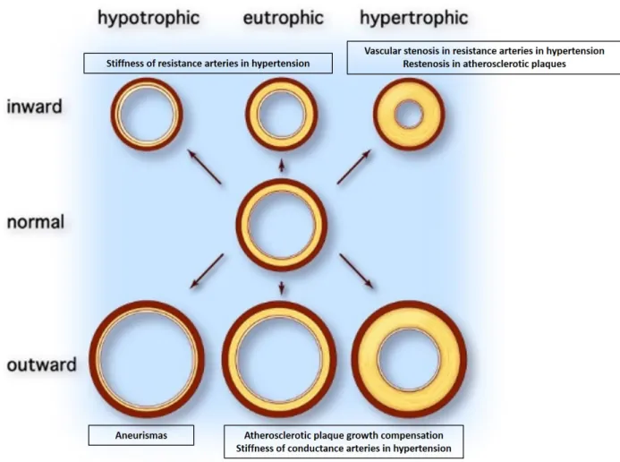
Figure 2: Types of vessel remodeling and associated pathologies ... 9
Figure 3: Inward eutrophic remodeling in restenosis ... 11
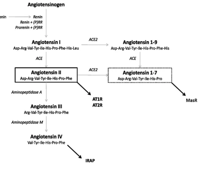
Figure 4: Components of the renin-angiotensin system (RAS) ... 15
Figure 5: Biological actions of Ang-II in the control of cardiovascular homeostasis ... 20
Figure 6: Schematic illustration of differential induction of G protein-mediated responses in VSMC ... 25
Figure 7: Topology and predicted domains of STIM-1 and Orai-1 ... 29
Figure 8: Model depicting Angiotensin-II-induced STIM-1/Orai-1-mediated Ca2+ entry and clearance by ATPases ... 30
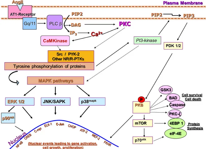
Figure 9: Ang-II-induced signalling ... 33
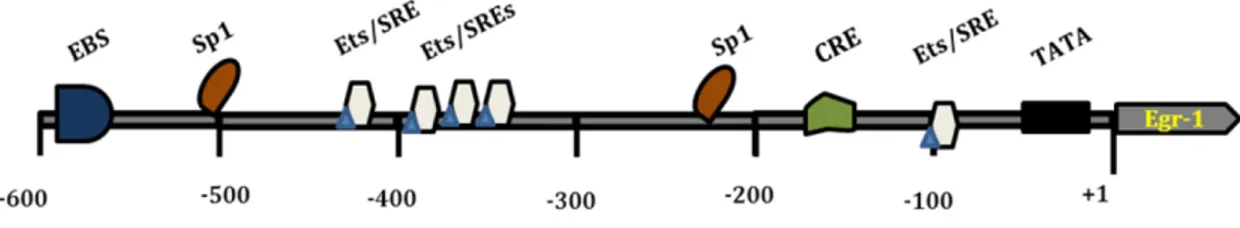
Figure 10: Structure of Egr-1 promoter present on target genes ... 38
Figure 11: Signaling pathways upstream of Egr-1 expression ... 41
Figure 12: Modulation of Ang-II-induced Egr-1 expression in A10 VSMC ... 132
Figure 13: Attenuation of Ang-II-induced Egr-1 expression by actinomycin D and cycloheximide ... 137
Figure 14: Inhibition of phospholipase C activity with U73122 attenuates Ang-II-induced Egr-1 expression ... 138 Figure 15: Attenuation of Ang-II-induced ERK and CREB phosphorylation by 2-APB . 139
List of Abbreviations
2-APB: 2-aminophenylborate AC: Adenylate cyclase
ACE: Angiotensin-converting enzyme
Ang: Angiotensin
Ang-II: Angiotensin-II
APA: Type A aminopeptidase APN: Type N aminopeptidase AT1R: Angiotensin-II type 1 receptor AT2R: Angiotensin-II type 2 receptor AT3R: Angiotensin-II type 3 receptor AT4R: Angiotensin-II type 4 receptor ATP: Adenylate triphosphate
ATPase: Adenylate triphosphatase
ATRAP: Angiotensin-II type 1 receptor-associated protein BNZ-cAMP: Benzoyl cyclic adenosine monophosphate
cAMP: Cyclic adenosine monophosphate Ca2+: Calcium
CAD: CRAC-activation domain CArD: Carotid artery disease
CaMKII: Calcium calmodulin-dependent protein kinase 2 CRAC: Ca2+ release-activated Ca2+ channel
CRE: Cyclic adenosine monophosphate response element
CREB: Cyclic adenosine monophosphate response element binding protein
CVD: Cardiovascular diseases DAG: Diacylglycerol
Db-cAMP: Dibutyryl cyclic adenosine monophosphate DBD: DNA binding domain
DBP: Diastolic blood pressure
EBS: Early growth response protein-1 binding site Elk-1: Ets domain-containing protein
Egr-1: Early growth response protein-1 eNOS: Endothelial nitric oxide synthase Epac: Exchange channel activated by cAMP ET-1: Endothelin-1
Ets: E-twenty-six
ER: Endoplasmic reticulum
ERK: Extracellular signal-regulated protein kinase FSK: Forskolin
FOXO/FKHR: Forkhead transcription factor GDP: Guanosine diphosphate GPCR: G-protein coupled receptor GRK: G-protein receptor kinase GSK: Glycogen synthase kinase GTP: Guanosine triphosphate IP3: Inositol-3-phosphate
IP3R: Inositol-3-phosphate receptor
JNK: c-Jun NH2-terminal kinase LPA: Lysophosphatidic acid LTCC: L-type calcium channel
MAPK: Mitogen-activated protein kinase
MEKK: Mitogen extracellular signal-regulated kinase kinase MSK: Mitogen and stress-activated kinase
mTOR: Mammalian target of rapamycin
NAB: Nerve growth factor-induced-A-Binding proteins NR-PTK: Non-receptor protein tyrosine kinase
PDK: Phosphoinositide- dependent kinases PI3-K: Phosphatidylinositol-3-kinase
PIP2: Phosphatidylinositol-4, 5-biphosphate
PIP3: Phosphatidylinositol-1, 4, 5-triphosphate
PKA: Protein kinase A
PKAcat: Protein kinase A catalytic subunit PKAR: Protein kinase A regulatory subunit PKAI: Protein kinase A type 1
PKAII: Protein kinase A type 2 PKB: Protein kinase B PKC: Protein kinase C PLA: Phospholipase A PLC: Phospholipase C PLD: Phospholipase D
PMCA: Plasma membrane calcium adenylate triphosphatase RAS: Renin-angiotensin-aldosterone system
R-PTK: Receptor protein tyrosine kinase
Sap: Serum response factor-associated accessory protein SAPK: Stress- activated protein kinase
SBP: Systolic blood pressure
SHR: Spontaneously hypertensive rats
SERCA: Sarco/endoplasmic reticulum calcium adenylate triphosphatase
SOAR: STIM-1/Orai-1 activating region
SOCC: Store-operated calcium channel SOCE: Store-operated calcium entry SRE: Serum-response element SRF: Serum-response factor STIM: Stromal interaction molecule SMC: Smooth muscle cell
TRPC: Transient receptor-activated channel VOCE: Voltage-operated calcium entry VSMC: Vascular smooth muscle cell
This thesis is dedicated to my exceptional parents, Dr Augustin Simo and Marguerite Yougo
For your limitless love, support, and trust,
Acknowledgements
First and foremost, my deep gratitude goes to my supervisor, Dr. Ashok Srivastava. I have truly been privileged to be welcomed and trained in your laboratory. I was young, and had no idea about the meaning of GPCR when I entered your office as an MSc student seven years ago. You opened your door to the stranger that I was. I felt at home in your lab. I was sure to have your full support for whatever I wanted to achieve. I felt safe. Your trust without limit has developed my independent thinking skills, I have learned a lot from you. Your friendship, your extreme patience and your humility, your support and your willingness to see your students succeed, have made this journey an extraordinary experience in my life. No word could ever tell how grateful I am, thank you for everything Dr. Ashok.
Merci aux Facultés de Médecine et des Études Supérieures et Postdoctorales de l’Université de Montréal, de même qu’aux encadreurs présents et passés du Département de Nutrition, pour le soutien académique et financier pendant mes études.
Je voudrais vous remercier membres du jury d’avoir accepté d’évaluer cette thèse, j’ai l’assurance que vos commentaires m’enseigneront davantage.
Mille mercis à Viktoria Youreva dont la contribution à mon rayonnement personnel et scientifique a été démesurée. Tu as été la partenaire de laboratoire parfaite et je remercie la vie pour cette amitié qui est née et sans laquelle je n’aurais pas surmonté les nombreux moments d’incertitude et de faiblesse. Ce travail n’est pas le mien, mais le nôtre. Merci Vicky, je te souhaite durant ta carrière de rencontrer des partenaires de travail et amis comme celle que tu es pour moi.
Merci à tous les anciens membres du laboratoire, Vincent Rondeau, Paulina Pietruczuk, Georgia Kapakos, Ahmed Mir, pour l’ambiance de travail agréable au laboratoire. A toi Dr Georges Vardatsikos, je tiens à dire ma reconnaissance pour tout ce que tu as fait pour faciliter mon intégration, merci pour ce que tu m’as légué de ton expérience personnelle et pour tous tes conseils d’ainé.
Merci à tous les membres des équipes du Centre de Recherche du Diabète de Montréal et du CRCHUM pour l’esprit de famille qui y règne et pour les discussions constructives. Merci à Ju Jing Tan, Henry Nchienzia, Yves Mugabo, Henry Leung, Valérie Bergeron et Hasna Maachi pour tous les bons moments de partage.
Merci au Dr Paul Désiré Dzeufiet ainsi qu’aux professeurs Kamtchouing Pierre et Wilfred Mbacham qui m’ont joyeusement introduit dans la recherche biomédicale.
Je voudrais profiter de cette occasion pour remercier tous mes amis à Montréal pour leur soutien inconditionnel. Un merci spécial à Emmanuelle Hoga et sa famille qui me semble mienne, en toi j’ai la présence d’une sœur pour qui je ne cesserai jamais d’exprimer de la gratitude.
À la Sims Crew, Lionel, Danielle et Joël, Patrick, Bruno, et nos parents Augustin Simo et Marguerite Yougo. Voyez en cette étape un des fruits de votre accompagnement pendant ces longues années. Merci pour vos milles attentions, pour les fous rires, pour les moments chaleureux, pour la confiance mutuelle, pour l’ambiance familiale que la distance n’a pas pu entamer. Tous les mots sont faibles …
À la dynamo force. Mes parents Pierre-Marie Nounamo et Marie-Noël Kapahwa, mes frere et sœurs Kanmeugne, Kuissu, Sikali et Dou. Merci pour vos encouragements constants et votre soutien moral. Merci pour votre désir ardent de me voir terminer avec succès mes études.
À ma famille toute entière, je voudrais dire à chacun un gros merci pour les paroles d’encouragement constantes.
Enfin, le soutien remarquable de mon mari m’a permis de compléter ce travail avec beaucoup de sérénité. À toi Kamdem, mon bijou, tu as vécu mes journées de laboratoire comme si tu y étais, ce travail est le tien également. Ta patience et ta compréhension pendant toute cette période, la valeur que tu accordes à ma passion pour la recherche, ton accompagnement moral 24h/7, la confiance, l’estime et l’amour que tu me portes ont fait de moi une épouse étudiante comblée, je ne remercierais jamais assez la vie pour nos deux vies unies.
1.1 V
ASCULAR DISEASES AND HYPERTENSIONThe past decades have witnessed global transition of causes of death from historical nutrient deficiencies and acute infectious diseases to the modern day nutrient excesses associated with chronic diseases (1). One of the consequences of this transition is an increase in the prevalence of metabolic conditions including diabetes, hypertension, and related cardiovascular diseases (CVDs) (2). In diabetic patients, heightened levels of circulatory fatty acids and hyperglycemia are associated with an aberrant vascular function (3). As a result, the elevated mortality and morbidity observed in these patients are in large part attributed to cardiovascular complications (4, 5). However, the main underlying cause of congestive heart failure, myocardial infarction and other vascular diseases is hypertension (6, 7). For this reason, hypertension is now considered as the number one risk factor for premature death (8). In 2012, 80 million adults over 20 years old were diagnosed with hypertension in the United States (9) whereas in Canada, one in five adults has hypertension (10). Globally, it is estimated that between 1980 and 2008, the number of adults with hypertension worldwide increased from 605 to 978 million (11); this number is predicted to rise to 1.56 billion by 2025 (12, 13).
Hypertension is a multifactorial disease generally classified into two types: primary (essential) hypertension and secondary hypertension. With a proportion of nearly 95% among the population diagnosed with hypertension, essential hypertension, characterized by the absence of an identifiable cause, is the most prevalent type (14, 15). Some determinants, linked with either the genetic background or the environment, have been identified as possible risk factors for the development of this type of hypertension (16). Those related to familial history, ethnicity (17), gender and aging are classified among the non-modifiable risk factors. Several types of hypertension due to genetic causes are regrouped in Mendelian hypertension with identified gene polymorphisms that are positively associated with the increase in blood pressure (18, 19). With regard to the environmental factors, many lines of evidence from epidemiological studies have reported the positive association between the growing incidence of essential hypertension and
behaviors such as excessive food and alcohol consumption, a high salt diet, physical inactivity and cigarette smoking (14). Prolonged stress and a low potassium diet are also associated with a high risk of developing high blood pressure. Thus, choices related to lifestyle are increasingly being considered as major modifiable variables for the prevention of hypertension and related end-organ damage (20, 21).
In contrast to essential hypertension, secondary hypertension is diagnosed by the presence of an underlying condition that contributes to the increase in blood pressure (22). 10% of the population diagnosed with hypertension belong to this category (22). For example, hypertension associated with sleep apnea, one type of secondary hypertension, where respiratory disorders facilitate an aberrant stimulation of the central production of vasoconstrictive hormones (23). Gestional hypertension is another type where the first onset of high blood pressure occurs during pregnancy possibly due to remodeling of the vascular system observed in that condition (24). Secondary hypertension also comes from metabolic conditions like diabetes and obesity, as well as from kidney diseases including glomerular dysfunction, renovascular stenosis, and polycystic kidney disease. Aberrant hormonal conditions such as the Cushing syndrome, hyperaldosteronism, pheochromocytoma or dysfunctional thyroid, have also been described as underlying causes of secondary hypertension (25). Hormonal disorders are mostly due to the presence of a tumor that may either enhance the secretion of prohypertensive substances or facilitate aberrant growth in cardiovascular relevant organs (26). Secondary hypertension can also occur as a side effect of medications like corticosteroids, contraceptive pills and several antidepressive drugs and pain killers (27).
By definition, hypertension is the condition of persistent non physiologic elevated blood pressure (28). Blood pressure is generally assessed by two values. The systolic blood pressure (SBP) reflects the force exerted by the blood on the arterial wall when the heart beats whereas the diastolic blood pressure (DBP) is the measure of the pressure when the heart relaxes and refills at the end of a cardiac cycle (28). These values are expressed in millimeters of mercury (mmHg) and are considered normal when they are respectively lower than 120 mmHg and 80 mmHg at the resting state (28). Systemic arterial
hypertension is currently defined as a resting SBP at 140 mmHg or greater and a DBP at 90 mmHg or greater (29). Noteworthy, beginning at 115/75 mmHg, the risk for developing CVD doubles for each increment of 20/10 mmHg (30) suggesting that vascular homeostasis is highly influenced by blood pressure dynamics even at prehypertensive stages.
1.2 V
ASCULAR DAMAGEVascular damage is a hallmark feature in the pathophysiology of hypertension (31) and often precedes the increase in blood pressure as observed in some types of genetic hypertension (32). This is in part due to heightened activity of vasoconstrictive and mitogenic hormones like angiotensin-II (Ang-II) or endothelin-1 (ET-1) present at elevated systemic and local concentrations. Alone or in concert with other stimuli, an exaggerated activity of these vasoactive peptides can induce structural and functional changes within the vessel wall (33, 34). The consequences of these changes termed as vessel remodeling mainly manifest as lumen narrowing and not only occur under conditions of chronic hypertension, but may also happens in response to temporary elevations of blood pressure. Since it is tightly related to the diameter of small vessels such as small arteries and arterioles neighbouring the capillarie beds, vascular resistance is highly increased by vessel remodeling. An increased peripheral vascular resistance is therefore the hallmark of vascular damage and the major feature in the pathogenesis of hypertension (7, 35, 36). Advances in the description of the tissular components of the vasculature as well as understanding how their functional properties are modulated by physical and chemical clues have contributed in linking blood pressure variations and other determinants of cardiovascular risk to vascular damage. Below is a brief description of the structural features of the vessel wall.
1.2.1 Structure of the vessel wall and cellular basis of vascular
damage
Similarly to other organs in the cardiovascular system, three main layers of tissue make up the wall of a vessel: an outer protective layer made of stromal tissue, a middle muscular layer that controls the tonus, and an innermost layer that consists of a single cell alignment directly in contact with the blood.
The outer layer (Figure 1) is the adventitia or tunica adventitia. It contains a mix of fibroblasts and smooth muscle cells (SMC) combined with an extracellular matrix rich in collagen (37). Small blood vessels that ensure oxygen-rich blood supply to the vessel wall can also be found in the adventitia of large vessels such as aorta and vena cava (38). Because of this structure, the adventitia plays a critical role in the maintenance of vessel integrity. In addition to the presence of connective tissue and differentiated SMC and fibroblasts, adventitia is also enriched in progenitor cells. As these cells can differentiate and give rise to cellular types that populate the other layers of the wall, their contribution in vascular damage is increasingly being considered (39-41) .
The medial layer (Figure 1) is the media or tunica media and essentially consists of a big population of vascular smooth muscle cells (VSMC) surrounded by elastin fibers organized into sheets that are intercalated by collagen fibers and proteoglycan. Contraction or relaxation of medial VSMC underlie the myogenic response to haemodynamic forces, this response is essential for the maintenance of a constant blood flow. In the capillaries, this role is played by the pericytes which replace VSMC in the wall of these small-diameter vessels and exhibit similar properties as well (42). In addition, based on specific cues, VSMC functional features can change from a contractile profile to a synthetic profile that allows them, among other actions, to synthesize and secrete the components of the extracellular matrix. This property is critical for vascular adaptation to external cues suggesting that VSMC functional integrity is a key determinant of vascular homeostasis (43). Similar to the progenitor cells found in the adventitia, multipotent stem cells are also
present in the medial layer and are able to differentiate into chondrogenic and SMC upon vascular injury (44).
The innermost layer (Figure 1), the tunica intima or intima, is bordered at the luminal side by endothelial cells directly in contact with the blood whereas at the peripheral side, a matrix of connective tissue combined with a lining of elastic fibres demarcates the intima from the media (37). Although the intima does not mechanically participate in the control of vessel conductance, endothelial cells are characterized by their secretory properties that enable them to recruit VSMC, immune and inflammatory cells during processes underlying vascular injury and neointima formation.
Figure 1: Cross sectional representation of the wall of an artery
(Modified from original figure in (45)).
Aberrant modifications in the physiology of vascular cells underlie persistent vessel remodeling which, along with prolonged vasoconstriction, represents the major contributor to the sustained vascular resistance associated with chronic hypertension (45, 46). Underlying processes are multicellular and converge towards changes in four main physiological responses within the vessel wall: cell growth in size or number, cell cycle
regulation (death or survival), cell motililty (migration, adhesion), and extracellular matrix turnover (secretion and degradation) (47). Endothelial dysfunction has been suggested to be the initial step that prompts susbsequent events in the progression of vascular dysfunction (48). In fact, the adluminal position of endothelial cells allows a direct contact with the bloodstream. This supports the idea that, hypertension and the other determinants of CVD such as hyperglycemia and dyslipidemia, by directly affecting endothelial cell physiology, are able to cause vascular damage (49). Deleterious consequences of chronic metabolic disturbances also involve aberrant expansion of inflammatory cells via hyperplasic and migratory responses. These inflammatory events participate in vascular lesion formation, an ultimate event in the atherogenic process (37). Because of the importance of the role played by inflammatory cells and determinants of innate immunity in vascular disease, it is also being considered as an immune disorder. This is supported by studies where animals bearing a deficiency in T or B lymphocytes (50), monocytes or macrophage (51), failed to develop vascular injury in disease promoting conditions. Finally, it is well documented that the pathophysiological responses exhibited by vascular smooth muscle cells (VSMC) underlie the structural changes observed in the vessel wall and further connect inflammatory responses and endothelial dysfunction to the development of vascular damage (52). Thus, together, individual properties beared by cells within the vasculature contribute to the pathogenesis of vascular damage. Accordingly, many vascular cell types in culture have been considered as in vitro models for the study of vascular disease. In the context of this thesis however, because of the central contribution of VSMC to vessel stiffness and reactivity, further description of the molecular basis of vessel remodeling will be focused on the signal transduction that control their physiology.
1.2.2 Pathophysiology of vessel remodeling
1.2.2.1 Determinants of vessel remodeling
The vasculature is sensitive to shear stress, the mechanical friction exerted by blood flow which results into an adaptative distension of the vessel (53). Additionally, it is
sensitive to blood internal pulsatility driven by blood pressure and which, despite being the key feature that ensures the continuity of the flow, also represents a stretch-promoting stimuli on the artery (53). Changes in blood volume or pressure are observed in some physiological states like in pregnancy, in response to transient vascular injury, or during transient metabolic disturbances accompanied with temporary changes in cardiac output or vascular resistance. Under such conditions, vascular homeostasis is maintained via either vasoconstrictive or vasodilatory responses resulting from stimulation by circulating and locally produced vasoactive peptides. This myogenic response is essential to accommodate transient flow-related disturbances while maintaining a stable vascular conductance (54). It requires a healthy functional cooperation between the cellular entities within the three layers of the vessel wall and the extracellular matrix. However, under circumstances of prolonged systemic hypertension or chronic metabolic disturbances as observed in obesity or type II diabetes, prolonged stimulation of vascular cells not only induces changes in the vasomotor tone, but also growth-promoting cascades. This is due to the mitogenic properties beared by vasoactive peptides that are able to trigger vascular hypertrophy, hyperplasia, as well as extracellular matrix formation. These events leading to structural modifications inside the wall define the process of vessel remodeling that is mostly reflected as aberrant thickening of the vessel wall and narrowing of the lumen. Increase in vascular resistance follows and mainly manifests as a decreased vessel capacity to appropriately adapt its size in response to environmental cues (46).
1.2.2.2 Types of vessel remodeling
Vessel remodeling is qualified as inward or outward based on whether the lumen diameter of the remodeled artery is smaller or bigger compared to the initial state (Figure 2). Due to prolonged wall tension and vasoconstriction, inward remodeling is very common in peripheral circulation during hypertension and contributes to the deleterious increase in peripheral vascular resistance (55). Outward remodeling usually accompanies the progression of atherosclerotic disease defined as the progressive accumulation of lipid and inflammatory cells toward plaque formation within the vessel lumen. In this situation,
the reduction in the lumen space due to atherogenic lesion is balanced by the outward remodeling resulting in a lumen dilation that serves as a counter mechanism to maintain a suitable blood flow (45). In addition to the size, the amount of material inside the vessel wall of the newly remodeled vessel with regard to the original state is also a parameter used to make a distinction between vessel remodeling events (Figure 2). Noteworthy, this criterion is closely linked to changes in mechanisms governing growth and survival of medial VSMC as well as extracellular matrix turn over. In this view, hypertension-induced inward remodeling can be eutrophic with no changes in the vessel wall mass. It can also be hypertrophic with an increase in the amount of cellular and non-cellular material in the vessel wall leading to increased stiffness. Neointima formation caused by aberrant hyperplasia and migration of VSMC toward the intima is the underlying mechanism of hypertrophic inward remodeling (56). It is moslty observed in hypertensive vascular stenosis or in restenosis following vascular interventions (57). In contrast, a decreased wall thickness due to matrix proteolysis and loss of medial cellular content defines outward hypotrophic remodeling. This process is at the basis of local aneurysm formation. Because of elevated mechanical forces exerted by high blood pressure, hypertension can cause aneurysm formation and rupture leading to deleterious consequences depending on the location of the organ (58).
Figure 2: Types of vessel remodeling and associated pathologies
How the structural changes affect the wall thickness or the lumen diameter depends on the function of the affected vessel and the underlying pathology. Large conductance arteries like aorta are prone to outward eutrophic or hypertrophic remodeling under circumstances of constantly elevated blood flow whereas small resistance arteries at the periphery are prone to inward remodeling. Atherosclerosis is accompanied by outward eutrophic remodeling later on followed by hypertrophic remodeling leading to vessel stenosis ( Adapted from (55) and (45)).
1.2.2.3 Clinical manifestations of vessel remodeling
Vessel remodeling is a hallmark of two major damages to the vascular system, arteriosclerosis and atherosclerosis. Arteriosclerosis affects the vasculature in a generalized fashion and is mainly associated with hypertension and/or other related determinants of CVD such as aging, obesity, diabetes, and inflammatory disorders. The main characteristic of atherosclerosis is that vessel remodeling accompanies the local formation of a lipid plaque leading to progressive vessel narrowing and eventual thrombus-mediated occlusion (37). Several hypertensive vascular diseases are defined based on the function of the damaged artery.
In coronary circulation: vessel remodeling within large coronary arteries alters the blood flow to the heart and clinically manifests as coronary heart disease. Angina and heart attack may occur under this condition where vessel narrowing or eventual occlusion leads to cardiac ischemia (59, 60). In some other cases of coronary disease, structural impairments in the wall of coronary ramifications can result in aberrant coronary vasomotor tone underlying ischemic symptoms (61). This other condition is known as microvascular coronary dysfunction.
In cerebral circulation: arteriosclerosis within the cerebral circulation mainly targets carotid arteries responsible for blood delivery to the brain through the neck. In this case, aberrant vessel remodeling manifests as cerebrovascular disease or carotid artery disease (CrAD)(62). Concomitant with atherosclerotic plaque formation, CrAD can cause disruption of blood flow to the brain causing stroke.
In peripheral circulation: Peripheral vascular disease (PVD) is the condition of remodeling within the wall of arteries that supply blood to legs, arms, pelvis, kidneys, and lungs. Pulmonary artery disease (PAD) and chronic kidney disease are types of (PVD) that are caused by similar risk factors and are associated with hypertension and chronic metabolic disorders.
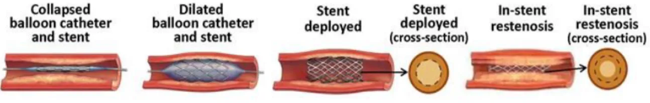
Another major clinical manifestation of aberrant vessel remodeling is restenosis following intravascular angioplasty. This is an intervention that consists of artificial distension of an arteriosclerotic artery using a stent inserted within the narrowed region. This approach has shown positive outcomes in the treatment of coronary heart disease (57). Following this surgery however, vessel re-narrowing usually occurs as a result of friction-induced inward wall restructuring in the surroundings of the stent. In such circumstances, exaggerated growth-promoting processes in VSMC underlie the formation of a neointima that elevates the risk of in-stent thrombotic complications (57) (Figure 3).
Figure 3: Inward eutrophic remodeling in restenosis
Schematic representation of aberrant VSMC hyperplasia during in-stent neointima formation. (Image from (57)).
1.2.2.4 Features of vascular smooth muscle cell physiology
Based on their capacity to contract and relax, the main function of VSMC within the vessel wall is to enable accurate vasomotor responses to haemodynamic stimuli. Maintenance of a contractile profile is essential for VSMC to fit this purpose. Determinants of VSMC profile have been the centre of intensive investigations since it is well demonstrated that depending on the nature of the vessel or even in the same vessel, VSMC are present in a diversity of profiles described as specific phenotypes (reviewed in (63)). This multiplicity can be explained by two factors: the diversity of embryonic sources of VSMC precursors (64, 65) and their capacity to adapt their physiological properties in response to specific conditions (56). This capacity designated as VSMC phenotypic
plasticity underlies the occurrence of VSMC in a spectrum of phenotypes that range from a normal quiescent contractile profile to a disease-prone synthetic profile. Proliferative, migratory, secretory or osteogenic phenotypes are intermediary profiles whose occurrence depends on the physiopathological condition. Phenotypic plasticity of VSMC represents a vital attribute for the vascular system as it enables the adaptative remodeling of the vessels in conditions of temporary modifications of vascular needs as observed during pregnancy where remarkable outward remodeling take place in the uteroplacental circulation (66). Also, adaptative remodeling occurs during vascular repair following injury, as well as in response to increased needs due to exercise training (reviewed in (67)).
Due to this phenotypic plasticity however, arterial wall exposed to hypertension and associated endo/paracrine stimulation is enriched with synthetic VSMC which, in contrast to contractile VSMC, exhibit exaggerated proliferative rate as well as increased migratory and secretory capacities (56). This is well demonstrated by early studies that showed that VSMC isolated from aorta of adult spontaneously hypertensive rat (SHR), which is a well-established rat model for essential hypertension (68), proliferate significantly more rapidly than those from normotensive Wistar Kyoto (WKY) rats (69). In several experimental models of vascular disease, phenotypic switch of VSMC toward a hyperproliferative profile forms the basis of neointima formation (43). In addition to exacerbated growth and motility responses, alterations in VSMC survival cycles also represent hallmarks of synthetic phenotype and result from major perturbations in intracellular signal transduction cascades triggered in a large part by vasoactive peptides (52). Several vasoactive peptides including angiotensin II (Ang-II) are well known inducers of VSMC hypertrophy and proliferation. In hypertensive conditions, Ang-II is present at heightened plasmatic levels and its concentrations near the cells could be much more elevated due tissular production (70). This elevation correlates with an augmentation in the level of molecular markers of remodeling and proliferation (68). Based on data showing that in addition to modulating vessel tone, Ang-II also exhibits growth promoting properties (71-73), its contribution to the pathophysiology of vessel remodeling has been widely explored making this peptide a relevant tool in investigating the intracellular
mechanisms involved in aberrant VSMC physiology. Noteworthy, Ang-II is the leading endocrine factor of the renin angiotensin aldosterone system (RAS) and its action on the vessel tone is an ultimate response in the regulation of cardiovascular homeostasis jointly controlled by the RAS and the sympathetic nervous system. Before an in-depth description of the molecular cascades induced by Ang-II in the control of VSMC physiology, here follows an overview of the components of the RAS.
1.2.3 Angiotensin-II in the renin angiotensin aldosterone system
The RAS represents the main system involved in sodium and body fluid regulation and thus, the principal regulator of blood pressure.
Renin is an aspartyl protease originally synthesized in an inactive form called pre-renin. Conversions of pre-renin into renin and subsequent cleavage of pro-renin are the sequential steps resulting in the production of active pro-renin. It has been demonstrated that pro-renin can be converted into renin through either enzymatic or receptor-mediated reactions respectively involving catalytic cleavage of the prosegment or pro-renin receptor-mediated conformational change (74).
Renin is released in response to three main physiological signals. First, a decrease in blood pressure within the kidney afferent arteriole is sensed by mechanoreceptors of juxtaglomerular pericytes lining the wall. Signal transduction from these cells leads to a paracrine stimulation of adjacent granular cells responsible for renin synthesis, storage and release. Secondly, a drop in blood pressure within the afferent arteriole induces a lowering of the glomerular filtration rate resulting in a drop in sodium concentration in the kidney distal convoluted tube. This is sensed by a third group of juxtaglomerular cells condensed as the macula densa. Upon sensibilization, these cells are also able to stimulate the granular cells to release renin. Thirdly, endocrine stimulation by cathecholamines released from the sympathetic nervous system is proposed to be the signal that triggers juxtaglomerular recruitment and subsequent increase in the number of renin-secreting cells arising from cellular differentiation resulting in an increase in renin production.
Although recent studies have reported alternative biological actions of pro-renin and renin, the principal role of renin is to convert the α-glycoprotein angiotensinogen into a decapeptide called angiotensin-1 (Ang-I) (Figure 4). Enzymatic cleavages of Ang-1 by two types of dipeptidyl carboxypeptidases named angiotensin-1 converting enzymes (ACE) give rise to the effectors of the RAS among which II (also called (1-8)) and Ang-(1-7) are the most studied and exert opposite activities (75, 76) (Figure 4). Additionally, ACE-independent reactions involving the participation of endopeptidases such as chymase, thimet oligopeptidase, endopeptidase, represent alternative pathways for the transformation of Ang-1 into specific angiotensins (77). Because angiotensinogen is constantly synthesized and since ACE synthesis by endothelial cells is ubiquitous, production of renin has been for a long time considered as the rate-limiting step that determines the function of the RAS. However, the discovery of an intracellular angiotensinogen-derived product named Ang-(1-12) has challenged this consideration since this 12 amino-acid product can be converted into Ang-1 following a renin-independent reaction (78). The strict endogenous occurrence of Ang-(1-12) and its capacity to give rise to angiotensins have generated a lot of questions regarding the enzymatic processes underlying its production from angiotensinogen and its conversion into Ang-1 (75, 79).
Figure 4: Components of the renin-angiotensin system (RAS)
The precursor peptide, angiotensinogen, is cleaved by renin to form the decapeptide angiotensin I. The catalytic activity of renin increases when bound to the (pro)renin receptor [(P)RR], and furthermore, the otherwise inactive prorenin can become catalytically active when bound to the (P)RR. The dipeptidase angiotensin-converting enzyme (ACE) cleaves angiotensin I to form the octapeptide angiotensin II (ANG II), the central active component of this system. ANG II can be catabolized by angiotensin-converting enzyme 2 (ACE2) into angiotensin-(1–7) [ANG-(1–7)], another active peptide of this system which typically opposes the actions of ANG II. ANG II can also be cleaved into smaller fragments, such as angiotensin III and angiotensin IV by aminopeptidases A and M, respectively. Most effects of ANG II are mediated by the angiotensin type 1 receptor (AT1R); however, ANG II can also bind to the angiotensin type 2 receptor (AT2R), which generally exhibits opposing effects to those at the AT1R. ANG-(1–7) acts via the Mas receptor and angiotensin IV can bind to the insulin-regulated aminopeptidase receptors (IRAP). (Figure and legend from (80) ).
1.2.3.1 Structure of angiotensin-II and receptor signaling
Ang-II is an octapeptide with two free N-terminal and C-terminal groups that enable further cleavage to form other types of angiotensins named on the basis of the number of remaining amino acids (Figure 4). The most biologically relevant angiotensin that can be made from Ang-II is Ang-(1-7) which is obtained from the ACE type 2 (ACE2)-catalyzed removal of the carboxyterminal amino-acid. The fact that Ang-(1-7) shows opposite physiological effects suggests a self regulatory mechanism within the RAS. Additionnally, from Ang-(1-8), Ang-III (Ang-(2-8)) is formed by the removal of the N-terminal aspartate; the reaction is catalyzed by a type A aminopeptidase (APA). Ang-III exhibits a biological activity close to that of Ang-II in terms of vasoactivity and aldosterone stimulation. Ang-III can further be a substrate for the generation of Ang-IV (Ang-(3-8)) following another aminopeptidase-catalyzed reaction (Type N, APN). Ang-IV acts mostly in the central nervous system where it is implicated in the control of synaptic plasticity and long term potention at the basis of learning and memory formation.
The characteristic of tissues that respond to Ang-II is the presence, at their cell surface, of two types of receptors, Ang-II type 1 and type 2 receptors (AT1R and AT2R), both of which have been cloned and characterized. AT3R and AT4R subtypes have also been described, yet these subtypes have not been fully characterized and do not account for the main vasoactive effects of Ang-II. The AT1R is widely distributed throughout the cardiovascular system and is also abundantly found in the renal, endocrine and nervous systems in humans. In the vasculature, VSMC exhibit high levels of AT1R (81, 82), while a certain amount is also found in the endothelium (33) and in the adventitia (83). Most of the vascular effects of Ang-II are mediated by the AT1R (84, 85). In terms of vascular damage however, eventhough AT1R-mediated responses of Ang-II are well demonstrated by the vasculo-protective consequences of whole body loss of function of AT1R, a cellular specificity of AT1R-mediated response remains a subject of controversy. Whether Ang-II-induced vascular damage is mediated by the AT1R present in VSMC, endothelial cells or fibroblasts is still unclear (86-88).
AT2R is mainly expressed in fetal mesenchyme, uterine smooth muscle, brain, ovary, adrenal medulla and heart, and plays an important modulatory role during embryonic development. AT2R expression decreases rapidly, however, after birth (89). In adults, this receptor is expressed mainly in pancreas, heart, kidney, adrenal brain and vascular tissues. AT2R can also bind Ang-(1-7) and similar to MasR-mediated signaling, transduction through AT2R generally antagonizes several of the Ang-II-induced AT1R activated events.
1.2.3.2 Angiotensin-II-mediated biological responses in cardiovascular relevant tissues
As depicted in Figure 5, Ang-II regulates cardiovascular homeostasis mainly by acting on the vasculature, the heart, the kidneys and adrenals, as well as in the central nervous system.
1.2.3.2.1 In the vasculature
Data revealing the distinct expression of the precursors of the RAS including renin (90), angiotensinogen and ACE (91) in the vasculature, where both AT1R and AT2R are expressed, have provided evidence of a strong local activity of Ang-II (92). The role of Ang-II in the modulation of vessel tone and thereby in cardiovascular homeostasis seems to be very critical since the respective activation of these two receptors produces opposite consequences. Ang-II induces vasoconstriction through a direct AT1R-mediated contraction of VSMC. Eventhough AT2R are also stimulated by Ang-II and mediate vasodilatory actions, AT1R-mediated effect dominates resulting in vasoconstriction as the overall net response to Ang-II. However, vasorelaxation could happen as a net response to Ang-II following AT1R blockade as observed in studies where a chronic administration of losartan to SHR unmasked Ang-II-induced activation of AT2R (93). Ang-II can exert its vasoconctrictive actions via its ability to either induce the endothelial secretion of the pressor peptides endothelins (94, 95) or reduce the release of endothelial vasodilatory molecules, such as nitric oxide, in a ROS-dependent fashion (96, 97). Furthermore, data
demonstrating an inhibition of the sympathetic activity following the use of AT1R have supported the notion that an additional mode of action of Ang-II in the modulation of vascular tone is via the adrenergic function (98). Ang-II stimulates vasoconstriction by improving VSMC sensitivity to norepinephrine whose release from vascular nerve endings is also facilitated by Ang-II (99). AT1R has indeed been shown to mediate norepinephrine release in mesenteric arteries from rat (100), rabbit (101), and guinea pig (102), as well as in thoracic aorta (103). Further vascular effects of Ang-II in cellular growth and fibrotic responses are mostly relevant to vessel remodeling and reviewed in details through section 1.2.3.3 of this thesis. Overall, prolonged Ang-II-induced contraction in the resistance vessels promotes the increase in systemic blood pressure (Figure 5).
1.2.3.2.2 In the cardiac tissue
Due to AT1R internalization by cardiac cells and the presence of a local RAS in the heart, cardiac Ang-II levels are found to be almost five times higher than plasmatic levels (104). Early studies reported that in SHR, left ventricular hypertrophy is accompanied by elevated levels of AT1R protein expression in cardiac tissue suggesting an increase in Ang-II-induced signaling responses in the pathogenesis of heart failure (105). Indeed, aberrant growth (106) and apoptotic (107) features observed in cardiomyocytes exposed to Ang-II indicate its implication in cardiac fibrosis and aberrant cardiac tissue remodeling in a failing heart. Additionally, Ang-II modulates the cellular events linked with calcium handling and excitation-contraction coupling in the heart (108). Heightened levels of Ang-II cause alterations in the function of intracellular calcium pumps and impairs diastolic relaxation (109). Ang-II exerts an influence on the conduction of action potential. This is underlied by the ability of Ang-II to alter junctional communication between cardiac cells (110, 111). In accordance with this, aberrant AT1R activation has been shown to cause arrhythmias (112) while ACE inhibition was earlier reported to ameliorate the electrophysiological responses in a failing heart (113).
1.2.3.2.3 In the central nervous system
In response to Ang-II, the release of vasopressin from the supraoptic nucleus is among the indirect actions that contribute to Ang-II-induced systemic vasoconstriction (114). Despite its inability to cross the blood brain barrier, circulating Ang-II is able to induce changes in brain function by acting on the circumventricular organs to regulate drinking behavior and sympathetic activity (115). In addition, similar to the heart and vascular tissues, the brain exhibits a local RAS and increasing body of evidence has revealed local Ang-II-induced pleiotropic actions in the brain. Ang-II is implicated in the brain response to stress where it regulates the release of the corticotrophin-releasing factor for cortisol production (116, 117) and the central sympathetic outflow towards the production of norepinephrine (118). A decrease in norepinephrine production following local brain AT1R blockade was associated with an attenuation of blood pressure and cardiac remodeling in SHR (119). Ang-II is additionally involved in the baroreflex deregulation (120), in brain response to inflammation (121) as well as in the control of cerebrovascular flow (122).
1.2.3.2.4 In the kidney and adrenals
Upon stimulation of the kidney AT1R, a contribution of intrarenal vasoconstriction has been demonstrated to account for a considerable proportion of the systemic increase in blood pressure (123). Additionally, actions of Ang-II on the kidney are critical for the maintenance of body fluids and electrolyte balance. Ang-II-induced contraction of the afferent arterioles initializes a sequence of events that result in an increase in tubular sodium and water reabsorption as well as a decrease in urinary volume, both phenomenons leading to a rise in blood pressure (124). While AT2R-mediated signaling has been suggested to reduce the synthesis of renin (125), Ang-II-AT1R-mediated signaling potentiates the release of kidney renin as well as it stimulates cortisol and aldosterone release from the adrenals (Reviewed by (126)).
Figure 5: Biological actions of Ang-II in the control of cardiovascular homeostasis
1.2.3.3 Angiotensin-II in vessel remodeling
Current therapeutical approaches involving the routine administration of anti RAS reagents along with other classes of drugs have shown positive outcomes in the regression of vessel remodeling in hypertensive patients (36, 128). The role of Ang-II in inflammatory responses, cellular hyperplasia, hypertrophy and adhesion, as well as in collagen and matrix deposition, has been extensively reported (129) indicating that Ang-II is an important molecular determinant for the vessel wall rearrangements observed under chronic disturbances. Specific regulatory actions of Ang-II in this context have been documented in reports from studies that used strategies aiming at RAS inhibition, targeting in particular the Ang-I/Ang-II/AT1R-mediated axis, in various models of vascular diseases. Since vascular remodeling manifests differently depending on the underlying pathology, these regulatory actions are discussed below distinctly based on whether the prevailing condition is atherosclerosis, hypertension, or neointimal thickening.
Administration of RAS antagonists to patients suffering from CAD reduces aberrant cellular adhesion and vessel stiffness as well as ischemic symptoms providing evidence of the pro-atherogenic action of Ang-II in these patients (130, 131). In animal models of atherosclerosis and hypertension, pharmacological blockade or depletion of AT1R inhibits the processes leading to increased vessel thickness, lesion progression and plaque rupture. In apolipoprotein-E knockout (ApoE-/-) mice for example, loss of function
of AT1R attenuates the signaling pathways underlying lipid accumulation, macrophage infiltration or low-density lipoprotein oxidization (132, 133). In the same model, concomitant administration of Ang-II with valsartan reduced the risk of thrombosis by attenuating the levels of the extracellular matrix metalloproteinase inducer, a protein that promote plaque rupture (133). These suggest a prominent role played by Ang-II in facilitating atherosclerotic plaque formation rupture. Indeed, a recent report assimilated the beneficial atheroprotective effects of AT1R antagonism to those of exercise training (134).
In arsenic-induced hypertensive rats, pharmacological blockade of AT1R blunted mitogenic signaling and thereby restored vessel wall integrity as exhibited by a regression
of intimal hyperplasia (135). Ang-II/AT1R-mediated signaling responses participate in the events linked with the incidence of restenosis following angioplasty. Indeed, in human, non-human primates and rodents that have undergone intracoronary stenting, AT1R blockade was able to prevent neointima formation via mechanisms involving attenuation of inflammatory and oxidative signaling (136, 137). Under conditions of vascular injury, Ang-II via AT1R stimulates the mechanisms at the basis of vessel wall restructuring such as collagen and elastin deposition together with neointima formation (138). By stimulating aberrant growth in VSMC, signaling pathways induced by Ang-II have been the center of several investigations in experimental models of neointima formation.
1.2.4 Molecular basis of vessel remodeling: cascades related to
angiotensin-II-induced signaling in vascular smooth muscle
cells
1.2.4.1 Angiotensin-II receptor signaling
As previously mentioned, Ang-II receptors are members of a large family of the seven transmembrane domains G-protein coupled receptors (GPCR). Classical G proteins possess three subunits α, β, and γ that form together an inactive complex maintained by the presence of a guanosine diphosphate (GDP) group bound to the α subunit (139). Upon binding of Ang-II to its receptor, a ligand-induced conformational change occurs and enables the transformation of GDP into GTP resulting in the release of α subunit (Gα) from the βγ complex (Gβγ). The GTP-bound α subunit (GTP-Gα) initializes the intracellular signal transduction by phosphorylating downstream enzymes or other effectors. This signal transduction is arrested by upstream mechanisms involving the phosphorylation of Ang-II receptor by particular proteins called G-protein receptor kinases (GRK) and the subsequent recruitment of β-arrestins to process the endocytosis of the phosphorylated receptor (140). In addition, AT1R is subject to endogenous regulation by a specific 18kDa protein called AT1R-associated protein (ATRAP) that colocalizes with the receptor in VSMC and has been shown to attenuate vascular remodeling by negatively regulating Ang-II-induced
VSMC proliferation, senescence and gene expression (141-143). The mode of action of ATRAP is based on a spontaneous binding with the intracytoplasmic portion of AT1R leading to its internalization and a subsequent decrease in the signal transduction (142, 144).
Depending on the nature of the ligand, the effector and the second messenger required for the signal transduction upon formation of GTP-Gα, G proteins are divided into four groups: Gi, Gs, Gq/11, and G12/13 (139). AT1R is coupled to Gαq/11 activity which is involved in the regulation of vascular tone and has been widely demonstrated to play a role in VSMC differentiation and vessel remodeling (145, 146). Activation of Gαq/11 prompts phospholipase activation and involves intracellular calcium as a second messenger. Gs/Gi proteins modulate the activity of adenylate cyclase (AC) and thereby the production of another second messenger, the cyclic-adenosine monophosphate (cAMP). Increased levels of cAMP as observed upon activation of Gαs protein and subsequent simulation of adenylate cyclase activity have been associated with a contractile phenotype in VSMC (147). In contrast to AT1R, AT2R activation was reported to be coupled with Gαi activity which exerts an inhibitory action on adenylate cyclase activity leading to a negative regulation of the formation of cAMP (148) (Figure 6). Similarly, the effects of the vasoactive peptide apelin-13, known to induce aberrant migration of VSMC, were recently reported to be mediated by Gi protein activation (149). Evidence additionally demonstrates that in hypertension, heightened activity of Ang-II induces an enhanced expression of Gi (150) suggesting an involvement of AC-dependent cAMP fluctuations in hypertensive vessel remodelling.
1.2.4.2 Adenylate cyclase and cyclic AMP-dependent pathway
AC is an effector for Gi and Gs proteins that respectively exert inhibitory and stimulatory actions. Therefore an increase in Gi protein expression as observed under conditions of exaggerated activity of Ang-II results in the attenuation of AC activity in VSMC. The principal role of AC is to convert molecules of intracellular adenosine triphosphate (ATP) into cAMP whose signal is transduced by downstream effector proteins
including protein kinase A (PKA) and the exchange protein-activated by cAMP (EPAC)). PKA is a tetrameric protein made of catalytic (PKAcat) and regulatory (PKAR) subunits each consisting of two α and β subunits. Although the PKAcat is constant, PKAI and PKAII isozymes were identified based on the differences between their regulatory subunits. Inactive PKA is characterized by the binding of all four subunits together into a stable holomere (151). This binding is made possible by the PKAcat-induced phosphorylation of the PKAR on serine 96 (151). Upon binding of cAMP to the four cAMP binding sites found on PKAR, this phosphorylation is inhibited enabling the detachment of the PKAcat from the holoenzyme (152-154). Released PKAcat is active and phosphorylates downstream kinases. PKA-dependent signaling attenuates VSMC proliferative responses and induces VSMC relaxation and vasodilation (155, 156). Together with cAMP, PKA is also able to stimulate the activity of intracellular phosphodiesterases leading to signal termination (157). PKA becomes inactive following degradation of cAMP that increases PKAR affinity for PKAcat, enhances serine 96 phosphorylation and subsequent holoenzyme formation (151, 154). This represents a negative feedback loop for cAMP-mediated pathway. While early studies showed that Ang-II-activated PKA activity induces AT1R downregulation (158), reports have demonstrated that aberrant VSMC proliferation (159) and senescence (160) induced by Ang-II can be significantly reduced by the activation of cAMP-PKA signaling.
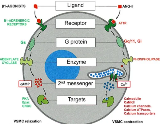
Figure 6: Schematic illustration of differential induction of G protein-mediated responses in VSMC
Angiotensin-II type 1 receptor (AT1R) and β-adrenergic receptor respectively activate Gαq11, Gαi and Gαs activities in VSMC. While Gi and Gs modulate the activity of adenylate cyclase and the production of the second messenger cyclic adenosine monophosphate (cAMP), Gq11 stimulates phospholipase activity toward the elevation of intracellular calcium levels. cAMP-dependent signalling involves the participation of protein kinase A (PKA), exchange protein activated by cAMP (Epac) and the cyclic nucleotide gated channels (CNGC). Calcium handling involves the participation of calmodulin, calcium/calmodulin- dependent protein kinase (CaMKII) calcium channels, transporters and pumps (ATPases). β-adrenergic receptor and AT1R exert opposite actions on VSMC physiology.
1.2.4.3 Phospholipase and calcium-dependent pathway
Several types of phospholipases have been identified and characterized depending on their site of action during phospholipids hydrolysis. Although activation of the types A, C and D (PLA, PLD, PLC) has been reported in VSMC stimulated with Ang-II (161), typical AT1R-GTP-Gα activation is coupled with the activation of PLC (PLC-β) that catalyzes the cleavage of membrane inositol phospholipid into diacylglycerol (DAG) and inositol-1,4,5-triphosphate (IP3) (161). Both IP3 and DAG-mediated activities lead to an
increase in intracellular calcium concentration ([Ca2+]i) by regulating two distinct pathways
(162). DAG is additionally known to bind to and activate isoforms of protein kinase C (PKC) which plays a prominent role in transducing Ang-II-induced proliferative responses in VSMC (163). IP3 activity mediates an increase in [Ca2+]i via its binding to specific
receptors (IP3R) distributed inside the membranes of intracellular organelles that act as
internal Ca2+ stores (164). These include the endoplasmic reticulum (ER) from where an
efflux of Ca2+ is produced upon IP3R activation. Early studies in VSMC reported a rapid
increase in [Ca2+]i in response to Ang-II(165). This increase in cytosolic [Ca2+] is handled
by a wide family of proteins including calmodulin and calcium/calmodulin-dependent protein kinases (CaMK) involved in contractile and growth responses in VSMC (166). The role of CaMKII in VSMC signaling and physiology has been addressed and evidences demonstrate that, under conditions of vascular injury, CaMKII expression and activity are upregulated (reviewed in (166)). CaMKII depletion has further been demonstrated to protect against experimentally induced hypertension, vessel wall thickening, and hypertrophic and proliferative responses of VSMC in in vivo and in vitro models of disease (167, 168). Additionally, an IP3R-mediated increase in cytosolic [Ca2+] triggers an influx
of external Ca2+ named store-operated calcium entry (SOCE) (169). The SOCE results
from the activation a group of store–operated calcium channels (SOCC), also known as Ca2+ release-activated Ca2+ channels (CRAC), defined as plasma membrane
voltage-independent calcium channels whose gating depends on the amount of Ca2+ inside the
intracellular stores (169-171). SOCE provides a sustained Ca2+ signal important to trigger