Plasticity following spinalization and step-training in the cat
par
Marie-Pascale Côté
Département de Physiologie Faculté de Médecine
Thèse présentée à la Faculté des études supérieures en vue de l’obtention du grade de Ph.D.
en Sciences neurologiques
Novembre 2006
Université
de Montréal
Direction des bibothèques
AVIS
L’auteur a autorisé l’Université de Montréal à reproduire et diffuser, en totalité ou en partie, par quelque moyen que ce soit et sur quelque support que ce soit, et exclusivement à des fins non lucratives d’enseignement et de recherche, des copies de ce mémoire ou de cette thèse.
L’auteur et les coauteurs le cas échéant conservent la propriété du droit d’auteur et des droits moraux qui protègent ce document. Ni la thèse ou le mémoire, ni des extraits substantiels de ce document, ne doivent être imprimés ou autrement reproduits sans l’autorisation de l’auteur.
Afin de se conformer à la Loi canadienne sur la protection des renseignements personnels, quelques formulaires secondaires, coordonnées ou signatures intégrées au texte ont pu être enlevés de ce document. Bien que cela ait pu affecter la pagination, il n’y a aucun contenu manquant.
NOTICE
The author cf this thesis or dissertation has granted a nonexciusive license aHowing Université de Montréal to reproduce and publish the document, in part or in whole, and in any format, solely for noncommerciat educationat and research purposes.
The author and co-authors if applicable retain copyright ownership and moral rights in this document. Neither the whole thesis or dissertation, nor substantial extracts from it, may be printed or otherwise reproduced without the author’s permission.
In complîance with the Canadian Privacy Act some supporting forms, contact information or signatures may have been removed from the document. While this may affect the document page count, it does flot represent any loss of content from the document.
Université de Montréal Faculté des études supérieures
Cette thèse intitulée
Plasticity following spinalization and step-trainïng in the cat
présentée par: Matie-Pascale Côté
A été évaluée par un jury composé des personnes suivantes: Dr Vincent Castellucci
Président-rapporteur Représentant du doyen de la FES
Dr Jean-Pierre Gossard
Directeur de recherche
Dr Trevor Drew
Membre du jury
Dc Susan JilI Harkema
RÉSUMÉ
L’entraînement locomoteur est fréquemment intégré au programme de réadaptation chez les blessés médullaires (SOI) afin de maximiser leurs capacités locomotrices résiduelles. Cette stratégie est directement inspirée de travaux effectués au laboratoire chez le chat spinal, un modèle pour lequel les voies réflexes et les réseaux locomoteurs sont bien décrits. En l’absence des voies supraspinales, la moelle épinière a la capacité de générer des patrons locomoteurs suite à une stimulation sensorielle répétée procurée par l’entraînement sur tapis roulant. Cependant, les mécanismes impliqués dans cette récupération sont peu connus.
Projet I. On assume fréquemment que les changements plastiques dans les réseaux locomoteurs spinaux sont responsables de cette récupération. Cependant, la stimulation sensorielle répétée liée à l’entraînement pourrait aussi modifier la transmission dans les voies réflexes qui contribuent aussi à l’activité musculaire durant la marche. Dans ce
projet, la transmission réflexe est évaluée pat la mesure de réponses intra
motoneuronales de muscles fléchisseurs et extenseurs qui innervent la cheville, le genou et la hanche évoquées par la stimulation d’un nerf musculaire ou cutané de la patte postérieure lors d’une expérience en aigu chez le chat décérébré. Les modifications possibles sont déterminées par une comparaison statistique des résultats provenant de deux groupes de chats dont la moelle a été complètement sectionnée à 113, dont un seul groupe est entraîné à la marche sur tapis roulant. Les résultats montrent que la
transmission dans les voies réflexes musculaires et cutanées est modifiée par
l’entraînement locomoteur. L’excitation monosynaptique dans les extenseurs est diminuée suite à l’entraînement et la modulation phasique normalement observée est récupérée. Aussi, l’inhibition de groupe lb est diminuée suite à l’entraînement et à l’injection de clonidine, un agoniste noradrénergique utile à la locomotion. De plus, il a été observé que
la plasticité dans les voies cutanées est spécifique : elle n’est présente que dans quelques
voies dans lesquelles la transmission est le plus souvent diminuée et les voies cutanées activées pat la plante du pied sont particulièrement modifiées. L’ensemble de ces données suggère que l’entraînement locomoteur diminue l’hyperexcitabilité réflexe observée chez les SCIs et qu’il facilite le recrutement des extenseurs importants pour le support de poids.
Projet II. Plusieurs études illustrent que la plasticité des circuits spinaux est affectée par divers mécanismes moléculaires qui dépendent de l’activité physique et qui influencent la
les maintenir. Plusieurs recherches récentes s’intéressent à des molécules impliquées dans la formation de la potentialisation à long-terme (LTP) afin de déterminer si les mécanismes responsables de l’apprentissage dans l’hippocampe sont similaires lors d’acquisition ou de la modulation de réflexes dans la moelle épinière. La protéine ERK joue un rôle reconnu lors de la plasticité synaptique et pour l’intégration des signaux de la surface cellulaire jusqu’aux facteurs de transcription. Elle apparaît donc comme un candidat idéal pour véhiculer les effets bénéfiques de l’entraînement et participer dans les événements synaptiques associés à la récupération de la marche suite à l’entraînement. Dans ce projet, des western blots ont été effectués pour mesurer l’expression de ERK et de sa forme activée, pERK, dans la moelle épinière de 3 groupes de chats intacts, spinaux, spinaux avec entraînement locomoteur. Les résultats montrent que l’activation de ERK est augmentée dans la majorité des segments lombaires chez les spinaux et qu’elle est spécifiquement diminuée au niveau L5 suite à l’entraînement locomoteur. Nos résultats suggèrent que la SOI peut augmenter l’activation de ERK et ceci, pendant plusieurs semaines et que l’activation de ERK est potentiellement nuisible à la récupération locomotrice si elle est présente dans certains segments spinaux.
Mots clés: CPG, CREB, entraînement sur tapis roulant, enregistrement intracellulaire,
ERK, lésion de la moelle épinière, locomotion, mise-en-charge (support de poids),
ABSTRACT
Locomotot training has gained in popuiarity and is more and more integrated in tehabilitative strategies to enhance stepping recovery in spinal cord injured (SOI) individuals. This strategy is ditectly inspired from several decades of work performed in the Iaboratory taking advantage cf a spinal cat model in which reflex and locomotor pathways are exhaustively described. Compietely isclated from supraspinai influences, the spinal cord has the capacity to recover stepping movements when given repetitïve and appropriate sensory feedback related to step-training on a treadmill. However, the underlying mechanisms for recuperating the appropriate motor patterns are stiil poorly understood and are the scope of this study.
Project I. It is generally assumed that plasticity in spinal locomotor circuits is responsible for the stepping recovery. However, the repetitive sensory stimulation related to step-training couid also modify transmission in reflex pathways, which are aise known to contribute significantly to the level cf muscle activity during stepping. In this project, transmission in reflex pathways was evaluated by measuring responses evoked by a stimulation cf a cutaneous or muscle nerve cf the hindpaw and recorded intracellularly in motoneurons from extensor and flexor muscles involved in ankle, knee and hip joint movements during an acute experiment in decerebrate cats. Possible modifications in reflex transmission were determined by the statistical comparison 0f responses between 2 groups cf spinal cats (complete transection at T13), but only one was assigned te a step-training regimen. Results showed that the synaptic transmission
in both group I muscle reflex pathways from extensors and cutaneous pathways were
modified following one month cf step-training. The monosynaptic excitation was decreased atter step-training and a normal pattern of modulation was recovered during locomotion. Moreover, group lb inhibition and polysynaptic group I excitation cf extensors were respectively decreased and increased after step-training and clonidine injection, a noradrenergic agonist useful for central pattern generation. t was further observed that plasticity in cutaneous pathways was highly specific: only certain pathways wete modulated (mostiy depressed). Transmission cf cutaneous input originating from the sole cf the foot was particularly modified. Overall, step-training is suggested te both decrease the hyperexcitability observed in reflex pathways after SCI and te facilitate the recruitment cf antigravity muscles te assist recovery and weight bearing.
Project II. There is now strong evidence that spinal circuits can be affected by activity
dependent biochemical processes that influence its ability to recover, perform and maintain an adequate Iocomotor pattern. Investigations have recentiy been oriented towatd molecules involved in LTP to determine if similar mechanisms are both implicated in hippocampal learning and spinal motor learning. Given the preponderant effect of ERK on synaptic piasticity and function and its role in integrating signais from the ceii surface to transcription factors, ERK appears to be a potentiai candidate for mediating the beneficial effects of step-training and may participate in the synaptic events associated with locomotor recovery aftet SOI. Ptotein expression was compared between 3 groups of cats (intact, SCI, SOI and step-trained) using western biot analysis of homogenates of spinal cord segments. The study focussed on assessing relative levels of ERK and pERK proteins. Resuits showed that ERK activation is up-regulated in a majority of lumbar segments following SCI and is specificaily down-reguiated in L5 by steptraining. These resuits suggest that ERK activation is invoived in long-term plasticity foilowing SOI and that it may be detrimental to iocomotor generation, at Ieast in specific spinal segments.
Keywords: cutaneous reflex pathways, CPG, CREB, ERK, intracellular recording, locomotion, muscle reflex pathways, trea dm111 training, spinal cord injury, spinal plasticity, weight-bearing, western blot.
vii TABLE 0F CONTENTS
RÉSUMÉ III
ABSTRACT V
TABLE 0F CONTENTS VIII
LIST 0F FIGURES IX
LIST 0F ABBREV1ATIONS X
INTRODUCTION 2
1. Spinal cord injury 2
1.1 General facts and probiematic 2
1.2 Recent advances in SCI research 2
2. The ABC of locomotion 4
2.1 The early description cf the locomotor cycle 4
2.2 Spinal locomotor networks in history 6
2.2.1 HaIf-center hypothesis 6
2.2.2 Central pattern generator 7
3. Control of locomotion 11
3.1 Supraspinal descending commands 12
3.2 Spinal interneurons 15
3.3 Sensoryfeedback 16
3.3.1 Muscle reflex pathways 17
3.3.1.1 Pathways from group la muscle afferents 18 3.2.1.2 Pathways from group lb muscle afferents 23 3.2.1.3 Pathways from other muscle afferents 25
3.3.2 Pathways from cutaneous afferents 27
3.3.3 Pathways from flexion reflex afferents (FRA) 31 3.3.4 Specific functional features cf sensory feedback 33
3.3.4.1 importance cf load signais 33
3.3.4.2 Regulation cf phase transition during stepping 35
3.3.4.3 Presynaptic inhibition 37
3.4 Motoneurons 38
3.4.1 Localization 38
3.4.2 Passive properties cf motoneurons 39
3.4.3 Dynamic properties cf motoneurons 40
4. Nervous system plasticity 42
4.1 Spinal piasticity 42
4.1.1 Reflex conditioning and denervation 43
4.1.2 Spinal cord injury and plasticity 43
4.1.3 Spinal plasticity and locomotion 45
4.1.3.1 Activity- and task-dependent 45
4.1.3.2 Neurotransmitters and neuromodulators 46
4.2 Neurotrophins 50
4.2.1 From the brain te the spinal cord 52
4.2.2.1 Ras/ERK MAPK pathway. 53
4.2.2.2 Pl3KIAkt pathway 55
4.2.2.3 cAMP response element binding 55
4.2.2.4 Selected molecules involved in synaptic transmission 56
4.2.3 Lesion-induced plasticity 56
4.2.4 Exercise-induced plasticity 58
4.2.5 Step-training induced plasticity after SOI 61
5. Model and hypothesis 63
5.1 The model: Step-training in chronic spinal cats 63 5.2 Project I: Plasticity cf spinal reflexes 63 5.3 Project Il: Modulation of intracellular signaling pathways associated with
activity-dependent plasticity 65
PuBLIcATIoN#J: SPINAL CAlS ON THE TREADMILL: CHANGES IN LOAD PATHWAYS 67
Introduction 67
Materials and Methods 68
Results 72
Discussion 79
Acknowledgments 82
PUBucATtON #2: STEP TRAINING-DEPENDENT PLASTICITY IN SPINAL CUTANEOUS
PATHWAYS 83
Introduction 83
Materials anç Methods 85
Results 88
Discussion 100
Acknowledgments 104
PuBLIcATIoN#3: LESION-INDUCED ERK ACTIVATION IS DECREASED FOLLOWING STEP
TRAINING IN CHRONIC SPINAL CATS 105
Introduction 106
Material and Methods 108
Results 111
Discussion 117
Acknowledgments 124
DISCUSSION 125
Step training-related plasticity does occur in both load and skin reflex pathways 126 Ptoteins involved in learning phenomena are ptesent in the cat spinal cord and
up-regulated after SOI 128
The specific character cf step ttaining-induced spinal plasticity 129 Potential mechanisms responsible for step training-induced plasticity in reflex
transmission 133
Functional considerations 134
Future directions 138
LIsT 0F FIGURES
Figure 1: Bookcover from Eadweard Muybridge 4
Figure 2: Neuronal organization of the mammalian locomotor system and
dynamic sensory integ ration during stepping 11
Figure 3: Proprioceptive pathways from extensors and flexors during
locomotion 18
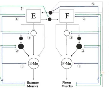
Figure 4: Cutaneous territories of the cat hindpaw 27
Figure 5: Plasticity of the spinal cord after SCI 44
Figure 6: Schematic diagram of afferents (supraspinal, segmental and
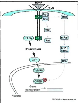
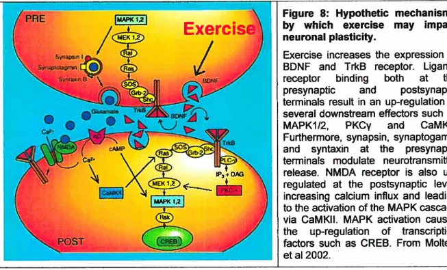
peripheral) to the spinal cord networks before and after SCI 45 Figure 7: Schematic diagram of BDNF-activated pathway via Trk receptor 51 Figure 8: Hypothetic mechanisms by which exercise may impact neuronal
LIsT 0F ABBREvIATI0Ns
5-HT: 5-hydroxytryptam me (serotonin) lp: Iliopsoas
AHP: Afterhyperpolarization I PSP: lnhibitory postsynaptic potential
AMPA: a-amino-3-hydroxy-5-methyl-4- LDP: Locomotor drive potential isoxazolepropionic acid
AP-5: 2-amino-5-phosphonovaleric acid LGS: Lateral gastrocnemius- soleus
APV: 2-amino-5-phosphonopentanoic LTP: Long-tetm potentiation
acid
BDN F: Brain-detived neutotrophic factor MAG: Myelin-associated glycoprotein
CaMKII: Calcium/calmodulin-dependent MAPK: Mitogen-activated protein kinase kinase Il
cAMP: Cyclic adenosine monophosphate MG: Medial gastrocnemius
Caspase: Cystein aspartate-specific protease MPL: Med lai plantar
CCS: Caudal cutaneous sural NA: Noradrenalin/noradrenergic
CDP: Cord dorsum potential NMDA: N-methyl-D-aspartate
CNQX: 6-cyano-7-nitroquinoxaiine-2-3- NT: Neurotrophins dione
CNS: Central nervous system OMgp: Oligodendrocyte myelin
glycoprotein
CPA: Canadian Paraplegic Association PI3K: Phosphatidylinositol-3-kinase
CPG: Central pattern generator PIC Persistent inward current
CREB: Cyclic AMP response element PKB: Protein kinase B binding
CSPG: Chondroitin sulfate proteoglycan PKC: Protein kinase C
DOPA: f3-3,4-dihydroxyphenylalan me PBSt: Posterior biceps-semitendinosus
DRG: Dorsal root ganglion PI: Plantaris
EDL: Extensor digitorum longus PLCy: Phospholipase Cy
ENG: Electroneurogram SCI: Spinal cord injury/injured
EPSP: Excitatory postsynaptic potential SmAB: Semimenbranosus-anterior biceps
ERK: Extracellular signal-regulated kinase SOL: Soleus
FDL: Flexor digitorum ongus SP: Superficial peroneal
FHL: Flexor hallucis longus Srt: Sartorius
FRA: Flexor reflex afferents St: Semitendinosus
GABA: Gamma-aminobutyric acid SynP: Synaptophysin
GAD: Glutamic acid decarboxylase TA: Tibialis anterior Grb2- Growth factor receptor-binding
SOS: protein 2 - the son of sevenless
GS: Gastrocnemii- Soleus
xi
Laura, Anne-Marie, Thomas-Xavier, Tristan,
Vos soutires, bisous et calins sans cesse me tappellent que l’essentiel est de s’éme,veiller d’un rien
Exp etience has shown, and a true phiosopfiy wiII aiways show, that a vast, perhaps the larger portion of the truth arises from the seemingly irrelevant.
- EdgarAllanPoe
The great tragedy of science - the slaying of a beautiful hypothesis by an ugly fact.
AcKN0wLEDGMENTs
Mes premiers remerciements vont au Dr Jean-Pierre Gossard, mon directeur de thèse. Son support et sa confiance ont été indispensables à la réalisation de ce projet; dès le départ il a cru en moi peut-être même plus que moi-même. Merci d’avoir accepté avec
enthousiasme mes projets les plus fous et de m’avoir laissé une indépendance si
précieuse. J’aimerais aussi remercier te Dr Tim Kennedy qui m’a acceuilli dans son laboratoire pendant près d’un an. Il a été d’une très grande générosité de mettre à ma disposition expertise et équipement.
Tous mes remerciements aux Dr Vincent Castellucci et Trevor Drew qui ont accepté d’évaluer et de critiquer mon travail tout au long de mes études supérieures en étant membre de mon comité pédagogique puis de mon comité de thèse. Leurs commentaires constructifs m’ont permis de garder le cap et la cadence!
Un merci tout particulier aux copains du Centre de Recherche en Sciences neurologiques
qui m’ont accompagné dans ce long périple et plus précisément à la vieille garde, Hugues, Ariane, Jean-François, Laurent et Annie. Vous avez été une très grande source d’inspiration et vos conseils et encouragements m’ont été précieux.
J’aimerais aussi remercier France Lebel et tous les voisins passés et présents du
laboratoire du Dr Rossignol sans qui ces nombreuses années auraient été bien solitaires tant scientifiquement que sur une note plus personnelle!
J’aimerais aussi souligner que grâce à leurs expertises techniques diverses, plusieurs personnes m’ont facilité la tâche dans ce parcours: Janine, Denis, France, Robert, Claude, Christian, René, Jeanne.
Amélie et Jessica, mes jumelles de thèse, que de discussions philosophiques sur le
pourquoi et le comment!? Et bien oui, nous y sommes enfin...
Puisque ceux qui nous accompagnent ont autant de mérite sinon plus que nous, mes plus sincères remerciements vont à ma famille.
À
mon amour, Julien, qui a fait preuve de très grande patience et m’a suivi de bon coeur dans cette éprouvante et cahoteuse aventure enpartageant mes succès, mes bonheurs et mes angoisses récurrentes.
À
mon petit homme,mentalement. Un merci tout spécial à mes parents pour leur accueil, leur disponibilité, leur support indéfectible et pour leurs petits plats qui ont été vivement appréciés et salvateurs.
Finalement, je me dois de mentionner que ce travail a été rendu possible grâce au support financier de la Fondation du Dr George Phénix, des Instituts de Recherche en Santé du Canada, de la Fondation pour la Recherche en Santé du Québec et de la Faculté des Études Supérieures de L’Université de Montréal.
1.1 General facts and problematic
Spinal cord injury (SCI) is a widespread condition which primarily affects young adult males between 15 and 34 years old. According to the Canadian Paraplegic Association
(CPA)1, 36000 Canadians live with SCI exciuding non-deficit or fatal injuries. Approximately 1050 new injuries occur every yeat (35 individuals/miflion) resulting in some level of permanent paralysis or neurological deficit. In Canada, car and motorcycle accidents are a leading cause of SCI, foltowed by falls, medical conditions, diving and sports. Approximately 80% of SCI occurs under the age of 30 and many of these individuals will live a normal lifetime generating important societal costs in terms of medical, surgical and rehabititative care. Hence, the financial care requirements, over this period, could vary from 1,25 million for a low thoracic paraplegic to 25 million Canadian dollars for a high cervical quadriplegic, such as Christopher Reeve, who required continuous ventilator support and 24/7 care2. Moreover, recent data collected by CPA suggests that there are a growing number of older adults being paralyzed as a resuit of disease and othet medical conditions. Given these facts and knowing that the population is ageing, that physical and psychological consequences of the paralysis have a devastating effect on the quality of lite of individuals and that cost will necessarily increase in the next years, research in the SCI field has gained support and popularity within the last ten years.
1.2 Recent advances in SCI research
Traumatic insults to the spinal cord induce both immediate mechanical damage and subsequent tissue degeneration. Hence, the outcome of SCI depends not only on the
initial tissue injury at the time of the trauma, but also on secondary injury processes that
may extend for hours, days, and even months. Incredible progress has been made in SCI tesearch over the last decade. Major improvements affecting the quality of life of SCI individuals including chronic pain and bladder function management were inconceivable only a few years ago. For example, paraplegic SCI individuals present a syndrome in which the posture of the legs as well as voluntary and locomotor movements are exten
1- canadian Paraplegic Association (http://www.canparaplegic.org) 2- International collaboration On Repaît Discoveries (http://wwwicord.org)
sively impaired. Many approaches have evolved to promote the recovery cf function. Noteworthy, combining multiple strategies to enhance functional improvements in an effort to teach a satisfactory daiiy life is thought to have a positive effect. Several reviews wete published recently and summarize the recent advances in SCI reseatch (Fouad et al 2001, Kwon et al 2002, David & Lacroix 2003, Dobkin 2004, Fouad & Pearson 2004, Hall & Springer 2004, Kiussmann & Martin-Villalba 2005). Years of fundamental and clinical research led to these conclusions and a detailed acknowledgment of achievements will be found in these reviews. The following section is solely aimed at drawing a succinct portrait.
From pre-clinical models to clinical application, therapeutic strategies are commonly divided in 4 subcategories: protection, regeneration, substitution, and management cf sublesional networks. These 4 categories are somehow intermingled; for example neuroprotection or regeneration can actually be achieved via a substitutive process.
Neuroprotection s the first-step strategy targeting secondary injury mechanisms and intending to limit neuronal loss and inflammation soon aftet the injury onset. The main rationale is to block secondary biochemical and cellular cascades initiated by tissue damage due to glutamate excitotoxicity, ischemia, oedema, Ca2 overload and oxidative stress. In animais, various pharmalocogical agents have been tested to prevent post traumatic secondary lesions and decrease lesion extent. Among them are antagonists of opioid receptors (naloxone) or gangiiosides, non-competitive antagonists of NMDA receptors (phencyclidine and ketamine) or massive doses cf steroids such as methyiprednisolone. Free radical scavengers have also been shown to preserve white and grey matter and to enhance motor performance. In human SOI, only methylprednisolone has been administered routinely. Triais are currently being held for other drugs but undesirable side effects often prevented therapeutic use.
The second type of intervention, regeneration, is aimed at re-establishing ascending and descending pathways. Promoting axonai regeneration and reconnection is currently a mainstream research field and has been the most dynamic in the iast ten years. The majority cf these interventions target eiements that prevent axonal regeneration and accumulate in the myelin (arretinin, CSPG, Nogo-A, MAG, OMgp) or around the guaI scar (CSPG, collagen-IV, tenascin, class-3-semaphorin, Eph3B) to neutralize them (see Kwon et al 2002, David & Lacroix 2003). Finally, a substitutive strategy can also be used te te express some factors that are absent or decreased in the subiesional part of the spinal cord. This can be achieved by various means: trophic factors and graif transplantation
such as stem ceils, Schwann ceils, embryonic raphe neurons, olfactory ensheathing celis, etc (Ribotta et al 2002, Bunge & Pearse 2003, Fouad & Pearson 2004). Recently, new alternatives to neuronal-celI grafts have started to draw attention. Among them figure non-neuronal cells ttansfected to express a gene coding for tyrosine hydroxylase in order to express serotonin (astrocytes, fibroblasts), non-neuronal stem celis (muscle or bone marrow), NT-2 human neuron (testicular tumoral cells) treated not to be tumoral (allows 30% cf differentiated serotoninergic cells).
Contrary te previous interventions aimed at trying to restore the before-SCI neural milieu, the 4th strategy is based on a different approach that is exclusively directed toward maximizing the residual function of the spinal cord. It takes advantage cf the intrinsic capabilities of the spinal cord through the activation ot the sublesional neural networks either with pharmacological intervention (reviewed in Rossignol et al 2001), transplantation
0f neurons or neural tissue (Ribotta et al 2000, Slawinska et al 2000) or with a specific
motor training regimen. This thesis is especially interested in the latter case and further details will be given in the following chapters.
2. The ABC of locomotion
Over the years, the study of rhythmic movements has covered a large range of behaviors existing ail over the animal kingdom. These stereotyped rhythmic pafterns are part of very primitive behaviors necessary to live in the wildlife such as breathing (respiration rhythm), eating (mastication and swallowing rhythm), escaping from predators or joining a companion for reproduction (locomotor rhythm as fiying, stepping, swimming, etc). A given behavior is produced by networks of interacting celis involving fine-tuning of molecuie/gene activation and interaction within the ceil, synapse and neural network. From invertebrate to human investigation, incredible progress has been made in understanding the fleurai control of such behaviors in the last 100 years. In this thesis, emphasis wiIi be given to stepping and its control in the cat. This section is a glance at eariy studies of locomotion and will deal with basic concepts and their evolution.
2.1 The eariy description of the locomotor cycle
Locomotion resuits from the sequentiai activation of numerous muscles. Their activation pafterns are nearly similar across individuals and to a lesser extent across vertebrates (Grillner 1981). Nowadays, the motor pattern of the stepping iimbs can be described using 3 different parameters: kinetics (force), kinematics (movements) and electromyographic activity (EMG). Pioneer experiments
investigating locomotion relied solely on anatomical studies of the legs. The improvements in photography and in the development of motion pictures in the late 19 century atlowed analysis of these images of animate motion for the first time. Scientists and artists such as Marey and Muybridge were early explorers of human and animal motion in images and image sequences. These pioneers first captured the sequential positions during gait and could then take precise measurements of the Ieg in motion. A few years later, Marey’s microphotographs were used by
I l(i :ii M( XI 1()\ ‘I1I U
( /—k4
\NF\1 \LS EN v1CYFIC)\ Figure 1: Bookcover from Eadweard MuybrîdgeThe Human Figure in Motion (1955) and Animais in Motion (1957) report a coilection of pictures taken by E. Muybridge at the beginning of the 19thcentury.
Philippson (1905) to precisely describe the step cycle, which he divided into 4 distinct portions. The first portion is a flexion of ail joints (flexion phase), followed by an extension cf the ankle and knee while the hip continues flexion (early extension phase). The thitd portion takes place at the very onset of the foot contact with the ground: knee and ankle joints are passiveiy flexed (weight acceptation). The step-cycie ends with an extension of ail joints to propei the body forward (propulsion phase). The extension phase is thus divided in 3 subcomponents: eariy extension (E1), weight acceptation (E2) and propulsion (E3). This nomenclature is still commonly used to describe the step-cycle and a detailed analysis of the motor pattern of the hindiimbs of the cat has been performed by a number cf investigators (reviewed in Rossignol 1996). When a detaiied description is not required, the iccomotor cycle is commonly defined as the period between two successive foot contacts and ccnsists of two principal parts: stance (support) and swing phase (transfer). The swing phase starts when the limb reaches the posterior extreme position in relation to the body. The limb is then lifted above the ground, moves forward until it teaches the anterior extreme position and the paw s piaced in contact with the ground. Then, the 11mb (still in contact with the ground) moves backward in relation to the body until it again reaches the posterior extreme position. During bipedai stepping, each iimb alternatively performs a cycle starting with foot contact with the ground (stance or extension phase) follcwed by a lift-off directed in front of the body (swing or flexion phase).
Aithough the activation cf each muscle has a specific temporal relationship with the step cycle, cf a general point of view, two functional groups of muscles are alternatively activated during stepping: extensors and flexors. Extensor muscles are generally active during stance and flexor muscles during the swing phase of locomotion. Extensor muscles have a very similar pattern cf activity (Pratt et al 1991) and are activated 20 to 80 ms before paw contact with the ground (Halbertsma 1983). However, they can have a different profile cf activation. For example, vastus lateralis peaks in E3 whereas both gastrocnemii (MG and LG) have an abrupt onset and peak in E2. The activation pattern cf flexor muscles is net as homogenecus as for extenscrs. Many cf the muscles related to the swing phase are biarticular (eg semitendinosus or St, sartorius or Srt) and may have two bursts cf EMG activity per step-cycie under some conditions (Engberg & Lundberg 1969, Perret & Cabeiguen 1980). A detailed description cf EMG activity during locomotion has been written by Rossignol (1996).
2.2 Spinal locomotor networks in history
Phiiippson (1905), Sherrington (1910), and Brown (1911) were pioneets in elucidating some basic features of motor control and locomotion more specificaiiy. Their experiments suggested that the spinal cord in itself is responsible for the genesis cf an alternate pattern cf muscle activation similar to locomotion. From these early experiments emerged two basic concepts: the haif-center hypothesis and the central pattern generator (CPG). The following 2 sections describe these concepts and their development over the yeats.
2.2.1 HaIf-center hypothesis
Brown’s haif-center hypothesis emerges from experiments demonstrating that the locomotor pattern could stiil be expressed after a rhizotomy in spinal animais suggesting that the locomotor pattern is generated centraily (Brown 1911). His modei assumes that each iimb is controlled by an independent interneuronal spinal circuit compcsed cf twc haif-centers driving either fiexor or extensor muscles. Moreover, simuitanecus activity in each half-center is prevented by mutual inhibitory connections. Strong evidence in favor of this hypcthesis were later obtained by Lundberg and colleagues: the activation cf the spinal networks with L-DOPA generated Iong-duration bursts aiternating between fiexcrs and extensors which were suggested ta correspond ta the half-center hypothesis (see section 3.3.3). According ta the half-center hypothesis, rhythmicity wouid arise from a decrease in activity of one half-center due te a fatigue process, ie refractoriness (synaptic fatigue, spike frequency adaptation) until the other haif-center is released from the opposing haif-center inhibition and takes over. The process repeats and the system osciilates.
Brcwn’s thecry s also supported by experiments in which a detailed fictive iocomotor pattern with a characteristic temporal organization is present and net fundamentaiiy modified in decerebrate and acute spinal cats injected with curare (ie ail afferent feedback is remcved) and L-DOPA (Griliner & Zangger 1979, Petret & Cabeiguen 1980, Fleshman et al 1984) and in chronic spinal cats injected with curare and cionidine (Pearson & Rossignol 1991). Eventually, the haif-center hypothesis became unsatisfactcry ta expiain the compiexity of the locomotor pattern observed in these reduced preparations. The simiiarity cf the pattern observed as compared to intact animais and the preservation cf complex features, such as double bursting within a single step-cycie or deiayed temporal
activation (or phase shift) of a single motor pool could flot be explained without modifications to the original version of the model (Griliner & Zangger 1979, 1984).
The term half-center is still in use today; not in its original and strict sense, rather to refer to the approximative alternance between flexor and extensor muscles during locomotion.
However, one needs to keep in mmd that a precise timing does exist for every single muscle during the step-cycle.
The original version of the half-center hypothesis led to the concept of the central pattern generator that is described in the following chapter.
2.2.2 CentraI pattern generator
From early studies of various rhythmic movements, a common characteristic emerges: the presence cf an interneuronal network located within the spinal cord and responsible for the basic locomotor commands, the so-called central pattern generator (CPG; Grillner 1981). This network is able to generate a given motor rhythmic behavior and te model the timing and amplitude of the output generated by motoneurons (Grillner 1981, Rossignol 1996, Pearson 2000, Rossignol et al 2006). During locomotion, for example, the CPG is responsible for the basic alternate activity between extensot and flexor muscles and for the very detailed muscle-specific temporal pattern (see section 2.1). Several excellent reviews with teference to the CPG and locomotion have been written recently (Grillner & Wallen 2002, Dietz 2003, Grillner 2002, 2003, Kiehn & Butt 2003, Kiehn 2006).
Here, the term CPG will refer exclusively tethe CPG for locomotion unless mentioned
A fascinating finding from studies of locomotion is the remarkable similarity in the neural solutions across species from fish to mammals (Grillner 1981, Prochazka 1996, Orlovsky et al 1999, Duysens et al 2000). Not only does this interneuronal network exist in invertebrates, primitive vertebrates and mammals but also in non-human primates (Fedirchuk et al 1998). Yet other evidence suggests the presence of a CPG in humans (Calancie et al 1994, Harkema et al 1997, Dimitrijevic et al 1998, Gerasimenko et al 2002). This is supported by the presence and characteristic features of the locomotor pattern found in young infants (immature descending control) and anencephalic babies (reviewed
in Yang et al 2004). However, irrefutable and conciusive reports are difficuit to obtain given the impcssibility te compieteiy discard the influence cf peripheral and supraspinal input to spinal cord networks. it is believed that the locomotor pattern is innate as t is expressed in infants and spinalized kittens (Forssberg et al 1980ab, Yang et ai 2004).
Modular organization of the CPG. Several hypotheses evolved concerning the organization cf the CPG and a variety cf conceptual modeis has been advanced (reviewed in Grillner 1981, Orlovsky et al 1999). in the 30’s, Von Hoist expressed the idea that each iimb might be driven by an independent 11mb controller in quadtupeds (Orlovsky et ai 1999). This hypothesis was later supported by experiments in which the hindlimbs of spinal animais (Forssberg et ai 1980b, Halbertsma 1983) and infants (Yang et ai 2005) walking on a treadmiii with spiit belts moving at different speeds exhibit appropriate rhythmic activity on each side. Noteworthy, this divergence is not aboiished foiiowing a longitudinal split cf the lumbosacral eniargement suggesting that the controiler for each 11mb is iocated within the ipsilateral half (Kato 1990). The Von Hoist model was later refined and the Unit burst generator concept was proposed by Griiiner (1981). This concept assumes that several interrelated unit burst generators interact together, le either one for each limb, for each joint or each group cf close synergists acting around a joint. In this model, each unit is independent but the global output is generated by the combined activity cf a series cf individuai, but coupled unit generators (see aiso Stem 2005). This ailcws several types cf output to be generated by the same networks from changes in excitabiiity cf a single or a set cf unit burst generators. The different units can be recombined te achieve different left-right or forelimb-hindiimb coordination to generate different gaits (walk, trot and gallop for animais or walk and run for humans) or to waik with a different speed for each iimb. This model impties that the interneurons constituting the CPG are responsible for the generation and timing cf muscle activity and for the excitatory drive te mctoneurons. The iatest evidence suggests that the organization cf the CPG must include a separation of the network for pattern-formation and rhythm generation (Lennard & Hermanson 1985, Burke et al 2001, Lafreniere-Roula & McCrea 2005). The two half centers impiied one ievel of control whereas the proposed organization invoives two interdependent levels cf controt for motoneuron activation (pattern formation) and step cycle timing (rhythm generation). This could explain, for exampie, an errer in pattern formation (eg deletion cf a burst) without any effect on the timing cf the subsequent bursts
Constitutive elements of the CPG. What is the constitution of the CPG? Among vertebrates, the CPG was first characterized in the iamprey, a primitive specie in which the spinal interneuronal networks are simpier to study because thete is a simple right-left alternation and a restricted number of neurons in each segment. In this preparation, the circuitry and neurotransmitters underlying iocomotor activity have been weli described and serve as building blocks for unraveling the complex mammalian CPG structure (Grillner & Wallen 2002, Griliner 2003). The CPG s controlled by both the reticulospinal pathways activated by varicus areas of the brainstem (section 3.1) and by monoaminergic pathways (section 4.1 .3.2). Moreover, glutamate and glycine are essential neurotransmitters for CPGs in virtually ail vertebrates whereas noradrenalin (NA) and serotonin (5-HT) are modulators cf the basic iccomotor pattern (Griiiner 2003).
In mammais, considerabie efforts have been made to identify fundamental elements of the CPG using a wide array cf methods (iesions, intra- or extracellular recordings, labeling cf Iocomotor-activated ceils with markers; reviewed in Kiehn 2006). StiIl, very little is kncwn about the identity, characteristics and organization cf the interneurons forming the CPG. lndeed, it is quite compiex te 1) avoid non specific labeling of celis, 2) distinguish cells Iabeled because cf sensory feedback cf those responsible for locomotion and 3) te discard celis specifically activated by supraspinal centers. However, a distinct population cf commissural inhibitory interneurons has been identified and is suggested te constitute part of the rhythm coordinating networks in the neonatal rat spinal cord (Kiehn & Butt 2003). The growing popuiarity cf the transgenic mice preparation and availability cf various genetic markers has enabied further investigations to precisely identify and characterize interneurons that might constitute a functionai ccmponent of the CPG. Recently, EphA4 (Kullander et aI 2003, Butt et al 2005) and HB9!GFP excitatcry interneurons (Hinckley et al 2005, Wilson et ai 2005) have been shown te be rhythmically active duting locomotion and aise suggested te be an integrai component cf the CPG in the mouse.
Segregation or distribution? Whether the neurai networks responsibie for generating
locomotion are segregated in a specific area cf the spinal cord or distributed along several spinal segments is still contreversiai. As first shown in the iamprey (Grillner 1981, 2003), there is evidence cf a distribution cf rhythm generating elements along several spinal segments in higher vertebrates (Deiiagina et ai 1983, Kremer & Lev-Tov 1997, Kiehn & Kjaeruiff 1998). However, ether wcrk fayots the concentration of these elements in the rostral segments cf the iumbosacrai eniargement (Cazalets et al 1995, Bertrand &
10
Cazalets 2002). From these experiments, t s believed that the rhythmogenic capacity of the mammalian hindlimb locomotor CPG s distributed along the lumbar spinal cord but with a rostrocaudal excitability gradient. The rostral segments (Ll-L3 in rodents, L3-L5 in cats) have a greater capacity to genetate rhythmic locomotor output than caudal segments. Recently, midlumbar segments (L3-L4) were shown to provide essential input to organize the locomotor pattetn and their integrity s critical to sustain locomotor activity in the cat (Marcoux & Rossignol 2000, Langlet et al 2005) and to induce locomotion by intraspinal microstimulation or dorsal root stimulation at L5 to SI in spinal cats (Barthélemy et al 2007). These results suggest that those segments may contain interneurons strongly involved in stepping generation in the cat. Noteworthy, a more caudal location of those interneurons in the cat might be expected given that hindlimb motoneurons are contained within L4-S1 segments (Vanderhorst & Holstege 1997) whereas these neurons are located in L1-L6 in the rat (Nicolopoulos-Stournaras & lIes 1983).
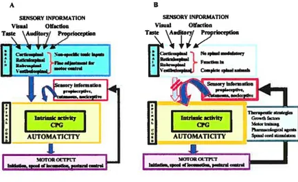
As illustrated in Figure 2, locomotor control relies on complex interactions between the CPG (blue) and supraspinal, spinal and multimodal sensory feedback to produce an appropriate temporal response and generate a highly adaptable motor pattern (reviewed in Armstrong 1986, Zehr & Stem 1999, Rossignol et al 2006). This regulation can be performed via actions on motoneurons, interneurons or primary afferents by means of presynaptic inhibition (yellow). Presynaptic inhibition may act on primary afferents, but also at othet selected areas of the central nervous system (CNS). Moreover, the motor output may depend on specific motoneuronal properties (gceen) emerging during locomotion. The following sections describe the different levels of control during locomotion: supraspinal descending commands, spinal interneurons, sensory feedback and motoneurons. The focus is essentially directed toward the description of muscle and cutaneous reflex pathways (section 3.3) because they constitute the basis of our investigation.
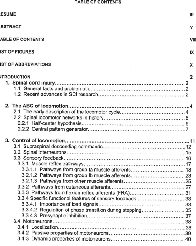
Figure 2: Neuronal organizatïon of the mammalian locomotor system and dynamic sensory integration during stepping. Dunng stepping, multimodal inputs
(supraspinal, cutaneous, muscular and joints afferents) reach both the brainstem and the spinal cord. In the spinal cord, some of these
afferents directly contact
motoneurons, but most of them synapse onto interneurons. The
activity of primary afferents can be
modulated by presynaptic inhibition before to reach a spinal target
(yellow). This allows to activate or
close some pathways, or to reverse the sign of the response (inhibition vs excitation) in different phases of
the step-cycle (phase
dependency). Phasic presynaptic
inhibition occurs at various levels of
the spinal cord including just before
the afferents contact the CPG
(blue), interneurons (pink) and
motoneurons (green). Moreover,
membrane properties of
motoneurons (green) are
modulated during locomotion and
may change the gain of the
response to a given sensory input.
3.1 Supraspinal descending commands
During movement, descending pathways can exert a direct control (monosynaptic) onto motoneurons (eg control cf fine movements cf the hand by corticospinal pathways) or an indirect control (polysynaptic) via interneurons that project to motoneurons (eg postural regulation). Several supraspinal pathways including the reticulospinal, vestibulospinal, rubrospinal and corticospinal tracts are involved in the control of locomotion and their rote has been well documented (reviewed in Armstrong 1986, Rossignol 1996, Jordan 1998, Orlovsky et al 1999, Drew et al 2002, 2004, Rossignol et al 2006). Most of the supraspinal inputs passes through a set cf interneurons to ensure their integration with the basic locomotor pattern generated by the CPG and leads te an adequate context-related locomotor response (Baldissera et al 1981, Jankowska 1992, McCrea 1996, Orlovsky et al 1999, Drew et al 2004).
Our model does flot involve supraspinal inputs to spinal pathways because a complete spinal cord transection has been performed. For that reason, the role cf supraspinal pathways in the control cf locomotion will be succinctly described and the functional
consequences cf disrupting these pathways will be emphasized.
Supraspinal pathways have extremely complex effects and interactions during locomotion. This paragraph is a general and simplified summary cf their role during stepping and will not deal with specificities and exceptions. The motor cortex exerts a powerful influence on locomotion and has been shown te be crucial during precise visually guided walking (Beloozerova & Sirota 1993, Armstrong & Marple-Horvat 1996, Dtew et al 2002, 2004). In the cat, the corticospinal pathway appears te contribute to the fine control and volitional positioning cf the limbs in a locomotor context requiring accurate and precise foot placement such as gait modification te step over an obstacle or walking on a narrow beam or the rungs cf a horizontal ladder (skilled locomotion). In the hindlimbs, the lateral corticospinal tract was shown te excite flexor and inhibit extensor motoneutons. Transcranial magnetic stimulation in human subjects showed that corticospinal inputs provide part of the drive te activate muscles for walking (Capaday et al 1999, Petersen et al 2001). The rubrospinal tract has been shown te control hindlimb flexion during swing when it evokes a facilitatory response in most flexor muscles (Orlovsky 1972, Rho et al 1999) and the vestibulospinal tract controls hindlimb extension during the stance phase of locomotion (Orlovsky 1972). Finally, beside its role in initiating locomotion (detailed in the
next paragraph), the reticulospinal tract may modify the activity in both flexor and extensor muscles tending to reinforce activity in muscles that are aiready active (Orlovsky 1972, Drew & Rossignol 1984, Drew 1991, Perreault et al 1994). This pathway is also suggested to be involved, together with the corticospinal pathway, in the control of posture when locomotion is disturbed (Drew et al 2004). These results confirm that the transmission in descending pathways to the spinal cord is modulated in a phase-dependent manner. Noteworthy, many supraspinal structures are also capable of resetting the locomotor rhythm. This suggests that they may act through interneurons that are part of the CPG (Orlovsky 1972, Perreault et al 1994, Rho et al 1999, Leblond et al 2000, 2001).
The spontaneous initiation of stepping, ie without drugs or electrical stimulation, requires the integrity of supraspinal structures. Decerebrated animais are only capable of spontaneous locomotion if the brainstem is flot transected beiow a specific level. For exampie, if the decerebration is performed between the rostral border of the superior colliculus dorsally and rostral to the mammillary bodies ventrally (referred to as pre mammillary cat), cats will preserve the ability to step spontaneously. Howevet, if the transection terminates caudally to the mammilary bodies ventrally (post-mammilary or mesencephalic cat), a 2 weeks period is necessary for the recovery 0f locomotor movements. No recovery has been reported with more caudal transections. Over years, severat areas of the brain and brainstem have been identified as being abie to induce locomotion in decerebrated or intact animais (reviewed in Armstrong 1986, Jordan 1998, Orlovsky et al 1999): the mesencephalic locomotor region (MLR), the subthalamic locomotor region (SLR), the pontine locomotor region (PLR) and the cerebeilar locomotor region (CLR, Mon et al 1999). AIl these areas converge on, and excite, reticulospinal neurons in the brainstem, which in turn exert their control in the lumbar spinal cord onto the CPG to initiate locomotion. The specific spinal targets cf the reticulospinal neurons have flot been clearly identified. However, maximal field potentiai following MLR stimulation occurs in the dorsomedial area cf the spinal gray matter (laminae V-VII) suggesting the presence of a concentration cf interneurons receiving reticulospinal input in this area (Noga et al 1995).
A lesion cf the spinal cord interrupts (complete) or compromises (incomplete transection, contusion, etc) supraspinal descending tracts and propriospinal pathways te lumbosacral segments. The observed deficits during stepping can mainly be attributed to the disruption of the control previously provided by these pathways as revealed by partial spinal lesion studies (Gorska et aI 1993, Jiang & Drew 1996, Brustein & Rossignol 1998, Rossignol et al
1999). The response of the spared pathways to the lack cf supraspinal input determines both the extent cf the recovery and the specific functions that may recover (Heigren & Goldberger 1993, Bregman et al 2002). Moreover, the amount cf fibers preserved in the ventral and lateral funiculi of the spinal cord, particularly the white matter associated with the reticulospinal tract, was shown to be directly related to locomotor performance after SOI (Schucht et al 2002). However, locomotion has been reported in the absence of ventral and ventrolateral quadrant in the cat (Brustein & Rossignol 1998). Depending on the pathways disrupted (corticospinal, rubrospinal, reticulospinal, vestibulospinal), serlous deficits impairing locomotion may be observed: incapacity to voluntary initiate stepping, lack of voluntary and anticipatory adjustment of locomotion (eg avoiding obstacles), impaired weight support, lateral stability and interlimb coordination (fore- vs hindpaw). Another functional consequence cf lacking supraspinal control is the paw dtag that is frequently observed at the onset of the swing phase in spinal cats. This behavior seems to be associated with an inappropriate timing of flexion movements in the hip, knee and ankle at the beginning of the swing phase. For example, the activation delay between St and Srt is absent so that the knee and hip joints flex simultaneously instead cf one after the other to clear the foot before hip flexion onset (Rossignol et al 2004). This could be due to a Iack of corticospinal and rubrospinal control, which are required for proper intralimb coordination (Jiang & Drew 1996). lndeed, paw dragging during stepping has been observed in cats with a restricted lesion to dorsolateral quadrants of the spinal cord (Jiang & Drew 1996, Rossignol et al 1999).
The disruption of descending input interferes with the ability to walk in a voluntary and controlled manner. After a complete spinal cord transection, aIl these deficits are observed. Indeed, the hindlimbs are flaccid and can barely perform weak and uncoordinated movements when placed over a treadmili beit. Ground contact will be performed on the dorsal surface of the paw. Moreover, the EMG is more clonic and cycle length is generally shorter for a given walking speed (Rossignol et aI 2004).
Incomplete SOI may stimulate the reorganization cf synaptic connections such as increasing collateral branching or shifting the representation of the hindlimbs in the motor
cortex. Here, a complete transection model was chosen in order to assess the plastic potential cf the spinal cord exciuding any plasticity driven by supraspinal pathways.
Notably, there is a persistent hyperexcitability of several reflexes following partial or complete SOI because of the removal of inhibitory descending input from the brainstem (Holmqvist & Lundberg 1961, Lundberg 1964, Hultborn & Malmsten 1983, Malmsten 1983). This wiIl be discussed in the appropriate section.
3.2 Spinal interneurons
Some afferent inputs may directly contact motoneurons. However, most of them will first transit thtough the interneuronal networks of the spinal cord. Interneurons are classified in 2 broad categories: segmental interneurons whose axons reside within the gray matter in the same or few nearby segments and propriospinal interneurons whose axons pass through the white matter to re-enter gtay matter in distant spinal segments. The latter are meant to coordinate activity across spinal cord segments. Thete are also interneurons specialized in relaying spinal and sensory information to the btain. Classically, spinal interneurons in the cat have been functionally identified according to their dominant synaptic input, intrinsic properties, target neurons and role in motor activity (reviewed in Jankowska 1992, 2001). A recent review illustrates that the properties and organization of the spinal interneuronal networks share several similarities in cats and humans (Jankowska & Hammar 2002).
Spinal interneurons are involved in mediating both simple reflexes and complex movements. Descending and peripheral input were assumed to travel along independent pathways to reach the motoneurons. This was denied by an elegant series of studies conducted by Lundberg and colleagues showing that the spinal interneuronal networks are
integrative centers, ie supraspinal and primary afferents of various modalities converge on common spinal interneurons before they reach the motoneurons (reviewed in Lundberg 1979, Baldissera et al 1981, Jankowska 1992). These interneurons then project in a divergent manner onto motoneurons, onto other spinal interneurons, and onto neurons projecting back to the supraspinal centers. This is a highly flexible network which includes mechanisms to select reflex pathways and allow the interaction between interneuronal populations. This resuits in the reconfiguration of the networks and provides a multifunctional character to a given set of interneurons. Spinal interneurons are crucial players involved both in the modulation of reflexes by supraspinal commands and in modulating the supraspinal command by sensory feedback before teaching the
motoneurons (Jankowska 1992). The integrated information is projected back to supraspinal centers whete multiple loops project back downward to control the spinal circuitry (Armstrong 1986). This allows a rapid adaptation of motoneuron activity to the central command and environmental constraints. During locomotion, the activity of interneurons will result from the mix of convergent input from the CPG, sensory feedback, descending commands and intrinsic membrane properties of the cells.
Obviously, none of the spinal interneurons are interposed in a pathway with input from a single type of afferents, but their name is meant to identify the dominant input.
Given the diversity of spinal interneurons (anatomical, functional, molecular, source of input and target neuron) and for the text to be intelligible, their description is located in the
appropriate section according to the reflex pathway in which they are involved
3.3 Sensory feedback
A reflex is a stereotyped motor response generated by the CNS in reaction to a particular sensory stimulus. Following a given peripheral afferent stimulation, a reproducible response s evoked (under similar conditions). Spinal reflexes were shown to be a great experimental tool to explore the organization of the CNS (Burke 1999) and are widely described in the literature (Baldissera et al 1981, Jankowska 1992, Zehr & Stem 1999, McCrea 2001). During movement, spinal segmental reflexes are highly flexible adjusting to the type, intensity and localization of the stimulus and also to the context. It can also be modulated in a task-dependent and phase-dependent manner. Recent reviews have been written to describe the spinal reflexes and their dynamic control during locomotion
(Hultborn 2006, Rossignol et al 2006).
Sensory inputs are not required to generate a basic locomotor pattern but does substantially contributes to the motor output and adapts the central activation to environmental constraints. This feedback can have a global influence in allowing, preventing and selecting motor patterns. Whether of muscular (Duysens & Pearson 1980, Pearson 1995, Dietz & Duysens 2000), cutaneous (Duysens & Pearson 1976) or articular (Grillner & Rossignol 1978) origin, this dynamic sensorimotor interaction powerfully influences the basic motor output acting directly or indirectly on the CPG (Grillner 1981,
Gossard & Hultborn 1991, Pearson et al 1998, Rossignol et al 2006). This process is performed in the spinal cord and can modify the frequency, amplitude and structure of the motor output, which is crucial for estabiishing the final stepping pattern. This is illustrated by the capacity of spinal animais (Forssberg et al 1980ab, Grillner 1981, Lovely et ai 1986, Pearson 2000, Leblond et al 2003) and babies with immature descending tracts (Yang et al 1998) to adapttheir locomotor pace to the treadmiil speed. When the locomotor pace is increased, the duration of extensor activity is decreased while the duration of flexor activity is relatively constant (Halbertsma 1983, Yang et al 1998, Oriovsky et al 1999). Fiexor bursts vary littie with change in step-cycle length as compared to extension and this basic feature of walking conserved in reduced preparation (Grillner & Zangger 1979, however see Yakovenko et al 2005). Weii-coordinated locomotion depends heavily on sensory inputs signaling 11mb kinematics and loading (Pearson 1996, Rossignol 1996, Duysens et al 2000).
Anatomical and behaviorai evidence suggest that sensory feedback piays a crucial role in the recovery of function after SCI in humans and animaIs to compensate for the loss of supraspinal input to spinal circuits. This is weil illustrated by the ability to regain rhythmic locomotor movements after repetitive sensory stimulation provided by step-training (section 4.1.3.1).
Here, we wiil emphasize the description of spinal refiexes under investigation, le reflexes evoked by muscle group la-lb afferents and by specific cutaneous afferents. Reflexes will
both be described in the absence 0flocomotion and when the spinal cord circuitry is conligured for locomotion.
3.3.1 Muscle reflex pathways
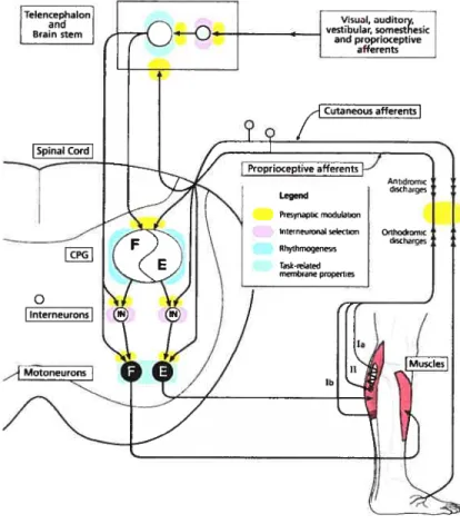
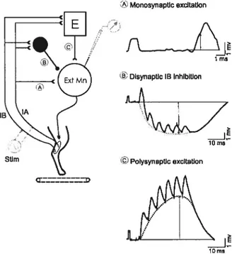
Group I afferents, large diameter and high conduction velocity fibers, carry information originating from muscle receptors. Group la fibers innervate muscle spindles and transmit information concerning the extent and velocity of muscle stretch. The lengthening of the intrafusal fibers increases la afferents firing frequency (together with group Il, see section 3.2.1.3). Group lb fibers innervate Golgi tendon organs and carry information related to changes in tension applied to a given muscle. As shown in Figure 3, muscle afferents synapse either directly on motoneurons in the ventral horn (pathway 1) or on interneurons
18
in the intermediate zone of the ventral horn gray matter. In the latter case, the motoneuron is either contacted via a single interneuron (disynaptic pathway; Fig.3 pathways 2-3) or a chain of intemeurons (polysynaptic pathway; Fig.3 pathways 4-5).
Figure 3: Proprioceptive pathways from
5 extensors and flexors during locomotion
Left: 4 pathways project to extensor motoneurons fE-Mn): monosynaptic pathway from la afferents (1), disynaptic group la÷lb inhibitory pathway (2), the disynaptic group la-i-lb excitatory pathway (3), polysynaptic excitatory pathway (4). Group lb afferents input are abie to interrupt flexion (5).
Right: Pathways to flexors are similarly organized to the pathways to extensors (left, pathways 1-4). However, most group I and II afferents from flexors can end the extension and reset the rhythm to flexion (5) while only a few can prolong the flexion phase (4).
Black circle, inhibitory interneuron; empty circle, excitatory interneuron; E-Mn, extensor motoneuron; F-Mn, flexor motoneuron; E, extensor generator; F, flexor generator; la afferents, blue; lb afferents, green; group Il afferents, brown. From Rossignol et al 2006.
3.3.1.1 Pathways from group la muscle afferents
The monosynaptic reflex. Ihe activation of muscle spindles mainly evokes a monosynaptic EPSP in homonymous and synergistic a-motoneurons (simplified to motoneuron in the text unless mentioned) acting at the same joint (Eccles et al 1957ab). According to Mendell and Henneman (1971), each la afferent from a given muscle directly projects to every motoneuron innervating the same muscle; conversely, each motoneuron is contacted by every la homonymous fiber (Fig3, pathway 1) representing 1-5% of ail afferent terminaIs on motoneurons. During muscle stretch, la fibers discharge and excite motoneurons producing contraction of the muscle to counter its own stretch. The monosynaptic stretch reflex is thought to make a major contribution to the level of EMG activity during stepping in cats walking on the treadmill (Stem et al 2000). Howevec, recent evidence in humans suggests otherwise: la afferent feedback generated during normal walking seems to make only a minor contribution to the SOL actïvity but would contribute significantly when the muscle is unexpectedly lengthened during walking (discussed in
4
EZF
I/2
Flexor I Musclesj
T I Extensor Muscles19
Sinkjaer et al 2000, see also Yang et aI 1991 b, Sinkjaer et al 1996). It is suggested that the ongoing la feedback is gated by presynaptic inhibition wheteas unexpected la signais trigger reflex activity to adjust the movement.
Task- and phase-dependency. The input-output relationship of the monosynaptic reflex is rather flexible and can be modulated to adapt to functional requirements of motor activity. The gain of the reflex is either increased to facilitate the motor task or reduced to ensure that the task is not compromised. In cats, the gain of the tricep surae stretch reflex is lower during stepping as compared to tonic contraction (state-dependent). Although gamma bias could contribute to modifying the gain of this reflex (Bennett et al 1996), intracellular recordings of lumbar motoneurons have shown a tonic reduction in la-EPSP amplitude during MLR-evoked fictive locomotion that was ascribed to an increased presynaptic inhibition of la afferents (Gosgnach et al 2000). Similarly, the H-reflex was first shown to be maximal during standing, decreased during walking and even more during running in humans (Capaday & Stem 1986, 1987, Edamura et al 1991) because of presynaptic inhibition of la afferents (Faist et al 1996). However, this was recently denied by Simonsen & Dyhre-Poulsen (1999). They demonstrated that there is no modulation of the H-reflex during various speed of running as compared to walking and that discrepancies with previous investigations arise from inadequacy of the stimulus intensity (Simonsen & Dyhre-Poulsen 1999).
Additionally, the monosynaptic reflex is modulated according to the phase in which it occurs during rhythmic behaviors. For example, the stretch reflex is modulated with a maximal amplitude during the stance phase of locomotion in SOL, when the motoneuronal pool is depolarized and the muscle active (Akazawa et al 1982, Capaday & Stem 1986, Crenna & Frigo 1987, Simonsen & Dyhre-Poulsen 1999). This is suggested to reinforce extensor activity during the ongoing stance phase (Guertin et aI 1995). Intracellular recordings also provided evidence for a phase-dependent modulation of la-EPSPs in hindlimb motoneurons during fictive locomotion in spinal, decerebrate or decorticated cats. The presence of modulation in reduced preparation suggests that this cannot be soiely attributed to the absence of reafference and depends, at Ieast partially, on spinal mechanisms (Schomburg & Behrends 1978b, Perret & Cabelguen 1980, Shefchyk et al 1984, Gossard 1996, Ménard et al 1999, 2003). The gain of this reflex is believed to depend on cyclic changes in the excitability of motoneurons (section 3.4) and strength of synaptic transmission by means of presynaptic inhibition (section 3.3.4.3).
la interneurons and Renshaw cells: last-order inhibitory interneurons. Not only does la afferents activation evokes an excitatory response in agonist motoneurons, but also an inhibition in antagonists, the so-called reciprocal inhibition (Lloyd 1946). This reciprocal inhibition is mediated by a group of glycinergic interneurons, referred to as la interneurons, that innervates motoneurons within a spinal segment or adjacent segments (Eccles et al 1956, Baldissera et al 1981, Jankowska 1992). la interneurons are characterized by a strong activation by la afferents and the ability to discharge at high frequency (Hultborn et al 1971). lndeed, most of la interneurons respond to a synchronous volley in la afferents with a single discharge; however, high frequency trains of action potentials can be evoked by the stimulation of other peripheral afferents such as FRA (section 3.3.3) or during walking (Hultborn et al 1971, McCrea et al 1980).
As exhaustively reviewed by Jankowska (1992), la interneurons receive multiple converging inputs from supraspinal (pyramidal tracts, corticospinal, rubrospinal, reticulospinal and vestibulospinal), spinal (propriospinal, CPG interneurons) and peripheral (cutaneous, muscle, joint, FRA) afferents. la interneurons have been shown to contribute to the inhibition of motoneurons of antagonists during muscle stretch, the crossed extensor reflex, postural reflex, centrally induced locomotion and voluntary movements. In humans, reciprocal inhibition can be evaluated by means of the H-reflex: an inhibition of the H-reflex of a given muscle is observed following a conditioning activation of the antagonistic motor nerve (Pierrot-Deseilligny et al 1981 b). Ia-IPSPs are also modulated in a phase-dependent manner with a maximum occurring during the hyperpolarized phase of fictive locomotion (Pratt & Jordan 1987, Degtyarenko et al 1998). Accordingly, reciprocal inhibition of extensors is maximal during swing in humans (Petersen et al 1999).
Notably, la interneurons are also contacted by Renshaw ceils associated with the agonist motoneuron and from la interneurons associated with the antagonist (Baldissera et al 1981). Most Renshaw cells are glycinergic and respond with a train of high frequency discharges by recurrent collaterals originating from motoneurons. The activation of Renshaw cells leads to the inhibition of surrounding synergistic o- and y-motoneurons, other Renshaw celis and la interneurons to antagonistic motor nuclei (Hultborn et al 1971, Baldissera et al 1981, Jankowska 1992). They also receive information originating from cutaneous afferents, group Il-III muscle afferents and descending pathways. Renshaw cells have been found to adjust the excitability of la inhibitory interneurons and o motoneurons to regulate the gain of motoneuron output. A strong stimulation of Renshaw cells both decreases the activation of the agonist muscle and the inhibition of the
antagonist te facilitate coactivation whereas a weaker stimulation leads to a selective activation cf agonist muscle. This system is organized in otder for the agonist meteneuren and antagenist la interneuron te be under the centrol cf the same afferent input. This organization leads te a clesely linked activation cf the agonist te the inhibition cf the antagonist during movement. Reciprecal and recutrent inhibitions have indissociable functions and are bcth invclved in the ccntrol cf mctcneurcn activity during vatious types cf reflex and rhythmic mcvements such as stepping.
la inhibitery interneurons and Renshaw cells are rhythmically active during fictive loccmeticn respectively during the inactive and active pericd cf the target mcteneurcn (McCrea et al 1980, Pratt & Jcrdan 1987). When strychnine is added te the system te blcck glycinergic transmission, fictive lcccmcticn still eccurs suggesting that la interneurens and Renshaw cells are net essential te locomotion (Ptatt & Jerdan 198f). Mcreever, their rhythmic activity cannet be attributed te the phasic activaticn cf peripheral recepters as shewn during fictive locomotion (McCrea et al 1980, Pratt & Jerdan 1987). Altheugh net essential, reciprecal inhibiticn is thcught te centribute te the generatien cf the basic leccmetcr pattern and recurrent inhibition is believed te help terminate the activity cf mcteneurens and la interneurcns and help fer the transition te the antagenist (McCrea et al 1980, Pratt & Jcrdan 1987).
Monosynaptic reflex and SCI. Inccnsistencies arise as te the effect cf SOI en the mcnesynaptic reflex and la-EPSPs. Seme studies report that synaptic transmission is increased by a cemplete spinal transectien enhancing the amplitude cf la-EPSPs and projection frequency and efficacy cf group la fibers (Cepe et al 1988). Hence, beth hemenymeus and hetercnymcus la-EPSPs were repcrted te be larger and have a faster rise time after a ccmplete acute or chrenic SCI (Ccpe et al 1986, 1988, Munscn et al 1986, Hcchman & McCrea 1994ab). On the ether hand, hcmcnymeus la-EPSP amplitude has aise been reperted te be unchanged after a cemplete SCI (Mayer et al 1984). In fact, changes in la-EPSP amplitude seem te be a very specific prccess. Fer example, Hcchman and McCrea (1994a) ebserved a general increase in hemenymcus la-EPSP amplitude in the triceps surae meteneurens. When PSPs were grcuped acccrding te mctcneurcnal pools, LG-EPSPs were cf larger amplitude whereas MG-EPSPs wete net mcdified in spinal animaIs as ccmpared te intacts. Unchanged hcmcnymeus EPSPs in MG meteneurons cf chrenic spinal cats were aise repcrted by cthers (Nelsen & Mendeli 1979, Mayer et ai 1984, Munsen et al 1986, Hechman & McCrea 1994a). Mereover, Munscn and