Spiroplasma affects host aphid
proteomics feeding on two
nutritional resources
Aline Sartori Guidolin
1, Thaís Regiani Cataldi
2, Carlos Alberto Labate
2, Frederic Francis
3&
Fernando Luis Cônsoli
1Bacterial symbionts are broadly distributed among insects, influencing their bioecology to different degrees. Aphids carry a number of secondary symbionts that can influence aphid physiology and fitness attributes. Spiroplasma is seldom reported as an aphid symbiont, but a high level of infection has been observed in one population of the tropical aphid Aphis citricidus. We used sister isolines of Spiroplasma-infected (Ac-BS) and Spiroplasma-free (Ac-B) aphids reared on sweet orange (optimum host) and orange jasmine (suboptimum host) to demonstrate the effects of Spiroplasma infection in the aphid proteome profile. A higher number of proteins were differently abundant in aphids feeding on orange jasmine, indicating an impact of host plant quality. In both host plants, the majority of proteins affected by Spiroplasma infection were heat shock proteins, proteins linked to cell function and structure, and energy metabolism. Spiroplasma also induced changes in proteins involved in antimicrobial activity, carbohydrate processing and metabolism, amino acid synthesis and metabolism in aphids feeding on orange jasmine. We discuss on how the aphid host proteome is differentially affected by Spiroplasma infection when the host is exploiting host plants with different nutritional values.
All multicellular animals have symbiotic microbes that establish biochemical and physiological interactions with their hosts. In insects, microorganisms are known to participate in many of the host physiological processes, e.g. by influencing the host capacity to use host plants or in pathogen transmission efficiency as vector of plant diseases1–3. The combination of two metabolic systems (symbiont + host) has a variety of potential consequences to the host, such as in aphids that may carry a great diversity of them. Aphids are known to harbor their obligate symbiont Buchnera aphidicola4,5, and yet be associated with several facultative symbionts6–10.
Buchnera aphidicola and aphids have a long co-evolutionary history. They share several metabolic pathways
related to amino acids synthesis, and B. aphidicola provides aphids with essential amino acids that are not pro-duced by the host nor are acquired from phloem sap, its natural diet. In many instances, B. aphidicola uses pre-cursors and enzymes from aphid metabolic pathways to contribute to aphid nutrition3,11. Facultative symbionts are diverse and the outcome of their interactions with their hosts are quite variable as they induce a plethora of phenotypes in host aphids12.
Secondary symbionts have also been related to host plant use in aphids, either by broadening or narrowing the insect host range13,14. However, the molecular and physiological mechanisms by which secondary symbionts interfere with host plant utilization and host range adaptation in aphids remains unclear. Proteomics is an inter-esting approach for the investigation and identification of metabolic processes and pathways differently expressed in an organism subjected to particular challenges. By example, proteomics of aphids saliva have been successfully applied to investigate aphid interactions with host plants and viruses aphids vector, as well as aphids response to stress conditions15–19. Aphid proteomics is also a growing area of research to investigate the effects of secondary symbionts that may have impact in the association with their hosts at different trophic levels15–19.
Spiroplasma is a common facultative symbiont reported in insects, but its role in host biology is still being
questioned and explored20. Spiroplasma are known to be associated with at least 16 species of Drosophila, but the relationship of this symbiont with their Drosophila hosts may be quite variable21–26. In aphids, this symbiont was
1Insect Interactions Laboratory, Department of Entomology and Acarology, ESALQ, University of São Paulo, Av.
Pádua Dias 11, 13418-900, Piracicaba, São Paulo, Brazil. 2Max Feffer Laboratory of Plant Genetics, Department of
Genetics, ESALQ, University of São Paulo, Av. Pádua Dias 11, 13418-900, Piracicaba, São Paulo, Brazil. 3Entomologie
Fonctionnelle et Evolutive, Gembloux Agro-Bio Tech, Université de Liège, 2 Passage des Déportés, 5030, Gembloux, Belgium. Correspondence and requests for materials should be addressed to F.L.C. (email: [email protected]) Received: 30 January 2017
Accepted: 21 January 2018 Published: xx xx xxxx
first shown in Acyrthosiphon pisum (Harris), and injections into “bacteria-free aphid lines” demonstrated that
Spiroplasma negatively affected host biology27. Later, Spiroplasma was discovered to confer protection against a fungal entomopathogen due to aphid behavior manipulation. Aphids carrying Spiroplasma would drop off the plant and sporulate away from siblings, limiting the spread of the disease28.
However, Spiroplasma is not so frequently found in aphids as other secondary symbionts29,30. Nevertheless, previous work reported high infections rates of Aphis citricidus (Kirkaldy) and Aphis aurantii31. Aphis citricidus is an oligophagous species, restricted to Citrus as suitable host plants, although it may survive and reproduce poorly in orange jasmine (Murraya paniculata)32. Previous comparative biology studies of A. citricidus showed that sweet orange is an optimum host, while orange jasmine is a suboptimum host plant for aphid development33.
Thus, we aimed to investigate the changes induced by Spiroplasma infection in the proteome of A. citricidus and associated symbionts when feeding on sweet orange (optimum host plant) and on orange jasmine (sub-optimum host plant) host plants. We report the overall effects of Spiroplasma infection on the proteomics of
A. citricidus and discuss on its implications for host fitness and host plant use in this aphid.
Results
The Spiroplasma-injected A. citricidus sister line was successfully infected as confirmed by diagnostic PCR (data not shown). Protein extraction and purification also yielded high quality protein samples with enough concen-tration for proteomic analyses.
Gel images passed to all quality filters, and spots were evenly distributed (Fig. 1). Initially, 174 spots were differently abundant between Spiroplasma infected and Spiroplasma free lines, but only 92 had minimum sample volume for further analysis. Out of the 92 spots analyzed, 80 of them were identified. 53 out of these 80 spots were differently abundant between Ac-B and Ac-BS using orange jasmine as a host plant, while 27 differently abundant spots were identified in aphids reared on sweet orange as a host (Tables 1 and 2).
Proteomic analysis of A. citricidus using the Ultraflex II MALDI Mass Spectrometer and the Synapt G2 HDMS systems rendered the identification of several putative proteins, which are associated with stress response, energy
metabolism, structural protein and other functional categories (Tables 1 and 2). Several proteins identified in A.
citricidus were present as proteoforms. In aphid samples from orange jasmine, we identified two differentially
expressed isoforms of the transitional endoplasmic reticulum ATPase-TER94 (spots 388 and 389), six of the repetitive proline-rich cell wall protein 2-like isoform X2 (spots 581, 587, 588, 589, 590 and 660), two of the heat shock protein cognate 3 precursor (spots 663 and 699b), two of the uncharacterized protein J9JJY7 (spots 687 and 693), five of the 60 kDa chaperone (spots 776, 832, 841, 855 and 937), and two for the ATP synthase subunit alpha (spots 959 and 1019) (Table 2). The overall number of differentially expressed proteins in the Ac-BS as compared to the Ac-B sister isoline was much reduced in sweet orange as a host plant if compared to orange jasmine. In this case, only two proteoforms each of the uncharacterized protein J9JWP2 (spots 193 and 194) and the uncharac-terized protein J9JJY7 (spots 215 and 217) were found (Table 1). We also had two spots rendering more than one protein hit each. Two different heat shock proteins were identified from spot 699 from aphids reared on orange jasmine (Table 2), and a heat shock protein (262a) and an unchacterized protein (262b) were obtained from mass data analysis of spot 262 from aphids reared on sweet orange (Table 1).
The overall analysis of the differently abundant proteins indicated that Spiroplasma regulated the levels of more proteins from the aphid host than from B. aphidicola (Tables 1 and 2). However, the proteomic profile obtained from aphids from the two host plants was quite different, indicating that Spiroplasma affected A.
citrici-dus proteomics differently depending on the host plant.
Changes in the proteome of A. citricidus induced by Spiroplasma infection was less evident in aphids reared in sweet orange, with 19 up-regulated and 9 down-regulated proteins belonging to protein-protein interaction,
energy metabolism, cell function and structure, ion transport and processing and degradation functional groups
(Table 1 and Fig. 2). Most of the proteins in aphids reared on sweet orange were uncharacterized, but analysis were made based on their domain. Up and down-regulated proteins were from several functional groups. Most of the eukaryote heat shock proteins and chaperonins from B. aphidicola were up-regulated (Fig. 2), and two eukaryote heat shock proteins (spots 274 and 262b), three proteins from cell function and structure, one protein
Figure 1. Representative 2D 12.5% SDS-PAGE gel of protein samples labeled with Cye3 from the Ac-B isoline
Spot Control Spiroplasma ANOVA (p-value) Fold change Protein Species Uniprot entry name Score Mass pI matched/searched % coverage Protein-Protein interactions
Heat shock 60
531 1.013 1.308 0.007 1.3 (+)* 60 kDa
chaperonin2** B. aphidicola Sg CH60_BUCAP 3615 57965 19/45 32.6 Heat shock 70
193 0.792 0.990 0.008 1.3 (+) uncharacterized protein2 Acyrthosiphon pisum J9JWP2_ACYPI 4044 72182 26/48 39.3
194 0.952 1.094 0.05 1.1 (+) uncharacterized protein2 Acyrthosiphon pisum J9JWP2_ACYPI 3291 72182 27/48 40.4
203 0.984 1.140 <0.001 1.2 (+) uncharacterized protein2 Acyrthosiphon pisum J9JWD2_ACYPI 940 73920 13/65 24.1
204 0.915 1.076 0.01 1.2 (+) chaperone protein DnaK2 B. aphidicola Sg DNAK_BUCAP 1285 70401 14/68 32.4
214 0.988 1.094 0.014 1.1 (+) heat shock protein cognate 3 precursor1
Acyrthosiphon
pisum gi|193716022 259 72993 5.2 28/45 47
215 0.819 1.082 0.009 1.3 (+) uncharacterized protein2 Acyrthosiphon pisum J9JJY7_ACYPI 1653 71670 15/49 22.6
217 0.948 1.130 0.03 1.2 (+) uncharacterized protein2 Acyrthosiphon pisum J9JJY7_ACYPI 2166 71670 17/49 31.9
262b 1.249 0.745 0.031 1.7 (−) heat shock protein cognate 3 precursor1
Acyrthosiphon
pisum gi|193716022 95 72993 5.2 15/42 28
274 1.105 0.979 0.002 1.1 (−) heat shock 70 kDa protein cognate 41 Acyrthosiphon pisum gi|193688192 169 72138 5.3 17/32 31
324 0.832 1.082 0.009 1.2 (+) heat shock 70 kDa protein cognate 4-like1
Acyrthosiphon
pisum gi|193603578 150 71626 5.3 15/25 33
Heat shock 90
131 0.875 1.074 0.03 1.2 (+) uncharacterized protein2 Acyrthosiphon pisum J9JP34_ACYPI 748 83759 10/69 16.2 Calcium binding
883 1.308 0.977 0.005 1.3 (−) ACYPI007505 protein2 Acyrthosiphon pisum C4WYG9_ACYPI 747 17432 5/22 33.7 Genetic transcription control
608 0.992 1.219 0.028 1.2 (−) uncharacterized protein2 Acyrthosiphon pisum J9JV14_ACYPI 2984 36544 17/28 55.8 Energy metabolism
440 0.993 1.117 0.017 1.1 (+) malyl-CoA lyase1 Granulibacter
bethesdensis gi|499950029 72 36167 5.8 6/14 21
788 1.327 1.078 0.022 1.2 (−) uncharacterized protein2 Acyrthosiphon pisum J9JQL8_ACYPI 1316 21268 4/17 23.0
801 0.812 0.952 0.02 1.2 (+) glyceraldehyde-3-phosphate dehydrogenase2
Acyrthosiphon
pisum C4WYF8_ACYPI 521 35735 6/24 24.4
Cell function and structure
363 0.878 1.025 0.01 1.2 (+) tubulin beta-1 chain2 Acyrthosiphon pisum D7RA98_ACYPI 4347 50558 28/29 54.4
610 1.1 1.25 0.025 1.1 (−) uncharacterized protein2 Acyrthosiphon pisum J9JLA2_ACYPI 3288 32571 9/27 25
637 1.2 1.214 0.047 1.2 (−) ACYPI003527 protein2 Acyrthosiphon pisum C4WTM0_ACYPI 215 22715 3/11 19.3
665 1.367 0.873 0.01 1.6 (−) uncharacterized protein2 Acyrthosiphon pisum X1XTT0_ACYPI 151 20386 2/8 12.8
893 0.927 1.08 <0.001 1.2 (+) ACYPI005394 protein2 Acyrthosiphon pisum C4WUV1_ACYPI 1374 20659 7/22 22.3
962 1.196 0.937 0.002 1.3 (−) ACYPI008348 protein2 Acyrthosiphon pisum C4WTD6_ACYPI 2421 11575 5/9 45.5
1038 1.1 0.968 0.019 1.1 (−) putative cofilin/actin depolymerizing factor-like protein2 Acyrthosiphon pisum Q1ZZP9_ACYPI 1411 17364 2/16 16.9 Ion transport
262a 1.249 0.745 0.031 1.7 (−) V-type proton ATPase catalytic subunit A1
Acyrthosiphon
pisum gi|193690627 116 68057 5 15/42 28
Processing and degradation Continued
from ion transport, one from energy metabolism and one from unknown functional group were down-regulated. Moreover, down-regulated proteins were all from eukaryote origin, while up-regulated proteins were from both eukaryote and prokaryote (Fig. 2).
But Spiroplasma infection of A. citricidus (Ac-BS) feeding on orange jasmine yielded broader changes in the host proteins. Differences in abundance of antimicrobial peptides, carbohydrate processing and metabolism, amino
acid synthesis and metabolism, anti-oxidants and biosynthesis of bactericides was observed only when comparing
infected and non-infected aphids fed on orange jasmine. Spiroplasma induced the up-regulation of 21 and the down-regulation of 33 proteins (Fig. 3) in orange jasmine-fed aphids. All of the differently abundant proteins of
cell function and structure and anti-oxidants, most of the amino acid synthesis and metabolism (except spot 975)
and ion transport (except spot 1872), and several proteins belonging to other functional groups were up-regulated in Spiroplasma-infected hosts (Fig. 3). Down-regulated proteins included mainly those in protein-protein
inter-action and antimicrobial peptides functional groups, with three 60 kDa chaperonins (spots 832, 855 and 937) and
one repetitive proline-rich cell wall protein 2-like isoform X2 (spot 587) being strongly down-regulated (Fig. 3). The functional groups energy metabolism, carbohydrate processing and metabolism, processing and degradation and
biosynthesis of bactericides also included proteins that were either up or down-regulated in Spiroplasma-infected
aphids in orange jasmine. Futhermore, we detected no pattern of protein regulation in regards to protein origin, if eukaryote or prokaryote (Fig. 3).
A comparison of the proteome of Spiroplasma-infected A. citricidus when exploiting sweet orange and orange jasmine indicated a number of different abundant proteins common to aphids reared on both host plants (Fig. 4), as the uncharacterized protein J9JP34 (spot 524 in orange jasmine and spot 131 in sweet orange), chaperone pro-tein DnaK (spot 685 in orange jasmine and 204 in sweet orange), uncharacterized propro-tein J9JJY7 (spots 687 and 693 in orange jasmine and spots 215 and 217 in sweet orange), 60 kDa chaperonin (spots 776, 832, 841, 855 and 937 in orange jasmine and spot 531 in sweet orange) and ACYPI003527 protein (spot 1541 in orange jasmine and spot 637 in sweet orange) (Tables 1 and 2). But although sharing similar differently abundant proteins in two dif-ferent host plants, only the uncharacterized protein J9JP34 and ACYPI003527 protein followed the same pattern of abundance in both plants (up-regulation), with the remaining proteins being up-regulated in sweet orange but down regulated in Spiroplasma-infected aphids feeding on orange jasmine (Figs 2 and 3). Differently abundant proteins common to both host plants, but with different patterns of expression were represented by heat shock proteins (protein-protein interaction) and the ACYPI003527 protein (cell function and structure).
Discussion
Comparative proteomic analysis between Spiroplasma-free (Ac-B) and Spiroplasma-infected (Ac-BS) sister iso-lines of A. citricidus on two host plants indicated Spiroplasma infection affects the host proteome, especially the abundance of proteins belonging to protein-protein interaction, cell function and energy metabolism categories. Changes in host proteome due to Spiroplasma infection were quite different in aphids feeding on different host plants - orange jasmine or sweet orange, suggesting that some of the differences observed between host plants for infected or uninfected aphids might be due to the interaction of the host plant and the infection status of the aphid. Due to a number of issues with aphids survival after microinjection and consequently the amount of protein obtained, we could not perform gels that would allow comparing differences in proteins abundances according to the host plant for a given infection status. Our data allows only comparisons between infection status for a given host plant. The huge differences in the aphid proteomics when feeding on sweet orange and orange jasmine certainly require additional studies to clarify the role of the host plants on the proteomics of A. citricidus.
One of the major groups of proteins affected by Spiroplasma infection, regardeless the host plant was the heat shock proteins (HSPs). HSPs are ubiquitous proteins common to prokaryotes and eukaryotes and are expressed either under stress or no-stress conditions34. HSPs are divided into families based on their molecular weight and sequence homology34,35. Function of HSPs proteins belonging to different families may depend on the develop-ment stage, subcellular location, and environdevelop-mental conditions36. Therefore, the role of HSPs depend on other proteins they may interact with, which changes depending on the physiological state of the aphid in a given moment.
Spiroplasma affected the expression of several HSPs (HSP10, HSP60, HSP70 and HSP90) from A. citricidus
and B. aphidicola. All of the identified HSP10 and HSP60 and one HSP70 (DnaK) were similar to those from B.
aphidicola, and the remaining HSP70 and HSP90 were similar to those from A. pisum. But the outcome of the Spot
Normalized volume
ANOVA
(p-value) Fold change Protein Species
NCBInr gi or Uniprot entry
name Score Mass pI
N. peptides matched/
searched % coverage Control Spiroplasma
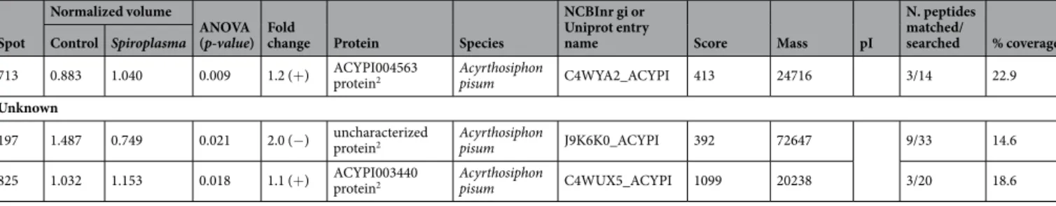
713 0.883 1.040 0.009 1.2 (+) ACYPI004563 protein2 Acyrthosiphon pisum C4WYA2_ACYPI 413 24716 3/14 22.9 Unknown
197 1.487 0.749 0.021 2.0 (−) uncharacterized protein2 Acyrthosiphon pisum J9K6K0_ACYPI 392 72647 9/33 14.6
825 1.032 1.153 0.018 1.1 (+) ACYPI003440 protein2 Acyrthosiphon pisum C4WUX5_ACYPI 1099 20238 3/20 18.6
Table 1. Identification of differently abundant spots from Spiroplasma-free (Ac-B) and Spiroplasma infected
(Ac-BS) isolines of Aphis citricidus feeding on sweet orange. *Protein abundance pattern in response to
Spiroplasma infection. (+) = up-regulated protein, (−) = down-regulated protein. **Number indicates the mass
Spot control Spiroplasma ANOVA (p- value) Fold change Protein Species Uniprot entry name Score Mass pI matched/searched % coverage Protein-Protein Interactions
Heat shock 10
2318 0.509 1.664 <0.001 3.3 (+)* 10 kDa chaperonin2** B. aphidicola 5 A CH10_BUCA5 1703 10243 2/6 28.1 Heat shock 60
776 1.651 0.759 0.004 2.0 (−) 60 kDa chaperonin2 B. aphidicola Sg CH60_BUCAP 4001 57965 29/45 46.7
832 1.998 0.432 <0.001 4.6 (−) 60 kDa chaperonin2 B. aphidicola Sg CH60_BUCAP 5376 57965 24/45 35.8
841 1.755 0.573 <0.001 3.1 (−) 60 kDa chaperonin2 B. aphidicola Sg CH60_BUCAP 3742 57965 21/45 35.8
855 2.107 0.490 <0.001 4.3 (−) 60 kDa chaperonin2 B. aphidicola Sg CH60_BUCAP 3200 57965 20/45 33.6
937 1.801 0.419 0.004 4.3 (−) 60 kDa chaperonin2 B. aphidicola Sg CH60_BUCAP 463 57965 11/45 29.1 Heat shock 70
417 1.344 0.472 0.002 2.8 (−) heat shock 70 kDa protein 41 Acyrthosiphon pisum gi|193596448 59 89426 5.5 6/11 9
663 1.532 0.634 0.003 2.4 (−) heat shock protein cognate 3 precursor1
Acyrthosiphon
pisum gi|193716022 257 72993 5.2 32/66 51
685 1.532 0.634 0.003 2.3 (−) chaperone protein DnaK2 B. aphidicola Sg DNAK_BUCAP 1404 70401 9/68 22.4
687 1.559 0.711 <0.001 2.2 (−) uncharacterized protein2 Acyrthosiphon pisum J9JJY7_ACYPI 2919 71671 19/49 33.8
693 1.453 0.529 <0.001 2.7 (−) uncharacterized protein2 Acyrthosiphon pisum J9JJY7_ACYPI 1246 71671 13/49 20.9
699a 1.543 0.641 0.007 2.4 (−) heat shock 70 kDa protein cognate 4-like1
Acyrthosiphon
pisum gi|193603578 112 71626 5.3 16/45 31
699b 1.544 0.642 0.007 2.4 (−) heat shock protein cognate 3 precursor1 Acyrthosiphon pisum gi|193716022 112 72993 5.2 17/45 29 Heat shock 90 397 1.298 0.529 <0.001 2.7 (−) endoplasmin1 Acyrthosiphon pisum gi|193643557 89 89250 4.8 14/35 16
524 0.579 2.168 0.014 3.7 (+) uncharacterized protein2 Acyrthosiphon pisum J9JP34_ACYPI 2221 83759 19/69 26.4 Calcium binding
2128 0.717 1.600 <0.001 2.2 (+) ACYPI000027 protein2 Acyrthosiphon pisum Q1ZZQ3_ACYPI 1093 17649 3/13 22.1 Energy metabolism
951 1.522 0.656 0.002 2.3 (−) ATP synthase subunit alpha mitochondrial1
Acyrthosiphon
pisum gi|193666827 84 59986 9.1 12/39 22
971 1.600 0.614 0.001 2.6 (−) uncharacterized protein2 Acyrthosiphon pisum J9JNB2_ACYPI 199 48183 8/40 19.6
959 1.623 0.739 <0.001 2.2 (−) ATP synthase subunit alpha2 Acyrthosiphon pisum J9JYX3_ACYPI 1469 60023 11/50 21.6
1019 0.468 2.045 0.006 4.4 (+) ATP synthase subunit alpha2 Acyrthosiphon pisum J9JYX3_ACYPI 472 60023 7/50 13.2
1331 1.446 0.593 <0.001 2.4 (−) malate dehydrogenase2 Acyrthosiphon pisum Q201V2_ACYPI 1007 36103 9/23 28.9
1353 1.574 0.662 <0.001 2.4 (−) glyceraldehyde-3-phosphate dehydrogenase2
Acyrthosiphon
pisum J9K9Z1_ACYPI 2993 35645 9/24 31.0
1760 0.709 1.631 <0.001 2.3 (+) triosephosphate isomerase2 Acyrthosiphon pisum C4WT45_ACYPI 293 27468 3/21 15.4 Anti-microbial peptides
581 1.892 0.597 <0.001 3.2 (−)
repetitive proline-rich cell wall protein 2-like isoform X21 Acyrthosiphon pisum gi|641669847 111 72602 7.7 15/47 22 587 1.838 0.411 <0.001 4.5 (−) repetitive proline-rich cell wall protein 2-like isoform X21
Acyrthosiphon
pisum gi|641669847 82 72602 7.7 10/35 16
Spot
Normalized volume
ANOVA
(p- value) Fold change Protein Species
NCBInr gi or Uniprot entry
name Score Mass pI
N. peptides matched/ searched % coverage control Spiroplasma 588 1.859 0.610 <0.001 3.0 (−) repetitive proline-rich cell wall protein 2-like isoform X21 Acyrthosiphon pisum gi|641669847 94 72602 7.7 15/60 21 589 1.703 0.577 <0.001 2.9 (−) repetitive proline-rich cell wall protein 2-like isoform X21 Acyrthosiphon pisum gi|641669847 115 72602 7.7 16/52 21 590 1.817 0.711 <0.001 3.0 (−) repetitive proline-rich cell wall protein 2-like isoform X21 Acyrthosiphon pisum gi|641669847 113 72602 7.7 17/70 21 660 1.821 0.751 0.016 2.4 (−) repetitive proline-rich cell wall protein 2-like isoform X21
Acyrthosiphon
pisum gi|641669847 79 72602 7.7 8/20 13
Cell function and structure
1264 0.641 1.474 <0.001 2.3 (+) actin related protein 12 Acyrthosiphon pisum gi|217330650 141 42158 5.3 12/32 40
1265 0.411 1.897 <0.001 4.6 (+) putative actin2 Acyrthosiphon
pisum Q201U9_ACYPI 175 42221 4/34 14.6
1356 0.460 1.591 <0.001 3.2 (+) uncharacterized protein2 Acyrthosiphon pisum C4WTU7_ACYPI 1192 32818 8/26 30.7
1541 0.606 1.584 <0.001 2.6 (+) ACYPI003527 protein2 Acyrthosiphon pisum C4WTM0_ACYPI 911 22715 6/11 40.4
1761 0.697 1.603 <0.001 2.3 (+) ACYPI005885 protein2 Acyrthosiphon pisum C4WYE4_ACYPI 810 24745 7/24 28.0
1781 0.422 1.675 <0.001 4.0 (+) ACYPI001342 protein2 Acyrthosiphon pisum C4WRW5_ACYPI 355 19792 3/11 18.4 Carbohydrate processing and metabolism
734 1.526 0.7499 0.004 2.0 (−) polysaccharide deacetylase1 Fusobacterium nucleatum gi|495968271 66 69834 9.1 15/62 24
1308 0.732 1.572 0.003 2.1 (+) ACYPI002851 protein2 Acyrthosiphon pisum C4WTT0_ACYPI 818 39779 8/23 28.9
1744 1.601 0.723 <0.001 2.2 (−) putative diacetyl/L-xylulose reductase2 Acyrthosiphon pisum Q201Y9_ACYPI 207 26334 2/17 10.4
1787 0.557 2.395 <0.001 4.3 (+) ACYPI000057 protein2 Acyrthosiphon pisum Q1ZZP5_ACYPI 188 25816 4/18 21.2 Amino acid synthesis and metabolism
913 0.495 1.718 <0.001 3.5 (+) glutamate dehydrogenase2 Acyrthosiphon pisum J9KB74_ACYPI 285 59967 9/44 20.7
975 1.527 0.669 0.005 2.3 (−) glutamyl-tRNA synthase1 Kyrpidia tusciae gi|502839225 78 55629 6.1 14/76 31
1215 0.705 1.690 0.007 2.4 (+) glutamine synthetase2 Acyrthosiphon pisum J9JML2_ACYPI 1783 41935 7/25 22.2
2302 0.615 1.289 <0.001 2.1 (+) aspartate kinase1 Nitrolancea
hollandica gi|495756139 58 8291 6.6 4/29 38
Ion transport
1607 0.400 1.895 <0.001 4.7 (+) ACYPI006090 protein2 Acyrthosiphon pisum C4WY80_ACYPI 40 26228 2/21 11.9
1872 1.689 0.802 <0.001 2.1 (−) ATP synthase oligomycin sensitivity conferral protein2 Toxoptera citricida Q5XUB9_TOXCI 157 22719 2/20 9.6
2293 0.457 1.703 <0.001 3.7 (+) V-type proton ATPase subunit F2 Acyrthosiphon pisum Q201X8_ACYPI 1251 13663 5/13 39.3 Processing and degradation
388 1.812 0.546 <0.001 3.3 (−) transitional endoplasmic reticulum ATPase TER941 Acyrthosiphon pisum gi|193617621 258 89914 5.1 26/36 32 389 1.753 0.748 0.001 2.3 (−) transitional endoplasmic reticulum ATPase TER941 Acyrthosiphon pisum gi|193617621 187 89914 5.1 27/68 32
1704 0.626 1.325 <0.001 2.1 (+) proteasome subunit alpha type2 Acyrthosiphon pisum C4WUA6_ACYPI 57 27982 2/22 8.3 Antioxidant
regulation of HSPs expression by Spiroplasma was quite different depending on the host plant the aphid was developing on. While the majority of the differently abundant HSPs were up-regulated in aphids developing on sweet orange, HSPs of aphids on orange jasmine were down-regulated.
The down-regulation of the B. aphidicola HSPs, GroEL (HSP60) and DnaK (HSP70) in infected aphids in a less suitable host (orange jasmine) demonstrate that Spiroplasma + orange jasmine can negatively interfere, directly or indirectly, with the exchange of molecules between the primary symbiont B. aphidicola and the aphid, even though a GroES was up-regulated. The regulation of different groups of HSPs demonstrate that the interaction between the aphid and B. aphidicola metabolism can differ depending on the changes induced by Spiroplasma in aphids feeding on food sources with different nutritional quality.
Two other abundant groups of proteins, energy metabolism and cell function and structure, were differentially expressed in response to Spiroplasma infection, regardless the host plant the aphid was feeding on. HSPs activity are closely linked to ATP availability35,36. Thus, down-regulation of HSPs in aphids feeding on orange jasmine were followed by down regulation of most of the ATP synthases produced, but no such correlation was observed in infected aphids feeding on sweet orange. Additionally, HSP are also reported to interact with cytoskeletal pro-teins37. We did observe up-regulation of proteins included in cell function and structure, including putative actins and actin-related proteins. But over-expression of these proteins in Spiroplasma-infected aphids was observed on both host plants, even under conditions in which HSP expression was reduced. Therefore, cell function on both host plant interacts with Spiroplasma in the same way to affect aphid’s actin proteins.
HSPs are also reported to play a role in immunity by interacting with antimicrobial peptides38,39. Proline-rich peptides are common antimicrobial peptides reported to produce pores in Gram- bacteria40 and play key roles in the immune response against microbials through interference with protein folding and protein expres-sion41–43. A strong down-regulation of HSP70 and antimicrobial peptides was observed in Spiroplasma-infected aphids developing on orange jasmine. Differentially expressed antimicrobial peptides were all isoforms of repetitive proline-rich cell wall protein 2-like isoform X2. Down-regulation of the host immune system in
Spiroplasma-infected aphids was also observed by the down-regulation of SAM binding domain–containing
pro-teins, which are linked to various bacteriocin biosynthesis proteins44.
Aphids are known to use a nutrition-based mechanism to control amino acid provisioning to regulate the growth of B. aphidicola45. Abundance of DnaK of B. aphidicola reflected the host plant suitability. DnaK abun-dance was higher in infected aphids feeding on sweet orange while in orange jasmine DnaK abunabun-dance was down-regulated in infected aphids.
Thus, it seems that Spiroplasma can affect the immune system of the host and/or the nutritional mechanism that regulates B. aphidicola growth. Glutamine is one of such nutrients provided by the host3,45. In orange jas-mine, we observed an increase in glutamine synthase and glutamate dehydrogenase proteins, which are enzymes involved in the recycling of ammonia and synthesis of glutamine and glutamate. Both are required for the syn-thesis of amino acids and purine metabolism in B. aphidicola-carrying bacteriocytes45. Regulation of these amino acids has been proposed as key in maintaining amino acids availability balanced for both the host and the sym-biont45. However, we did not detect up-regulation of glutamine transporters in the protein spots we were able to analyze to demonstrate enhanced transport of glutamine from the hemolymph to the bacteriocyte46,47.
Aspartate is also a non-essential amino acid provided by the host that has to be transported into B.
aphidi-cola cells. Aspartate is an important source of nitrogen for the synthesis of essential amino acids45. Additionally, we observed an increase in a prokaryote aspartate kinase, an enzyme involved in the synthesis of essential amino acids (lysine and threonine) through aspartate phosphorylation45,48. Up-regulation of aspartate kinase is suggestive of increased synthesis of essential amino acids from aspartate, but malate dehydrogenase was down-regulated in Spiroplasma-infected aphids in orange jasmine. Malate dehydrogenase is the enzyme involved Spot control Spiroplasma ANOVA (p- value) Fold change Protein Species Uniprot entry name Score Mass pI matched/searched % coverage
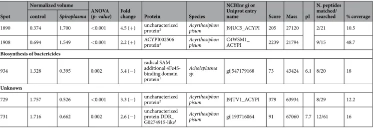
1890 0.374 1.700 <0.001 4.5 (+) uncharacterized protein2 Acyrthosiphon pisum J9JUC5_ACYPI 205 27120 2/21 10.5
1908 0.694 1.549 <0.001 2.2 (+) ACYPI002506 protein2 Acyrthosiphon pisum C4WSM1_ACYPI 2239 21794 9/15 48.7 Biosynthesis of bactericides 934 1.328 0.395 0.002 3.4 (−) radical SAM additional 4Fe4S-binding domain protein1 Acholeplasma sp. gi|547179168 73 43424 6.1 8/20 18 Unknown
729 1.757 0.526 <0.001 3.3 (−) uncharacterized protein2 Acyrthosiphon pisum J9JTV1_ACYPI 379 63934 8/29 12.2
731 1.716 0.662 0.002 2.6 (−) uncharacterized protein DDB_ G0274915-like1
Acyrthosiphon
pisum gi|193716064 91 67060 7.7 12/61 16 Table 2. Identification of differently abundant spots from Spiroplasma-free (Ac-B) and Spiroplasma infected
(Ac-BS) isolines of Aphis citricidus feeding on orange jasmine. *Protein abundance pattern in response to
Spiroplasma infection. (+) = up-regulated protein, (−) = down-regulated protein. **Number indicates the mass spectrometer used in the spot identification, 1 = Ultraflex II MALDI and 2 = Synapt G2 HDMS.
in the transamination of oxaloacetate to produce aspartate, but in aphid bacteriocytes synthesis of aspartate is based on the precursor glutamine. However, down-regulation of the aphid malate dehydrogenase enzyme may be correlated with the observed reduction in aphid energy metabolism, as oxaloacetate is also an intermediate of the citric acid cycle.
Moreover, up-regulation of antioxidants and proteasome subunit alpha type proteins in Spiroplasma-infected aphids feeding on orange jasmine may also indicate increased cell damage and the requirement for cell protection and degradation of unneeded or degraded proteins. Antioxidants protects cells by reducing the negative effects of the accumulation of reactive oxygen species49, while proteasomes degrade damaged and misfolded proteins50,51. Proteasome subunit alpha type proteins belong to a selective proteolytic protein machinery (proteasome) acting on ubiquitin-tagged proteins, even though proteins from the ubiquitinylation pathway were not identified among the analyzed proteins. Up-regulation of proteasome subunit alpha type proteins indicates the increased need of protein degradation, as proteasome deficiency could lead to the accumulation of damaged and misfolded pro-teins and result in a number of pathologies50,51. The requirement of high levels of proteasome activity may also be associated with the reduced availability of HSPs, which in turn may have led to an accumulation of damaged and/or misfolded proteins.
In conclusion, Spiroplasma infection affects the aphid-primary symbiont proteome mainly by regulating the activity of heat shock proteins and other processes that HSPs participate, such as energy metabolism and immune system. However, the effects of Spiroplasma infection on the host aphid proteomics differed between host plants, suggesting that some of the differences could be a result of the interaction between the infection status of the host and the nutritional quality of the host plant. We propose that Spiroplasma-infected aphids feeding on orange jasmine invest to provide a better nutritional environment to its primary symbiont, B. aphidicola and reduce any fitness cost associated with the Spiroplasma infection under such nutritional stress condition in a controlled trade-off between immune system regulation and nutritional deficiency.
Further studies to investigate the fitness costs of A. citricidus association with Spiroplasma in response to immune challenges under different nutritional stress conditions and the impact of nutrition alone will help to shed light on the role this secondary symbiont may play in aphid biology and on the costs involved in such association.
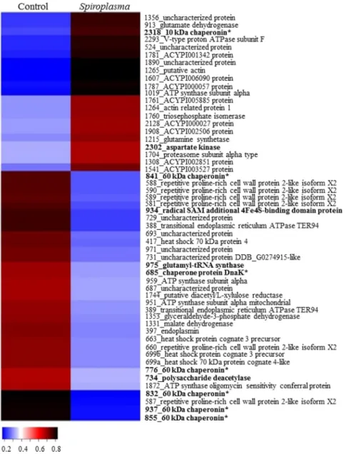
Figure 2. Heatmap of identified proteins on orange jasmine. Proteins are identified by their spot number
followed by Blast2Go or Uniprot identification. Regular letters represents proteins from eukaryote, bold letters represents proteins from prokaryote origin, and bold letters with an asteristic (*) represents proteins from B.
aphidicola aphidicola. Fold change was transformed in a proportional scale in which blue represents the smallest
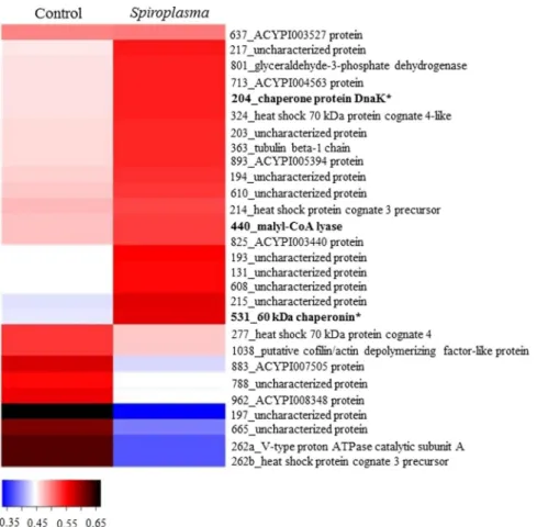
Figure 3. Heatmap of identified proteins on sweet orange. Proteins are identified by their spot number followed
by Blast2Go or Uniprot identification. Regular letters represents proteins from eukaryote, bold letters represents proteins from prokaryote origin, and bold letters with asteristic (*) represents protein from B. aphidicola
aphidicola. Fold change was transformed in a proportional scale in which blue represents the smallest fold
change and black represent the largest fold change.
Material and Methods
Insect isolines.
Sister isolines of A. citricidus infected (Ac-BS) or not (Ac-B) with Spiroplasma were estab-lished as described elsewhere33. Briefly, part of an isoline of A. citricidus free of secondary symbionts was micro-injected with hemolymph from Spiroplasma-infected A. aurantii, while the other part was micromicro-injected with PBS buffer (137 mM NaCl, 2.7 mM KCl, 10 mM Na2HPO4, 2 mM KH2PO4). Thus, infected and control lines of A.citricidus had the same genetic background.
Ac-B and Ac-BS sister lines were each maintained on seedlings of sweet orange (Citrus sinensis var. Pera) and orange jasmine (Murraya paniculata) inside rearing cages (50 cm high × 15 cm diameter), containing two lateral openings closed with cloth for ventilation, and maintained under controlled condition (25 ± 2 °C; 60 ± 10% UR; photoperiod 14 L:10D h).
Protein extraction.
Adults from Ac-B and Ac-BS were collected from their host plants and subjected to protein extraction. Three biological replicates of two-hundred and fifty aphids each per host plant and isoline were crushed in 1 mL of extraction buffer (8 M Urea, 4% DTT, 4% CHAPS) and incubated in ice for 40 min. The material was centrifuged (12,000 g × 15 min × 4 °C) and the supernatant was transferred to a new tube for puri-fication with the commercial system 2D - Clean up (GE Healthcare®
). Briefly, purification was made by adding 300 μL of the precipitant buffer followed by incubation on ice for 15 min. Afterwards, 300 μL of co-precipitant was added and the sample centrifuged (12,000 g × 5 min × 4 °C) once again. The supernatant was discarded and 40 μL of co-precipitant was layered on top of the pellet. The pellet was allowed to sit on ice for 5 min. The sample was centrifuged (12,000 g × 5 min × 4 °C) and the pellet recovered in 25 μL of Milli-Q, 1 mL of wash buffer and 5 μL of wash additive. The purification procedure was finalized after centrifugation (12,000 g × 5 min × 4 °C) and resus-pension of the pellet in UT buffer (7 M Urea, 2 M Thiourea, 20 mM Tris, 2% CHAPS). The pH was measured using Neutralit pH 5.5–9 strips (Merck) and adjusted to pH 8.5 with 1 M NaOH. Total protein concentration was veri-fied by using the commercial system RC DC Protein Assay (Bio-Rad®
), following the manufacturer instructions.2D-gel electrophoresis.
Proteomic analyses were made separately for protein samples collected from aphids from different host plants; hence, the effect of Spiroplasma infection on the aphid proteome was individu-ally analyzed within each host plant. For each biological replication (3) four technical replication were performed; thus a total of 12 images were analyzed per host plant for each status of infection.To perform analytic 2D gels, protein extracts were labeled with one of the three CyeDye (GE healthcare) in a total of 25 µg of protein followed by first dimension protein separation by electrofocalisation on IPG strip (pH3-10NL, 24 cm, GE Healthcare), following the manufacturer instructions. Therefore, each Ac-B and Ac-BS sample from both experimental host plants was labeled in duplicates with Cye3 and Cye5 leading to a total of 4 replicates per treatment. All IPG strips also carried the internal standard labeled with Cye2. The internal standard was com-posed of an equimolar mixture of Ac-B and Ac-BS.
The solution with labeled proteins was adjusted to a final volume of 450 µL of UT buffer, 2 M dithiothreitol (DTT) and 0.8% v/v IPG ampholyte (SERVA). IPG strips were set on a PROTEAN i12 IEF (Bio-Rad) for active rehydration for 11 h at 50 V, followed by isoelectric focalization (IEF) at 200 V for 200 Vh, 500 V for 500 Vh, 100 V for 100 Vh, and 8000 V for 60000 Vh. Both steps were conducted at 15 °C and 50 µA maximum/IPG strip.
After IEF, IPG strips were prepared for second dimension electrophoresis. First, IPG strips were equilibrated for 15 min in 375 mM Tris (pH 8.8), 6 M Urea, 20% glycerol (v/v), 2% SDS and 130 mM dithiothreitol (DTT), followed by an extra 15 min in a similar buffer, in which DTT was replaced by 135 mM iodoacetamide (IAA).
Once treated, IPG strips were positioned onto a 2D 12.5% HPE large gel (SERVA
®
). Second dimension elec-trophoresis used the HPE FlatTop tower (GelCompany) under the following conditions: phase I − 100 V, 7 mA, 1 W for 30 min, 200 V, 13 mA, 3 W for 30 min, and 300 V, 40 mA, 5 W for 10 min; phase II − 1500 V, 40 mA, 30 W for 3 h 50 min, and 1500 V, 45 mA, 40 W for 50 min. IPG strips were removed after phase I, and only 2D gel was used in phase II.Image analysis and spot selection.
2D gel images were captured with Typhoon Fluorescence Imager (Amersham) at wavelengths corresponding to each dye. A total of 12 images were analyzed with the software Progenesis SameSpots (Nonlinear Dynamics). Statistical analysis of protein expression levels were determined for each spot based on mean spot volume (n = 12), and differences in protein expression between control andSpiroplasma-infected aphids were detected using ANOVA. Spots with a p ≤ 0.05 were selected for inclusion in
the results. In the experiment with orange jasmine as a host plant, only spots with a significant p value and with a change in ratio higher than two (fold change ≥ 2) were selected, once spots with a fold change lower than 2 were considered dubious.
After the list of spots was defined, a preparative gel was made, and another IPG strip loaded with internal standard labeled with Cye2 and 500 µg of unlabeled proteins (250 µg Ac-B and 250 µg Ac-BS) was subjected to electrofocusing. This IPG strip was later used for a second dimension electrophoresis as earlier mentioned. The image of this preparative gel was aligned with the previously obtained gels to locate the spots of interest. Spot picking was made in duplicate using an Ettan Spot Picker Robot (GE Healthcare).
Mass spectrometry using an Ultraflex II MALDI Mass Spectrometer.
Excised spots were designated for the Proteineer dp automated Digester (Bruker, Bremen, Germany). Briefly, gel pieces were washed with three incubations in 100% of 50 mM ammonium bicarbonate, followed by incubations in a 1:1 mixture of acetonitrile (ACN) and 50 mM ammonium bicarbonate. Two additional washes were performed with 100% acetonitrile to dehydrate the gel. Gel pieces were first soaked in freshly activated trypsin (Porcine, Proteomics Grade, Roche) at 8 °C for 30 min, and later subjected to protein trypsinization for 3 h at 30 °C. Peptide extractions were performed with 10 µL of 1% formic acid (FA) for 30 min at 20 °C.aton. Spots were washed on-target using 10 mM ammonium dihydrogen phosphate in 0.1% trifluoroacetic acid (TFA) and desalted after washing with MilliQ water (Millipore). High throughput spectra were acquired using an Ultraflex II MALDI Mass Spectrometer (Bruker) in positive reflectron mode with close calibration enabled.
Successful spectra were summed, treated, and de-isotoped in line with an automated SNAP algorithm using Flex Analysis 2.4 software (Bruker), and subsequently submitted in batch mode to the Biotools 3.0 software suite (Bruker) with an in-house hosted MASCOT search engine (www.MatrixScience.com). Three databases were used: (1) the public National Center for Biotechnology Information (NCBI) non-redundant database, (2) the public National Center for Biotechnology Information (NCBI) non-redundant database with parameters set for Arthropoda, and (3) a homemade database containing all available aphid and aphid-symbiont protein sequences. A mass tolerance of 100 ppm with close calibration and one missing cleavage site were allowed. Partial oxidation of methionine residues and complete carbamylation of cystein residues were considered. The probability score calculated by the software was used as a criterion for correct putative identification.
In order to confirm identifications, experimental molecular weight (MW) and pI were compared to the pre-dicted values resulting from the MASCOT analysis.
Mass spectrometry using Synapt G2 HDMS.
Another range of excised spots were dehydrated with 100% ACN, followed by reduction and alkylation. Reduction was performed in 20 mM DTT and 50 mM ammo-nium bicarbonate for 40 min at 55 °C. The supernatant was removed and alkylation performed in 55 mM IAA and 50 mM ammonium bicarbonate for 30 min in the dark at room temperature. The supernatant was removed and gel fragments were washed in 25 mM ammonium bicarbonate.Gel fragments were dehydrated with 100% ACN and rehydrated with 20 ng/µL trypsin (Promega V5111) in 50 mM ammonium bicarbonate. Protein digestion with trypsin was carried out at 37 °C for 16 h; after this period, enzymatic activity was blocked by a solution of 50% ACN and 5% FA in ultrapure water. Peptides were first washed in 60% methanol (MeOH) and 1% FA in ultrapure water, followed by a wash in 50% ACN and 1% FA in ultrapure water, with a final wash in 100% ACN. In each wash, peptides were sonicated for 20 min at 45 °C (Ultrassom, Thronton). The supernatant was transferred to a new vial, and dried at room temperature in a vac-uum centrifuge (Eppendorf 5301).
Dried samples were rehydrated in 10 µL 0.1% TFA in water and purified using a reverse phase column (Reverse phase Zip-Tip C18, Millipore) following manufactorer’s instructions. Samples were eluted in 15 µL of 0.1% TFA acidified water and 8 µL were injected in a NanoAcquity UPLC (Waters, Manchester, UK) connected to a Synapt G2 HDMS. Peptides were passed through a C18 Symmetry capture column (5 μm, 180 μm × 20 mm) and sepa-rated on a silica-based, reverse phase C18 HSS T3 column (1.8 μm, 75 μm × 100 mm) (Waters, Manchester, UK). Mobile phase was composed of solution A (100% H2O + 0.1% FA) and solution B (100% ACN + 0.1% FA) starting
with a gradient of 99% A and 1% B for 90 min, and then 60% A and 40% B for 2 min and 15% A and 85% B for 3 min. The original condition of the column was reestablished for 25 min, totalling 120 min/run at 300 µL/min.
MS data acquisition used a Q-TOF in the Synapt G2 HDMS equipped with an ion mobility cell and a nano lock spray source in the positive ion and ‘V’ mode. The human (Glu1)-fibrin-peptide (GFP) (1 pmol.μL−1) was
used to calibrate the Synapt G2 HDMS with data post-acquisition lock mass using the GFP double charged pre-cursor ion, with sampling of the reference spray at a frequency of 30 s. MS experiments were performed by switch-ing between low (3 eV) and high collision energies (15–50 eV) applied to the T-wave cell trap filled with argon. The low and high energy scans from m/z 50 to 2000 used a scan time of 0.8 s.
Analysis of MS data was performed using ProteinLynx GlobalServer version 3.1 (Waters) with reverse Uniprot databases: Acyrtosiphum pisum, Myzus persicae, Toxoptera sp., B. aphidicola and Spiroplasma sp., accessed in August 14th, 2015. Uniprot database of aphids included their secondary symbionts. Analysis followed parameters automatic tolerance for precursor and ion product, minimum of three consecutive fragment-ions for peptide, minimum of seven consecutive fragments-ions for protein, minimum of two peptides for protein, and only one missed cleavage for trypsin, carbamylation of cysteine with fixed modification and methionine oxidation with variable modification; and false discovery rate (FDR) maximum of 4%.
Spots that yield good spectra in both methods adopted not always rendered identical putative protein iden-tification due to differences in databases. Spots that rendered diverse hits on both mass spectrometry systems were assigned to the best hit by considering coverage (%), expected mass in both analysis, and the estimated error (ppm) obtained with the Ultraflex II-MALDI System.
All identified proteins were classified into functional groups. Groups were set based on ontology, protein domain and homology after similarity searches in the Uniprot, String, InterPro and Pfam databases.
References
1. Moran, N. A. Symbiosis as an adaptive process and source of phenotypic complexity. P. Natl. Acad. Sci. USA 104, 8627–8633 (2007). 2. Cilia, M. et al. Genetics coupled to quantitative intact proteomics links heritable aphid and endosymbiont protein expression to
circulative polerovirus transmission. J. Virol. 85, 2148–2166 (2011).
3. Hansen, A. K. & Moran, N. A. The impact of microbial symbionts on host plant utilization by herbivorous insects. Molec. Ecol. 23, 1473–1496 (2014).
4. Buchner, P. Endosymbiosis of animals with plant microorganisms. (ed. Wiley, J) 909p (Interscience Publishers, 1965).
5. Douglas, A. E. Nutritional interactions in insect-microbial symbioses: Aphids and their symbiotic bacteria B. aphidicola. Ann. Rev.
Entomol. 43, 17–37 (1998).
6. Chen, D. Q. & Purcell, A. H. Occurrence and transmission of facultative endosymbionts in aphids. Curr. Microbiol. 34, 220–225 (1997).
7. Tsuchida, T., Nikoh, N., Kawai, R. & Koga, R. Secondary endosymbiotic microbiota of the pea aphid, Acyrthosiphon pisum, in natural populations. Aphids in a New Millennium, 93–96 (2004).
8. Moran, N. A., Degnan, P. H., Santos, S. R., Dunbar, H. E. & Ochman, H. The players in a mutualistic symbiosis: Insects, bacteria, viruses, and virulence genes. P. Natl. Acad. Sci. USA 102, 16919–16926 (2005).
9. Douglas, A. E. Multiorganismal insects: diversity and function of resident microorganisms. Ann. Rev. Entomol. 60, 17–34 (2014). 10. Zytynska, S. E. & Weisser, W. W. The natural occurrence of secondary bacterial symbionts in aphids. Ecol. Entomol. 41, 13–26 (2016). 11. Chandler, S. M., Wilkinson, T. L. & Douglas, A. E. Impact of plant nutrients on the relationship between a herbivorous insect and its
symbiotic bacteria. Proc. R. Soc. London-B 275, 565–570 (2008).
12. Su, Q., Zhou, X. & Zhang, Y. Symbiont-mediated functions in insect hosts. Commun. Integr. Biol. 6, 23804–23804 (2013). 13. McLean, A. H. C., van Asch, M., Ferrari, J. & Godfray, H. C. Effects of bacterial secondary symbionts on host plant use in pea aphids.
Proc. R. Soc. London - B 278, 760–766 (2011).
14. Ferrari, J., West, J. A., Via, S. & Godfray, H. C. J. Population genetic structure and secondary symbionts in host-associated populations of the pea aphid complex. Evolution 66, 375–390 (2012).
15. Francis, F. et al. Proteomics in Myzus persicae: Effect of aphid host plant switch. Insect Biochem. Molec. Biol. 36, 219–227 (2006). 16. Nguyen, T. T. A., Michaud, D. & Cloutier, C. Proteomic profiling of aphid Macrosiphum euphorbiae responses to
host-plant-mediated stress induced by defoliation and water deficit. J. Insect Physiol. 53, 601–611 (2007).
17. Francis, F. et al. Tritrophic interactions among Macrosiphum euphorbiae aphids, their host plants and endosymbionts: Investigation by a proteomic approach. J. Insect Physiol. 56, 575–585 (2010).
18. Elzinga, D. A., De Vos, M. & Jander, G. Suppression of plant defenses by a Myzus persicae (Green Peach Aphid) salivary effector protein. Mol. Plant Microbe 27, 747–756 (2014).
19. Vandermoten, S. et al. Comparative analyses of salivary proteins from three aphid species. Insect Molec. Biol. 23, 67–77 (2014). 20. Gasparich, G. E. Spiroplasmas and Phytoplasmas: Microbes associated with plant hosts. Biologicals 38, 193–203 (2010). 21. Haselkorn, T. S. The Spiroplasma heritable bacterial endosymbiont of Drosophila. Fly 4, 80–87 (2010).
22. Ventura, I. M. et al. Spiroplasma in Drosophila melanogaster populations: prevalence, male-killing, molecular identification, and no association with Wolbachia. Microb. Ecol. 64, 794–801 (2012).
23. Jaenike, J., Unckless, R. L., Cockburn, S. N., Boelio, L. M. & Perlman, S. J. Adaptation via symbiosis: recent spread of a Drosophila defensive symbiont. Science 329, 212–215 (2010).
24. Cockburn, S. N. et al. Dynamics of the continent-wide spread of a Drosophila defensive symbiont. Ecol. Lett. 16, 609–616 (2013). 25. Xie, J., Vilchez, I. & Mateos, M. Spiroplasma bacteria enhance survival of Drosophila hydei attacked by the parasitic wasp Leptopilina
heterotoma. PLoS One 5, https://doi.org/10.1371/journal.pone.0012149 (2010).
26. Xie, J., Butler, S., Sanchez, G. & Mateos, M. Male killing Spiroplasma protects Drosophila melanogaster against two parasitoid wasps.
Heredity 112, 399–408 (2014).
27. Fukatsu, T., Tsuchida, T., Nikoh, N. & Koga, R. Spiroplasma symbiont of the pea aphid, Acyrthosiphon pisum (Insecta: Homoptera).
Appl. Environ. Microbiol. 67, 1284–1291 (2001).
28. Lukasik, P., Guo, H., van Asch, M., Ferrari, J. & Godfray, H. C. 2013. Protection against a fungal pathogen conferred by the aphid facultative endosymbionts Rickettsia and Spiroplasma is expressed in multiple host genotypes and species and is not influenced by co-infection with another symbiont. J. Evol. Biol. 26, 2654–2661 (2013).
29. Russell, J. A. et al. Uncovering symbiont-driven genetic diversity across North American pea aphids. Molec. Ecol. 22, 2045–2059 (2013).
30. Brady, C. M. et al. Worldwide populations of the aphid Aphis craccivora are infected with diverse facultative bacterial symbionts.
Microb. Ecol. 67, 195–204 (2014).
31. Guidolin, A. S. & Cônsoli, F. L. Diversity of the most commonly reported facultative symbionts in two closely-related aphids with different host ranges. Neotr. Entomol. https://doi.org/10.1007/s13744-017-0532-0 (2017).
32. Halbert, S. E. & Brown, L. G. Brown Citrus Aphid, Toxoptera citricida (Kirkaldy) (Insecta: Hemiptera: Aphididae). Florida: University of Florida IFAS Extension (2011).
33. Guidolin, A. S. Multipartite interactions of Aphis (Toxoptera) and their associated symbionts. Thesis: Escola Superior de Agricultura “Luiz de Queiroz”. available In, http://www.teses.usp.br/teses/disponiveis/11/11146/tde-26092016-095921/ (2016).
34. Feder, M. E. & Hofmann, G. E. Heat-shock proteins, molecular chaperones, and the stress response: Evolutionary and ecological physiology. Ann. Rev. Physiol. 61, 243–282 (1999).
35. Kaufmann, S. H. E. Heat-shock proteins and the immune-respose. Immunol. Today 11, 129–136 (1990).
36. King, A. M. & Macrae, T. H. Insect heat shock proteins during stress and diapause. Ann. Rev. Entomol. 60, 59–75 (2015). 37. Schlesinger, M. J. Heat-shock proteins. J. Biol. Chem. 265, 12111–12114 (1990).
38. Otvos, L. O. I. Jr. et al. Interaction between heat shock proteins and antimicrobial peptides. Biochem. 39, 14150–14159 (2000). 39. Pockley, A. G. Heat shock proteins as regulators of the immune response. Lancet 362, 469–476 (2003).
40. Li, Y., Xiang, Q., Zhang, Q., Huang, Y. & Su, Z. Overview on the recent study of antimicrobial peptides: origins, functions, relative mechanisms and application. Peptides 37, 207–215 (2012).
41. Kragol, G. et al. The antibacterial peptide pyrrhocoricin inhibits the ATPase action of DnaK and prevents chaperone-assisted protein folding. Biochem. 40, 3016–3026 (2004).
42. Roy, R. N., Lomakin, I. B., Gagnon, M. G. & Steitz, T. A. The mechanism of inhibition of protein synthesis by the proline-rich peptide oncocin. Nat. Struct. Mol. Biol. 22, 466–469 (2015).
43. Taniguchi, M. et al. Pyrrhocoricin, a proline-rich antimicrobial peptide derived from insect, inhibits the translation process in the cell-free Escherichia coli protein synthesis system. J. Biosci. Bioeng. 121, 591–598 (2016).
44. Haft, D. H. & Basu, M. K. Biological systems discovery in silico: radical S-Adenosylmethionine protein families and their target peptides for posttranslational modification. J. Bacteriol. 193, 2745–2755 (2011).
45. Hansen, A. K. & Moran, N. A. Aphid genome expression reveals host–symbiont cooperation in the production of amino acids. P.
Natl. Acad. Sci. USA 108, 2849–2854 (2011).
46. Price, D. R. G. et al. Aphid amino acid transporter regulates glutamine supply to intracellular bacterial symbionts. P. Natl. Acad. Sci.
USA 111, 320–325 (2014).
47. Russell, C. W. et al. Matching the supply of bacterial nutrients to the nutritional demand of the animal host. Proc. R. Soc. London - B
281, https://doi.org/10.1098/rspb.2014.1163 (2014).
48. Viola, R. E. The central enzymes of the aspartate family of amino acid biosynthesis. Acc. Chem. Res. 34, 339–349 (2001).
49. Dowling, D. K. & Simmons, L. W. Reactive oxygen species as universal constraints in life-history evolution. Proc. R. Soc. London-B
276, 1737–1745 (2009).
50. Wolf, D. H. & Hilt, W. The proteasome: a proteolytic nanomachine of cell regulation and waste disposal. Biochim. Biophys. Acta
Molec. Cell Res. 1695, 19–31 (2004).
51. Kleiger, G. & Mayor, T. Perilous journey: a tour of the ubiquitin-proteasome system. Trends Cell Biol. 24, 352–359 (2014).
Acknowledgements
To Fundação de Amparo à Pesquisa do Estado de São Paulo (FAPESP) for providing fellowships to ASG (grans# 2012/04287-0 and 2014/12067-5) and a research grant to the senior author (2011/50877-0). FLC and FF are also thankful to CNPq-FNRS (grant# 490474/2011-0) for fundings.
A.S.G. and F.L.C. designed the experiments; F.L.C., F.F. and C.A.L. provided funds to all experiments; A.S.G. collected the data; A.S.G., T.R.C., C.A.L., F.F. performed data analysis; A.S.G. and F.L.C. interpreted the data and drafted the manuscript; F.F., C.A.L., T.R.C. commented and all authors approved the submission of the manuscript.
Additional Information
Competing Interests: The authors declare that they have no competing interests.
Publisher's note: Springer Nature remains neutral with regard to jurisdictional claims in published maps and
institutional affiliations.
Open Access This article is licensed under a Creative Commons Attribution 4.0 International
License, which permits use, sharing, adaptation, distribution and reproduction in any medium or format, as long as you give appropriate credit to the original author(s) and the source, provide a link to the Cre-ative Commons license, and indicate if changes were made. The images or other third party material in this article are included in the article’s Creative Commons license, unless indicated otherwise in a credit line to the material. If material is not included in the article’s Creative Commons license and your intended use is not per-mitted by statutory regulation or exceeds the perper-mitted use, you will need to obtain permission directly from the copyright holder. To view a copy of this license, visit http://creativecommons.org/licenses/by/4.0/.