DOI: 10.1111/j.1540-8175.2009.00962.x
ORIGINAL INVESTIGATIONS
Usefulness and Limitation of Dobutamine Stress
Echocardiography to Predict Acute Response to
Cardiac Resynchronization Therapy
Mario S´en´echal, M.D., F.R.C.P.C.,∗ Patrizio Lancellotti, M.D., Ph.D.,† Patrick Garceau, M.D.,
F.R.C.P.C.,∗Jean Champagne, M.D., F.R.C.P.C.,∗Michelle Dubois, B.Sc.,∗Julien Magne, Ph.D.,∗
Louis Blier, M.D., F.R.C.P.C.,∗ Frank Molin, M.D., F.R.C.P.C.,∗ Franc¸ois Philippon, M.D.,
F.R.C.P.C.,∗Jean G. Dumesnil, M.D., F.R.C.P.C.,∗Luc Pierard, M.D., Ph.D., F.E.S.C.†,
and Gilles O’Hara, M.D., F.R.C.P.C.∗
∗Department of Cardiology, Institut de Cardiologie de Qu´ebec, Hˆopital Laval, Qu´ebec, Canada; and
†Department of Cardiology, CHU de Li`ege, University Hospital, Sart Tilman, Li`ege, Belgium Background: It has been hypothesized that a long-term response to cardiac resynchronization therapy (CRT) could correlate with myocardial viability in patients with left ventricular (LV) dysfunction. Contractile reserve and viability in the region of the pacing lead have not been investigated in regard to acute response after CRT. Methods: Fifty-one consecutive patients with advanced heart failure, LV ejection fraction ≤ 35%, QRS duration > 120 ms, and intraventricular asynchronism ≥ 50 ms were prospectively included. The week before CRT implantation, the presence of viability was evaluated using dobutamine stress echocardiography. Acute responders were defined as a ≥15% increase in LV stroke volume. Results: The average of viable segments was 5.8 ± 1.9 in responders and 3.9 ± 3 in nonresponders (P = 0.03). Viability in the region of the pacing lead had an excellent sensitivity (96%), but a low specificity (56%) to predict acute response to CRT. Mitral regurgitation (MR) was reduced in 21 patients (84%) with acute response. The presence of MR was a poor predictor of response (sensibility 93% and specificity 17%). However, combining the presence of MR and viability in the region of the pacing lead yields a sensibility (89%) and a specificity (70%) to predict acute response to CRT. Conclusion: Myocardial viability is an important factor influencing acute hemodynamic response to CRT. In acute responders, significant MR reduction is frequent. The combined presence of MR and viability in the region of the pacing lead predicts acute response to CRT with the best accuracy. (ECHOCARDIOGRAPHY, Volume **, ******** ****)
dobutamine stress echocardiography, resynchronization therapy, ventricular dyssynchrony, heart failure, myocardial viability, mitral regurgitation
Cardiac resynchronization therapy (CRT) im-proves ventricular dyssynchrony, and in term is associated with an improvement in symp-toms and prognosis in patients with severe heart failure.1–6Echocardiographic assessment
of the acute hemodynamic response to CRT Dr Mario S´en´echal is a recipient of a Grant from Institut de Cardiologie de Qu´ebec
Address for correspondence and reprint requests: Mario S´en´echal, M.D., F.R.C.P.C., Institut Universitaire de Cardi-ologie et de PneumCardi-ologie de Qu´ebec, 2725, Chemin Sainte-Foy, Qu´ebec, Qu´ebec G1V 4G5, Canada. Fax: 418-656-4581; E-mail: mario.senechal@crhl.ulaval.ca
predicts long-term clinical outcome in both is-chemic and nonisis-chemic cardiomyopathy.7
Af-ter CRT, about 50% of patients have an acute increase in stroke volume ≥15% and are iden-tified as acute responders.8 An acute increase
in stroke volume is related to reduction of left ventricle (LV) dyssynchrony and correspond-ing stress–strain disparities and inefficient con-traction of the ventricle.9Resynchronization of
the LV improved coordinated timing of the me-chanical activation of papillary muscles and appears to be the main mechanistic contribu-tor to immediate MR reduction and increase in stroke volume.10–17 Response to CRT might
be modulated by the presence of functional mitral regurgitation before implantation. In the CARE-HF study, it was shown that pa-tients whose conditions did not improve were likely to have no significant mitral regurgita-tion as compared with responders.18 However,
the presence of MR seems not to predict acute response to CRT.8 To date, the main approach
identifying CRT candidates has been QRS du-ration and mechanical dyssynchrony.19–21
How-ever, between 30% and 40% of patients with congestive heart failure and QRS >120 ms do not clinically improve after CRT.6,22
More-over, even in patients with QRS >120 ms and significant intraventricular asynchronism re-sponse to CRT may not occur,23 CRT
nonre-sponse is likely a diverse phenomenon. Con-tractile reserve may represent a key element in the resynchronization process. Because elec-trical conduction and regional wall thickening are influenced by the extent of myocardial fibro-sis, it has been hypothesized that a long-term response to CRT could correlate with myocar-dial viability in patients with LV dysfunction. Using nuclear5,6 myocardial perfusion
imag-ing (2C1Ti),24,25 magnetic resonance imaging
(MRI),23,26 or dobutamine stress
echocardiog-raphy (DSE),27–31 studies have demonstrated
the importance of LV viability in predicting re-sponse to CRT. Furthermore, scar tissue in the LV pacing lead region may prohibit response to CRT.32However, contractile reserve and
viabil-ity in the region of the pacing lead have not been investigated in regard to acute response after CRT. We therefore hypothesized that the combined presence of viability in the region of the pacing lead and MR is the best echocar-diographic parameter to predict acute response following CRT.
Methods
From May 2005 to March 2008, 51 patients (mean age 66 ± 11 years, 35 (67%) male) pro-vided informed consent and were prospectively enrolled. Inclusion criteria were as follows: (1) NYHA functional classes III and IV heart fail-ure, (2) QRS duration ≥120 ms, (3) chronic LV systolic dysfunction (LV ejection fraction ≤ 35%), (4) basal LV dyssynchrony ≥50 ms, (5) optimal medical treatment for heart fail-ure including angiotensin-converting enzyme inhibitors or AT1 receptor antagonists diuret-ics, beta-receptor blockers and spironolactone when tolerated, and (6) sinus rhythm. Pa-tients with recent myocardial infarction, with
coronary revascularization (<6 months), and presenting standard contraindications to DSE were excluded. All patients underwent coro-nary angiograms before implantation to ex-clude treatable ischemic heart disease. Etiology was considered ischemic in the presence of sig-nificant coronary artery disease (≥50% stenosis in one or more of the major epicardial coronary arteries) and/or a history of myocardial infarc-tion or prior revascularizainfarc-tion. All patients pro-vided informed consent, and the study protocol was approved by local ethics committee. Study Design
The patients underwent a clinical examina-tion, a 12-lead electrocardiography (ECG), and a resting and DSE, within the week before CRT implantation. Resting echocardiography was also performed within 24 hours follow-ing device placement. Acute responders to CRT were defined as a ≥15% increase in LV stroke volume.8
Echocardiographic Assessment
Echocardiographic measurements were per-formed by two observers blinded to patients’ status using Philips Sonos 5500 or 7500 instru-ment with a 2.5-MHz transducer (Philips Med-ical Systems, Amsterdam, The Netherlands). LV volumes and ejection fraction were mea-sured using the modified biplane Simpson’s rule. LV stroke volume was calculated by mul-tiplying the LV outflow tract area by the LV outflow tract velocity–time integral measured by pulsed-wave Doppler.
The proximal isovelocity surface area (PISA) was used to assess the MR severity and to measure the effective regurgitant orifice (ERO) area and regurgitant volume.33Aortic and
pul-monary Doppler flows were recorded in the pulsed mode from the apical four-chamber view and parasternal short-axis view, respectively. Aortic and pulmonary ejection delays were de-fined as the delay between the onset of the QRS complex on the surface ECG and the onset of the aortic and pulmonary waves. The interventric-ular delay was defined as the time difference between the aortic and pulmonary electrome-chanical delay.34
Intraventricular Asynchronism Measurement Tissue Doppler imaging (TDI) was performed in the pulsed-wave Doppler mode from apical
views to assess longitudinal myocardial re-gional function, analyzing the septal, inferior, lateral, anterior, and posterior walls.34Velocity
profiles were recorded with a sample volume placed in the middle of the basal segment of each wall. Gain and filters were adjusted as needed to eliminate background noise and to allow for a clear tissue signal. TDI signals were recorded at a sweep of 100 mm/s. The electrome-chanical delay defined as the delay between the onset of the QRS complex on the surface ECG and the onset of the systolic TDI wave was measured by MS or PG. Intraventricular asyn-chronism was defined as the time difference be-tween the shortest and longest electromechan-ical delay among the five LV walls.34
Assessment of Contractile Reserve
All patients underwent DSE according to a low-dose infusion protocol. The patients re-ceived 5, 10, 15, and 20 µg/kg per minute of dobutamine in a 3-minute stage, with echocar-diographic images recorded at each stage.35,36
Heart rate and blood pressure were moni-tored during each stage. Criteria for stopping the dobutamine infusion included (1) hypoten-sion (systolic blood pressure < 90 mmHg), (2) angina, (3) significant arrhythmias (atrial fibrillation, bigeminy, ventricular tachycardia), and (4) attainment of 85% maximal predicted heart rate. The regional wall motion was as-sessed by the 16-segment model recommended by the American Society of Echocardiography.37
Thus, a normal or hyperkinetic segment was graded as 1, hypokinetic as 2, akinetic as 3, and dyskinetic as 4. The stress images at the dobutamine dose showing the maximum aug-mentation of wall motion were compared with baseline images. A segment was considered to have contractile reserve if after dobutamine the wall motion improved by one grade. Viability in the region of the LV pacing lead was defined as the presence of viability in two contiguous segments. DSE was interpreted by MS or PG. CRT Implantation and LV Lead Position
A coronary sinus venogram was obtained us-ing balloon catheter, followed by the insertion of the LV pacing lead (Guidant Corporation, St Paul, NM or Medtronic Inc., Minneapolis, MN, USA) in the coronary sinus. The preferred position was a lateral or posterolateral vein. The right atrial and ventricular leads were positioned conventionally. All leads were
con-nected to a dual-chamber biventricular pac-ing (Guidant Corporation, or Medtronic Inc., Milwaukee, WI, USA). One day after implanta-tion, the LV lead position was assessed from a chest x-ray. Using the frontal and lateral views (scored anterior, lateral, or posterior), we deter-mined the LV lead locations.38
Statistical Analysis
Results are expressed as mean ± SD or per-centages unless otherwise specified. The pa-tients were separated into two groups (respon-ders and nonrespon(respon-ders) according to the early post-CRT change in LV stroke volume (>15%).8
Interobserver and intraobserver variabilities for the measurement of inter- and intraventric-ular asynchronism as well as for the quantifi-cation of the wall motion score index (WMSI) were determined from the analysis of Doppler echocardiographic images of 15 randomly se-lected patients by two independent observers (MS and PG). The results were compared with a one-way analysis of variance, Pearson’s correla-tion coefficient, and the Bland–Altman method. Baseline data of the responder group versus the nonresponder group were compared for statis-tical significance using the t-test or chi-square test, as appropriate. Baseline and post-CRT MR severity were compared within groups using the paired-t test or chi-square test, as appropri-ate. Sensitivity and specificity for prediction of CRT response were determined for various cut-off values of the echocardiographic parameters using receiver-operating characteristic curves. Linear regression analyses were used to eval-uate the relationship between CRT response, assessed as the percentage of change in LV stroke volume, and the percentage of change in echocardiographic data.
Results
Patients
The day after CRT implantation, 28 patients (55%) were responders and compared to non-responders (n = 23, 45%) there was no signifi-cant difference with regards to baseline demo-graphic and clinical characteristics (Table I). However, the patients in the nonresponder group tended to have higher LV stroke volume (46 ± 2 ml vs. 39 ± 12 ml, P = 0.06). The num-ber of akinetic segments in each patient ranged from 1 to 15 segments (mean 9.5 ± 3.3). De-vice implantation was successful in all patients and one patient developed pneumothorax after
TABLE I
Demographic and Clinical Data
All Patients Responders Nonresponders
Variables (n = 51) (n = 28, 55%) (n = 23, 45%) P-Value Demographic data Age (years) 66 ± 11 67 ± 10 65 ± 13 0.52 Male, n (%) 35 (69) 19 (68) 16 (70) 0.90 CAD, n (%) 35 (69) 18 (65) 17 (74) 0.46 Clinical data QRS duration (ms) 161 ± 30 159 ± 27 163 ± 33 0.67 LBBB, n (%) 32 (63) 20 (71) 12 (52) 0.16 RBBB, n (%) 3 (6) 2 (7) 1 (4) 0.67 IVCD, n (%) 8 (17) 3 (12) 5 (22) 0.36 PR (ms) 184 ± 41 176 ± 32 194 ± 49 0.14 Pre-CRT pacing, n (%) 8 (16) 3 (11) 5 (22) 0.28 NYHA III/IV, n (%) 35 (69)/16 (31) 21 (75)/7 (25) 14 (60)/9 (39) 0.28 Medication Diuretic, n (%) 48 (94) 26 (93) 22 (96) 0.67 β-blockers, n (%) 48 (94) 26 (94) 22 (96) 0.67 ACEi, n (%) 35 (69) 19 (68) 16 (70) 0.90 AR blockers, n (%) 15 (30) 9 (33) 6 (26) 0.58 Digoxin, n (%) 14 (27) 5 (18) 9 (39) 0.09 Spironolactone, n (%) 33 (63) 15 (54) 17 (74) 0.14
CAD = coronary arteries disease; LBBB = left bundle branch block; RBBB = right bundle branch block; IVCD = intraventricular conduction defect; ACEi = angiotensin-converting enzyme inhibitors; AR = angiotensin receptors. CRT implantation. LV pacing threshold was not
different between responders and nonrespon-ders (1.18 ± 0.70 vs. 1.75 ± 0.5, P = 0.17). In the subgroup of patients with CAD, no patients experienced angina, electric, or regional wall motion modification at peak stress (20 µg/kg per minute) suggesting ischemia.
Reproducibility of Asynchronism and WMSI There were excellent correlations (r ≥ 0.96) between intra- and interobserver analyses of viability in the region of the pacing lead and for WMSI. Intra- and interobserver relative differences were <3% for all parameters. The Bland–Altman method showed excellent agree-ment between inter- and intraobserver mea-surements in both low and high values of asyn-chronism or WMSI.
Contractile Reserve to Predict Response
All patients completed the DSE protocol without complications. During low-dose dobu-tamine infusion, responders tended to have less akinetic segments (7.7 ± 3 vs. 9 ± 3, P = 0.06) and a significantly higher number of viable
seg-ments (5.8 ± 1.94 vs. 3.87 ± 2.99, P = 0.007) than nonresponders. The presence of more than four viable segments and viability in the region of the pacing lead were statistically more fre-quent in responders (96% vs. 52%, P < 0.0001 and 96% vs. 43%, P < 0.0001, respectively) (Table II). LV stroke volume changes after CRT were directly related to the improvement in WMSI during dobutamine infusion (r = 0.45, P = 0.0012) (Fig. 1A). A similar correlation was also observed between the change in ERO af-ter CRT and the improvement of WMSI during DSE (r = 0.41, P = 0.0057) (Fig. 1B).
Global Viability and Local Viability versus Response to CRT
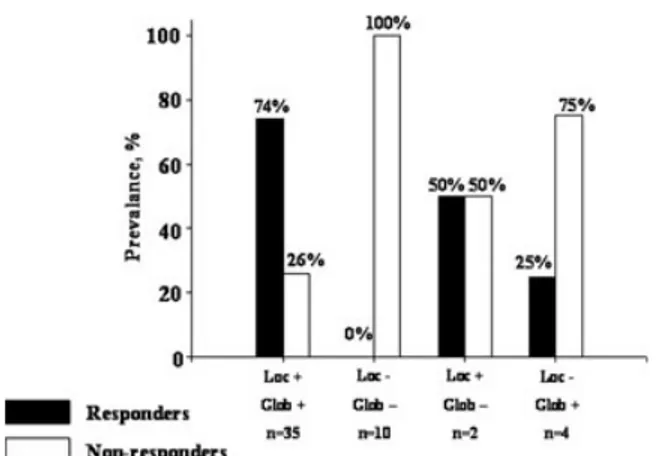
Among patients with local viability (i.e., via-bility in the region of the pacing lead), 27 (73%) were responders corresponding to 96% of all re-sponders. Conversely, in patients with global viability (i.e., ≥ 4 viable segments) without lo-cal viability (n = 4), only one patient (25%) was identified as a responder. In the absence of lo-cal and global viabilities, all patients (n = 10, 100%) were nonresponders (Fig. 2).
TABLE II Echocardiographic Data
All Patients Responders Nonresponders
Variables (n = 51) (n = 28, 55%) (n = 23, 45%) P-Value
LV geometry and function
LV end-diastolic volume (ml) 214 ± 67 211 ± 69 217 ± 65 0.75 LV end-systolic volume (ml) 178 ± 63 177 ± 67 180 ± 67 0.90 LV end-diastolic diameter (mm) 67 ± 8 66 ± 8 68 ± 8 0.26 LV end-systolic diameter (mm) 59 ± 9 61 ± 9 57 ± 3 0.13 End-systolic SI (%) 63 ± 9 64 ± 9 63 ± 9 0.87 End-diastolic SI (%) 69 ± 9 66 ± 2 65 ± 2 0.82 LV stroke volume (ml) 42 ± 12 39 ± 12 46 ± 2 0.06 LV ejection fraction (%) 19 ± 7 18 ± 8 19 ± 6 0.70 Asynchronism Interventricular (ms) 45 ± 27 43 ± 26 47 ± 28 0.63 Intraventricular (ms) 83 ± 25 87 ± 25 79 ± 24 0.23
No. of akinetic segments
Rest 9.5 ± 3 9.4 ± 3 10 ± 3 0.26
Dobutamine 8.4 ± 3 7.7 ± 3 9 ± 3 0.06
Wall motion score index
Rest 3.5 ± 0.4 3.41 ± 0.42 3.55 ± 0.25 0.15
Dobutamine 3.1 ± 0.5 2.97 ± 0.41 3.25 ± 0.48 0.03
Viability
No. of viable segments 4.9 ± 3 5.8 ± 1.94 3.87 ± 2.99 0.007
More than viable segments, n (%) 39 (76) 27 (96) 12 (52) <0.0001
Viability in the region of the lead, n (%) 37 (73) 27 (96) 10 (43) <0.0001 Lead placement
Posterior, n (%) 34 (66) 20 (71) 14 (61) 0.83
Lateral, n (%) 15 (30) 8 (29) 7 (30) 0.87
Anterior, n (%) 2 (4) – 2 (9) –
LV = left ventricular; ED = end-diastolic; ES = end-systolic; SI = sphericity index. Impact of Viability and Mitral Regurgitation
on CRT Response
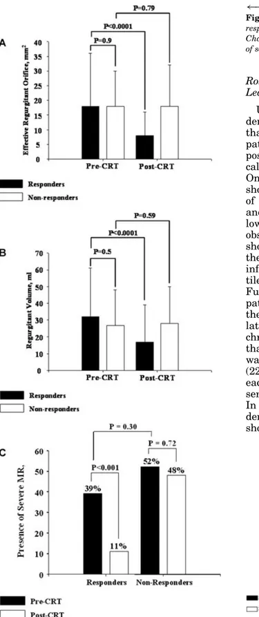
The prevalence of MR between responders and nonresponders was not statistically dif-ferent before and after CRT (pre-CRT: 93% vs. 83%, P = 0.26, post-CRT: 83% vs. 85%, P = 0.96). Moreover, there was no significant difference in baseline MR severity between groups (Fig. 3A), and whereas in responders ERO and regurgitant volume were significantly reduced following CRT, there was no significant change in nonresponders (Fig. 3A and B). In-deed, in responders, ERO was reduced by 57 ± 24% (from 18 ± 12 mm2 to 8 ± 8 mm2, P =
< 0.001). The percentage of patients with se-vere MR (ERO ≥ 20 mm2) was also not
sta-tistically different between groups before CRT (Fig. 3C). Only four responders had no MR be-fore CRT. In receiver-operating characteristics curves, the presence of MR, as well as the pres-ence of viability on the region of the pacing lead, was associated with excellent sensitivity
(93% and 96%) but with low specificity (17% and 56%) in predicting acute CRT response. Com-bining the presence of MR and viability in the region of the pacing lead yield the best combi-nation of sensitivity and specificity (89%, 70%)
(Fig. 4).
Discussion
The main finding of the present study showed that for acute benefit in ischemic and non-ischemic cardiomyopathies, CRT requires the presence of myocardial viability. A direct re-lationship between improvement in WMSI as assessed during low-dose dobutamine infusion and the improvement in LV stroke volume and reduction in ERO after CRT was described. This study shows the ability of DSE to predict acute response to CRT in patients with drug-refractory systolic heart dysfunction and signi-ficative intraventricular dyssynchrony.
Lastly, the combined presence of viability in the region of the pacing lead defined as viability
Figure 1. A. Correlation between changes in WMSI
(rest/dobutamine) and changes in stroke volume after CRT.
B. Correlation between changes in WMSI (rest/dobutamine)
and changes in ERO after CRT.
in two contiguous segments and MR predicts acute response with the best accuracy.
Effect of Global Viability
Wall motion response during dobutamine infusion is useful in predicting functional myocardial recovery in patients with ischemic and nonischemic heart diseases.35,39–41 The
clinical response to treatments such as β-blockade or revascularization in patients with LV systolic dysfunction has been shown to be dependent on the presence and extent of viable myocardium. Although CRT improves cardiac function by other mechanisms than revascu-larization or by up-regulation of sarcoplasmic reticulum calcium ATPase (beta-blockade), the relationship between viability and CRT bene-fit still holds.27–31 When myocytes have been
supplanted by replacement fibrosis because of cell death and interstitial remodeling, medical
Figure 2. Percentage of responders to CRT for four different
patient categories based on the presence or the absence of viability in the region of the pacing lead (local +/local −) in combination with the presence or the absence of four or more viable segments (global +/global −).
therapy and CRT may not improve LV func-tion. However, studies evaluating the relation-ship between CRT and myocardial viability are scarce. Ypenburg et al. evaluated and demon-strated that besides the presence of LV dyssyn-chrony, myocardial contractile reserve (result-ing in a ≥7.5% increase in LV ejection fraction during dobutamine infusion) predicts LV re-verse remodeling and improvement in LV function, 6 months after CRT implantation. An-other study in 67 patients (34% ischemic) re-vealed that the presence of contractile myocar-dial reserve was an independent predictor of event-free survival after CRT. Using a cutoff value of 25% increase in dobutamine LV ejec-tion fracejec-tion exhibits a sensitivity of 70% and a specificity of 62% for predicting major cardiac events.31 Hummel et al., in 21 CRT patients
(100% ischemic), evaluated myocardial viabil-ity by myocardial contrast echocardiography.30
The LV systolic performance was assessed by echocardiography on the day after implanta-tion. In that study, acute improvement in LV stroke volume was significantly correlated with the degree of viability as determined by the per-fusion score index.30 In our study, we related
global viability to acute response to CRT. Im-provement in LV stroke volume correlated (r = 0.45, P = 0.0012) with improvement in WMSI during the dobutamine test. Moreover, respon-ders showed a greater number of viable seg-ments (5.8 ± 1.94 vs. 3.87 ± 2.99, P = 0.007).
The presence of more than four viable seg-ments predicted acute response to CRT with a high sensitivity but with a low specificity.
←−−−−−−−−−−−−−−−−−−−−−−−−−−−−−−−−−−−−− Figure 3. Quantification of MR in responders and
non-responders before and after CRT. A. Changes in ERO; B. Changes in regurgitant volume; C. Changes in the presence of severe MR (≥20 mm2)
Role of Viability in the Region of the Pacing Lead
Using contrast-enhanced MRI, Bleeker et al. demonstrated in 40 patients (100% ischemic) that CRT did not reduce LV dyssynchrony in patients with transmural scar tissue in the posterolateral LV segments, resulting in clini-cal and echocardiographic nonresponse to CRT. Only 14% of patients with a posterolateral scar showed response to CRT. Even in the subset of patients with intraventricular asynchrony and postero-lateral scar, the response rate was low (n = 2, 18%).32 Ypenburg et al. recently
observed in 31 CRT patients that responders showed an increase in strain in the region of the LV pacing lead during low-dose dobutamine infusion while nonresponders had no contrac-tile reserve (absence of an increase in strain).27
Furthermore, Lim et al. demonstrated that only patients (n = 19) with contractile reserve in the LV target site for pacing (lateral, postero-lateral) presented a decrease in LV dyssyn-chrony with CRT.28 The authors also showed
that the mean increase of LV stroke volume was greater in patients with contractile reserve (22% vs. 0%). The number of viable segments in each wall showing viability (i.e., contractile re-serve) was however not stated in that study. In line with these results, the present study demonstrated that acute responders to CRT showed viability in the region of the pacing lead
Figure 4. Performance of viability and MR evaluated
significantly more often than nonresponders. It appears likely that viability of the paced seg-ments is the crucial factor mediating the influ-ence of viability (local viability vs. global) on re-sponse to CRT. Of interest, in the present study, in the absence of viability in the region of the pacing lead, three patients (75%) did not show acute response even if they had more than four viable segments. Moreover, one patient (50%) was a responder in the presence of only three viable segments (i.e., only in the region of the pacing lead). Therefore, it is likely that acute improvement in LV stroke volume after biven-tricular pacing is driven mainly by viability of the paced myocardial segments. However, viability in the region of the pacing lead pre-dicts acute response with an excellent sensitiv-ity (96%) but with a low specificsensitiv-ity (56%). Com-bining viability in the region of the pacing lead and MR predicts acute response after CRT with the best accuracy suggesting that reduction of MR is almost mandatory in acute responders. The principal mechanism explaining this acute change in LV stroke volume is probably, at least in part, related to the diminution of functional MR. CRT appears to increase mitral closing force, coordinate tethering forces on papillary muscles, and increase the leaflet coaptation sur-face to reduce MR.13–15 Previous studies have
reported conflicting results regarding the pres-ence and severity of MR and response to CRT. Of interest, none of those studies have evalu-ated LV viability and its relationship with MR presence or severity and LV remodeling.42–45In
our study, only four responders did not have MR in pre-CRT. Of interest, those patients had nonischemic cardiomyopathy, viability in the region of the pacing lead, and a mean of 8 viable segments, which suggests that acute response may occur in patients without MR before CRT but only in the presence of substantial viable myocardium.
Clinical Implications
Our present results confirm earlier sugges-tions that the absence of viability in the region of the LV pacing lead may prohibit response to CRT. This study focuses on acute response and its relationship with viability and MR. Even if acute response after CRT underestimates long-term effects, identification of such patients may be important since acute hemodynamic re-sponse to CRT predicts long-term clinical out-come and acute responders may represent more than 70% of all eventual responders.8 Our
re-sults support that the presence of viability in the region of the pacing lead is a better predic-tor of acute response over the burden of global viability. Also, in the presence of local viability, a decrease of MR seems mandatory in most pa-tients with acute response. Of interest, in our study, the criterion used to define the presence of “significant” viability in the region of the LV pacing lead (presence of viability in two con-tiguous segments) is simple, rapid, and easily applicable in the context of clinical evaluation before CRT. This underlines the importance of assessing local viability in order to guide LV positioning. The region of myocardium without viability should be avoided as a final resting place for LV lead placement to maximize the possibility of therapeutic benefit.
Study Limitations
These results should be regarded cautiously, and some limitations should be underlined. First, the lack of difference between responders and nonresponders regarding the presence of CAD may be the result of the small number of patients, and results should be confirmed by a larger study. Second, although the difference was not statistically different, more nonrespon-ders took digoxin and spironolactone than re-sponders and showed a higher incidence of class IV NYHA; therefore, because of the sample size (n = 51) and the heterogeneity of the popula-tion studied, those data should be interpreted cautiously until confirmed by suitably powered clinical trials that are undoubtedly needed. Third, dyssynchrony was defined by longitudi-nal tissue Doppler imaging using a cutoff value of 50 ms as inclusion criterion. Combining lon-gitudinal and radial dyssynchrony indices as inclusion criteria could have been helpful in choosing a more homogenous population prone to CRT response.46
Conclusion
In this study, we demonstrated that myocar-dial viability is an important factor influenc-ing acute hemodynamic response to CRT. In acute responders, significant MR reduction is frequent. The combined presence of MR and vi-ability in the region of the pacing lead defined as two contiguous viable segments, determined by DSE predicts acute response to CRT with the best accuracy.
References
1. Cazeau S, Leclercq C, Lavergne T, et al: Effects of multisite biventricular pacing in patients with heart failure and intraventricular conduction delay. N Engl
J Med 2001;344:873–880.
2. Auricchio A, Stellbrink C, Sack S, et al: Pacing Ther-apies I Congestive Heart Failure (PATH-CHF) Study Group: Long-term clinical effect of hemodynamically optimized cardiac resynchronization therapy in pa-tients with heart failure and ventricular conduction delay. J Am Coll Cardiol 2002;39:2026–2033. 3. Higgins SL, Hummel JD, Niazi IK, et al: Cardiac
resynchronization therapy for the treatment of heart failure in patients with intraventricular conduction delay and malignant ventricular arrhythmias. J Am
Coll Cardiol 2003;42:1454–1459.
4. Abraham WT, Fisher WG, Smith AL, et al. MIRACLE Study Group: Multicenter InSync randomized clini-cal evaluation. Cardiac resynchronization in chronic heart failure. N Engl J Med 2002;346:1845–1853. 5. Bristow MR, Saxon LA, Boehmer J, et al. Comparison
of Medical Therapy, Pacing and Defibrillation in Heart Failure (COMPANION) Investigators: Cardiac resyn-chronization therapy with or without an implantable defibrillator in advanced chronic heart failure. N Engl
J Med 2004;350:2140–2150.
6. Cleland JG, Daubert JC, Erdmann E, et al: The effect of cardiac resynchronization on morbidity and mor-tality in heart failure. N Engl J Med 2005;352:1539– 1549.
7. Tournoux FB, Alabiad C, Fan D, et al: Echocardio-graphic measure of acute haemodynamic response af-ter cardiac resynchronization therapy predicts long term clinical outcome. Eur Heart J 2007;28:1143– 1148.
8. Gorscan III J, Kanzaki H, Bazaz R, et al: Usefulness of echocardiographic tissue synchronization imaging to predict acute response to cardiac resynchronization therapy. Am J Cardiol 2004;93:1178–1181.
9. Bleeker GB, Mollema SA, Holman ER, et al: Left ventricular resynchronization is mandatory for re-sponse to cardiac resynchronization therapy: Anal-ysis in patients with echocardiographic evidence of left ventricular dyssynchrony at baseline. Circulation 2007;116:1440–1448.
10. Nof E, Glikson M, Bar-Lev D, et al: Mechanism of diastolic mitral regurgitation in candidates for cardiac resynchronization therapy. Am J Cardiol 2006;97:1611–1614.
11. Ypenburg C, Lancellotti P, Tops LF, et al: Mechanism of improvement in mitral regurgitation after cardiac resynchronization therapy. Eur Heart J 2008;29:757– 765.
12. Breithardt OA, Sinha AM, Schwammenthal E, et al: Acute effects of cardiac resynchronization therapy on functional mitral regurgitation in advanced sys-tolic heart failure. J Am Coll Cardiol 2003;41:765– 770.
13. Kanzaki H, Bazaz R, Schwartzman D, et al: A mecha-nism for immediate reduction in mitral regurgitation after cardiac resynchronization therapy. J Am Coll
Cardiol 2004;44:1619–1625.
14. Vinereanu D, Turner MS, Bleasdale RA, et al: Mech-anisms of reduction of mitral regurgitation by cardiac resynchronization therapy. J Am Soc Echocardiogr 2007;20:54–62.
15. Karvounis HI, Dalamaga EG, Papadopoulos CE, et al:
Improved papillary muscle function attenuates func-tional mitral regurgitation in patients with dilated cardiomyopathy after cardiac resynchronization ther-apy. J Am Soc Echocardiogr 2006;19:1150–1157. 16. Agricola E, Oppizzi M, Galderisi M, et al: Role of
re-gional mechanical dyssynchrony as a determinant of functional mitral regurgitation in patients with left ventricular systolic dysfunction. Heart 2006;92:1390– 1395.
17. Madaric J, Vanderheyden M, Van Laethem C, et al: Early and late effects of cardiac resynchronization therapy on exercise-induced mitral regurgitation: Re-lationship with left ventricular dyssynchrony, remod-eling and cardiopulmonary performance. Eur Heart J 2007;28:2134–2141.
18. Diaz-Infante E, Mont L, Leal J, et al: Predictors of lack of response to resynchronization therapy. Am J
Cardiol 2005;95:1436–1440.
19. Nelson GS, Curry CW, Wyman BT, et al: Predic-tors of systolic augmentation from left ventricular preexcitation in patients with dilated cardiomyopa-thy and intraventricular conduction delay. Circulation 2000;101:2703–2709.
20. Auricchio A, Stellbrink C, Butter C, et al. Pacing ther-apies in Congestive Heart Failure II Study Group, Guidant Heart Failure Research Group: Clinical ef-ficacy of cardiac resynchronization therapy using left ventricular pacing in heart failure patients stratified by severity of ventricular conduction delay. J Am Coll
Cardiol 2003;42:2109–2116.
21. Kass DA: Predicting cardiac resynchronization re-sponse by QRS duration: The long and short of it. J
Am Coll Cardiol 2003;42:2125–2127.
22. Leclercq C, Kass DA: Retiming the failing heart: Prin-ciples and current status of cardiac resynchronization.
J Am Coll Cardiol 2002;39:194–201.
23. White JA, Yee R, Yuan X, et al: Delayed enhance-ment magnetic resonance imaging predicts response to cardiac resynchronization therapy in patients with intraventricular dyssynchrony. J Am Coll Cardiol 2006;48:1953–1960.
24. Ypenburg C, Schalij MJ, Bleeker GB, et al: Extent of viability to predict response to cardiac resynchroniza-tion therapy in ischemic heart failure patients. J Nucl
Med 2006;47:1565–1570.
25. Henneman MM, Van Der Wall EE, Ypenburg C, et al: Nuclear imaging in cardiac resynchronization ther-apy. J Nucl Med 2007;48:2001–2010.
26. Chalil S, Foley PWX, Muyhaldeen SA, et al: Late gadolinium enhancement-cardiovascular magnetic resonance as a predictor of response to cardiac resyn-chronization therapy in patients with ischaemic car-diomyopathy. Europace 2007;9:1031–1037.
27. Ypenburg C, Sieders A, Bleeker GB, et al: Myocar-dial contractile reserve predicts improvement in left ventricular function after cardiac resynchronization therapy. Am Heart J 2007;154:1160–1165.
28. Lim P, Bars C, Mitchell-Hegg L, et al: Importance of contractile reserve for CRT. Europace 2007;9:739– 743.
29. Ypenburg C, Schalij MJ, Bleeker GB, et al: Impact of viability and scar tissue on response to cardiac resyn-chronization therapy in ischaemic heart failure pa-tients. Eur Heart J 2007;28:33–41.
30. Hummel JP, Lindner JR, Belcik JT, et al: Extent of myocardial viability predicts response to biventricu-lar pacing in ischemic cardiomyopathy. Heart Rhythm 2005;2:1211–1217.
31. Da Costa A, Th´evenin J, Roche F, et al: Prospective validation of stress echocardiography as an identi-fier of cardiac resynchronization therapy responders.
Heart Rhythm 2006;3:406–413.
32. Bleeker GB, Kaandorp TAM, Lamb JH, et al: Ef-fect of posterolateral scar tissue on clinical and echocardiographic improvement after cardiac resyn-chronization therapy. Circulation 2006;113:969–976. 33. Zoghbi WA, Enriquez-Sarano M, Foster E, et al:
Rec-ommendations for evaluation of the severity of na-tive valvular regurgitation with two-dimensional and Doppler echocardiography. J Am Soc Echocardiogr 2003;16:777–802.
34. Bader H, Garrigue S, Lafitte S, et al: Intra-left ven-tricular electromechanical asynchrony: A new inde-pendent predictor of severe cardiac events in heart failure patients. J Am Coll Cardiol 2004;43:248–256. 35. deFilippi CR, Willett DL, Irani WN, et al: Compari-son of myocardial contrast echocardiography and low-dose dobutamine stress echocardiography in predict-ing recovery of left ventricular function after coronary revascularization in chronic ischemic heart disease.
Circulation 1995;92:2863–2868.
36. Cigarroa CG, deFilippi CR, Brickner ME, et al: Dobu-tamine stress echocardiography identifies hibernating myocardium and predicts recovery of left ventricular function after coronary revascularization. Circulation 1993;88:430–436.
37. Schiller NB, Shah PM, Crawford M, et al: Recommen-dations for quantitation of the left ventricle by two-dimensional echocardiography. J Am Soc
Echocar-diogr 1989;2:358–367.
38. Butter C, Auricchio A, Stellbrink C, et al: Effect of resynchronization therapy stimulation site on the sys-tolic function of heart failure patients. Circulation 2001;104:3026–3029.
39. Qureshi U, Nagueh SF, Afridi I, et al: Dobu-tamine echocardiography and quantitative rest-redistribution201TI tomography in myocardial
hiber-nation: Relation of contractile reserve to201TI uptake
and comparative prediction of recovery of function.
Circulation 1997;95:626–635.
40. Ramahi TM, Longo MD, Cadariu AR, et al: Dobutamine-induced augmentation of left ventricular ejection fraction predicts survival of heart failure pa-tients with severe non-ischemic cardiomyopathy. Eur
Heart J 2001;22:849–856.
41. Eichhorn EJ, Grayburn PA, Mayer SA, et al: Myocar-dial contractile reserve by dobutamine stress echocar-diography predicts improvement in ejection fraction with β-blockade in patients with heart failure. The β-blocker evaluation of survival trial (BEST).
Circu-lation 2003;108:2336–2341.
42. Cappola TP, Harsch MR, Jessup M, et al: Predictors of remodeling in the CRT Era: Influence of mitral regur-gitation, BNP and gender. J Card Fail 2006;12:182– 188.
43. Cabrera-Bueno F, Garcia-Pinilla JM, Pena-Hernandez J, et al: Repercussion of functional mitral regurgitation on reverse remodelling in cardiac resynchronization therapy. Europace 2007;9:757–761. 44. Diaz-Infante E, Mont L, Leal J, et al: Predictors of lack of response to resynchronization therapy. Am J
Cardiol 2005;95:1436–1440.
45. Reuter S, Garrigue S, Barold SS, et al: Comparison of characteristics in responders versus non-responders with biventricular pacing for drug-resistant conges-tive heart failure. Am J Cardiol 2002;89:346–350. 46. Gorcsan J, Tanabe M, Bleeker GB, et al: Combined
longitudinal and radial dyssynchrony predicts ven-tricular response after resynchronization therapy.


