HAL Id: dumas-01154337
https://dumas.ccsd.cnrs.fr/dumas-01154337
Submitted on 21 May 2015
HAL is a multi-disciplinary open access archive for the deposit and dissemination of sci-entific research documents, whether they are pub-lished or not. The documents may come from teaching and research institutions in France or abroad, or from public or private research centers.
L’archive ouverte pluridisciplinaire HAL, est destinée au dépôt et à la diffusion de documents scientifiques de niveau recherche, publiés ou non, émanant des établissements d’enseignement et de recherche français ou étrangers, des laboratoires publics ou privés.
Early brain MRI interest in infective endocarditis
clinical management: a prospective study
Julia Champey
To cite this version:
Julia Champey. Early brain MRI interest in infective endocarditis clinical management: a prospective study. Human health and pathology. 2015. �dumas-01154337�
AVERTISSEMENT
Ce document est le fruit d'un long travail approuvé par le
jury de soutenance et mis à disposition de l'ensemble de la
communauté universitaire élargie.
Il n’a pas été réévalué depuis la date de soutenance.
Il est soumis à la propriété intellectuelle de l'auteur. Ceci
implique une obligation de citation et de référencement
lors de l’utilisation de ce document.
D’autre part, toute contrefaçon, plagiat, reproduction illicite
encourt une poursuite pénale.
Contact au SICD1 de Grenoble :
thesebum@ujf-grenoble.fr
LIENS
LIENS
Code de la Propriété Intellectuelle. articles L 122. 4
Code de la Propriété Intellectuelle. articles L 335.2- L 335.10
http://www.cfcopies.com/juridique/droit-auteurAnnée : 2014/2015
UNIVERSITE JOSEPH FOURIER FACULTE DE MEDECINE DE GRENOBLE
THESE PRESENTEE POUR L'OBTENTION DU DOCTORAT EN MEDECINE
DIPLOME D'ETAT
Early brain MRI interest in infective endocarditis clinical management:
A prospective study
Julia, Hélène, CHAMPEY née le 07 mars 1985 à Toulon
Thèse soutenue publiquement à la Faculté de Grenoble* le 30 avril 2015
devant le jury composé de :
Monsieur le Professeur Jean-François Payen, président du jury
Madame le Professeur Carole Schwebel
Monsieur le Professeur Patrice François
Monsieur le Docteur Gilles Francony
Madame le Docteur Hélène Bouvaist
Madame le Docteur Patricia Pavese, directrice de thèse
* La faculté de Médecine de Grenoble n’entend donner aucune approbation ni improbation aux opinions émises dans les thèses ; ces opinions sont considérées comme propres à leur auteurs.
SERMENT D’HIPPOCRATE
En présence des Maîtres de cette Faculté, de mes chers condisciples et devant l’effigie d’HIPPOCRATE,
Je promets et je jure d’être fidèle aux lois de l’honneur et de la probité dans l’exercice de la Médecine.
Je donnerais mes soins gratuitement à l’indigent et n’exigerai jamais un salaire au-dessus de mon travail. Je ne participerai à aucun partage clandestin d’honoraires.
Admis dans l’intimité des maisons, mes yeux n’y verront pas ce qui s’y passe ; ma langue taira les secrets qui me seront confiés et mon état ne servira pas à corrompre les mœurs, ni à favoriser le crime.
Je ne permettrai pas que des considérations de religion, de nation, de race, de parti ou de classe sociale viennent s’interposer entre mon devoir et mon patient.
Je garderai le respect absolu de la vie humaine.
Même sous la menace, je n’admettrai pas de faire usage de mes connaissances médicales contre les lois de l’humanité.
Respectueux et reconnaissant envers mes Maîtres, je rendrai à leurs enfants l’instruction que j’ai reçue de leurs pères.
Que les hommes m’accordent leur estime si je suis fidèle à mes promesses. Que je sois couvert d’opprobre et méprisé de mes confrères si j’y manque.
LISTE DES PROFESSEURS DES UNIVERSITES ET
MAITRES DE CONFERENCE DES UNIVERSITES –
PRATICIENS HOSPITALIERS
ANNEE 2014-2015
Université Joseph Fourier Faculté de Médecine Domaine de la Merci 38700 La Tronche
Doyen de la Faculté de Médecine :
Monsieur le Professeur Jean-Paul ROMANET
TABLE OF CONTENTS
ABSTRACT ... 9 1. RATIONAL (context and hypothesis) ... 13 2. PATIENTS AND METHODS ... 15
A. Population B. Data collection
C. Magnetic Resonance Imaging (MRI) D. Assessment of MRI utility
E. Statistical analysis
3. RESULTS ... 19 F. Study population
G. IE characteristics H. MRI results
I. Assessment of diagnosis and treatment by panels of experts
4. DISCUSSION ... Erreur ! Signet non défini. 5. CONCLUSION ... 26 6. REFERENCES ... 28 7. FIGURES AND TABLES ... 31
Abstract
IntroductionIn Infective Endocarditis (IE), brain Magnetic Resonance Imaging (MRI) is helpful to diagnose clinically silent neurologic events, especially micro ischemic and micro haemorrhagic lesions. Brain MRI may be fitted in a complete imaging check-up including, at an early stage, chest abdomen, pelvis Computered Tomography (CT) and echocardiography for a thoroughly asymptomatic lesions mapping.
Objective
We aimed to assess the usefulness of systematic early brain MRI in IE diagnosis and medico-surgical cares management.
Methods
During one year, all patients admitted in one of the 3 hospitals participating and fulfilling Duke’s criteria for definite or possible IE, underwent cerebral MRI with angiography within the 7 days of IE-suspicion.
To assess the MRI contribution, records were analyzed a posteriori by 8 panels of experts. For each case, one record with and one record without the MRI results, were randomly assigned to 2 panels who determined theoretical diagnosis and treatment. Inter-rater concordances were assessed through Cohen Kappa test.
Results
Thirty-seven brain MRI were realized in a median time of 5 days after inclusion. MRI was pathological in 26 patients (70%) showing 62 % of micro ischemia and 58% of microbleeds.
Experts advices were significantly concordant between the 2 evaluations with or without the MRI results. Diagnosis differed in 2 cases (5.4%) with an upgrading of diagnosis from possible to definite IE using MRI results. Discrepancies concerning therapeutic attitudes occurred in 8 cases (21.5 %) including one change in the indication to operate, 3 modifications of timing of surgery namely 2 earlier and one postponed surgery with the MRI results and 1 disagreement over the choice of prosthesis. Changes concerned anticoagulation in 2 cases. Rifampicin was added to the antimicrobial therapy in 1 case.
Conclusion
Because it does not seem to plenty affect diagnosis and medico-surgical cares; our results do not support the need for a systematic use of early brain MRI in IE. Further studies are necessary to clearly define if MRI is mandatory in IE management within a multidisciplinary approach; the better timing to realize it and the subset of patients in which it could be beneficial.
Keywords
Infective endocarditis, brain magnetic resonance imaging, brain micro ischemia, cerebral microbleeds, cardiac surgery procedures.
Résumé
IntroductionL’Imagerie encéphalique par Résonance Magnétique (IRM) est utile pour le diagnostic des lésions neurologiques silencieuses, particulièrement pour la mise en évidence des micro-ischémies et microhémorragies dans l’Endocardite Infectieuse (EI). Associée au scanner thoraco-abdomino-pelvien et à l’échocardiographie, l’IRM permet de dresser une cartographie précoce et exhaustive des lésions emboliques septiques.
Objectifs
Cette étude avait pour objectif d’évaluer la place de l’IRM cérébrale dans le diagnostic et la prise en charge médico-chirurgicale de l’EI.
Méthode
Tous les patients hospitalisés pendant un an dans un des 3 hôpitaux participants, pour une EI certaine ou probable, étaient éligibles pour l’étude. Il était proposé de réaliser une IRM encéphalique avec angiographie avant le 7ème jour d’évolution.
Pour évaluer l’apport de l’IRM, les dossiers ont été analysés, a posteriori, par 8 panels d’experts. Pour chaque patient, un dossier avec IRM et un dossier sans IRM étaient assignés au hasard à 2 panels qui statuaient sur le diagnostic et proposaient un traitement. La concordance des avis sur un même cas était évaluée à l’aide du test Kappa de Cohen.
Résultats
Trente-sept patients ont bénéficié d’une angio-IRM cérébrale dans un délai médian de 5 jours. Vingt-six IRM étaient pathologiques (70%) montrant 62% de micro-ischémies et 58% de microbleeds.
Les avis des experts étaient significativement concordants entre les deux évaluations avec ou sans IRM. Le diagnostic d’EI différait dans 2 cas (5.4%) passant de probable sans IRM à certain avec IRM. Des discordances concernant les traitements ont été notées dans 8 cas (21.5 %). Dans l’évaluation avec IRM : une indication de chirurgie valvulaire n’était pas retenue ; le délai opératoire était raccourci dans 2 cas et retardé dans un cas ; un choix différent de prothèse a été fait ; Deux choix différents d’anticoagulation ont été fait. la rifampicine était ajoutée à l’antibiothérapie dans 1 cas.
Conclusion
L’utilisation précoce de l’IRM encéphalique ne semble pas influencer de façon majeure le diagnostic ou le traitement médico-chirurgical des EI. D’autres études sont nécessaires pour clairement définir l’intérêt de l’IRM cérébrale dans la prise en charge des endocardites au sein d’une concertation multidisciplinaire, le délai de réalisation le plus approprié et les patients pour lesquels cet examen serait plus particulièrement utile.
Mots-clés
Endocardite infectieuse, imagerie encéphalique par résonance magnétique, micro-ischémies cérébrales, microbleeds cérébraux, chirurgie cardiaque.
1. RATIONAL (context and hypothesis)
Infective Endocarditis (IE) is a serious disease carrying a considerable risk of morbi-mortality (1–5). Despite improvements in therapy, mortality remains high from 15 to 22 % (5). Mortality rates vary among subgroups of patients reaching 40 % for Staphylococcus aureus prosthetic valve IE patients and 50% in Intensive Care Unit (ICU) (1).
With regards to extra-cardiac complications, cerebral lesions are frequent with an incidence rate ranging from 10 to 65 % (5,6). Recent data suggest that neurological failure is a major determinant for short-term prognosis (2). Neurologic damages are numerous and various (7,8). The IE neuroimaging findings can be classified as follows: ischemic stroke, intra parenchyma macro and micro-haemorrhages, subarachnoid haemorrhage, mycotic aneurysm, brain abscess and meningitis (5). Because a part of them are clinically silent (9,10), some medical teams suggest to perform neuroimaging for all IE patients, even those without clinical neurologic symptoms (11–14). Additionally, since embolic risk and neurologic worsening remain high during the first week of anti-microbial therapy (15,16), neuroimaging diagnosis need to be performed early in care management (17,18).
First, we performed a systematic review of the literature regarding the overall brain MRI interest in acute IE. [Annex 1, to be published]. Because Brain Magnetic Resonance Imaging (MRI) is more sensitive and less toxic than Computered Tomography (CT) scan, this exam appears to have more value to detect cerebral micro lesions. Diffusion-weighted echo planar imaging (DWI) and T2*-weighted Gradient Recalled Echo (GRE) sequences are especially used in this field (17,19).
Secondly, in order to clarify the prevalence and the type of cerebral MRI lesions in IE (including cerebral micro-haemorrhages, also call Cerebral Microbleeds (CMBs)) and to compare them with CT scan lesions, we achieved a retrospective observational study, from 2010 to 2012, in the Grenoble University hospital [Annex 2, in revision]. In the 62 IE patients of this series, CT-scan was abnormal in 26 (49%) patients whereas MRI demonstrated lesions in 32 (74%) cases. Regarding asymptomatic patients, 37 of 43 (74 %) had MRI lesions while 12 of 53 (46%) had CT-scan lesions. MRI shown more ischemia lesions (48% vs. 35%) and micro bleeds (34% vs. 0%) than CT-scan. This preliminary study was concordant with literature data (9,11,12,20). Allowing an early and precise neurological status, MRI may influence therapeutic plans (14) and lead to faster surgical decision (7,11).
Actually, if urgent surgery is well agreed for patients with poor hemodynamic tolerance, embolic events remain a controversial indication for surgery (21,22). Risk factors of embolic events are well known: Staphylococcus aureus infection (23) more so in a mitral location, large (> 10 mm) and highly mobile vegetation, other systemic embolisms (13–15). The change in the vegetation size with antibiotics is also relevant (23).
For those patients, several studies have shown that in order to prevent recurrent embolic events and to decrease mortality (24,25), surgery has to be performed without delay (13,20,26).
If cerebral bleeding or major ischemia is a temporary surgical contra-indication, cardiac surgery can be safely performed after silent cerebrovascular complications (16,27,28). In this way, brain MRI fitted in a complete imaging check-up could lead clinicians to change the surgical strategy with regards to cardiac surgery indication and timing of valve replacement (14).
The additional information provided by cerebral MRI may also impact diagnosis classification (14). If small ischemic lesions, as peripheral manifestations of IE, obviously upgrade the Duke’s classification, the role of CMBs is still unclear. This small foci of haemorrhage, only visible by using T2*- weighted GRE MRI sequences, have a strong IE association (9) with a specific distribution in cortical areas (9,19). The value of this phenomenon as a pyogenic vasculitis (7) may testify the infection severity (19). As an IE feature, CMBs could also be considered like a new imaging minor criterion (19).
Overall, few studies analyzed the interest of brain MRI in the management of IE. The effect on clinical decision-making and the influence of micro ischemia and CMBs on diagnosis have not been fully evaluated. Prospective data are limited. IE guidelines are still mainly based on experts opinions (5). [Annex 1]
The present study aims to evaluate the impact of systematic early brain MRI in IE management: diagnosis of IE according to Duke’s modified classification [Annex 3] and treatment (surgical indication, time of surgery, anticoagulation management and antimicrobial choice).
2. PATIENTS AND METHODS
A. Study design
Between November 2013 and November 2014, an observational prospective multicentric study was conducted at the Grenoble University Hospital and at the Annecy and Chambery General Hospitals, France. IE patients underwent brain angio-MRI within the 7 days of diagnosis.
In a second step, records, with or without the MRI results, were randomly assigned to panels of experts whotheoretically chose diagnosis and treatments.
The protocol was approved by the local ethic committee.
B. Population
A consecutive series of patients hospitalized with IE was prospectively achieved during one year. Only adult patients (≥18 years) with definite or possible IE according to the modified international Duke’s criteria, and presenting at least a major criterion (evidence of endocardial involvement, repeated positive blood cultures) were enrolled in the study. After the diagnosis confirmation by an expert (infectious disease specialist or cardiologist), brain MRI was quickly performed.
The patients were screened thanks to daily blood cultures in collaboration with the bacteriology department and through contacts with physicians in the more concerned departments (cardiology, infectious disease, intensive care, cardiac surgery).
Patients were excluded if they had undergone cardiac surgery in emergency (during the first 24 hours after diagnosis), if they had a MRI absolute contraindication [Annex 5], if they presented an IE on an implantable device incompatible with a MRI achievement, or if IE initial cares were done in another hospital where more than a week of adapted antibiotic treatment was received.
C. Data collection
Demographic, clinical, biological and imaging data [Annex 3] were collected on a standardized form, at the inclusion and during the hospitalization. Chest abdomen pelvis and brain CT, transthoracic and/or transoesophageal echocardiographies were performed as clinically indicated as soon as possible. After discharge, a follow-up care was organized with an infectious disease specialist by a consultation at 3 months. In cases of missing data, the information was collected by calling the patient’s general practitioner.
D. MRI
Cerebral MRI with angiography was performed before the seventh day after diagnosis. The following type sequences were included in a standardized neuroimaging protocol:
Axial diffusion, axial FLAIR, axial T2 Gradient Echo Recalled, axial or sagittal T1, axial or sagittal T1 3D with gadolinium injection, intracranial Angiography MRI in time of fly and with gadolinium.
E. Assessment of MRI utility
Eight multidisciplinary panels of experts (Infectious Diseases Specialist, Cardiologist and Intensivist) met, a posteriori, and worked independently from each other. The panels were blinded for real care. The patient data, with or without the MRI results were randomly assigned to one panel of experts. Experts reviewed the whole patient’s medical history, biological and imaging results. Completing a standardized questionnaire, panels established the diagnosis and the best medico-surgical treatment for each case [Annex 4]. Treatment was supposed to be determined on the basis of available guidelines (22).
F. Statistical analysis:
Continuous variables were presented as median and interquartile ranges (IQR). Categorical variables were presented as numbers and percentages.
Cohen’s kappa coefficient was used to quantify the concordance between expert advices on diagnosis and therapeutic strategies, with and without MRI results, for each cases.
The level of 0.05 was used for statistical significance. Statistical analyses were performed with
3. RESULTS
A.
Study population
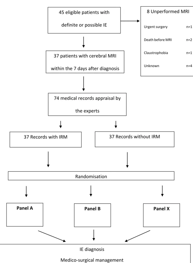
During the inclusion period, 247 patients with a possible or definite IE were recorded on the hospital database of the Grenoble University Hospital (155), the general hospital of Annecy (54) and Chambery (38) and 45 patients were eligible for the study. Among them, 37 underwent brain angio-MRI examination within the 7 days of management. The median duration from diagnosis to MRI was 5 days (IQR 3-7) (figure 1).
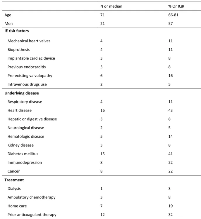
Patients’ baseline characteristics are detailed in table 1. The median age was 71 years (IQR 66-81).Twenty-six cases (70%) of IE developed on native valves. 11 patients (30%) had prosthetic material IE. The median Charlson comorbidity score (29) was 5 (IQR 3-6) and the forecast surgical risk (median Euroscore I) (30) was 14 (IQR 11-17).
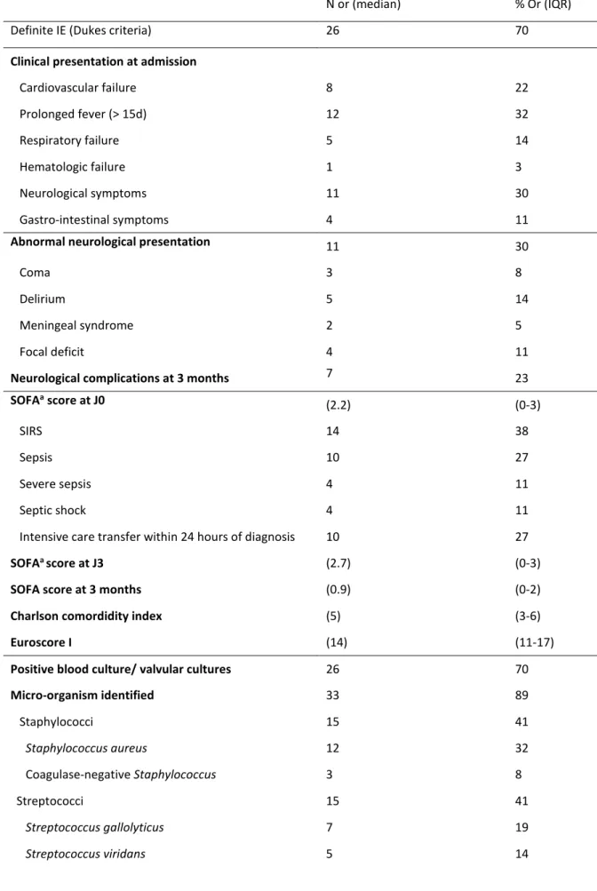
Clinical characteristics are listed in table 2. Twenty-six patients (70%) had a definite IE according to Duke’s criteria.Neurologic symptoms were present in eleven patients (30%): 5 deliriums (14%), 4 focal deficits (11%) 3 impaired consciousnesses (8%) with Coma Glasgow Scale under 14 and 2 meningeal syndromes (5%). IE was health-care associated in 10 cases (29%). Staphylococcus aureus was the most common microorganism, identified in 12 cases (32%). Echocardiography showed 30 vegetations (81%) with a 10 mm median length (IQR 5-14). Mitral and aortic valves were involved in respectively 11 (41%) and 13 (48%) cases. Three right-IE happened (8%). Five patients (17%) had severe regurgitation. Five trigon abscesses (15%) and 1 acute valve obstruction (3.5%) were diagnosed.
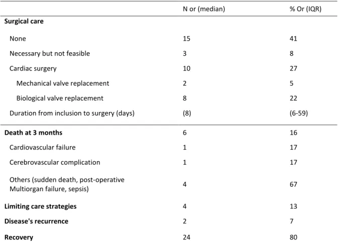
Six patients (16%) died within the 3 months follow-up.One patient died after cardiac surgery on the grounds of a multi-organ failure. Seven patients (23%) suffered from neurologic complications within the 3 months follow-up but only one died from this complication per se.
B. Neuroimaging findings
Neuroimaging data are summarized in table 4. IE-related lesions were highlighted in 26 patients (70%). MRI showed acute ischemia for 16 patients (62%). All but one were punctiform lesions. MRI detected CMBs in 15 cases (58%). Four patients (9%) had hemorrhagic lesions; 1 (4%) had a microbial aneurysms and 1 (4%) had a brain abscess. Among the 26 patients without neurological symptoms, silencious MRI lesions were detected in 8 (31%) cases.
C. Assessment of diagnosis and treatment by panels of experts
After analysis of expert’s reports, IE management was finally changed in 10 patients (27%) including diagnosis amendment for 2 patients (5.4%) and therapeutic change in 8 patients (21.5%).
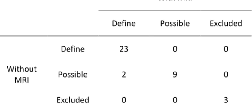
The IE diagnosis, according to Duke’s criteria, was upgraded in 2 cases (5.4%) from possible to define. Agreement for diagnosis evaluation was high between panels (ĸ coefficient 0.89 (95% CI, 0.74 to 1)) (Table 5)
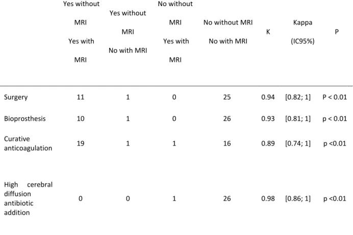
Variations in surgical strategies concerned 1 indication, 3 variations in timing of surgery out of 12 theoretical surgical patients (25%) i.e. one postponed and 2 earlier surgery with the MRI results, and the choice of 1 type of prosthesis. Medical treatment differed in 3 cases (8%):
anticoagulant therapy in 2 cases (5.4%) and antimicrobial therapy in 1 case (use of rifampicin). Inter-expertise concordance of panels was high concerning indication of surgery (ĸ coefficient of 0.94 (95% CI, 0.82 to 1), choice of kind of prosthesis (ĸ coefficient of 0.93 (95% CI, 0.81 to 1)), anticoagulation and antibiotic management (respectively ĸ coefficient of 0.89 (95% CI, 0.74 to 1) and of 0.98 (95% CI, 0.86 to 1)) (Table 6). Inter-expertise concordance was moderate for surgery delay (ĸ coefficient of 0.53 (95% CI, 0.17 to 0.84)) (Table 7).
Hence, IE management was not statistically influenced by MRI results.
4. DISCUSSION
Decisions of the experts differ in 27 % of patients in the light of MRI results but concordance between experts advices were high according to Cohen Kappa test results. Thus, there is no statistical argument proving that brain MRI could be associated with a major change on IE clinical decision-making.
In the literature, this approach had already yielded opposite advices. A recent French trial conducted on 130 IE patients demonstrated that the MRI findings before the seventh day of evolution changed diagnosis and therapeutic plans in 28% of patients including medical and surgical plan modifications in 18 % of cases (11). Accurately, excluding CMBs and solely basis on MRI results, Duval et al, achieved 14% of surgical plan adjustments. In this study, the methodology differed since a pair of 2 experts established IE diagnosis and therapeutic strategy 24 hours before and 24 hours after MRI achievement and the decision were compared. Experts may have overestimated the MRI usefulness by a confirmation bias. In the present study panels were blinded for the opinion of other groups and results are probably more reliable.
Some others elements could provide explanations for these results.
First, because of the major risk of neurologic worsening due to extracorporeal circulation, per operative hypotension and anticoagulant use (7,11), appropriate timing of surgery remains controversial; it is team-dependent. The earlier surgery policy in our referral center could explain the minor role of MRI in this study.
Moreover, new European guidelines incite to prevent recurrent embolisms and support the routine use of an early valve replacement (22). The early removal of vegetation is, beyond obvious contra-indications, the best way to impede neurologic injury.
Surgery is overall fostered before the end of the first week of antimicrobial treatment in cases of isolated large vegetation (15 mm) or in presence of a 10 mm vegetation following one or more clinical or even asymptomatic embolic events (15).
In the last thirty years, surgeon’s behavior changed and they operate earlier with less fear of relapse, complications and postoperative neurological worsening. Nonetheless, optimal therapeutic strategy may vary for individual patient and precocious surgical decision remains difficult as proved by the low inter-observer concordance rate concerning the timing of surgery in our study.
A multidisciplinary team should discuss the benefit-risk ratio and the period of surgery in litigious cases, with regards to the risk of fatal embolic recurrence and the risk of major cardiac surgery (31). The root role of comorbidities has assuredly to be considered. Thus, a randomized study comparing conventional treatment and early valve replacement revealed that, for patients with large vegetation, early surgery decreases the composite end point of death and embolic events though declining embolism. In this trial, patients were young with a low operative risk. MRI should maybe be more beneficial for this sub-group of patients (24).
Otherwise, as silent neurologic lesions do not represent a major risk of bleeding, no great difference were observed on anticoagulation and type of surgery (12). The prominence of bio prosthesis in our ageing population is causing a less use of anticoagulants in the immediate postoperative period (32).
The low rate of cerebral abscess in our sample, which could lead to the addition of a high cerebral diffusion antimicrobial agent, may also explain why we did emphasized the interest of MRI for an IE antibiotic adjustment.
The discovery of silent lesions might also impact diagnosis classification. As Duke’s criteria have some limitations in clinical practice (15), IE diagnose is not always easy and is frequently delayed inducing progress of systemic damages. The diagnosis is even more difficult as echocardiography can be negative in around 5 to 10 % of cases, mostly for intra-cardiac device IE, prosthetic valve IE or pre-existent valvular severe lesions (20). Rarely, negative blood cultures are also still a diagnosis challenge. Identification of cerebral micro lesions (micro infarctions, micro abscesses and CMBs) as minor criteria in the Duke classification may allow upgrading the diagnosis. In the prospective study IMAGE, solely based on MRI results and excluding CMBs, they upgraded the IE classification in 32 % of cases to non- defined to either defined or possible IE (14). Including CMBs, the authors upgraded the diagnoses in 51% of patients with initially non-defined endocarditis (11). In our study, taking CMBs into account, the diagnosis changes prompted by MRI were modest as demonstrated by the high inter-observer concordance.
However, using highly sensitive cerebral imaging, CMBs detection should become an additional diagnosis marker forasmuch as these non-pathognomonic IE lesions have a specific endocarditis pattern (19,27,33). This small vessel vasculitis marker witnesses for tissue damages (34,35). In the same way as Positron Emission Tomography Scan using, CMBs may
represent the IE activity (20). Future approaches for elucidating the role of these common lesions are necessary. More generally, no sufficient data are available to set up CMBs clinical implication. [Annex 1]
Lastly, demographic and clinical characteristics of our population are consistent with the IE featuring in industrialized country (3,36). Nowadays IE occurred mainly in elderly patients with significant co-morbid medical illness like hemodialysis and diabetes (15,37). As a result of increasing cardiac surgery indications and invasive intravascular procedures the proportion of health-care associated IE (nosocomial or not) is rising. The present series shows a substantial proportion of prosthetic valve and pace-maker IE. The IE mortality around 20% is still high as historically described (5,36) as are the 27 % surgical rate (16,36) and the 16 % systemic embolic events rate (15). Our bacteriological data support recent epidemiological changes: the increasing frequency of nosocomial IE is correlated to the growth of Staphylococcus aureus,
Staphylococcus epidermidis and enterococci (36). Given Staphylococcus aureus propensity to
affect the brain (2), our cerebral lesion rate is relatively low.
Thirty per cent of patients had neurologic symptom as in recent studies (38). Using brain MRI in 37 patients at an early stage of IE, the present study shows 70 % of brain lesions like in previous research (12,14,16,19). On the contrary, the 32% rate of silencious brain damages was not so high. Hess et al, in a 109 patients study found 71% of occult cerebral lesions including CMBs (19). As expected according to the pathogenesis of IE stroke, MRI infarction were mainly small lesions disseminated in watershed territories (junctional cortical and sub-cortical arterial territories) (19).
Study limitations:
The small sample size is the first weakness of this study. A lack of statistical power is possible and our results should be interpreted with caution.
Secondly, the subjective featuring of group expertise, especially in link with some leadership skills, can originate some variations in expert’s decisions.
Thirdly, the single institution expertise may be not representative of all current cardiac surgery and antimicrobial IE management.
Some other advantages of brain MRI use in endocarditis must be taken to account: non-irradiation and, above all, non-iodine injection have to be considered in these frail patients who often have a renal failure.
With practical experience, this sensitive exam can be done even in critically unstable patients.
Obviously, clinicians are always limited by the availability of early MRI and cost-effectiveness studies have to be done in this field.
To date, the usefulness of brain MRI remains unclear and this one should not be systematically included in an infective endocarditis algorithm management
5. CONCLUSION
Thèse soutenue par: Julia, Hélène CHAMPEY
Titre: « Early brain MRI interest in infective endocarditis clinical management: A prospective study »
As confirmed by our 70 % rate of IE brain lesions discovered through MRI, endocarditis broader affected the brain.
Recent reports suggest that brain MRI may lead to diagnose advances in this elusive systemic disease, still severe and misdiagnosed. MRI could contribute to a more accurate risk-benefit estimation of the therapeutic approach as well.
The present study, conducted on a prospective cohort of 37 patients benefiting from early brain MRI within 7 days of IE-suspicion, compared two theoretical strategies, one with and one without the MRI results, and provided no convincing evidence for a major effect of cerebral MRI in endocarditis management. With expertises, IE clinical decision was finally changed in 10 patients (27 %) including diagnosis changed in 2 patients (5.4 %) and therapeutic changed in 8 patients (21.5 %). This contrasts with the excellent inter-group concordances calculated with Kappa over than 0.9 for all the variables expected for the period of surgery in which the Kappa was lower at 0.5.
Early brain MRI examination appears to have a subordinate role in the clinical IE decision-making. Yet, when available, it should be preferred to CT scan, for a clearer risk stratification of embolism mainly useful in the early surgical controversial decision. Such exam may be especially indicated in cases of doubt about diagnosis and for young and severe IE patients. Even more, non-iodine injection advantages of MRI imaging should be beneficial for frail patients. In any case, early therapeutic decision in a multidisciplinary approach must be undertaken.
To date, the usefulness of brain MRI remains unclear and this one should not be systematically included in an infective endocarditis algorithm management
6. REFERENCES
1. Sonneville R, Mourvillier B, Bouadma L, Wolff M. Management of neurological complications of infective endocarditis in ICU patients. Ann Intensive Care. 2011;1(1):10.
2. Sonneville R, Mirabel M, Hajage D, Tubach F, Vignon P, Perez P, et al. Neurologic complications and outcomes of infective endocarditis in critically ill patients: the ENDOcardite en REAnimation prospective multicenter study. Crit Care Med. juin 2011;39(6):1474.81.
3. Corral I, Martín-Dávila P, Fortún J, Navas E, Centella T, Moya JL, et al. Trends in neurological complications of endocarditis. J Neurol. sept 2007;254(9):1253.9. 4. García-Cabrera E, Fernández-Hidalgo N, Almirante B, Ivanova-Georgieva R,
Noureddine M, Plata A, et al. Neurological complications of infective endocarditis: risk factors, outcome, and impact of cardiac surgery: a multicenter observational study. Circulation. 11 juin 2013;127(23):2272.84.
5. Hoen B, Duval X. Infective endocarditis. N Engl J Med. 22 août 2013;369(8):785. 6. Corr P, Wright M, Handler LC. Endocarditis-related cerebral aneurysms: radiologic
changes with treatment. AJNR Am J Neuroradiol. avr 1995;16(4):745.8.
7. Goulenok T, Klein I, Mazighi M, Messika-Zeitoun D, Alexandra JF, Mourvillier B, et al. Infective endocarditis with symptomatic cerebral complications: contribution of cerebral magnetic resonance imaging. Cerebrovasc Dis Basel Switz. 2013;35(4):327.36.
8. Grabowski M, Hryniewiecki T, Janas J, Stępińska J. Clinically overt and silent cerebral embolism in the course of infective endocarditis. J Neurol. juin 2011;258(6):1133.9. 9. Klein I, Iung B, Labreuche J, Hess A, Wolff M, Messika-Zeitoun D, et al. Cerebral
microbleeds are frequent in infective endocarditis: a case-control study. Stroke J Cereb Circ. nov 2009;40(11):3461.5.
10. Klein I, Iung B, Wolff M, Brochet E, Longuet P, Laissy J-P, et al. Silent T2* cerebral microbleeds: a potential new imaging clue in infective endocarditis. Neurology. 5 juin 2007;68(23):2043.
11. Duval X, Iung B, Klein I, Brochet E, Thabut G, Arnoult F, et al. Effect of early cerebral magnetic resonance imaging on clinical decisions in infective endocarditis: a prospective study. Ann Intern Med. 20 avr 2010;152(8):497.504, W175.
12. Cooper HA, Thompson EC, Laureno R, Fuisz A, Mark AS, Lin M, et al. Subclinical brain embolization in left-sided infective endocarditis: results from the evaluation by MRI of the brains of patients with left-sided intracardiac solid masses (EMBOLISM) pilot study. Circulation. 18 août 2009;120(7):585.91.
13. Thuny F, Avierinos J-F, Tribouilloy C, Giorgi R, Casalta J-P, Milandre L, et al. Impact of cerebrovascular complications on mortality and neurologic outcome during infective endocarditis: a prospective multicentre study. Eur Heart J. mai 2007;28(9):1155.61.
14. Iung B, Klein I, Mourvillier B, Olivot J-M, Détaint D, Longuet P, et al. Respective effects of early cerebral and abdominal magnetic resonance imaging on clinical decisions in infective endocarditis. Eur Heart J Cardiovasc Imaging. août 2012;13(8):703.10.
15. Habib G. Management of infective endocarditis. Heart Br Card Soc. janv 2006;92(1):124.30.
16. Snygg-Martin U, Gustafsson L, Rosengren L, Alsiö A, Ackerholm P, Andersson R, et al. Cerebrovascular complications in patients with left-sided infective endocarditis are common: a prospective study using magnetic resonance imaging and neurochemical brain damage markers. Clin Infect Dis Off Publ Infect Dis Soc Am. 1 juill
2008;47(1):23.30.
17. Morofuji Y, Morikawa M, Yohei T, Kitagawa N, Hayashi K, Takeshita T, et al.
Significance of the T2*-weighted gradient echo brain imaging in patients with infective endocarditis. Clin Neurol Neurosurg. juin 2010;112(5):436.40.
18. Derex L, Bonnefoy E, Delahaye F. Impact of stroke on therapeutic decision making in infective endocarditis. J Neurol. mars 2010;257(3):315.21.
19. Hess A, Klein I, Iung B, Lavallée P, Ilic-Habensus E, Dornic Q, et al. Brain MRI findings in neurologically asymptomatic patients with infective endocarditis. AJNR Am J Neuroradiol. août 2013;34(8):1579.84.
20. Thuny F, Gaubert J-Y, Jacquier A, Tessonnier L, Cammilleri S, Raoult D, et al. Imaging investigations in infective endocarditis: current approach and perspectives. Arch
Cardiovasc Dis. janv 2013;106(1):52.62.
21. Barsic B, Dickerman S, Krajinovic V, Pappas P, Altclas J, Carosi G, et al. Influence of the timing of cardiac surgery on the outcome of patients with infective endocarditis and stroke. Clin Infect Dis Off Publ Infect Dis Soc Am. janv 2013;56(2):209.17.
22. Guidelines on the prevention, diagnosis, and treatment of infective... - PubMed - NCBI http://www.ncbi.nlm.nih.gov/pubmed/19713420
23. Vilacosta I, Graupner C, San Román JA, Sarriá C, Ronderos R, Fernández C, et al. Risk of embolization after institution of antibiotic therapy for infective endocarditis. J Am Coll Cardiol. 1 mai 2002;39(9):1489.95.
24. Kang D-H, Kim Y-J, Kim S-H, Sun BJ, Kim D-H, Yun S-C, et al. Early surgery versus conventional treatment for infective endocarditis. N Engl J Med. 28 juin
2012;366(26):2466.73.
25. Funakoshi S, Kaji S, Yamamuro A, Tani T, Kinoshita M, Okada Y, et al. Impact of early surgery in the active phase on long-term outcomes in left-sided native valve infective endocarditis. J Thorac Cardiovasc Surg. oct 2011;142(4):836.42.e1.
26. Piper C, Wiemer M, Schulte HD, Horstkotte D. Stroke is not a contraindication for urgent valve replacement in acute infective endocarditis. J Heart Valve Dis. nov 2001;10(6):703.11.
27. Ruttmann E, Willeit J, Ulmer H, Chevtchik O, Höfer D, Poewe W, et al. Neurological outcome of septic cardioembolic stroke after infective endocarditis. Stroke J Cereb Circ. août 2006;37(8):2094.9.
28. Kim SJ, Lee JY, Kim TH, Kim SC, Choi YH, Pai H, et al. Imaging of the neurological complications of infective endocarditis. Neuroradiology. févr 1998;40(2):109.13. 29. Charlson ME, Pompei P, Ales KL, MacKenzie CR. A new method of classifying
prognostic comorbidity in longitudinal studies: development and validation. J Chronic Dis. 1987;40(5):373.83.
30. Roques F, Nashef SA, Michel P, Gauducheau E, de Vincentiis C, Baudet E, et al. Risk factors and outcome in European cardiac surgery: analysis of the EuroSCORE
multinational database of 19030 patients. Eur J Cardio-Thorac Surg Off J Eur Assoc Cardio-Thorac Surg. juin 1999;15(6):816‑22; discussion 822.3.
31. Botelho-Nevers E, Thuny F, Casalta JP, Richet H, Gouriet F, Collart F, et al. Dramatic reduction in infective endocarditis-related mortality with a management-based approach. Arch Intern Med. 27 juill 2009;169(14):1290.8.
32. Nishimura RA, Otto CM, Bonow RO, Carabello BA, Erwin JP, Guyton RA, et al. 2014 AHA/ACC Guideline for the Management of Patients With Valvular Heart Disease: executive summary: a report of the American College of Cardiology/American Heart Association Task Force on Practice Guidelines. Circulation. 10 juin
2014;129(23):2440.92.
33. Loitfelder M, Seiler S, Schwingenschuh P, Schmidt R. Cerebral microbleeds: a review. Panminerva Med. sept 2012;54(3):149.60.
34. Greenberg SM, Vernooij MW, Cordonnier C, Viswanathan A, Al-Shahi Salman R, Warach S, et al. Cerebral microbleeds: a guide to detection and interpretation. Lancet Neurol. févr 2009;8(2):165.74.
35. Subramaniam S, Puetz V, Dzialowski I, Barber PA. Cerebral microhemorrhages in a patient with mycotic aneurysm: relevance of T2-GRE imaging in SBE. Neurology. 14 nov 2006;67(9):1697.
36. Murdoch DR, Corey GR, Hoen B, Miró JM, Fowler VG Jr, Bayer AS, et al. Clinical presentation, etiology, and outcome of infective endocarditis in the 21st century: the International Collaboration on Endocarditis-Prospective Cohort Study. Arch Intern Med. 9 mars 2009;169(5):463.73.
37. Moreillon P, Que Y-A. Infective endocarditis. Lancet. 10 janv 2004;363(9403):139.49. 38. Grecu N, Tiu C, Terecoasa E, Bajenaru O. Endocarditis and stroke. Mædica. déc
7. TABLES AND FIGURES
Figure 1. Flow shart
45 eligible patients with definite or possible IE
8 Unperformed MRI
Urgent surgery n=1 Death before MRI n=2 Claustrophobia n=1 Unknown n=4
37 patients with cerebral MRI within the 7 days after diagnosis
74 medical records appraisal by the experts
37 Records without IRM 37 Records with IRM
Randomisation
Panel A Panel B Panel X
IE diagnosis
Table 1. Baseline characteristics of patients
N or median % Or IQR
Age 71 66-81
Men 21 57
IE risk factors
Mechanical heart valves 4 11
Bioprothesis 4 11
Implantable cardiac device 3 8
Previous endocarditis 3 8
Pre-existing valvulopathy 6 16
Intravenous drugs use 2 5
Underlying disease
Respiratory disease 4 11
Heart disease 16 43
Hepatic or digestive disease 3 8
Neurological disease 2 5 Hematologic disease 5 14 Kidney disease 3 8 Diabetes mellitus 15 41 Immunodepression 8 22 Cancer 8 22 Treatment Dialysis 1 3 Ambulatory chemotherapy 3 8 Home care 7 19
Table 2. Clinical features and IE characteristics
N or (median) % Or (IQR) Definite IE (Dukes criteria) 26 70
Clinical presentation at admission
Cardiovascular failure 8 22 Prolonged fever (> 15d) 12 32 Respiratory failure 5 14 Hematologic failure 1 3 Neurological symptoms 11 30 Gastro-intestinal symptoms 4 11
Abnormal neurological presentation 11 30
Coma 3 8
Delirium 5 14
Meningeal syndrome 2 5
Focal deficit 4 11
Neurological complications at 3 months 7 23
SOFAa score at J0 (2.2) (0-3) SIRS 14 38 Sepsis 10 27 Severe sepsis 4 11 Septic shock 4 11
Intensive care transfer within 24 hours of diagnosis 10 27
SOFAa score at J3 (2.7) (0-3)
SOFA score at 3 months (0.9) (0-2)
Charlson comordidity index (5) (3-6)
Euroscore I (14) (11-17)
Positive blood culture/ valvular cultures 26 70
Micro-organism identified 33 89 Staphylococci 15 41 Staphylococcus aureus 12 32 Coagulase-negative Staphylococcus 3 8 Streptococci 15 41 Streptococcus gallolyticus 7 19 Streptococcus viridans 5 14
Streptococcus agalactiae 1 3
Streptococcus pyogenes (group A) 2 5
Enterococci 2 5 Escherichia coli 2 5 Others microorganism 3 8 No microorganism identified 4 11 Polymicrobial infection 3 8 Health-care associated IE 10 29 Echocardiography data Regurgitation > 3/41 5 17 Vegetation2 30 81 Mitral 11 41 Aortic 13 48 Tricuspid 3 11 Mobile vegetations 17 77 Vegetation length (cm) 10 5-14 Size ≥ 1 cm 13 50 Cardiac abscess 5 15 Valvular perforation 2 6
Ultrasound heart failure 8 24
MRI abnormalities 26 70
Chest-abdomino-pelvic CT scan abnormalities 12 33
Embolic lesions 7 19
Systemic abscesses 5 16
a Sequential Organ failure Assessment
b Systemic Inflammatory Response Syndrome
c Simplified Severity Index
1Valvular regurgitation was assessed semi quantitatively from 0 to 4
Table 3. Surgical cares and patient’s outcomes
N or (median) % Or (IQR)
Surgical care
None 15 41
Necessary but not feasible 3 8
Cardiac surgery 10 27
Mechanical valve replacement 2 5 Biological valve replacement 8 22 Duration from inclusion to surgery (days) (8) (6-59)
Death at 3 months 6 16
Cardiovascular failure 1 17
Cerebrovascular complication 1 17 Others (sudden death, post-operative
Multiorgan failure, sepsis) 4 67
Limiting care strategies 4 13
Disease's recurrence 2 7
Table 4. Neuroimaging data
Angio-MRI data N = 37 or (median) % Or (IQR) Duration from diagnosis to MRI (days) (5) (3-7)
MRI abnormalities 26 70
MRI lesions without neurologic symptom 8 31
Ischemia 16 62
Ischemia: territorial lesion 1 6 Ischemia: punctiform lesions 15 94
Haemorrhage 4 15 Microbleeds 15 58 Microbial aneurysm 1 4 Brain abscess 1 4 Others 5 19 With MRI
Define Possible Excluded
Without MRI Define 23 0 0 Possible 2 9 0 Excluded 0 0 3 Kappa=0.89 (IC95%:[0.74;1]) p=<0.01
Table 6. Difference between indication of surgery, type of prostheses and choice of curative anticoagulation Yes without MRI Yes with MRI Yes without MRI No with MRI No without MRI Yes with MRI No without MRI No with MRI K Kappa (IC95%) P Surgery 11 1 0 25 0.94 [0.82; 1] P < 0.01 Bioprosthesis 10 1 0 26 0.93 [0.81; 1] p < 0.01 Curative anticoagulation 19 1 1 16 0.89 [0.74; 1] p <0.01 High cerebral diffusion antibiotic addition 0 0 1 26 0.98 [0.86; 1] p <0.01 With MRI Emergency (within 24H) Urgent (after 48H of antibiotherapy) Postponed Without MRI Emergency 4 2 0 Urgent 1 3 0 Postponed 0 0 1 Kappa = 0.53 (IC 95%: [0.17; 0.84]) p=0.01
Abstract
Introduction
In Infective Endocarditis (IE), brain Magnetic Resonance Imaging (MRI) is helpful to diagnose clinically silent neurologic events, especially micro ischemic and micro haemorrhagic lesions. Brain MRI may be fitted in a complete imaging check-up including, at an early stage, chest abdomen, pelvis Computered Tomography (CT) and echocardiography for a thoroughly asymptomatic lesions mapping.
Objective
We aimed to assess the usefulness of systematic early brain MRI in IE diagnosis and medico-surgical cares management.
Methods
During one year, all patients admitted in one of the 3 hospitals participating and fulfilling Duke’s criteria for definite or possible IE, underwent cerebral MRI with angiography within the 7 days of IE-suspicion.
To assess the MRI contribution, records were analyzed a posteriori by 8 panels of experts. For each case, one record with and one record without the MRI results, were randomly assigned to 2 panels who determined theoretical diagnosis and treatment. Inter-rater concordances were assessed through Cohen Kappa test.
Results
Thirty-seven brain MRI were realized in a median time of 5 days after inclusion. MRI was pathological in 26 patients (70%) showing 62 % of micro ischemia and 58% of microbleeds. Experts advices were significantly concordant between the 2 evaluations with or without the MRI results. Diagnosis differed in 2 cases (5.4%) with an upgrading of diagnosis from possible to definite IE using MRI results. Discrepancies concerning therapeutic attitudes occurred in 8 cases (21.5 %) including one change in the indication to operate, 3 modifications of timing of surgery namely 2 earlier and one postponed surgery with the MRI results and 1 disagreement over the choice of prosthesis. Changes concerned anticoagulation in 2 cases. Rifampicin was added to the antimicrobial therapy in 1 case.
Conclusion
Because it does not seem to plenty affect diagnosis and medico-surgical cares; our results do not support the need for a systematic use of early brain MRI in IE. Further studies are necessary to clearly define if MRI is mandatory in IE management within a multidisciplinary approach; the better timing to realize it and the subset of patients in which it could be beneficial.
Keywords
Infective endocarditis, brain magnetic resonance imaging, brain micro ischemia, cerebral microbleeds, cardiac surgery procedures.